Year : 2019Thesis N°234
Adhesive small bowel obstruction in children
THESIS
PRESENTED AND DEFENDED PUBLICLY THE 01/10/2019
BY
Mr.
YOUNESS EL-KHADIR
Born on the 27 September 1993 in Kasba Tadla
TO OBTAIN THE DEGREE OF DOCTOR OF MEDECINE
KEYWORDS :
Adhesions - Adhesive small bowel obstruction - ASBO - Children – Pediatric –
conservative management – Water-soluble contrast - Gastrographin
JURY
Mr. S. YOUNOUS
Professor of Anesthesiology and Intensive Care Medicine
Mr. M. OULAD SAIAD
Professor of General Pediatric Surgery
Mr. E.E. KAMILI
Professor of General Pediatric Surgery
Mr. H. JALAL
Professor of Radiology
Mr. M. BOURROUS
Professor of Pediatrics
PRESIDENT
SUPERVISOR
JUDGES
FACULTE DE MEDECINE ET DE PHARMACIE
MARRAKECH
DoyensHonoraires : Pr. Badie AzzamanMEHADJI
: Pr. Abdelhaq ALAOUI YAZIDI
ADMINISTRATION
Doyen : Pr. Mohammed BOUSKRAOUI
Vice doyen à la Recherche etla Coopération : Pr. Mohamed AMINE Vice doyen auxAffairesPédagogiques : Pr. Redouane EL FEZZAZI
SecrétaireGénérale : Mr. Azzeddine ELHOUDAIGUI
Professeurs de l’enseignementsup
érieur
Nom et Prénom Spécialité Nom et Prénom Spécialité
ABKARIImad Traumato- orthopédie FINECHBenasser Chirurgie –
générale
ABOU EL HASSANTaoufik Anésthésie- réanimation FOURAIJI Karima Chirurgiepédiatriq
ue ABOUCHADIAbdeljalil Stomatologie et chir maxillo
faciale GHANNANEHoussine Neurochirurgie ABOULFALAHAbderrahim Gynécologie- obstétrique GHOUNDALE Omar Urologie ABOUSSAIRNisrine Génétique HAJJIIbtissam Ophtalmologie ADERDOURLahcen Oto- rhino- laryngologie HOCAROuafa Dermatologie
ADMOUBrahim Immunologie JALALHicham Radiologie AGHOUTANE El Mouhtadi Chirurgiepédiatrique KAMILI El Ouafi El Aouni Chirurgiepédiatriq
ue
AIT AMEURMustapha Hématologie Biologique KHALLOUKIMohammed Anesthésie-
réanimation AIT BENALISaid Neurochirurgie KHATOURI Ali Cardiologie
AIT-SABImane Pédiatrie KISSANINajib Neurologie AKHDARINadia Dermatologie KOULALI IDRISSIKhalid Traumato-
orthopédie ALAOUIMustapha Chirurgie- vasculaire
péripherique KRATIKhadija Gastro- entérologie AMALSaid Dermatologie KRIETMohamed Ophtalmologie AMINEMohamed Epidémiologie- clinique LAGHMARIMehdi Neurochirurgie
AMMAR Haddou Oto-rhino-laryngologie LAKMICHI Mohamed
Amine Urologie AMROLamyae Pneumo- phtisiologie LAOUADInass Néphrologie
ARSALANELamiae Microbiologie -Virologie LOUZIAbdelouahed Chirurgie –
générale ASMOUKIHamid Gynécologie- obstétrique MADHAR Si Mohamed Traumato-
orthopédie ASRIFatima Psychiatrie MANOUDIFatiha Psychiatrie BEN DRISSLaila Cardiologie MANSOURINadia Stomatologie et
chiru maxillo faciale BENCHAMKHAYassine Chirurgie réparatrice et
plastique MOUDOUNISaid Mohammed Urologie BENELKHAIAT
BENOMARRidouan Chirurgie - générale MOUFIDKamal Urologie BENJILALILaila Médecineinterne MOUTAJ Redouane Parasitologie BOUAITY Brahim Oto-rhino- laryngologie MOUTAOUAKILAbdeljalil Ophtalmologie BOUCHENTOUFRachid Pneumo- phtisiologie NAJEBYoussef Traumato-
orthopédie BOUGHALEM Mohamed Anesthésie - réanimation NARJISSYoussef Chirurgiegénérale BOUKHIRAAbderrahman Biochimie - chimie NEJMI Hicham Anesthésie-
réanimation BOUMZEBRADrissi Chirurgie Cardio-Vasculaire NIAMANE Radouane Rhumatologie
BOURROUSMonir Pédiatrie NOURIHassan Oto rhino laryngologie BOUSKRAOUIMohammed Pédiatrie OUALI IDRISSIMariem Radiologie CHAFIKRachid Traumato- orthopédie OULAD SAIADMohamed Chirurgie
pédiatrique CHAKOUR Mohamed Hématologie Biologique QACIF Hassan Médecineinterne CHELLAKSaliha Biochimie- chimie QAMOUSSYoussef Anésthésie-
CHOULLI MohamedKhaled Neuro pharmacologie RAFIK Redda Neurologie DAHAMI Zakaria Urologie RAJIAbdelaziz
Oto-rhino-laryngologie EL ADIB Ahmed Rhassane Anesthésie- réanimation SAIDI Halim Traumato-
orthopédie EL ANSARINawal Endocrinologie et maladies
métaboliques SAMKAOUI Mohamed Abdenasser Anesthésie- réanimation EL BARNIRachid Chirurgie- générale SAMLANI Zouhour Gastro- entérologie
EL BOUCHTIImane Rhumatologie SARFIsmail Urologie EL BOUIHIMohamed Stomatologie et chir maxillo
faciale SORAANabila Microbiologie - Virologie ELFEZZAZI Redouane Chirurgie pédiatrique SOUMMANIAbderraouf Gynécologie-
obstétrique EL HAOURYHanane Traumato- orthopédie TASSINoura Maladiesinfectieu
ses
EL HATTAOUIMustapha Cardiologie YOUNOUSSaid Anesthésie-réanimation EL HOUDZIJamila Pédiatrie ZAHLANEMouna Médecineinterne EL KARIMISaloua Cardiologie ZOUHAIR Said Microbiologie ELFIKRIAbdelghani Radiologie ZYANI Mohammed Médecineinterne ESSAADOUNILamiaa Médecineinterne
Professeurs Agrégés
Nom et Prénom Spécialité Nom et Prénom Spécialité
ABIR Badreddine Stomatologie et Chirurgie
maxillo faciale GHAZI Mirieme Rhumatologie ADALIImane Psychiatrie HACHIMIAbdelhamid Réanimationmédi
cale ADARMOUCH Latifa Médecine
Communautaire (médecine préventive, santé publique et hygiène) HAROUKaram Gynécologie- obstétrique
AISSAOUIYounes Anesthésie - réanimation HAZMIRI Fatima Ezzahra Histologie –
Embryologie - Cytogénéque AIT BATAHAR Salma Pneumo- phtisiologie IHBIBANE fatima Maladies
ANIBAKhalid Neurochirurgie LAHKIM Mohammed Chirurgiegénérale ATMANE El Mehdi Radiologie LAKOUICHMIMohammed Stomatologie
et Chirurgie maxillo faciale BAIZRIHicham Endocrinologie et
maladies métaboliques LOUHABNisrine Neurologie BASRAOUIDounia Radiologie MAOULAININE Fadl mrabih
rabou Pédiatrie (Neonatologie) BASSIRAhlam Gynécologie- obstétrique MARGAD Omar Traumatologie
-orthopédie BELBACHIR Anass Anatomie- pathologique MATRANEAboubakr Médecinenucléair
e
BELBARAKARhizlane Oncologiemédicale MEJDANEAbdelhadi Chirurgie Générale BELKHOUAhlam Rhumatologie MLIHA TOUATI Mohammed Oto-Rhino -
Laryngologie BENHIMA Mohamed Amine Traumatologie -
orthopédie MOUAFFAKYoussef Anesthésie réanimation - BENJELLOUN HARZIMI Amine Pneumo- phtisiologie MOUHSINE Abdelilah Radiologie
BENLAIAbdeslam Psychiatrie MSOUGGARYassine Chirurgiethoraciq ue
BENZAROUELDounia Cardiologie NADER Youssef Traumatologie -
orthopédie BOUKHANNILahcen Gynécologie- obstétrique OUBAHA Sofia Physiologie
BOURRAHOUATAicha Pédiatrie RADANoureddine Pédiatrie
BSISS Mohamed Aziz Biophysique RAISHanane Anatomiepatholo gique
CHRAA Mohamed Physiologie RBAIBI Aziz Cardiologie DAROUASSIYoussef Oto-Rhino - Laryngologie ROCHDIYoussef Oto-rhino-
laryngologie DRAISSGhizlane Pédiatrie SAJIAIHafsa Pneumo-
phtisiologie EL AMRANI MoulayDriss Anatomie SALAMATarik Chirurgiepédiatriq
ue EL HAOUATIRachid Chirurgie Cardio-
vasculaire SEDDIKI Rachid Anesthésie - Réanimation EL IDRISSI SLITINENadia Pédiatrie SERGHINI Issam Anesthésie - Réanimation EL KHADER Ahmed Chirurgiegénérale TAZI Mohamed Illias Hématologie-
clinique EL KHAYARIMina Réanimationmédicale TOURABI Khalid Chirurgie
réparatrice et plastique
EL MGHARI TABIBGhizlane Endocrinologie et
maladies métaboliques ZAOUISanaa Pharmacologie EL OMRANIAbdelhamid Radiothérapie ZARROUKI Youssef Anesthésie -
Réanimation FADILIWafaa Néphrologie ZEMRAOUI Nadir Néphrologie FAKHIR Bouchra Gynécologie- obstétrique ZIADIAmra Anesthésie -
réanimation FAKHRIAnass Histologie- embyologie
cytogénétique ZIDANE Moulay Abdelfettah Chirurgie Thoracique
Professeurs Assistants
Nom et Prénom Spécialité Nom et Prénom Spécialité
ABDELFETTAH Youness Rééducation et Réhabilitation Fonctionnelle
ELOUARDIYoussef Anesthésie réanimation ABDOU Abdessamad Chiru Cardio vasculaire ELQATNI Mohamed Médecineinterne AIT ERRAMIAdil Gastro-entérologie ESSADI Ismail Oncologie Médicale AKKA Rachid Gastro - entérologie FDIL Naima Chimie de
Coordination Bio- organique
ALAOUI Hassan Anesthésie -
Réanimation FENNANE Hicham Chirurgie Thoracique AMINE Abdellah Cardiologie GHOZLANI Imad Rhumatologie ARABI Hafid Médecine physique et
réadaptation fonctionnelle
HAJJIFouad Urologie
ARSALANE Adil Chirurgie Thoracique HAMMI Salah Eddine Médecine interne ASSERRAJI Mohammed Néphrologie Hammoune Nabil Radiologie AZIZZakaria Stomatologie et chirurgie
maxillo faciale JALLAL Hamid Cardiologie BAALLAL Hassan Neurochirurgie JANAH Hicham Pneumo-
phtisiologie BABA Hicham Chirurgiegénérale LAFFINTI Mahmoud
Amine Psychiatrie BELARBI Marouane Néphrologie LAHLIMI Fatima Ezzahra Hématologie
clinique BELFQUIH Hatim Neurochirurgie LALYA Issam Radiothérapie BELGHMAIDI Sarah OPhtalmologie LOQMAN Souad Microbiologie et
toxicologie environnementale BELHADJ Ayoub Anesthésie -Réanimation MAHFOUD Tarik Oncologiemédicale
BENNAOUI Fatiha Pédiatrie NAOUI Hafida Parasitologie Mycologie BOUCHENTOUF Sidi
Mohammed Chirurgiegénérale NASSIHHouda Pédiatrie BOUKHRIS Jalal Traumatologie -
orthopédie NASSIM SABAH Taoufik Chirurgie Réparatrice et Plastique
BOUTAKIOUTEBadr Radiologie NYA Fouad Chirurgie Cardio - Vasculaire
BOUZERDA Abdelmajid Cardiologie OUERIAGLI
NABIHFadoua Psychiatrie CHETOUI Abdelkhalek Cardiologie OUMERZOUKJawad Neurologie CHETTATIMariam Néphrologie RAISSI Abderrahim Hématologie
clinique DAMIAbdallah Médecine Légale REBAHI Houssam Anesthésie -
Réanimation DOUIREKFouzia Anesthésie- réanimation RHARRASSI Isam
Anatomie-patologique EL- AKHIRIMohammed Oto- rhino- laryngologie SAOUAB Rachida Radiologie EL AMIRI My Ahmed Chimie de Coordination
bio-organnique SAYAGH Sanae Hématologie EL FAKIRIKarima Pédiatrie SEBBANI Majda Médecine
Communautaire (médecine préventive, santé publique et hygiène)
EL HAKKOUNIAwatif Parasitologiemycologie TAMZAOURTE Mouna Gastro - entérologie EL HAMZAOUIHamza Anesthésie réanimation WARDAKarima Microbiologie EL KAMOUNI Youssef Microbiologie Virologie ZBITOU Mohamed Anas Cardiologie ELBAZ Meriem Pédiatrie ELOUARDIYoussef Anesthésie réanimation
LISTE ARRÉTÉÉ LE 22/04/2019
First and foremost praise is to ALLAH, the Almighty, the Greatest
of all, on whom ultimately we depend for sustenance and guidance.
I would like to thank Almighty ALLAH for giving me the
opportunity, determination and strength to complete my thesis. His
To the beloved memory of my Father Mohamed EL-KHADIR
(1958-2019), may ALLAH bless his soul and grant hem the
highest levels of Jannah.
I could write a million words, but still be unable to say just how
much I love and miss him every single day.
I will always remember him as a Superhero, a kind and loving father
who had a heart of gold .and I will never forget all he taught me.
His absence hurt me, but I won’t be sad, because I know for sure
that he is inbetter place, watching me and proud of me. I will
All the existing words of love in this universe can’t express the
endless gratitude and the love that I carry for you. If I could write a
story, it would be the best story of a brave mother who protects and
provides for her children, a perfect mother with the most beautiful
soul and the purest heart on this earth .every step I took was made
easier with your blessed prayers and your supports. You have always
been, my source of tenderness, my inspiration, my guidance that
whispers wisdom in my ears and enlightens my path.
My dear mom, I owe you everything in my life, and I hope that I
have been up to your expectations and I have made you so proud as
you have always been my source of pride and my example. May
Almighty ALLAH protect you and grant you health, long life, and
happiness.
MAROUA
It is not a lie when they said that there is nothing more beautiful in
this life than having sisters. During my whole life you have been my
angels that support me and give me strength. You are the most
beautiful, brave, and kind sisters that everyone dreams to have. I
feel very lucky and proud of you and I hope I have been a good
brother for you and a source of pride. You are always in my heart my
beloved angels. I wish you all the best and may ALLAH bless and
protect you and guide your steps.
To my dearest aunts HALIMA and AICHA
Since my birth and for all those years You have been a second moms
for me, you raised me and you looked after me , you showed me
affection and tenderness. I think that all the words of love and
gratitude wont be enough to express how grateful I am to you . I
love you my Moms and I thank you for everything . I ask ALLAH
To my beloved little birds, my niece Rayhane Sakhi and my
nephew Mohamed Reda Sakhi
Of the many blessings in my life, I count you as a main one. If
nephews and nieces were jewels, I would have the most beautiful
gems ever. I am very lucky and proud of you. You have brought for
me and for all the family joy and happiness. I ask ALLAH to
protect you, May you have a wonderful year ahead of you and may
all your dreams come true.
To the memory of my beloved Grandmothers Rabha
Moustabchir and Fettouma El boukhari and my Grandfather
Moha El-khadir
Your special love has always submerged my life. Thank you for your
sincere prayers that helped me through life. I pray Allah to grant you
To all my adorable family members :
My Grandfather Ibrahim ASSAKAIE,
My Uncles and Aunts,
Mr Othmane Sakhi,
my cousins Hasna, Mehdi, Khaled, Badr, Imane, Aya,
Hidaya, Manal, fakhereedine, Tawab and Sojoud
my childhood best friends and brothers: Abdelkabir Douali,
Marouane Intissar, Hakim El Amrani, Ayoub Aguelmam and
Hamza Recharg.
Families are the first in our lives, as we grow we always returning
to those who love, encourage, and support us the most, our family.
Throughout the years I had many challenges, but you were still there
offering support and encouragement. Your kindness, your generosity,
and your help have been an inspiration for me to overcome all those
challenges, Please find in this modest work the expression of my deep
My brothers ;Younes, Youssef, Abdellah, Mustapha, Zakaria,
Walid, Samir, Salah, Othmane, Mohamed, Youness,
Abderrahim, Hamza, Adnane, Abdelkarim, Abdelhamid,
Anouar,Abdelali ,Mouad, Tawfiq, Abdelwahed, redoine, Imad,
Ayoub …
My sisters; Fadoua, Fatimezzahra, Mounia, Nisserine,
Soukaina,Rania, Laila, Rihane, Sara, Nassima, jihade,
Naima,Hajar, Joumana, Asma, Yasmine, Rokaya, Ichrak, Hanae,
Majda ….
Throughout those past years we shared memorable moments
We
worked together, we laughed and cried together, we made up and
shared many more precious moments of life
. Our friendship
overpassed the university benches and hospital wards, and it
continues to bring me joy and happiness in my life.
To all of you, my friends that I have mentioned and my friends that
I forgot to mention unintentionally
.
I am honored and so grateful
that I had the opportunity to meet you.
Thank you for giving me
memories that I am going to hold close to my heart and cherish
forever . and
May you shine in your career and your personal lives
To all medical and paramedical staff of the Mohamed VI
teaching Hospital of Marrakech , Hassan II hospital of
Khouribga , & Moulay Ismail hospital of Kasba Tadla.
To all the people who have supported me during these years of
medical studies.
To Sci-hub community
To all medical students and doctors of the Stethoscope
Revolution
Please find this work the testimony of my sincere gratitude ,and my
deep appreciation and respect.
YOUNOUS, Professor of Anesthesiology and Intensive care
medicine
Thank you for granting me this great honor by accepting the
presidency of this honorable jury. You are the example of the
professor with great human and professional qualities .your
seriousness, your competence, and your sense of duty have always
been a source of inspiration for me and all your students. Please
accept through this work the expression of my sincere gratitude and
my deep consideration and respect.
To my Dear Master and Thesis supervisor
,
Professor
Mohamed OULAD SAIAD Professor of General Pediatric
Surgery
I would like to express my sincere gratitude and my deep respect for
trusting me to Conduct this study. Your kindness, your human and
professional qualities deserve all of admiration, and they make of
you a role model for me and for all your students and trainees.
Thank you for your patience, your pertinent advices, and for your
guidance in every step during this work. I owe you this research
experience, and I hope that I have been up to your expectations.
KAMILI Professor of General Pediatric Surgery
I would like to express my sincere gratitude and my deep consideration
for
your valuable participation and help in the development of this work
and
for your tireless encouragement
. Your kindness, your generosity and your
humility that you share your knowledge with, deserves all of respect and
esteem. thank you for accepting to be part of this jury .and please find here,
the testimony of my high consideration and deep appreciation
To my Dear Master and thesis judge, Professor Mounir
BOURROUS , professor of Pediatrics
I sincerely thank you for granting me the honor by accepting to be part
of this honorable committee. I always appreciated your kindness, your
humility and your competence that make of you a great Master that every
medical student and trainee doctor dream to have.
Please find this work,
the testimony of my sincere gratitude and my high esteem and respect
To my Dear Master and thesis judge, Professor Hicham JALAL ,
Professor of Radiology
You have granted me a great honor by accepting being part of this respectful
jury.
The kindness you have shown while receiving this thesis was
particularly touching. I thank you for your availability, your kindness and
your professional dedication that make you a great Master to follow. please
List of abbreviations :
ASBO : Adhesive small bowel obstruction AXR : Abdominal X-ray
NOM: Non operative management AFL : air-fluid levels
DLB : Dilated loops of bowel
CT scan : Computed tomography scan WSCA : Water-soluble constrast agent CRP : C-Reactive Protein
BUN : Blood urea nitrogen LDH :Lactate dehydrogenase CK : Creatine kinase
PATIENTS & METHODS
……….………...….-3-I. TYPE OF STUDY ……….…..…-4- II. PURPOSE OF THE STUDY ………...…-4- III. PATIENTS ………-4- 1. Inclusion criteria………....…-4- 2. Exclusion criteria ………..……….-4- IV. METHODS ………..……..……….-5-RESULTS
……….……….………-6-I. EPIDEMIOLOGICAL DATA ……….………-7- 1. Incidence ………...………-7- 2. Gender……...………-7- 3. Age ………..………-8-II. PAST MEDICAL HISTORY ……….. -8-1. Medical history ………-8-
2. Surgical history ………-8-
III. HISTORY OF PRESENTING ILLNESS ………..…-10-
1. Symptoms ………..…-10-
2. the onset of symptoms ………..…-10-
3. Patient delay………. ……….-11-
4. the Time period between the previous surgery and development of ASBO symptoms
……-11-IV. PHYSICAL EXAMINATION ………...………-12-
1. General examination………..…-12- 2. Abdominal examination ……….…-12- V. PARACLINICAL INVESTIGATIONS………..…-13- 1. RADIOLOGICAL INVESTIGATIONS………..…-13- 2. BIOLOGICAL TESTS ………..……-15- VI. MANAGEMENT………..…-16- 1. Conservative management ………-16- 2. Operative management………..…-19- VII. EVOLUTION ……….…-20-
1. Length of hospital stay ………..…-20-
2. Recurrence………..……-21-
………...…………-21-I. EPIDEMIOLOGICAL DATA ……….…..…………...-23- 1. Gender……….…-23- 2. Age ………-24- 3. PAST MEDICAL HISTORY ………-25- II. PATHOPHYSIOLOGY ………-29- 1. Etiopathogenesis of adhesions ………-29- 2. Mechanisms of obstruction………-33- 3. Consequences of intestinal obstruction………...…-33- III. CLINICAL FEATURES ………-37- 1. Symptoms……….…-37- 2. Physical signs ………-38- IV. PARACLINICAL FEATURES ……….-39- 1. Radiological investigations ………...…-39- 2. Biological tests ………..-47- V. MANAGEMENT………-48- 1. The Goals of treatment ………..-48- 2. Therapeutic approaches ………-48- 2.1. Conservative management………..…-48- 2.2. Operative management………-55- VI. EVOLUTION ………-64-
1. Length of hospital stay ………..…-64- 2. Recurrence………..…-65- 3. Mortality ……….………-66-VIII. PREVENTION………-67-
CONCLUSION
………-70-APPENDIX
……….……-73-ABSTRACT
……….…-85-BIBLIOGRAPHY
………..………-89-- 1………..………-89--
- 2-
Adhesive small bowel obstruction (ASBO) is a mechanical disruption in the patency of the gastrointestinal tract, caused by peritoneal adhesions and resulting in a combination of emesis, obstipation, and abdominal pain.
Peritoneal adhesions can be defined as abnormal fibrous bands between organs or tissues or both in the abdominal cavity that are usually separated. They may be acquired or congenital. However, most are acquired as a result of abdominopelvic surgery, Less commonly, adhesions may form as the result of inflammatory conditions, intraperitoneal infection, radiation, or abdominal trauma [1]
Adhesive small bowel obstruction (ASBO) is very common in adults and considered the second cause of intestinal obstruction after obstructed abdominal wall hernia [2]. Moreover,it was reported that up to 25% of adult patients who have undergone abdominal surgery, subsequently developed ASBO [3]. Unfortunately, this condition does not spare children; the available data reported an incidence of ASBO in children that range between 1% and 9% [4-7].
ASBO should be suspected in any child with surgical history, who is presenting persistent vomiting, distension, and abdominal pain. Moreover, further investigations must be performed to confirm the diagnosisbecause delayed diagnosis and treatment can lead to devastating consequences. [8]
In adult patients with ASBO, conservative management found to be effective in a large proportion of cases [10-12], as well as operative management which can be effective and even essential in some cases, though it carries a risk of associated morbidity and mortality[9] . in children , there is a lack of consensus,or available guidelines, on the management of ASBO ; furthermore a small number of existing studies in the broader literature that have reviewed those therapeutic approaches in this group age .
The present study was designed to review the epidemiological, the clinical, and the paraclinical features, and mainly, tooutline and compare the different aspects of ASBO management in children.
- 3-
- 4-
I. TYPE OF STUDY :
This is a retrospective study, that involved 61 cases of adhesive small bowel obstruction (ASBO) in 57 children admitted in the pediatric surgery department of the Mohamed VI Teaching Hospital of Marrakech, within a period of 7 years, between January 2012 and December 2018
II. PURPOSE OF THE STUDY
This study aimed to review the epidemiological, the clinical, and the paraclinical features in children with ASBO .and also to outline and compare the existing therapeutic approaches for managing ASBO in pediatric patients .( conservative management Vs. operative management), and review their outcomes
III. PATIENTS
1. INCLUSION CRITERIA
Our study involved children under 15 years old, who were admitted in the pediatric surgery department of the Mohamed VI Teaching Hospital of Marrakech, and they were diagnosed with adhesive small bowel obstruction, basing on the past medical history, the clinical presentation and the abdominal X-ray
2. EXCLUSION CRITERIA
- 5-
IV. METHODS
to make this work, we followed those steps : Step1 :
Using the registry of our pediatric surgery department, we searched for medical records of children with ASBO that fit our criteria
Step2 :
We made a medical summary sheet (see appendix I ) which contains different parts for our studied parameters including ; Identity, past medical history, history of the present complain, clinical and preclinical features, management, outcome, and evolution of the patient.
Step 3 :
we collected the essentialdata from medical records then we gathered them in the summery medical sheets.
step 4 :
- 6-
- 7-
I. EPIDEMIOLOGICAL DATA
1. Incidence :
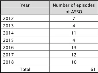
During a 7 years period, between January 2012 and December 2018. We have identified 61 episodes of ASBO in 57 patientsadmitted in our pediatric surgery department.
Table I: Distribution of the number of Episodes of ASBO by years Year Number of episodes
of ASBO 2012 7 2013 4 2014 11 2015 4 2016 13 2017 12 2018 10 Total 61
2. Gender
Out of a total of 57 patients included in our study, 46 (80.7%) were males, and 11 (19.3%) were females. the sex ratio was 4.18
Figure 01: Gender distribution of the studied group 81%
19%
GENDER
MALE FEMALE
- 8-
3. Age :
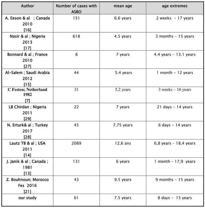
The mean age of our patients was 7 years and 5 months . and the age extremes ranged between 8 days and 15 years.
Table II: distribution ofpatients by age groups
Age group Number (N) %
Newborn (0 - 1 month) 1 1.75%
infant+ toddler ( > 1 month - 3 years ) 9 15.79% preschooler child ( > 3 - 6 years ) 12 21.05% school age child ( > 6 - 12 years ) 23 47.37%
Adolescent ( > 12 years ) 6 14.04%
II. PAST MEDICAL HISTORY
1. Medical history
Only one patient of our studied group had a medical condition, he was treated for abdominal tuberculosis, and he had a peritoneal biopsy.
2. Surgical history
3 out of our 57 patients had neither medical nor surgical history. The remaining 54 patients were operated on for different surgical conditions.
- 9-
36 of the 54 patients were operated on for appendicitis (63.16%). their surgical records have shown, that 27 patients havehad perforated appendicitis (47.37%) and 9 patients with no perforation (15.79%). Four patients were operated on for Intussusception (7.02%). 3 had perforated bowel after abdominal trauma (7.02%). 3 patients had midgut volvulus (5.26%) . two patients had small bowel atresia (3.51%) . two others had omphalocele (3.51%). Diaphragmatic hernia (1.75%), sigmoid volvulus (1.75%) and peritoneal biopsy (1.75%) were found one time for each one. (the results are shown in the figure 03 bellow).
In our studied group, all patients who had surgical history have been operated on by laparotomy.
53 of 54 surgical procedures were performed in infracolic compartment while only one was performed in supracolic compartment for diaphragmatic hernia cure
Figure02: distribution of the previous surgical conditions leading to ASBO 5,26% 47,37% 15,79% 7,02% 7,02% 5,26% 3,51% 3,51% 1,75% 1,75% 1,75% 0,00% 5,00% 10,00% 15,00% 20,00% 25,00% 30,00% 35,00% 40,00% 45,00% 50,00%
- 10-
III. History of presenting Illness
1. Symptoms :
Table III: distribution of ASBO symptoms in our patients
Symptoms Number of ASBO
episodes Percentage Abdominal pain • diffused pain • Umbilical 61 56 5 100.00% 92 % 7 % Vomiting • Bilious vomiting • Nonbilious vomiting 61 37 24 100.00% 61 % 39 % Obstipation 59 97 %
2. the onset of symptoms :
Figure 03: Distribution of the onset of symptoms 68%
32%
- 11-
3. Patient delay:
it ranged from 6 hours to 7 days, and the mean delay was 54 hours.
Figure04
:
Distribution of the time interval between the symptom’s beginning and doctor visit4. the Time period between the previous surgery and development of ASBO
symptoms
The mean time period for the development of ASBO symptoms was 1 year. the extremes ranged between 7 days and 10 years
Table IV: Distribution of the time period between the surgery and the development of symptoms 3,28% 34,43% 31,15% 29,51% 0,00% 10,00% 20,00% 30,00% 40,00% Under 6 hours 6 - 24 hours 24 - 48 hours beyond 48 hours Série1
Time period Number of patients percentage [0 - 6 weeks] 13 24.07% ]6 weeks - 3 months] 16 29.63% ]3 months - 12 months ] 16 29.63% beyond 1 year 9 16.67%
- 12-
IV. PHYSICAL EXAMINATION
1. General examination
Figure 05: Distribution of physical signs found in the general examination
2. Abdominal examination :
Table V: Distribution of the abdominal examination findings Physical signs Number of ASBO episodes Percentage
Abdominal distension 35 57 %
Abdominal tenderness • Diffused
• Umbilical
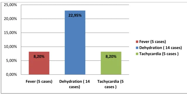
• Right Iliac region 61 - 49 - 9 - 3 100% - 80 % - 15 % - 5 % Abdominal guarding 3 5% 8,20% 22,95% 8,20% 0,00% 5,00% 10,00% 15,00% 20,00% 25,00%
Fever (5 cases) Dehydration ( 14
cases) Tachycardia (5 cases )
Fever (5 cases)
Dehydration ( 14 cases) Tachycardia (5 cases )
- 13-
V. PARACLINICAL INVESTIGATIONS
1. Radiological investigations
1.1. Abdominal X-r ay (AXR)
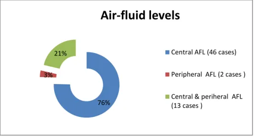
The AXR has been performed in all patients of our studied group, and it showed distended loops of bowels and air-fluid levels (AFL) mostly central.
Figure 06: Distribution of the Air-fluid levels types found in our studied group
Figure 07: AXR of a 13 years old child showing central Air-fluid levels and dilated loops of bowel 76%
3% 21%
Air-fluid levels
Central AFL (46 cases) Peripheral AFL (2 cases ) Central & periheral AFL (13 cases )
- 14-
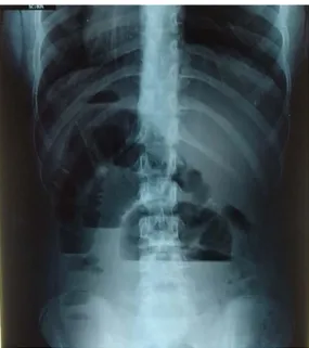
Figure 08: AXR of a 7 years old showing peripheral Air-fluid levels and DLB
- 15- 1.2. Abdominal ultr asonogr aphy
The abdominal ultrasonography was performed in 9 patients. It showed dilated loops of bowel in all of them,and peritoneal effusion in 6 patients.
2. Biological tests
9 patients have Hyperleucocytosis, one patient had severe anemia that needed a blood transfusion, and 14 patients have electrolytes imbalances including hyponatremia, hypochloremia and hypokalemia.
- 16-
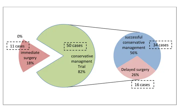
VI. MANAGEMENT
out of 61 ASBO cases, 11 cases (18%) underwent immediate surgery without any previous conservative therapy trial . 50 cases ( 82 % ) were treated conservatively, and out of those patients, 16 cases (26% ) underwent delayed surgical exploration after unsuccessful conservative management.
Figure 10: distribution of the types of management in our studied group
1. Conservative management
In all the 50 episodes where the patients have been managed conservatively, the patients were treated according to the standard decompressive therapy which included nasogastric decompression, exclusion of oral intake, intravenous fluid, correction of electrolytes imbalance, analgesics, monitoring and frequent reassessment. 33 patients among them received water-soluble contrast agent (Gastrographin ) just after their admission along with the decompressive therapy, without waiting for a 48 hours delay.
immediate surgery 18% 0% successful conservative management 56% Delayed surgery 26% conservative managment Trial 82% 11 cases
50 cases
16 cases 34 cases- 17-
Figure 11: distribution of conservative management protocols performed in our patients
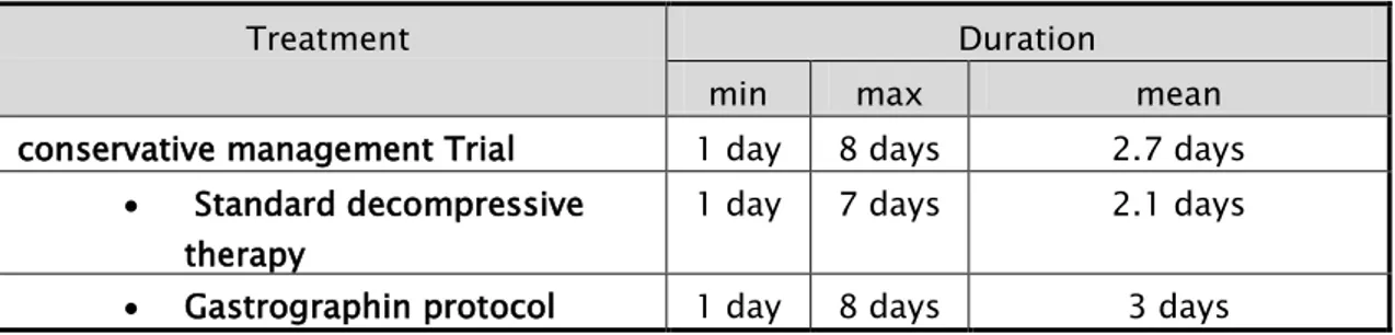
1.1. Dur ation of the conser vative management tr ial
Table VI: minimum, maximum, and the mean duration of the conservative management trial
Treatment Duration
min max mean
conservative management Trial 1 day 8 days 2.7 days • Standard decompressive
therapy
1 day 7 days 2.1 days • Gastrographin protocol 1 day 8 days 3 days
1.2. The outcome of the conser vative management
Table VII: Distribution of conservative management outcomes
Treatment Number of episodes success rate Successful
outcome
Unsuccessful outcome
conservative management trial 34/50 16/50 68 % • Standard decompressive therapy 13 /17 4/17 76 % • Gastrographin prtocol 21 / 33 12 /33 64 % 34% 66%
conservative managment protocols
standard decompressive therapy (17 patients)
- 18-
Figure 12:Abdominal Radiographs of a 13 years old male patient. A- AXR obtained 3 hours after Gastrographin administration showing contrast in the stomach. B- AXR obtained 12 hours after
Gastrographin administration showing the presence of the contrast in the large bowel
Figure 13 :Abdominal Radiographs of a 9 years old male patient , A- AXR showing Air-fluid levels and dilated loops of bowel . B- AXR obtained 12 hours after Gastrographin administration. C- AXR Obtained 18 hours after Gastrographin administration showing the contrast in the large bowel and
the rectum .
A B
- 19-
2. Operative management
2.1. Timing
27 patients (44 % ) out of 61caseshave been managed operatively, eleven (18 % ) of them have had immediate surgical exploration after their admission, the remaining 16 patients (26 % ) underwent delayed surgery after unsuccessful conservative management trial.
Figure 14: distribution of the operative management timing
2.2. Sur gical appr oach
All operative managements in our studied group have been performed using Laparotomy 2.3. Sur gical explor ation
Table VIII: distribution of the surgical exploration findings Successful
conservative management trial
Early surgery Delayed surgery 56%
18% 26%
Successful conservative management trial Early surgery
Delayed surgery
Surgical exploration findings Number of patients Percentage
Single bowel band 15 56%
Multiple bowel bands 12 44%
Adhesions 17 63%
Volvulus 6 24%
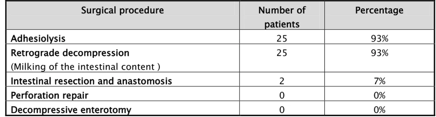
- 20- 2.4. Sur gical pr ocedur e
Table IX: Distribution of the surgical procedures performed in our studied group Surgical procedure Number of
patients
Percentage
Adhesiolysis 25 93%
Retrograde decompression (Milking of the intestinal content )
25 93%
Intestinal resection and anastomosis 2 7%
Perforation repair 0 0%
Decompressive enterotomy 0 0%
The intestinal resection was performed in two patients . for the first patient, it took away 25 cm of the ileum length and left out 7 cm of terminal ileum downstream . for the second, the resection took away 15 cm of the ileumlength and left away the last 10 cm of the terminal ileum.
2.5. Post-oper ative outcomes
One patient had postoperative sepsis that needed a transfert to critical care departement .
VII. Evolution :
1. Length of hospital stay :
Table X: Distribution of the length of hospital stay by therapeutic groups
Group length of hospital stay
min max mean
Conservatively managed group 1 day 8 days 3 days • Standard decompressive
therapy
1 day 7 days 2.1 days
• Gatrographin protocol 1 day 8 days 3.5 days
Operatively managed group 4 days 13 days 6.1 days All the studied group 1 day 13 days 4.4 days
- 21-
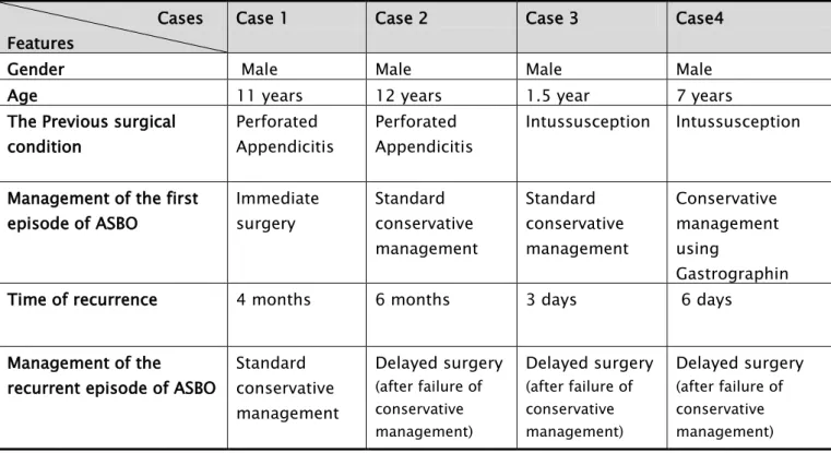
2. Recurrence
There have been 4 cases (6.5 % ) of ASBO recurrence, 3 of them were previously managed conservatively ( 2 patients were managed with standard decompressive therapy and one received WSCA), the remaining case was managed operatively. During the recurrent episodes, the 3 patients who have been previously managed non operatively underwent surgery after unsuccessful conservative trial, while the patient who had been previously managed operatively was successfully managed non operatively.
Table XI : Features of the recurrent cases of ASBO Cases
Features
Case 1 Case 2 Case 3 Case4
Gender Male Male Male Male
Age 11 years 12 years 1.5 year 7 years
The Previous surgical condition Perforated Appendicitis Perforated Appendicitis Intussusception Intussusception Management of the first
episode of ASBO Immediate surgery Standard conservative management Standard conservative management Conservative management using Gastrographin Time of recurrence 4 months 6 months 3 days 6 days Management of the
recurrent episode of ASBO
Standard conservative management Delayed surgery (after failure of conservative management) Delayed surgery (after failure of conservative management) Delayed surgery (after failure of conservative management)
3. Mortality
Two cases of death (3.5% ) were registered in our studied group.One of themdied after 24 hours of conservative management, from a sudden cardiopulmonary arrest secondary to massive inhalation of gastric content. The second one was managed operatively after 72 hours of unsuccessful conservative management; he diedin the critical care unit from postoperative sepsis.
- 22-
- 23-
I. Epidemiological data
1. GENDER
Table XII: the sex ratio reported by authors
Author number of cases with ASBO sex ratio (M/F) J. Janik & al ; Canada ; 1981
[13] 131 1,2
Lautz TB & al ; USA 2011
[14] 2089 1,7
Al-Salem ; Saudi Arabia 2012
[15] 44 1,7
A. Eeson & al ; Canada 2010
[16] 151 2,2
Nasir & al ; Nigeria 2013
[17] 618 1,8
Akgur & al ; Turkey 2017
[18] 181 2
CY Lee & al ; Taiwan 2014
[19] 33 1.4
F. Linden; USA 2019
[20] 12 5
Z. Bouhnoun ; Morocco (Fes) 2016
[21] 43 1.2
our study
61 4.1
Studies conducted in adults, noted that gender was not a risk factor of ASBO . some studies reported males predominance [22-24], while others recorded predominance of females [25,26]
in pediatric patients, all the reviewed studies reported a predominance of males with various sex-ratio.
- 24-
2. AGE
Table XIII : the mean age and age extremes reported by authors
Author Number of cases with
ASBO mean age age extremes A. Eeson & al ; Canada
2010 [16]
151 6.6 years 2 weeks - 17 years
Nasir & al ; Nigeria 2013
[17]
618 4.5 years 3 months - 15 years
Bonnard & al ; France 2010
[27]
8 7 years 4.4 years - 13.1 years
Al-Salem ; Saudi Arabia 2012
[15]
44 5.4 years 1 month - 12 years
C Festen; Netherland 1982
[7]
31 5,2 years 3 weeks – 16 years
LB Chirdan ; Nigeria 2011
[29]
22 7 years 21 days - 14 years
N. Erturk& al ; Turkey 2017
[28]
45 7.75 years 6 days - 14 years
Lautz TB & al ; USA 2011
[14]
2089 12,6 ans 6,8 years – 18,4 years
J. Janik & al ; Canada ; 1981
[13]
131 6 years 1 month – 17,9 years
Z. Bouhnoun; Morocco Fes 2016
[21]
43 9.5 years 9 months - 15 years
- 25-
Adhesive small bowel obstruction can occur at any age [30]. Young et al.[31] reported a higher incidence of ASBO in infants under 1 year of age, irrespective of the initial site or indication for laparotomy. Conversely, the study conducted by N. Erturk et al [28] showed that 82.5% of ASBO occurred among children older than one year, while only 17.7% occurred in infant under 1 year . in the other hand Grant et al [6] found that there is no difference in readmission rates for ASBO between younger and older children when comparing the organ on which surgery was initially performed.
In our study, the mean age of the children with ASBO was 7.5 years, though some studies reported a mean age older or younger than our patients. Nasir et al [17] reported a mean age of 4.5 years, and in the series of Lautz [14] the mean age was 12.6 years.
3. PAST MEDICAL HISTORY
3.1. Medical histor yFewer are the studies that reported the existence of aprevious medical condition in their patients with ASBO. XY Cao [32] Reported 51 cases of ASBOin patients that were being treated for abdominal tuberculosis,and he also reported that Abdominal tuberculosis is an important and increasingly common cause of acute bowel obstruction.
In our study, only one child among our young patients had a previous medical condition; he was treated for abdominal tuberculosis. The same patient also had a peritoneal biopsy two months before the beginning of ASBO symptoms, which would most likely be the cause of those adhesions.
- 26- 3.2. Sur gical histor y
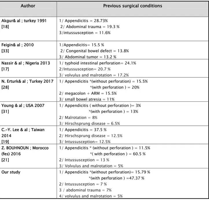
Table XIV: the mean previous surgical conditions leading to ASBO in children reported by authors
Author Previous surgical conditions
Akgur& al ; turkey 1991 [18] 1/ Appendicitis = 28.73% 2/ Abdominal trauma = 19.3 % 3/intussusception = 11.6% Feigin& al ; 2010
[33] 1/Appendicitis= 15.5 % 2/ Congenital bowel defect = 13.8% 3/ Abdominal tumor = 13.2 % Nassir & al ; Nigeria 2013
[17] 1/ typhoid intestinal perforation= 24.1% 2/Intussusception= 20.7 % 3/ volvulus and malrotation = 17.2% N. Erturk& al ; Turkey 2017
[28] 1/ Appendicitis *(without perforation) = 15.5% *(with perforation ) = 20% 2/ megacolon + ARM = 15.5%
3/ small bowel atresia = 11% Young & al ; USA 2007
[31]
1/ Appendicitis ( without perforation )= 3% *(with perforation ) = 13% 2/ Malrotation = 8%
3/ Hirschsprung disease = 6.5% C.-Y. Lee & al ; Taiwan
2014 [19] 1/ Appendicitis = 37.5 % 2/ Hirschsprung disease = 12.5% 3/ Intussusception= 12.5% Z. BOUHNOUN ; Morocco (fes) 2016 [21]
1/ Appendicitis * (without perforation ) = 11.5% *( with perforation ) = 60.5 % 2/ Intussusception = 13 %
3/ Volvulus and malrotation = 5%
Our study 1/ Appendicitis *(without perforation)= 15.79 % *(with perforation ) =47.37 % 2/ Intussusception = 7 %
3 / abdominal trauma = 7% 4/ volvulus and malrotation = 5%
Various previous surgical conditions have been reported in children with ASBO. Vijay [34] found that Hirshprung disease and Intussusception were associated with high rates of ASBO . in Nasir’s Study[17] typhoid intestinal perforation was the frequent previous condition.
- 27-
Appendicitis was reported by the majority of authors [18,33,28,19,21] as the commonest previous condition associated with ASBO in children. Moreover the incidence of ASBO was higher after perforated appendicitis than non perforated one . some studies [13, 31] conducted in children to assess the risk after appendectomy , they noted a low incidence of ASBO after non perorated appendicitis ,elseways the incidence after perforated appendicitis was relatively higher , Young & al [31]reported a rate of 0.3% after non perforated appendicitis versus 3% after perforated appendicitis . this can be explained by the high level of inflammation that occurs during perorated appendicitis and the large laparotomy incision that is performed in this case, which raises the risk of adhesions formation [1]
In our study, the majority of the children with ASBO have had appendicitis.47.37% of cases have had perforated appendicitis and 15.79 % without perforation.
Many studies conducted in African countries [17,21, 34] reported Intussusception as the second previous condition in children with ASBO. Nasir & al [17]reported that 20.6% of the children with ASBO have had previous Intussusception . in our study intussusception was noted in 7 % of the children.
Grant et al [6] found that operation on the ileum of children is followed by a high risk of readmission for adhesionrelated problems. This risk is much higherand it can reach up to 25% when Ileostomy formation and closure is performed, that goes further to confirm our findings of high incidence of ASBO in children with previous midgut malrotation or small bowel perforation resulting of abdominal trauma, their incidence in our study were respectively 5% and 7% . those findings are consistent with other studies reports[17,18,21,31]
- 28-
Aguayo & al [4] conducted a study on the risk of bowel obstruction after treatment of abdominal tumours; he concluded that Bowel obstruction is relatively uncommon after intra-abdominal malignancies in children, with an overall incidence of 3.7 %. However, Wilms ’ tumour, rhabdomyosarcoma and Burkitt’ s lymphoma appear to be associated with the highest risk of bowel obstruction in comparison with other intra-abdominal malignancies.
Tree children (5%) enrolled in our study have no medical nor surgical history, and though they presented ASBO . all of them underwent surgical exploration which has confirmed the presence of congenital adhesions. The incidence of adhesions without previous surgery has been reported to range between 3% and 28 % as determined by autopsy [36,37,38]. Congenital adhesion band is a very rare condition[38] which make a few existing studies that reviewed small bowel obstruction by Congenital adhesions. KH Yang [38] reported that 5.9 % of the patients with ASBO included in his research had congenital adhesions, including 2/3 of them that were children. And unlike adult patients, pediatric patients with congenital ASBO showed a high proportion of early operation and bowel resection [38].
3.3. The time inter val between the pr evious sur ger y and the development of ASBO
Obstruction can occur anytime from the early post‐operative period to many decades later after the surgical procedure [39], a Swedish study conducted by RE. Anderson in adult patients showed that ASBO can occur even 30 years after laparotomy[40]. In pediatric patients, most of ASBO occur within the first year after surgery [31,34,41,42]. Akgur & al [18] reported that most of the ASBO occurred less than 3 months after laparotomy, and those episodes were associated with a high success rate of conservative management which was 81% versus 59 % when the time elapsed after the laparotomy is more than 18 months.
In our study, 53.7% of ASBO episodes occurred in less than three months after the surgical procedures, and about 80.3% occurred in less than one year, which is consistent with the previous findings in the literature.
- 29-
II. PATHOPHYSIOLOGY
1. Etiopathogenesis of adhesions
1.1. Post-oper ative adhesions for mation [43,35,45,48]
Peritoneal adhesions may be acquired or congenital; however, most are acquired as a result of peritoneal injury, which occurs by inflammatory conditions, intraperitoneal infection, abdominal trauma, or most commonly abdominopelvic surgery.
The mechanisms of post-operativeadhesion-genesis are common to all acquired peritoneal adhesions. They involve mesothelial surface disruption with subsequent fibrino-coagulative and inflammatory signalling processes.
When peritoneal serous is injured, the process of repair or healing occurs in two ways; the first one is theusual way which leads to the resolution of the injury and peritoneal repair. The second way is pathological and it leads to persistent adhesions formation.
Immediately after injury, there is bleeding and an increase in vascular permeability with fluid leakage from injured surfaces. Simultaneously, a posttraumatic inflammatory response occurs, with infiltration of inflammatory cells, the release of pro-inflammatory cytokines and activation of the complement and coagulation cascades. The activation of the coagulation cascade results in the formation of thrombin, which is necessary for the conversion of fibrinogen to fibrin, which functions to restore injured tissues and, once generated, is deposited along peritoneal surfaces. Fibrin is a tacky substance and causes adjacent organs or injured serosal surfaces to coalesce.
- 30-
Under normal circumstances, the formation of a fibrin matrix during wound healing is only Temporary and degradation of these filmy fibrinous adhesions by locally released proteases of the fibrinolytic system occurs within 72 hours of injury. Fibrinolysis allows mesothelial cells to proliferate and the peritoneal defect to be restored within 4 to 5 days, preventing the permanent attachment of adjacent surfaces. This process has the key role in tissue remodelling and repair.
If fibrinolysis does not occur within 5 to 7 days of peritoneal injury, or if the local fibrinolytic activity is reduced, the fibrin matrix persists.If this occurs, the temporary fibrin matrix gradually becomes more organized as collagen-secreting fibroblasts and other reparative cells infiltrate the matrix.The organization of fibrin bands over time and their transformation into mature fibrous adhesions is what enables them to persist These“mature” adhesions are not simply composed of connective tissue; studies have demonstrated that, over time, they become highly organized cellular structures that contain arterioles, venules, capillaries and nerve fibres in addition to collagen.
- 31-
- 32-
- 33- 1.2. Congenital Adhesions for mation[38,47]
The congenital adhesions andbands have no relation to an intra-abdominal process (previous laparotomy, inflammatory diseases, peritonitis, embryogenic remnants, etc.), they are considered to have embryologic bases, such as incomplete regression of the fetal vitelline circulation, or a genetic defects that impair embryogenesis. Furthermore, congenital adhesions might also be a result of intrauterine exposure to certain infectious agents or ischemic events.
2. Mechanisms of obstruction
[ 44,45,46 ]The ASBO is a mechanical obstruction, the mechanism of this obstruction can occur either by direct extrinsic compression . or by volvulus, during which, the loop of intestine twists around itself and the mesentery that supports it, resulting in bowel obstruction and later on ischemia and gangrene due to arterial occlusion
3. Consequences of intestinal obstruction
[ 43,8,47,48 ]The pathophysiologic alterations produced by the intestinal obstruction are both local and general. With mechanical occlusion of the lumen,the intestine above becomes distended, resulting from an accumulation of gas and fluid within its lumen. This distention is enhanced by swallowed air, diffusion of gas via the bloodstream, multiplication of bacteria within the stagnant intestinal contents, and hypersecretion of the injured gut.
- 34-
When intraluminal pressure exceeds capillary and venous pressure in the bowel wall, the intestinal absorption and lymphatic drainage decrease, the bowel becomes ischemic when capillary blood flow stops, allowing bacteria to pass into the peritoneum; from that point, it passes into the bloodstream, leading to septicemia by a process known as bacterial translocation. The peritoneal fluid is continuously secreted by the visceral peritoneum and absorbed by the parietal peritoneum, mostly in the diaphragmatic abdominal surface, where the diaphragmatic pores can be distended as much as 3 times their normal size to allow the passage of bacteria. The colonized fluid is then transported via the lymphatic channels into the thoracic duct, which drains in the jugulosubclavian angle, allowing bacteria to enter the circulation and causing septicemia.
Perforation can develop as the ischemia leads to bowel necrosis. First, lymphatic obstruction occurs because of the lesser pressure in these vessels. This is followed by a venous obstruction, which accelerates the edema process because blood enters the affected bowel segment but does not have a drainage route. Finally, the continuous increase in the bowel wall pressure blocks the arterial vessels, leading to ischemic necrosis and perforation.
The massive third spacing of fluids results from peritoneal reflex hypersecretion. Rapidly leads to shock, contributing to morbidity and mortality. This sequence may occur more quickly in a closed-loop obstruction with no proximal escape for bowel contents.
In addition to the local effects produced by obstruction, most profound systemic alterations occur. These may be summarized as a rapidly occurring dehydration with oliguria, hemoconcentration, hypochloremia, hypopotassemia, alkalosis, retention of certain nitrogenous substances, and alterations of the serum proteins.
- 35-
- 36-
- 37-
III. CLINICAL FEATURES
[8,44,45,46]
1. SYMPTOMS
1.1. Abdominal pain
Pain is the earliest symptom to appear; it can be acute or progressive . and most commonly of a colicky character. Furthermore, in children, the pain is associated With motor agitation and crying, and as the obstruction progress, The child becomes increasingly lethargic.
Neither the site nor the type of pain can determine the exact location of the obstruction. 1.2. Vomiting
Vomiting is as early as the site of obstruction is more proximal; initially, the emesis may be nonbilious, but with time it progresses to bilious or event feculent emesis . in 30% of cases the vomiting is associated with relief of abdominal pain.
1.3. Obstipation
With partial obstruction, there still be a continuous passage of flatus and small stool. But in children with complete obstruction both of flatus and stool ceases. Interestingly, in early stages, diarrhea may be present and might confuse the clinical picture.
In our study, abdominal pain and vomiting were present in all children (100% ), and the obstipation was found in 59/61 patients (97%)
- 38-
2. Physical signs
2.1. Systemic signs
In the early stage, children with SBO could present Tachycardia along with palor and motor agitation, which can be related to high pain intensity. Later on, when the patients are seen in a late-stage, they can present tachycardia, Fever, hypotension, and dehydration, those signs indicate commonly circulatory collapse and bowel gangrene.
In our study fever was found in 5/57 patient ( 8%), dehydration in 12/57 patients (24.5%) and the tachycardia in 5 patients (8%)
2.2. Abdominal examination Inspection
The inspection of the abdomen notices the amount of distension which is greater in case of lower small bowel or colonic obstruction. While in proximal bowel obstruction it might not be so marked .furthermore visible peristalsis under the abdominal wall can be seen . and also abdominal scars related to previous surgery but in case of congenital ASBO abdominal scar is missing.
Palpation
Palpation usually finds abdominal tenderness, diffused or localized . Less commonly, abdominal guarding can be found, which highly refers to bowel necrosis or event peritonitis in case of diffused abdominal guarding.
Percussion
Is of a little value except for tympanic resonance that confirms abdominal distension or dullness over the flanks which suggestive of peritoneal effusion.
- 39- Auscultation
Also, it is of a very little help. Initially, it noticed hyperactive bowel sounds due to hyperperistalsis then decrease.
2.3. Rectal examination
A rectal exam must be performed. This gives important information, including the absence of stool, and possibly blood.
IV. PARACLINICAL FEATURES
1. Radiological investigations
Radiologic imaging plays a crucial role in the diagnosis and management of patients with suspected SBO. A myriad of radiologic investigations are available, and the evaluating physician must choose in a reliable, cost-effective way.
[46]
1.1. Abdominal X-r ay
[44, 50,51,52,53]
After a past medical history and physical exam, the initial radiographic study would be plain abdominal radiographs in orthostatic position .even though an upright film is optimal, lateral decubitus or cross-table lateral films could be acceptable substitutes in bedridden patients.
In ASBO, the abdominal radiographs can classically show the following features.: • dilated loops of small bowel proximal to the obstruction >2.5-3 cm • predominantly central dilated loops
• valvulae conniventes are visible
• air-fluid levels in erect view study, especially suspicious if >2.5 cm in length and in the same loop of bowel but at different heights
- 40-
Obviously, any or all of these findings may be present, but the skilled clinician should be able to correlate these radiograph findings with the clinical picture. A recent study evaluating plain radiographs in patients with suspected SBO demonstrated good accuracy, with a mean sensitivity of 83% for correctly identifying a SBO[51]. while it has a low specificity in diagnosing an SBO since both mechanical obstruction and functional bowel disorders may appear identical [46]
1.2.
Abdominal Ultr asonogr aphy[52,54,55]
Ultrasound is a Bedside test also an alternate modality, but really of limited use, especially when intestinal dilation is present and air-fluid levels limit the effectiveness of the study. Adhesions and band are not seen in ultrasound images, but it shows many suggestive findings of small bowel obstruction :
• dilated bowel loop (diameter >2.5 cm)
• ineffective peristalsis (results in "whirling" appearance of intra-luminal contents) • the prominence of the valvulae conniventes (present in dilated jejunal loops)
Furthermore, ultrasound can help to find suggestive signs of bowel ischemia/ infarction that will need urgent surgical evaluation :
• extra-luminal free fluid ( the "pointy" triangular appearance of inter-loop free fluid is often referred to as the tanga sign )
• loss of peristalsis
• bowel wall thickening >3 mm (with effacement of mural architecture) • intramural bowel gas
- 41-
Figure 19: abdominal ultrasound image showing, dilated small bowel loop, with diameter 3.24 cm (> 2.5 cm ) [104]
Figure 20: Ultrasound image showing a dilated loop of bowel and extra-luminal free fluid known as “Tanga sign “[56]
- 42- 1.3. Abdominal CT scan
[52,57]
Abdominal CT scan, especially with administration of oral or intravenous contrast, performs better than plain Xray in finding the transition point, evaluating the severity of obstruction, identifying the cause of obstruction, and recognizing complications such as ischemia, necrosis, and perforation [58]. The sensitivity, specificity, and accuracy of CT scans for ASBO diagnosis are very high; they are, respectively, from 90% to 94%, 96%, and 95% [59].The ASBO Features on CT may include:
• dilated small bowel loops >2.5-3 cm • normal caliber or collapsed loops distally
• small bowel feces signwhich can be defined by the presence of feculent matter mingled with gas bubbles in the lumen of dilated loops
Figure 21: Axial abdominal CT showing the transition point between dilated bowel loop and collapsed loop (red arrows)
[61]
- 43-
Figure 22: Axial abdominal CT showing small bowel feces sign (blue arrow) [62]
CT scan can also show characteristic features of Closed-loop obstruction ( when a bowel loop of variable length is occluded at two adjacent points along its course) :
• radially distributed, C or U-shaped small bowel loops
• " Fat notch sign":reflects insinuation of mesenteric fat at an area of adhesions
with focal caliber change
• “beak sign” corresponding abrupt luminal transition and luminal constriction • two adjacent collapsed loops of bowel
- 44-
Figure 23: Axial abdominal CT of an adult patient showing closed-loop obstruction caused by adhesive bands[63]
Figure 24:coronal reconstruction of an abdominal CT scan showing the “Fat notch sign” ( white arrow) [64]
- 45-
Figure 25 : coronal reconstruction of an abdominal CT scan showing the “beak sign” (double arrow)[64]
When the diagnosis of closed-loop obstruction is delayed, it may be associated with intestinal ischemia which is known as bowel strangulation. It is considered as a dangerous complication since it is associated with high mortality. Strangulation presents with non-specific features ,it include[60]:
• thickened and increased attenuation of the bowel wall • halo or target sign
• pneumatosis intestinalis: gas in the intestinal wall
• pneumatosis portalis: gas in the portal vein or in the mesenteric vein • localized fluid or hemorrhage in the mesentery
- 46-
Figure 26: Axial Abdominal CT showing the difference between normal bowel wall enhancement and ischemic bowel wall enhancement [62]
- 47-
However, abdominal CT scan should not be routinely implemented in the diagnosis making process except when clinical history, physical examination, and plain film were not convincing for ASBO diagnosis, since these are readily available, less expensive, expose the patient to less radiation, and may highlight the need for abdominal CT in some patients . [66]
2. Biological tests
The minimum of laboratory tests includes; blood cell count, lactate, electrolytes, CRP, and BUN/creatinine. Currently, available laboratory tests do not contribute to the diagnosis of SBO, but they can confirm the clinical suspicion of volume depletion, guide volume resuscitation, and assist recognition of bowel ischemia.
[67]
Elevated Leukocytosis count and CRP has been implicated as a marker of strangulation, although sensitivity and specificity of these tests are relatively low
[68]
Electrolytes are often disturbed in patients with a bowel obstruction; in particular, hypochloremia, hypokalemia, and metabolic alkalosis are frequently found.Dehydration and Hypovolemia may manifest as azotemia with an elevated blood urea nitrogen (BUN) to creatinine ratio (BUN/creatinine), and it could result in acute kidney injury [71]
L-lactate, lactate dehydrogenase (LDH) and creatine kinase (CK) can rise due to hypoperfusion of the intestinal tissue. L-lactate level increases sensitivity for detecting bowel ischemia and is considered a reliable indicator for emergency surgical intervention. In contrast, LDH and CK raised levels are therefore unspecific[69,70]
- 48-
VI. MANAGEMENT
1. The Goals of management [44,46,47,71]
The management of Adhesive small bowel obstruction in children consists of the following goals :
Resuscitation, restoring of normal fluid volume and the correction of electrolytes imbalances
Relief of pain and discomfort
Achieving resolution of the obstruction and minimize morbidity and mortality
2. Therapeutic approaches and Indications
2.1. Conser vative management2.1.1. Standar d conser vative management without WSCA
The cornerstone of non-operative management is gastro-intestinal decompression with a nasogastric tube and bowel rest by nil peros. Moreover it consists of, intravenous fluid hydration, correction of electrolytes imbalances, nutritional support, Strict input and output measurements, symptomatic treatment, and Prophylactic antibiotics that should be administered as the clinical situation dictates .in addition to that patients should be Frequently reassessed to make sure that the patient is not developing suggestive signs of complications .