Morphological and functional characterization of
placenta during gestation in bovine clones
derived by somatic nuclear transfer
Par
Hamid Reza Kohan-ghadr
Département de sciences cliniques Centre de recherche en reproduction animale
Faculté de médecine vétérinaire
Thèse présentée à la Faculté de médecine vétérinaire en vue de l’obtention du grade de
Philosophiae Doctor (Ph.D.) en sciences vétérinaires
option reproduction
Décembre, 2010
Université de Montréal Faculté de médecine vétérinaire
Cette thèse intitulée
Morphological and functional characterization of placenta during gestation in bovine clones derived by somatic nuclear transfer
présenté par
Hamid Reza Kohan-ghadr
a été évalué par un jury composé des personnes suivantes
Alan K. Goff, président-rapporteur Réjean C. Lefebvre, directeur de recherche
Bruce D. Murphy, codirecteur Lawrence C. Smith, membre du jury W. Allan King, examinateur externe Jean Sirois, représentant du doyen
Résumé
La technique de clonage par transfert nucléaire de cellules somatiques (SCNT) présente une page importante dans les annales scientifiques, mais son application pratique demeure incertaine dû à son faible taux de succès. Les anomalies placentaires et de développement fœtal se traduisent par des pertes importantes de gestation et des mortalités néonatales.
Dans un premier temps, la présente étude a caractérisé les changements morphologiques des membranes fœtales durant la gestation clonée en les comparant à des gestations contrôles obtenues à partir de l’insémination artificielle. Les différentes anomalies morphologiques des placentomes telles que l’œdème chorioallantoique, la présence de zones hyperéchoiques et irrégulières dans la membrane amniotique et la présence de cellules inflammatoires dégénérées compromettent le développement fœtal normal de la gestation clonée. L’examen ultrasonographique représente une technique diagnostique importante pour faire le suivi d’une gestation et de caractériser les changements placentaires dans le cadre d’évaluation globale du bien-être fœtal.
Le profil hormonal de trois stéroïdes (progestérone (P4), estrone sulfate (E1S), et œstradiol (E2)) et de la protéine B spécifique de gestation (PSPB) dans le sérum des vaches porteuses de clones SCNT a été déterminé et associé aux anomalies de gestations clonées. Une diminution de la P4 sérique au jour 80, une élévation du niveau de la concentration de la PSPB au jour 150, et une augmentation de la concentration d’E2 sérique durant le
deuxième et troisième tiers de la gestation clonée coïncident avec les anomalies de gestation déjà reportées. Ces changements du profil hormonal associés aux anomalies phénotypiques du placenta compromettent le déroulement normal de la gestation clonée et gênent le développement et le bien-être fœtal.
Sur la base des observations faites sur le placenta de gestation clonée, le mécanisme moléculaire pouvant expliquer la disparition de l’épithélium du placenta (l’interface entre le tissue maternel et le placenta) a été étudié. L’étude a identifié des changements dans l’expression de deux protéines d’adhérence (E-cadhérin et β-catenin) de cellules épithéliales pouvant être associées aux anomalies du placenta chez les gestations clonées. Le tissu de cotylédons provenant de gestations clonées et contrôles a été analysé par Western blot, RT-PCR quantitatif, et par immunohistochimie. Les résultats présentaient une diminution significative (p<0.05) de l’expression des dites protéines dans les cellules trophoblastiques chez les gestations clonées. Le RT-PCR quantitatif démontrait que les gènes CCND1, CLDN1 et MSX1 ciblés par la voie de signalisation de la Wnt/β-catenin étaient significativement sous exprimés. La diminution de l’expression des protéines E-cadherin et β-catenin avec une réduction de l’activation de la protéine β-catenin durant le période d’attachement de l’embryon peut potentiellement expliquer l’absence totale ou partielle de l’attachement des membranes fœtales au tissu maternel et éventuellement, l’insuffisance placentaire caractéristique des gestations clonées chez la vache.
La caractérisation morphologique et fonctionnelle du placenta durant les gestations clonées à haut risque est essentielle pour évaluer le statut de la gestation. Les résultats de la présente étude permettront de prédire le développement et le bien-être fœtal de façon critique à travers un protocole standardisé et permettre des interventions médicales pour améliorer le taux de succès des gestations clonées chez les bovins.
Abstract
Although somatic cell nuclear transfer (SCNT) has been shown to be successful, there are still problems with this technique that is inhibiting its use in industry. Altered placental formation and development results in the high incidence of pregnancy loss and prenatal morbidity and mortality found in SCNT pregnancies.
The objective of the initial study was to characterize morphological changes in fetal membranes of a group of bovine recipients carrying cloned fetuses and comparing them with control AI fetuses at various stages of gestation. Several morphological anomalies of placentomes such as edematous chorioallantoic membrane, hyper-echodense spikes or irregularities in the amniotic membrane as well as the pathological presence of degenerated inflammatory cells accompanied by disappearance of the placental epithelium were observed. It was concluded that these anomalies compromise fetal development. The results of this work also showed that ultrasonography may be a reliable technique to monitor and to characterize the placental changes in bovine pregnancies that can be used to assess fetal well-being.
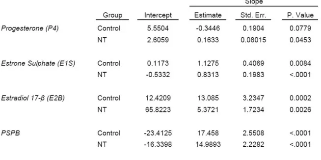
The next study compared the functionality of the placenta of cloned fetuses with controls. The concentration of three steroids (progesterone (P4), estrone sulphate (E1S), and estradiol (E2)) and pregnancy-specific protein B (PSPB) in maternal peripheral circulation were assessed and their associations with gestational anomalies were determined. The hormones profiles in the SCNT recipients deviated from the control group
at certain stages of pregnancy. We observed higher concentrations of E2 throughout the study period, lower levels of P4 at day 80 as well as elevated PSPB concentrations at day 150 in SCNT recipients which coincided with high rate of abortion in these animals shortly after this stage. So, it is proposed that these hormonal changes together with the morphological anomalies of the placenta result in compromised fetal development.
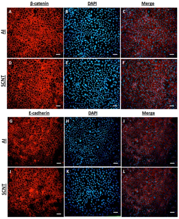
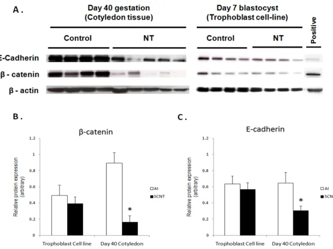
Finally, the molecular mechanism that could be responsible for the abnormal disappearance of the epithelial layer observed in SCNT placenta was investigated. To do so, we measured the expression of two major epithelial adherens junction proteins (E-cadherin and β-catenin) and determined if their expression is altered in relation to the aberrant placentation in SCNT embryos. Cotyledonary tissues from SCNT gestations and control pregnancies were analyzed by Western blot, quantitative RT-PCR and immunohistochemical analysis. Both candidates were significantly (P < 0.05) under-expressed in SCNT trophoblast cells at the protein level. Also, qRT-PCR confirmed that the Wnt/β-catenin signaling pathway target genes CCND1, CLDN1 and MSX1 were significantly down-regulated in SCNT placentas. So, we inferred that impaired E-cadherin and β-catenin protein expression, along with defective β-catenin signaling during embryo attachment, specifically in the window of placentation, results in loose attachment and contributes to insufficient placentation in bovine SCNT-derived embryos.
Overall, we concluded that during the high-risk pregnancy of cloned fetuses, characterization of the morphological and functional changes of the placenta is critical to
enable us to predict normal fetal development and wellbeing through a standardized procedure during clone gestations and to intervene medically in emergency cases to improve the overall efficiency of cloning in cattle.
Table of Contents
Résumé ... i
Abstract ... iv
Table of Contents ... vii
List of tables ... xi
List of figures ... xii
List of Abbreviations ... xiv
Acknowledgments ... xviii
Introduction ... 1
Literature Review ... 4
Article I: Somatic Cell Nuclear Transfer Clones and Placental Anomalies in Cattle – a review ... 5
Abstract ... 7
Introduction ... 8
Placental development ... 9
Morphology of the placenta ... 12
Placenta: an endocrine organ ... 13
Placental consequences of SCNT cloning ... 16
Conclusion ... 22
References ... 23
Mini-review: Role of Adherens junctions during placentation in ruminants ... 32
Introduction ... 32
Placental Development in Ruminants ... 32
Adherens junction’s structure ... 35
Cadherins ... 35
Catenins ... 38
Conclusion ... 43
Hypothesis/Objectives ... 44
Problem ... 44
Hypothesis ... 45
Objectives ... 45
MATERIALS, METHODS AND RESULTS ... 46
Article II: Ultrasonographic and histological characterization of the placenta of somatic nuclear transfer-derived pregnancies in dairy cattle ... 47
Abstract ... 49
Introduction ... 50
Materials and methods ... 51
Animals ... 51
Embryo production ... 52
Ultrasonographic monitoring ... 54
Calving ... 55
Tissue collection ... 56
Experimental design and statistical analysis ... 56
Results ... 57
Placentomes ... 58
Umbilical cord ... 59
Amniotic and allantoic membranes ... 60
Fetal fluid echodensity ... 61
Histopathological observations ... 61
Discussion ... 62
Acknowledgements ... 69
Tables and Figures ... 70
References ... 81
Article III: Endocrine Profiles of Somatic Nuclear Transfer-Derived Pregnancies in Dairy Cattle ... 85
Abstract ... 87
Introduction ... 88
Materials and methods ... 91
Animals and embryo production ... 91
Fetal ultrasonography assessment ... 92
Blood samples collection and serum extraction ... 92
Progesterone and estradiol 17-β (E2) RIA ... 93
PSPB RIA ... 93
Estrone sulfate (E1S) ELISA ... 94
Experimental design and statistical analysis ... 94
Results ... 95
Discussion ... 98
Conclusion ... 105
Acknowledgements ... 106
Tables and Figures ... 107
Article IV: Aberrant Expression of E-Cadherin and β-Catenin Proteins in Placenta of Bovine Embryos Derived from Somatic Cell Nuclear Transfer ... 122
Abstract ... 124
Introduction ... 126
Materials and Methods ... 129
Animals, Embryo Production and in vivo tissue collection ... 129
Cell Culture ... 130
RNA isolation and cDNA synthesis ... 130
Real-time PCR ... 131
Preparation of protein lysates ... 132
Immunoblot Analysis ... 132
Immunohistochemistry ... 133
Immunocytochemistry in trophoblast cell culture ... 134
Results ... 135
Expression analysis and immunolocalization of E-cadherin and β-catenin proteins 135 Analysis of E-cadherin (CDH1) and β-catenin (CTNNB1) mRNA expression ... 137
Expression profile of WNT/β-catenin signalling target genes ... 137
Discussion ... 137
Acknowledgements ... 142
Tables and Figures ... 143
References ... 150
General Discussion... 156
General Conclusion and Future perspective ... 165
List of tables
Article II:
Ultrasonographic and histological characterization of the
placenta of somatic nuclear transfer derived pregnancies in dairy cattle
Table I - Morphology in control vs. clone bovine pregnancies ... 72
Article III:
Endocrine Profiles of Somatic Nuclear Transfer-Derived
Pregnancies in Dairy Cattle
Table II - Relationship between hormones concentration in maternal circulation and placentome size. ... 108
Article IV:
Aberrant Expression of E-Cadherin and β-Catenin Proteins in
Placenta of Bovine Embryos Derived from Somatic Cell Nuclear Transfer
List of figures
Mini-review:
Role of Adherens junctions during placentation in ruminants
Figure 1 - Illustration of bovine placenta. ... 34
Figure 2 - Adherens junction’s structure ... 37
Figure 3 - Canonical WNT signalling pathway. ... 41
Article II:
Ultrasonographic and histological characterization of the
placenta of somatic nuclear transfer derived pregnancies in dairy cattle
Figure 4 - Ultrasonography of fetal membranes during gestation in both cloned and control pregnancies. ... 74Figure 5 - Macroscopic observations of collected tissues in necropsy ... 76
Figure 6 - Light micrograph of fetal tissues... 78
Figure 7 - Comparison of polynomial regression curves of cotyledon length ... 79
Figure 8 - The comparison of percentage of persisting pregnancies ... 80
Article III:
Endocrine Profiles of Somatic Nuclear Transfer-Derived
Pregnancies in Dairy Cattle
Figure 9 - Changes in the hormones progesterone (P4), Estrone sulphate (E1S), Estradiol-17β (E2) and bovine pregnancy specific protein-B (PSPB) during second and final thirds of gestation in NT and control recipients (LSM±SEM). 110 Figure 10 - Comparison of hormone concentrations in two groups of alive NT and aborted NT vs. controls categorized by different stages when the failure occurred. ... 112Article IV:
Aberrant Expression of E-Cadherin and β-Catenin Proteins in
Placenta of Bovine Embryos Derived from Somatic Cell Nuclear Transfer
Figure 11 - Immunohistochemical studies of total β-catenin, active dephosphorylated
β-catenin and E-cadherin in SCNT bovine placenta... 146
Figure 12 - Bovine Trophectoderm (BT) cells ... 147
Figure 13 - Western blot analysis. ... 148
List of Abbreviations
3βHSD Hydroxy-delta-5-steroid dehydrogenase, 3 beta- and steroid delta-isomerase
AI Artificial insemination
ART Assisted reproduction techniques
bHLH Basic helix-loop-helix
BNC Binucleate trophoblast cell
bPAG Bovine pregnancy-associated glycoprotein bPL Bovine prolactin-related protein
BSA Bovine serum albumen
CCND1 Cyclin D1
CDH1 cadherin 1, E-cadherin (epithelial) CDH2 cadherin 2, N-cadherin (neuronal) CDX2 Caudal type homeobox 2
CK1α Casein kinase Iα CKII Casein kinase II
CL Corpus luteum
CLDN1 Claudin 1
COC Cumulus oocyte complexe
CTNNB1 Catenin (cadherin-associated protein), beta 1 DAB 3,3'-diaminobenzidine
DAPI 4',6-diamidino-2-phenylindole
E1S Estrone Sulphate
E2 Estradiol EC Extracellular
FBS Fetal bovine serum
FSH Follicle-stimulating hormone FZD5 Frizzled homolog 5
GAPDH Glyceraldehyde-3-phosphate dehydrogenase
GH Growth hormone
GSK3-β Glycogen synthase kinase 3- β H2AFZ H2A histone family, member Z
HES Haematoxylin-eosin-saffran
HRP Horseradish peroxidase
I.M. Intra-muscular
ICM Inner cell mass
IFNt Interferon-tau IGF-1 Insulin-like growth factor 1 IVP In vitro produced
LEF1 Lymphoid enhancer factor 1
LH Luteinizing hormone
LOS Large offspring syndrome
LRP5/6 Low-density lipoprotein receptor-related protein 5/6
LSM Least squares mean
MDCK Madin-Darby Canine Kidney MNC Mononucleate trophoblast cell
MSX1 Msh homeobox 1
NT Nuclear transfer
P4 Progesterone
P450scc Cytochrome P450, family 11, subfamily A, polypeptide 1 PAF Paraformaldehyde
PAS periodic acid-Schiff PBS Phosphate buffered saline
PPIA Peptidylprolyl isomerase A (cyclophilin A) PSPB Pregnancy-specific protein B
PSTT Placental site trophoblastic tumour
PVDF Polyvinylidene fluoride
qRT-PCR Quantitative reverse transcriptase PCR SCNT Somatic cell nuclear transfer
SEM Standard error of the mean
StAR Steroidogenic acute regulatory protein
TCF T-cell factor
TGC Trophoblast binucleate giant cell TGF-β Transforming growth factor, beta
TGF-βR Transforming growth factor, beta receptor
TNC Trinucleate cell
TP53 Tumour protein p53
This is dedicated to my wife for her love and support in all my endeavours. Your unwavering support was essential in getting me to this point.
Acknowledgments
This dissertation would not have been possible without the work, efforts, and support of many people. Foremost, I would like to thank my PhD supervisor, Dr. Rejean Lefebvre, for his excellent supervision during these five years. His unwavering enthusiasm and steady stream of encouragement guided me through my doctorate training. I would also like to thank my co-supervisor, Dr. Bruce Murphy, for welcoming me into his lab. I greatly appreciate his ability to guide us when we needed it, but at the same time, allowed us to learn and grow on our own. He quickly became for me the role model of a successful researcher and leader.
I would like to express my sincerest thanks to Mira Dobias-Goff not only for her invaluable guidance and her technical wizardry in RIA technique but also for her kind friendship. Also, I am grateful to Vickie Roussel for her technical expertise and assistance.
My lab mates through the years were some of the most valuable resources. There have been many colleagues through the years on whom I have leaned upon for support. I remember and appreciate everyone.
I would like to thank my advisory committee members, Dr. Lawrence Smith and Dr. Gilles Fecteau for their support, advice, and insight. I would also like to thank my thesis committee for their attention, time, and useful suggestions for my research and this dissertation.
I must acknowledge the guidance and support from all people in “Centre de Recherche on Reproduction Animale (CRRA)” and “Faculty of Veterinary Medicine”. I
would like to particularly thank Micheline Sicotte and Micheline St-Germain for their help with administrative concerns throughout the years.
Finally, I must thank my dear wife, for her understanding, endless patience and unconditional love. Without her support, I would not have any chance here writing this document.
Introduction
The development of placenta is a critical prerequisite for development of the embryo past the blastocyst stage. The placenta has a specialized pregnancy-specific structure that functions as a source of nutrients and hormones and protection for embryo/fetus throughout gestation. Placenta shape and form is largely variable among the mammals. The bovine placenta was described anatomically as synepitheliochorial (Wooding et al. 2008) that is initially formed by trophectodermal cells invasion to caruncular endometrium around 4th week of gestation to form villi or cotyledons. At the later stages of pregnancy, each caruncle and its associated cotyledon will be referred to as a placentome (Schlafer et al. 2000). Embryos that fail to accomplish proper placentation suffer from various morphologic defects and die. The complications found during normal gestation are rarely reported. However, since the introduction of assisted reproduction techniques (ART) such as in vitro production of embryos and nuclear transfer cloning, several reports have shown on low survival rate and high pregnancy failure due to abnormal placental development (Diskin et al. 1980; Hill et al. 2001; Heyman et al. 2002; Constant et al. 2006). A broad spectrum of anatomical and physiological anomalies was reported to be related to poor placentation in somatic cell nuclear transfer (SCNT) derived embryos.
It seems that the first trimester of gestation is a critical period for bovine SCNT embryos as the majority of implanted embryos are lost in this window (Willadsen et al. 1991) mostly accompanied with flattened cotyledons and abnormally shaped placentomes (Stice et al. 1996; Hill et al. 2000; Hashizume et al. 2002). Beyond day 90, placentome hypertrophy is the most frequent anomaly in bovine SCNT pregnancies (Hill et al. 1999;
DeSouza et al. 2001). Similar abnormalities were also reported in ovine SCNT gestations (DeSouza et al. 2001). Reduction in placental vascularisation was also reported in SCNT gestations (Palmieri et al. 2007). Although few bovine SCNT gestations reach full term, many give birth to overweight malformed neonates with placental abnormalities such as oedematous placenta, placentomegaly, hydroallantois and enlarged umbilical vessels (Hill et al. 1999; Wells et al. 1999; Constant et al. 2006).
In addition to abnormal gross placental morphology, changes in cellular and sub-cellular population were reported repeatedly in placenta of SCNT embryos (Hill et al. 2000; Hashizume et al. 2002; Arnold et al. 2006). Histologically, hypoplasia of trophoblastic cells, reduced numbers of the trophoblast binucleate cells (BNCs) and sub-epithelial haemorrhages were observed in different studies on SCNT pregnancies (Stice et al. 1996; Hashizume et al. 2002; Loi et al. 2006; Palmieri et al. 2007).
The atypical placentation in SCNT also endangers fetal well-being by alteration of normal placenta function. Many proteins involved in the regulation of feto-maternal interface such as bovine pregnancy-associated glycoproteins (bPAG) 1 and 9 and bovine prolactin-related protein (bPL) 1 are deregulated throughout pregnancy in SCNT placenta (Hashizume et al. 2002; Patel et al. 2004; Hirayama et al. 2008). Studies have demonstrated that the perturbations in steroidogenic activity of placenta in bovine SCNT pregnancies are related with some late gestational problems such as higher birth weight, abortion and prolonged gestations (Shah et al. 2007; Hirayama et al. 2008). Generally, there is aberrant molecular biology in SCNT pregnancies, as shown with an abnormal protein profile (Kim et al. 2005). Together, it seems that during high risk pregnancy, characterization of the
morphological and functional changes of fetal membranes is critical. Also, by defining the fetal growth parameters and morphologic characterization of the placenta, we will be able to predict through a standardized protocol the stages when the normal fetal development and wellbeing is compromised during clone gestations and to intervene medically in emergency cases to improve the overall efficiency of cloning in cattle.
Article I
: Somatic Cell Nuclear Transfer Clones and Placental
Anomalies in Cattle – a review
Somatic Cell Nuclear Transfer Clones and Placental Anomalies in Cattle – a
review
Kohan-Ghadr HRa, Lefebvre RCab, Fecteau Ga, Smith LCb, Murphy BDb
Address: aDepartment of Clinical Sciences, bCentre de recherche en reproduction animale,
cDepartment of pathology and microbiology, of the Faculty of Veterinary Medicine,
University of Montreal, 3200 Sicotte, Saint-Hyacinthe, Québec, Canada, J2S 2M2.
(a)Corresponding address:
Réjean C. Lefebvre,
Department of Clinical Sciences, College of Veterinary Medicine, University of Montreal, 3200 Sicotte, Saint-Hyacinthe, Québec, Canada; Tel: 1-450-773-8521;
Abstract
In ruminants, somatic cell nuclear transfer (SCNT) can result in healthy offspring however, it is often associated with pathological changes in the fetus and the placental phenotype. The fetal membranes are essential in maintaining the fetus during pregnancy, any pathological changes of the placenta have potentially serious consequences for embryonic and fetal development and neonatal survival. The low efficiency of SCNT in producing embryos that develop into normal and healthy offspring has triggered research interest to understand mechanisms associated with morphological and functional placenta abnormalities to improve cloning technology. Although current evidence implicates aberrant reprogramming by epigenetic mechanisms of the donor chromatin as a cause, the association between morphological, functional and clinical abnormalities is poorly understood. Given the great variability in phenotype observed in SCNT, even with the same nuclear genetics, several aspects of cloning need to be explored. The present review summarizes current understanding of the normal placental structure in cattle and discusses developmental, morphological and functional abnormalities in placenta associated with SNT cloning.
Introduction
While still mobile in the uterus, the outermost layer of the embryo (the trophoblast) has acquires some specialization toward its eventual role in the transport of nutrients and fluids, synthesis of proteins and hormones, and excretion of waste products. The early embryo initiates communication with the mother to ensure maternal recognition of the pregnancy, achieve implantation, establish the interface for exchanges between maternal and fetal circulation, and alter the local immune environment. The feto-maternal interface, the placenta, is responsible for maintenance of the fetus, including sustaining the favorable nutrient partitioning essential to fetal growth and its physical protection. Any placental abnormality is expected to increase the risk of death of the embryo or fetus or compromise the life and development of the neonate.
The somatic cell nuclear transfer (SCNT) cloning opened a new chapter in placenta research field. This reproductive assisted technique has undeniable potential benefit for research of genetic and epigenetic mechanisms underlying developmental biology, aging, carcinogenesis (Meissner et al. 2006), generation of organs for xenotransplantation (Baguisi et al. 1999; Dai et al. 2002), developing transgenic animals for production of valuable recombinant proteins, saving genetically valuable animals (McClintock 1998) and for preserving endangered breeds and species (Loi et al. 2001). However, practical advantages are reduced by frequent developmental, morphological and functional abnormalities of the placenta. Clinical observations, macroscopic and histological evidence, gene expression studies and hormone profiles, all incriminate placental dysfunction as a substantial factor in
the failure of SCNT derived pregnancies. This gives fresh impetus to determine the association between placental dysfunction and pregnancy loss.
The purpose of this review is to summarize current knowledge on normal placental and discuss developmental, morphological and functional abnormalities in cattle associated with somatic cell nuclear transfer-derived gestation. Practical approaches to remediation rising from the knowledge of placental dysfunction should increase embryo and fetal survival and reduce economic losses.
Placental development
Embryogenesis in cattle has been well documented. At Day 7, the bovine embryo (Day 0 = fertilization) is in the blastocyst stage, characterized by a hollow central cavity and a plaque of cells lining in the inner blastocoel at one pole (inner cell mass; ICM), all surrounded by an outer layer of trophoblast cells (trophectoderm). The ICM develops into the embryo proper and the trophoblast cells into the fetal component of the placenta (McLaren 1972). At Day 8, a specialized cell layer (the endoderm) will grow from the ICM, inside the trophoblast cell layer and forms the yolk sac (Patten 1964). In addition, a third layer of cells (the mesoderm) grows from the ICM between the endoderm and the trophoblast layer. After hatching or emergence from the zona pellucida at 150 µm in diameter (Day 9), the growth of the embryo is rapid and reaches about 15 cm long, occupying the entire uterine lumen by Day 20 (Constant et al. 2006). This embryonic
elongation establishes the first direct contact between the embryo and the mother prior to implantation. By day 12, the trophoblast cell and mesoderm layers combine to form the chorion (Patten 1964). The chorion grows around the embryo forming a fluid-filled space around the embryo called the amniotic sac. Prior closure of the embryo abdominal wall, a new sac (allantois) extends between the yolk sac and the chorion. As the chorion extends out into the uterine lumen, the allantois grows rapidly and fuses with the chorion to form the chorioallantois (Schlafer et al. 2000). In ruminants, the outer layer of trophoblast cells (chorion) proliferates and differentiates into binucleate (BNC) and mono-nucleate cells (MNC) in a ratio of one to five (Wooding 1992). The MNCs synthesize placental interferon-tau (IFNt), a pregnancy recognition protein in cattle (Roberts et al. 1992) and BNCs synthesize several pregnancy associated proteins (Wooding et al. 1994).
Early during the process of implantation of the embryo (Day 20, apposition phase), the trophoblastic papillae (Guillomot et al. 1982) of the chorion (trophoblastic cells and connective tissues) enter into the uterine glands of the endometrium to achieve more intimate interaction and to anchor the primitive placenta to the uterus. This results in a diffuse and transitional placentation that enables the fetus to absorb the uterine secretion. This intimate contact between the placenta and the mother is first observed in the regions nearest the embryo and progressively continues to the tip of the elongated sac. By Day 25, the BNCs have migrated into the endometrium and fuse with uterine epithelial cells to form a multinucleated fetomaternal cell, a syncytium that is critical to villous formation and maintenance of a closer adhesion between microvilli and the trophoblastic cell membranes of the attachment phase (Wooding et al. 1980; Wooding 1992; Guillomot 1995). The
syncytial cells produce placental lactogen beginning on Day 18, pregnancy-associated glycoproteins (PAG’s); beginning on Day 25. They also synthesize prolactin-related proteins (Wooding 1981; Kessler et al. 1991; Zoli et al. 1992; Green et al. 1998; Patel et al. 2004), and release them into the uterine connective tissue.
The number of BNCs within the trophoblast increases from Days 20 to 30 of the gestation (Morgan et al. 1989), whereas the interface between the outer layer of the chorion formed by the single layer of trophoblast cells and the endometrium is maintained as a simple epitheliochorial attachment. At Day 22 of gestation, the amniotic sac is formed by folding of the trophoblastic cells and mesodermal cells (inside) and begins to accumulate fluid, whereas the allantoic sac is completed at Day 24 and the attachment of the placenta by Day 27 (King et al. 1981; Schlafer, Fisher et al. 2000). The allantoic membrane is a thin and translucent and well vascularized by arteries originating from the aorta and umbilical veins. While the amniotic and allantoic membranes are formed, the third fetal membrane, the vitellin sac, disappears. Adhesion and attachment between the placenta and the endometrium becomes stronger by Day 30 (King, Atkinson et al. 1981; Guillomot 1995). The more intimate contact of the placenta with the maternal tissues induces a localized thickening on the fetal side that becomes the cotyledon (Li et al. 2005) where it covers preexisting uterine caruncles. The interdigitated chorionic villi and uterine crypts develop by Day 45 of gestation (King et al. 1979) and appear macroscopically as placentome; the union of the cotyledon and the caruncle. In cows, the placentation is classified as synepitheliochorial (Wooding 1992) because of the BNC-derived syncytium and the
epithelio-chorial interaction, representing large areas of simple apposition of maternal tissues known as the intercotyledonary space.
Morphology of the placenta
The surface of the endometrium in pregnant cow has four rows of specialized endometrial regions known as caruncles along the long axis of both horns, with a total of 60 to 80 convex and ovoid structures (Duello et al. 1986). Hydrostatic pressure of the allantoic and trophoblastic membrane against the uterine wall is believed to facilitate chorioallantoic attachment to the caruncle and stimulate the fetal chorion to vascularise and hypertrophy. As the cotyledons mature, the remodelling of the endometrium that leads to the development of the caruncles is necessary to accommodate the specialized folding of the chorioallantois. The surface of contact between maternal and fetal tissues is increased by the development of outgrowths on the surface of chorion, known as the villi. These chorionic villi consist of vascular mesenchymal cones surrounded by cuboidal trophoblastic and giant binucleate cells bring the fetal (allantoic) vessels into proximity with the maternal blood vessels. The temporal and spatial changes of the extra cellular matrix (ECM) during apposition, adhesion and attachment increase the complexity of placental formation and implantation process (Johnson et al. 2001; MacIntyre et al. 2002; Xiang et al. 2002).
Placentomes are larger and more numerous closer to the fetus and in the horn containing the fetus relative to those located in the uterine extremities and those found in the contralateral horn. Total placentome weight and length increase until Day 190, attaining
approximately 4.5 kg and 10 to 12 cm (Schlafer, Fisher et al. 2000) with most acquiring a mushroom-like shape with an occasional flat configuration (Miles et al. 2004). The total number of placentomes does not correlate with the increased fetal nutritional demands of late pregnancy suggesting that there is an alteration in the pattern of vasculature to increase feto/maternal exchange without an increase in placentome number (Leiser et al. 1997).
Placenta: an endocrine organ
The bovine placenta is an autocrine, paracrine and endocrine organ, in that it synthesizes a broad range of steroids and peptide hormones that regulate the development of the feto-placental unit (Gootwine 2004). Among hormones produced are steroids and protein factors including growth hormone (GH), insulin like growth factor-I (IGF-I), cytokines (Bauer et al. 1998) lactogenic hormones (PLs), and relaxin (Sjaastad et al. 2003). Placental protein hormones stimulate ovarian hormone production and fetal growth while contributing to mammary development and parturition. Growth hormone and IGF-1 have an anabolic effect and regulate fetal growth through cell growth and differentiation (Creasy et al. 2004). Relaxin, produced by both the placenta and ovaries, prevents uterine contractions during pregnancy and causes connective tissue in the cervix and pelvic ligaments to depolymerize and relax before parturition, facilitating expulsion of the fetus (Sjaastad, Hove et al. 2003).
Pregnancy in cattle depends, for the most part, on progesterone produced by the corpus luteum (Sjaastad, Hove et al. 2003) however; pregnancy can be maintained by the
placenta in the absence of ovarian progesterone after 200 days of gestation (Johnson et al. 1962; Estergreen et al. 1967; Johnson et al. 1981). Progesterone is synthesized within the placenta from circulating maternal cholesterol (Creasy and Resnik 2004), even though the total amount produced by the placenta seems to be small (Melampy et al. 1959). During steroidogenesis in the placenta, a series of catalytic reactions decrease the size of the basic 27-carbon-unit structure of the cholesterol to 21-carbon molecules (progesterone) (Thorburn et al. 1994). The bovine placenta has both P450scc and 3βHSD activity (Shemesh et al. 1989) in addition to StAR mRNA (Pescador et al. 1996; Verduzco et al. 2011). Progesterone is necessary for secretion of the histiotroph, comprised of various uterine proteins formation prior to completion of placentation, maintenance of myometrial quiescence, stimulation of mammary gland development and, suppression of immune rejection of the conceptus (Thorburn and Harding 1994). Other steroids, e.g. estrogens, are believed to be important in placental steroidogenesis. Some estrogens are mediators of implantation, uterine growth, mammary duct development, pelvic and cervical relaxation, induction of myometrial oxytocin receptors and, development of maternal behaviour (Thorburn and Harding 1994). The source of steroids in the ruminant placenta is not well known, although there is evidence that the mononucleate trophoblastic cells are the main source of placental estrone (Matamoros et al. 1994; Verduzco. et al. 2011).
The bovine placenta also produces a large family of glycoproteins named the pregnancy-associated glycoproteins (PAGs) which is specifically expressed by BNC of fetal cotyledonary tissue (Zoli, Guilbault et al. 1992). The PAGs are also known under a variety of names as pregnancy-specific protein 60 (Mialon et al. 1993) or pregnancy
specific protein B (Butler et al. 1982; Lynch et al. 1992). The PAGs are secreted continuously throughout gestation, with concentrations in maternal serum rising from Days 24 to 28 to the end of pregnancy and persisting after parturition (Sasser et al. 1986; Humblot et al. 1988; Humblot et al. 1988). From Day 33 of gestation, cows can be correctly diagnosed pregnant and non-pregnant by radioimmunoassay with about 87 % accuracy rate (Szenci et al. 1998). Therefore, a PAG assay can be used to detect and monitor pregnancy and to determine factors influencing late embryonic mortality (Humblot, Camous et al. 1988; Humblot, Jeanguyot et al. 1988). In the case of embryonic mortality, PAG concentrations were depressed before disappearance of the corpus luteum(Humblot, Camous et al. 1988; Humblot et al. 1991).
The synthesis and the secretory pattern of each PAG may differ as some proteins can be measured as early as Day 19 and others only appear by Day 45 of the gestation. Pregnancy-associated glycoprotein-1 can be measured between Day 20 and Day 30. Its concentration remains stable between Day 40 and 70 and slowly increases until Day 150, after which it increases rapidly to reach a peak about 20 days before calving. The plasma level remains elevated for about 3 months after calving because of its long half-life. Also synthesized by the BNCs, PL has a similar structure and function to the growth hormone and the relaxin (Constant, Guillomot et al. 2006). As expected for a protein derived from the fetus, the PL concentration of the mother is very low (1 to 2 ng/ml) compared to the fetal serum levels (Constant, Guillomot et al. 2006).
Placental consequences of SCNT cloning
Since the first bovine nuclear transfer (Prather et al. 1987) and the first demonstration that a nucleus derived from differentiated tissues from adult mammals may support fetal development to term (Wilmut et al. 1997), serious anomalies of gestation have been reported (Wells et al. 1999). In ruminants, these anomalies appear more extreme in clones produced from somatic cells relative to those produced from embryonic cells (Chavatte-Palmer et al. 2002). It is noteworthy that the rates of embryonic, fetal and perinatal survival are variable in cloned ruminant species (Baguisi, Behboodi et al. 1999; Lee et al. 2005; Galli et al. 2008). Although the specific pathologies vary among fetuses, the predominant cause of pregnancy failure associated with SCNT cloned embryos is believed to be associated with aberrant placenta, either morphologic or functional failures (Hill et al. 2000; Hill et al. 2001; Lee et al. 2004).
Four lines of evidence implicate abnormal placentas as a major cause of pregnancy failure and poor reproductive performance. First, the multitude, severity and the consistency of the placental lesions reported in multiple species could account for the high rate of pregnancy losses. Secondly, chimera studies with tetraploid blastomere NT cloned embryos in rodents further support the important role of placental anomalies in pregnancy losses (Eggan et al. 2001). Eggan (2001) showed that tetraploid blastomeres contributed only to formation of the placenta but not to the embryo proper in mice (Eggan, Akutsu et al. 2001). In this experiment, the neonatal SCNT mice cloned from embryonic stem cells had mean placental weight of 0.32 g that was significantly higher than those of tetraploid-produced pups (0.1 g) which were intermediate between SCNT and traditionally derived
pregnancies (Eggan, Akutsu et al. 2001). The third line of evidence can be found in similarities in placental and endometrial lesions between NT pregnancy and the interspecies pregnancies between the goat and the sheep (Hancock et al. 1968; Oppenheim et al. 2001). Early pregnancy losses were associated with reduced villi and crypts and reduced vascularity and presence of inflammatory cells in placentas (Hancock, McGovern et al. 1968; Allen 1982) which are also observed in NT pregnancies (Hill, Burghardt et al. 2000; Hashizume et al. 2002). The interspecies chimeric pregnancies seem to be similar to NTs in that in both gestations, the embryo is from a different genetic background and the maternal immunological rejection of the conceptus could result in implantation failure in early stage, or later, be manifest as placental anomalies (Dent et al. 1971; Oppenheim, Moyer et al. 2001; Davies et al. 2004). A fourth line of evidence is found in observations made on the effects of in vitro culture on placenta. The in vitro culture environment affects not only embryonic development and survival, but also alters placental development, morphology and function later in gestation (Farin et al. 2001). Placentas from in vitro produced (IVP) embryos that developed in the presence of serum were heavier, had a smaller caruncular surface, decreased villous volume density and reduced number of BNC compared to those produced in vivo (Farin et al. 2000). Alteration of placental development has been observed in SCNT cloned embryos with higher frequency and severity than in IVP embryos, providing further evidence for correlation of aberrant placentation with the low rates of gestational success in SCNT embryos (Cibelli et al. 1998; Hill et al. 1999; Hill, Edwards et al. 2001; Lee, Peterson et al. 2004).
Thus, the major impediment to the use of SCNT technique is its low efficiency, with less than 5% of SCNT pregnancies reaching term (Heyman et al. 2002). The majority of SCNT pregnancies in which the embryo survives and attaches to the uterus are lost between Days 30 and 90 of gestation (Willadsen et al. 1991) due to poorly developed placentomes (Stice et al. 1996; Hill, Burghardt et al. 2000; Hashizume, Ishiwata et al. 2002). Distinct placental morphology accompanies these losses. In the first trimester, cotyledons appeared flattened, abnormally shaped and reduced in numbers (Hashizume, Ishiwata et al. 2002). Placentomes in later pregnancy are frequently hypertrophied (Hill, Roussel et al. 1999; DeSouza et al. 2001). This hypertrophy is believed to be due, in part, to a mechanism to compensate for the reduced number of placentomes. This has been shown in an intrauterine growth restriction experiment in sheep where placental attachment sites were removed by carunclectomy, resulting in a significant increase in the size of the remaining placentomes (Robinson et al. 1979). In one bovine study, only 12 large functional cotyledons were found in the gravid horn of the uterus bearing a SCNT embryo (Hill, Edwards et al. 2001).
Alternatively, the mechanism of compensatory hypertrophy may be related to differential expression of transforming growth factors (TGF-βs) which are involved in all phases of development of the placenta that may alter the size of the placentomes in pregnancies derived from SCNT cloning. Between Days 50 and day 150 of gestation TGF-β1, TGF-β2, TGF-β3 mRNA abundance increased whereas expression of TGF-βR1 and TGF-βR2 receptors transcripts decreased significantly in SCNT placentomes, compared to pregnancies derived from artificial insemination (Ravelich et al. 2006). Consequently, the
placentomes of SCNT placentas may become resistant to the suppressive effects of the TGF-βs and become much larger.
The presence of smaller placentomes may be explained by a mechanism of compensation for the poor efficiency of larger placentomes by the formation of confluent smaller cotyledons around an already existing cotyledon (adventitial placentation). This has been observed in cases of chronic uterine conditions (Kennedy et al. 1993) and may be associated with development of hydrallantois, a condition observed frequently in SCNT cloned pregnancy. Enlarged umbilical vessels and edematous fetal membranes have also been reported in SCNT cloned fetuses (Willadsen, Janzen et al. 1991; Hill, Roussel et al. 1999; Wells, Misica et al. 1999).
In addition to the typical gross malformations of the placenta associated with SCNT pregnancies, the endometrial-trophoblast interface displays a reduced epithelial height with underdeveloped vascularity upon histological examination (Hill, Burghardt et al. 2000). Hashizume et al. (2002) reported the presence of fewer BNC at Day 60 in placenta of somatic NT pregnancy compared to controls derived by artificial insemination (Hashizume, Ishiwata et al. 2002). Similar observations were reported by Arnold et al. (2006) at Day 40 (Arnold et al. 2006). The question that arises is whether the reduced number of placentomes is a consequence of invasion of fewer BNC in cloned pregnancies, or whether fundamental differences in placentome formation result in few BNC. More investigation is clearly warranted. In addition to differences in placentome number, placental septae were more irregular and sparsely arranged in SCNT pregnancies (Hashizume, Ishiwata et al. 2002).
Morphometric analysis revealed that placentomes from IVP pregnancies have a smaller volume of fetal villi (Miles, Farin et al. 2004).
As proposed by Hill et al. (2000), the stage of pregnancy loss seems to be associated with different kinds of placental anomalies (Hill, Burghardt et al. 2000). Early pregnancy loss, occurring before complete placental establishment (less than 45 days) is believed to result from severely insufficient or delayed development of the chorionic epithelium and its vascularisation. The initial contact between the fetal trophoblast and the maternal caruncles does not induce the necessary hypertrophy of the fetal chorion and the formation of the normal cotyledon. Since fewer BNC migrate with the endometrial epithelium, the villous formation is perturbed and the maintenance and expansion of close adhesion between microvilli and the trophoblastic cell membrane is not maintained. This results in failure of the pregnancy. Fetuses dying between Day 45 to 90 of pregnancy appear to have compensated for earlier delays in chorionic development but do not develop a minimum threshold number of placentomes (Cross 2006) to allow a sufficient supply of nutrients, and gas exchange; therefore, the fetus eventually dies by starvation or other result of insufficient vascularization. This view is based on observations that, in the second trimester, fetal growth rate increases rapidly (Hubbert et al. 1972; Prior et al. 1979); the functional capacity of placentomes becomes critical because of the increased fetal demand for nutrients. If the compensatory mechanism (increasing placentome size) is not sufficiently rapid, the fetal demand is not met and the fetus died by the end of the second trimester (150 days of pregnancy). In addition to this, compensatory effects of increasing size, placentomes become resistant to the inhibitory effect of TGF-βs because of the
reduced number of receptors (TGF-βR1 and TGF-βR2), get even larger and allow pregnancy to be maintained. This escape strategy from growth inhibitory effect of TGF-β was reported in human cancers (Filmus et al. 1993; Markowitz et al. 1996). The loss of cell surface TGF-βR2 is correlated with functional resistance to growth inhibition by TGF-β in human colon neoplasia and result tumor progression (Markowitz et al. 1995). The compensatory growth of the reduced number of placentomes has some consequences that can result in pregnancy loss. These placentomes become so large in the last trimester that growth is disorganized and function of the tissue is impaired and eventually they undergo necrosis. This was observed in a case report of an SCNT cloned pregnancy, where fetal death occurred, the chorioallantois was mineralized and degenerating and necrotic cotyledons were observed (Hill, Edwards et al. 2001). In addition, the necrotic tissue, the vessels and the degenerating epithelium were mineralized.
Pregnancies with placental anomalies resulting from SCNT do not always result in fetal death. It is possible to predict calf size, and from there, their chance for survival so that the fetus can be delivered as soon as it is viable. If the calf grows an average of 0.35 kg/d in the last trimester (Prior and Laster 1979), birth of an extremely large fetus may be reduced and its chance of survival would be improved. The birth weight of the calf is predictable in the last trimester of pregnancy because of the linear correlation between minimal metacarpal or metatarsal thickness and body weight (Takahashi et al. 2001). These dimensions can be measured by transrectal ultrasonography, rendering it possible to detect oversized fetuses in cloned pregnancy and thus allowing intervention (Takahashi et al. 2005).
Conclusion
Placental membranes support the fetus throughout gestation however, anomalies in placental structure and function often result in SCNT pregnancy loss. These losses appear during the embryonic and fetal periods and are a major cause of decreased reproductive efficiency in cattle and a substantial cause of economic loss for the industry. Even though causes have been proposed to explain pregnancy loss, under field conditions, the etiology is often elusive. Currently, the frequent occurrence of placental anomalies in pregnancies obtained by SCNT derived pregnancy, points to an important role of placental dysfunction in pregnancy loss. Diagnostic tools like ultrasonography, hormonal profiles and molecular techniques could be used to monitor and study high-risk pregnancies derived from SCNT and better elucidate the role of placental dysfunction in pregnancy loss and potentially improve fetal survival.
References
Allen, W. R. (1982). "Immunological aspects of the endometrial cup reaction and the effect of xenogeneic pregnancy in horses and donkeys." J Reprod Fertil Suppl 31: 57-94. Arnold, D. R., V. Bordignon, R. Lefebvre, B. D. Murphy and L. C. Smith (2006). "Somatic
cell nuclear transfer alters peri-implantation trophoblast differentiation in bovine embryos." Reproduction 132(2): 279-90.
Baguisi, A., E. Behboodi and D. Melican (1999). "Production of goats by somatic cell nuclear transfer." Nat Biotechnol 17: 456-461.
Bauer, M. K., J. E. Harding, N. S. Bassett, B. H. Breier, M. H. Oliver, B. H. Gallaher, P. C. Evans, S. M. Woodall and P. D. Gluckman (1998). "Fetal growth and placental function." Mol Cell Endocrinol 140(1-2): 115-20.
Butler, J. E., W. C. Hamilton, R. G. Sasser, C. A. Ruder, G. M. Hass and R. J. Williams (1982). "Detection and partial characterization of two bovine pregnancy-specific proteins." Biol Reprod 26(5): 925-33.
Chavatte-Palmer, P., Y. Heyman, C. Richard, P. Monget, D. LeBourhis, G. Kann, Y. Chilliard, X. Vignon and J. Renard (2002). "Clinical, hormonal, and hematologic characteristics of bovine calves derived from nuclei from somatic cells." Biol Reprod 66: 1596-1603.
Cibelli, J. B., S. L. Stice, P. J. Golueke, J. J. Kane, J. Jerry, C. Blackwell, F. A. Ponce de Leon and J. M. Robl (1998). "Cloned transgenic calves produced from nonquiescent fetal fibroblasts." Science 280(5367): 1256-8.
Constant, F., M. Guillomot, Y. Heyman, X. Vignon, P. Laigre, J. Servely, P. Renard and P. Chavatte-Palmer (2006). "Large offspring or large placenta syndrome? Morphometric analysis of late gestation bovine placentomes from somatic nuclear transfer pregnancies complicated by hydrallantois." Biol Reprod 75(1): 122-130. Creasy, R. and R. Resnik (2004). Maternal-Fetal Medicine - principals and practices,
Cross, J. C. (2006). "Placental function in development and disease." Reprod Fertil Dev 18(1-2): 71-6.
Dai, Y., T. Vaught, J. Boone, S. Chen, C. Phelps, S. Ball, J. Monahan, P. Jobst, K. McCreath, A. Lamborn, J. Cowell-Lucero, K. Wells, A. Colman, I. Polejaeva and D. Ayares (2002). "Targeted disruption of the alpha1,3-galactosyltransferase gene in cloned pigs." Nat Biotechnol 20(3): 251-255.
Davies, C. J., J. R. Hill, J. L. Edwards, F. N. Schrick, P. J. Fisher, J. A. Eldridge and D. H. Schlafer (2004). "Major histocompatibility antigen expression on the bovine placenta: its relationship to abnormal pregnancies and retained placenta." Anim Reprod Sci 82-83: 267-80.
Dent, J., P. T. McGovern and J. L. Hancock (1971). "Immunological implications of ultrastructural studies of goat X sheep hybrid placentae." Nature 231(5298): 116-7. DeSouza, P., T. King, L. Harkness, L. Young, S. Walker and I. Wilmut (2001). "Evaluation
of gestational deficiencies in cloned sheep fetuses and placentae." Biol Reprod 65(1): 23-30.
Duello, T. M., J. C. Byatt and R. D. Bremel (1986). "Immunohistochemical localization of placental lactogen in binucleate cells of bovine placentomes." Endocrinology 119(3): 1351-5.
Eggan, K., H. Akutsu, J. Loring, L. Jackson-Grusby, M. Klemm, W. M. Rideout, 3rd, R. Yanagimachi and R. Jaenisch (2001). "Hybrid vigor, fetal overgrowth, and viability of mice derived by nuclear cloning and tetraploid embryo complementation." Proc Natl Acad Sci U S A 98(11): 6209-14.
Estergreen, V. L., Jr., O. L. Frost, W. R. Gomes, R. E. Erb and J. F. Bullard (1967). "Effect of ovariectomy on pregnancy maintenance and parturition in dairy cows." J Dairy Sci 50(8): 1293-5.
Farin, C., P. Farin, P. Blondin and A. Crosier (2000). "Fetal development of in vitro-produced embryos: Possible association with uterine function." J Anim Sci 77: 1-16. Farin, P. W., A. E. Crosier and C. E. Farin (2001). "Influence of in vitro systems on embryo
Filmus, J. and R. S. Kerbel (1993). "Development of resistance mechanisms to the growth-inhibitory effects of transforming growth factor-beta during tumor progression." Curr Opin Oncol 5(1): 123-9.
Galli, C., I. Lagutina, R. Duchi, S. Colleoni and G. Lazzari (2008). "Somatic cell nuclear transfer in horses." Reprod Domest Anim 43 Suppl 2: 331-7.
Gootwine, E. (2004). "Placental hormones and fetal-placental development." Anim Reprod Sci 82-83: 551-66.
Green, J. A., S. Xie and R. M. Roberts (1998). "Pepsin-related molecules secreted by trophoblast." Rev Reprod 3(1): 62-9.
Guillomot, M. (1995). "Cellular interactions during implantation in domestic ruminants." J Reprod Fertil Suppl 49: 39-51.
Guillomot, M. and P. Guay (1982). "Ultrastructural features of the cell surfaces of uterine and trophoblastic epithelia during embryo attachment in the cow." Anat Rec 204(4): 315-22.
Hancock, J. L., P. T. McGovern and J. T. Stamp (1968). "Failure of gestation of goat x sheep hybrids in goats and sheep." J Reprod Fertil Suppl 3: Suppl 3:29-36.
Hashizume, K., H. Ishiwata, K. Kizaki, O. Yamada, T. Takahashi, K. Imai, O. Patel, S. Akagi, M. Shimuzu, S. Takahashi, S. Katsuma, S. Shiojima, A. Hirasawa, G. Tsujiumoto, J. Todoroki and Y. Izaike (2002). "Implantation and placental development in somatic cell clone recipient cows." Cloning Stem Cells 4(3): 197-209.
Heyman, Y., P. Chavatte-Palmer, D. LeBourhis, S. Camous, X. Vignon and J. Renard (2002). "Frequency and occurrence of late-gestation losses from cattle cloned embryos." Biol Reprod. 66(1): 6-13.
Hill, J., R. Burghardt, K. Jones, C. Long, C. Looney, T. Shin, T. Spencer, J. Thompson, Q. Winger and M. Westhusin (2000). "Evidence for placental abnormality as the major cause of mortality in first-trimester somatic cell cloned bovine fetuses." Biol Reprod. 63(6): 1787-1794.
Hill, J., J. Edwards, N. Sawyer, C. Blackwell and J. Cibelli (2001). "Placental anomalies in a viable cloned calf." Cloning 3(2): 83-88.
Hill, J., A. Roussel, J. Cibelli, J. Edwards, N. Hooper, M. Miller, J. Thompson, C. Looney, M. Westhusin, J. Robl and S. Stice (1999). "Clinical and pathologic features of cloned transgenic calves and fetuses (13 case studies)." Theriogenology 51(8): 1451-1465.
Hubbert, W. T., O. H. Stalheim and G. D. Booth (1972). "Changes in organ weights and fluid volumes during growth of the bovine fetus." Growth 36(3): 217-33.
Humblot, F., S. Camous, J. Martal, J. Charlery, N. Jeanguyot, M. Thibier and R. G. Sasser (1988). "Pregnancy-specific protein B, progesterone concentrations and embryonic mortality during early pregnancy in dairy cows." J Reprod Fertil 83(1): 215-23. Humblot, P., N. Jeanguyot, C. Ruder, I. Leriche, M. Thibier and R. Sasser (1988).
Accuracy of pregnancy diagnosis by bPSPB RIA in the plasma of dairy cows 28 days after IA. 11th Int. Congr. Animal Reproduction and Artificial Insemination, Dublin
Humblot, P., B. Payen, N. Jeanguyot, M. Thibier and R. Sasser (1991). "Progesterone and pregnancy specific protein B concentrations in serum and plasma 28-30 days after AI and their relationship with embryonic mortality in French beef breeds." J Reprod Fertil Suppl. 43: 302-303.
Johnson, G. A., F. W. Bazer, L. A. Jaeger, H. Ka, J. E. Garlow, C. Pfarrer, T. E. Spencer and R. C. Burghardt (2001). "Muc-1, integrin, and osteopontin expression during the implantation cascade in sheep." Biol Reprod 65(3): 820-8.
Johnson, K. and R. Erb (1962). "Maintenance of pregnancy in ovariectomized cattle with progestin compounds and their effect on progestin levels in the corpus luteum." J Dairy Sci 45: 633-639.
Johnson, W. H., J. G. Manns, W. M. Adams and R. J. Mapletoft (1981). "Termination of pregnancy with cloprostenol and dexamethasone in intact or ovariectomized cows." Can Vet J 22(9): 288-90.
Kennedy, P. and R. Miller (1993). The female genital system. Pathology of Domestic Animals. K. Jubb, P. Kennedy and N. Palmer. Toronto: 349-527.
Kessler, M. A. and L. A. Schuler (1991). "Structure of the bovine placental lactogen gene and alternative splicing of transcripts." DNA Cell Biol 10(2): 93-104.
King, G. J., B. A. Atkinson and H. A. Robertson (1979). "Development of the bovine placentome during the second month of gestation." J Reprod Fertil 55(1): 173-80. King, G. J., B. A. Atkinson and H. A. Robertson (1981). "Development of the
intercaruncular areas during early gestation and establishment of the bovine placenta." J Reprod Fertil 61(2): 469-74.
Lee, B. C., M. K. Kim, G. Jang, H. J. Oh, F. Yuda, H. J. Kim, M. S. Hossein, J. J. Kim, S. K. Kang, G. Schatten and W. S. Hwang (2005). "Dogs cloned from adult somatic cells." Nature 436(7051): 641.
Lee, R., A. Peterson, M. Donnison, S. Ravelich, A. Ledgard, N. Li, J. Oliver, A. Miller, F. Tucker, B. Breier and D. Wells (2004). "Cloned cattle fetuses with the same nuclear genetics are more variable than contemporary half-siblings resulting from artificial insemination and exhibit fetal and placental growth deregulation even in the first trimester." Biol Reprod 70(1): 1-11.
Leiser, R., C. Krebs, B. Ebert and V. Dantzer (1997). "Placental vascular corrosion cast studies: a comparison between ruminants and humans." Microsc Res Tech 38(1-2): 76-87.
Li, N., D. N. Wells, A. J. Peterson and R. S. Lee (2005). "Perturbations in the biochemical composition of fetal fluids are apparent in surviving bovine somatic cell nuclear transfer pregnancies in the first half of gestation." Biol Reprod 73(1): 139-48.
Loi, P., G. Ptak, B. Barboni, J. J. Fulka, P. Cappai and M. Clinton (2001). "Genetic rescue of an endangered mammal by cross-species nuclear transfer using post-mortem somatic cells." Nat Biotechnol 19(10): 962-964.
Lynch, R., B. Alexander and R. Sasser (1992). "The cloning and expression of the pregnancy-specific protein B (bPSPB) gene." Biol Reprod 46(Suppl. 1).
MacIntyre, D. M., H. C. Lim, K. Ryan, S. Kimmins, J. A. Small and L. A. MacLaren (2002). "Implantation-associated changes in bovine uterine expression of integrins and extracellular matrix." Biol Reprod 66(5): 1430-6.
Markowitz, S., J. Wang, L. Myeroff, R. Parsons, L. Sun, J. Lutterbaugh, R. S. Fan, E. Zborowska, K. W. Kinzler, B. Vogelstein and et al. (1995). "Inactivation of the type II TGF-beta receptor in colon cancer cells with microsatellite instability." Science 268(5215): 1336-8.
Markowitz, S. D. and A. B. Roberts (1996). "Tumor suppressor activity of the TGF-beta pathway in human cancers." Cytokine Growth Factor Rev 7(1): 93-102.
Matamoros, R. A., L. Caamano, S. V. Lamb and T. J. Reimers (1994). "Estrogen production by bovine binucleate and mononucleate trophoblastic cells in vitro." Biol Reprod 51(3): 486-92.
McClintock, A. (1998). "Impact of cloning on cattle breeding systems." Reprod Fertil Dev. 10(7-8): 667-669.
McLaren, A. (1972). The embryo. Reproduction in mammals, Cambridge Press. 2.
Meissner, A. and R. Jaenisch (2006). "Mammalian nuclear transfer." Dev Dyn 235(9): 2460-9.
Melampy, R., W. Hearn and J. Rakes (1959). "Progesterone Content of Bovine Reproductive Organs and Blood during Pregnancy." J Anim Sci 18: 307-313.
Mialon, M. M., S. Camous, G. Renand, J. Martal and F. Menissier (1993). "Peripheral concentrations of a 60-kDa pregnancy serum protein during gestation and after calving and in relationship to embryonic mortality in cattle." Reprod Nutr Dev 33(3): 269-82.
Miles, J. R., C. E. Farin, K. F. Rodriguez, J. E. Alexander and P. W. Farin (2004). "Angiogenesis and morphometry of bovine placentas in late gestation from embryos produced in vivo or in vitro." Biol Reprod 71(6): 1919-26.
Morgan, G., F. B. Wooding, J. F. Beckers and H. G. Friesen (1989). "An immunological cryo-ultrastructural study of a sequential appearance of proteins in placental binucleate cells in early pregnancy in the cow." J Reprod Fertil 86(2): 745-52.
Oppenheim, S. M., A. L. Moyer, R. H. BonDurant, J. D. Rowe and G. B. Anderson (2001). "Evidence against humoral immune attack as the cause of sheep-goat interspecies and hybrid pregnancy failure in the doe." Theriogenology 55(7): 1567-81.
Patel, O. V., O. Yamada, K. Kizaki, T. Takahashi, K. Imai and K. Hashizume (2004). "Quantitative analysis throughout pregnancy of placentomal and interplacentomal expression of pregnancy-associated glycoproteins-1 and -9 in the cow." Mol Reprod Dev 67(3): 257-63.
Patten, B. (1964). Foundations of Embryology. New York, McGraw-Hill Book Co.
Pescador, N., K. Soumano, D. M. Stocco, C. A. Price and B. D. Murphy (1996). "Steroidogenic acute regulatory protein in bovine corpora lutea." Biol Reprod 55(2): 485-91.
Prather, R., F. Barnes, M. Sims, J. Robl, W. Eyestone and N. First (1987). "Nuclear transplantation in the bovine embryo: assessment of donor nuclei and recipient oocyte." Biol Reprod 37(4): 859-866.
Prior, R. L. and D. B. Laster (1979). "Development of the bovine fetus." J Anim Sci 48(6): 1546-53.
Ravelich, S. R., A. N. Shelling, D. N. Wells, A. J. Peterson, R. S. Lee, A. Ramachandran and J. A. Keelan (2006). "Expression of TGF-beta1, TGF-beta2, TGF-beta3 and the receptors TGF-betaRI and TGF-betaRII in placentomes of artificially inseminated and nuclear transfer derived bovine pregnancies." Placenta 27(2-3): 307-16.
Roberts, R. M., D. W. Leaman and J. C. Cross (1992). "Role of interferons in maternal recognition of pregnancy in ruminants." Proc Soc Exp Biol Med 200(1): 7-18.
Robinson, J. S., E. J. Kingston, C. T. Jones and G. D. Thorburn (1979). "Studies on experimental growth retardation in sheep. The effect of removal of a endometrial caruncles on fetal size and metabolism." J Dev Physiol 1(5): 379-98.
Sasser, R. G., C. A. Ruder, K. A. Ivani, J. E. Butler and W. C. Hamilton (1986). "Detection of pregnancy by radioimmunoassay of a novel pregnancy-specific protein in serum of cows and a profile of serum concentrations during gestation." Biol Reprod 35(4): 936-42.
Schlafer, D., P. Fisher and C. Davies (2000). "The bovine placenta before and after birth: placental development and function in health and disease." Anim Reprod Sci. 60-61: 145-160.
Shemesh, M., W. Hansel, J. F. Strauss and L. S. Shore (1989). "Regulation of side-chain cleavage enzyme and 3 beta-hydroxysteroid dehydrogenase by Ca2+ second messenger and protein kinase C systems in the placenta of the cow." J Reprod Fertil Suppl 37: 163-72.
Sjaastad, Ø. V., K. Hove and D. Sand (2003). Physiology of Domestic Animals, Scandinavian Veterinary Press.
Stice, S., N. Strelchenko, C. Keefer and L. Matthews (1996). "Pluripotent bovine embryonic cell lines direct embryonic development following nuclear transfer." Biol Reprod 54(1): 100-110.
Szenci, O., J. F. Beckers, P. Humblot, J. Sulon, G. Sasser, M. A. Taverne, J. Varga, R. Baltusen and G. Schekk (1998). "Comparison of ultrasonography, bovine pregnancy-specific protein B, and bovine pregnancy-associated glycoprotein 1 tests for pregnancy detection in dairy cows." Theriogenology 50(1): 77-88.
Takahashi, M., T. Goto, H. Tsuchiya, A. Ueki and K. Kawahata (2005). "Ultrasonographic monitoring of nuclear transferred fetal weight during the final stage of gestation in Holstein cows." J Vet Med Sci 67(8): 807-11.
Takahashi, M., A. Ueki, K. Kawahata and T. Goto (2001). "Relationships between the Width of Metacarpus or Metatarsus and the Birth Weight in Holstein Calves." J Reprod Dev 47(2): 105-108.
Thorburn, G. and R. Harding (1994). Textbook of Fetal Physiology, Oxford Medical Publications.
Verduzco, A., G. Fecteau. , R. Lefebvre, L. Smith and B. Murphy (2011). "Expression of steroidogenic proteins in bovine placenta during the first third of the gestation " Reprod Fertil Dev In press.
Wells, D., P. Misica and H. Tervit (1999). "Production of cloned calves following nuclear transfer with cultured adult mural granulosa cells." Biol Reprod 60(4): 996-1005.
Willadsen, S., R. Janzen, R. McAlister, B. Shea, G. Hamilton and D. McDermand (1991). "The viability of late morulae and blastocysts produced by nuclear transplantation in cattle " Theriogenology 35(1): 161-170.
Wilmut, I., A. Schnieke, J. McWhir, A. Kind and K. Campbell (1997). "Viable offspring derived from fetal and adult mammalian cells." Nature 385(6619): 810-813.
Wooding, F. (1992). "Current topic: the synepitheliochorial placenta of ruminants: binucleate cell fusions and hormone production." Placenta 13(2): 101-113.
Wooding, F. B. (1981). "Localization of ovine placental lactogen in sheep placentomes by electron microscope immunocytochemistry." J Reprod Fertil 62(1): 15-9.
Wooding, F. B. and A. P. F. Flint (1994). Placentation. Marshall’s Physiology of Reproduction. G. H. Lamming. London, Chapman and Hall: 233–460.
Wooding, F. B. and D. C. Wathes (1980). "Binucleate cell migration in the bovine placentome." J Reprod Fertil 59(2): 425-30.
Xiang, W. and L. A. MacLaren (2002). "Expression of fertilin and CD9 in bovine trophoblast and endometrium during implantation." Biol Reprod 66(6): 1790-6. Zoli, A. P., L. A. Guilbault, P. Delahaut, W. B. Ortiz and J. F. Beckers (1992).
"Radioimmunoassay of a bovine pregnancy-associated glycoprotein in serum: its application for pregnancy diagnosis." Biol Reprod 46(1): 83-92.
Mini-review
:
Role of Adherens junctions during placentation in
ruminants
Introduction
In eutherian species, establishment of close contact with mother is necessary to maintain pregnancy. This feto-maternal interface has been defined as placenta and it develops throughout pregnancy in all mammals. This is a critical prerequisite for development of the embryo past the blastocyst stage. Embryos that fail to accomplish this will die because of various morphologic defects. The anatomical features of final form of this organ are the basics for placental classification. In ruminants, placentation is of the epitheliochorial and diffuse placentomal type.
The objective of this brief review is to highlight the new information on the role of adherens junction’s proteins during placentation in eutherian mammals that is focused primarily on the ruminants.
Placental Development in Ruminants
The tissue source of placenta in eutherian mammals is the combination of two fetal membranes, the allantois and the chorion to form chorio-allantoic placenta. From the gross