Music Recognition in Frontotemporal Lobar Degeneration and
Alzheimer Disease
Julene K Johnson, PhD1,2, Chiung-Chih Chang, MD3, Simona M Brambati, PhD4, Raffaella Migliaccio, MD, PhD5, Maria Luisa Gorno-Tempini, MD, PhD2, Bruce L Miller, MD2, and Petr Janata, PhD6
1Institute for Health and Aging, Department of Social and Behavioral Sciences, University of
California San Francisco, CA USA
2Department of Neurology, University of California San Francisco, CA USA 3Department of Neurology, Chang Gung Memorial Hospital, Kaohsiung, Taiwan 4University of Montreal, Montreal, Canada
5Centre de Recherche de l’Institut du Cerveau et de la Moëlle Epinière (CRICM), Hôpital de la
Salpêtrière, Paris, France
6Department of Psychology, Center for Mind and Brain, University of California Davis, CA, USA
Abstract
Objective—To compare music recognition in patients with frontotemporal dementia, semantic dementia, Alzheimer disease, and controls and to evaluate the relationship between music recognition and brain volume.
Background—Recognition of familiar music depends on several levels of processing. There are few studies about how patients with dementia recognize familiar music.
Methods—Subjects were administered tasks that assess pitch and melody discrimination, detection of pitch errors in familiar melodies, and naming of familiar melodies.
Results—There were no group differences on pitch and melody discrimination tasks. However, patients with semantic dementia had considerable difficulty naming familiar melodies and also scored the lowest when asked to identify pitch errors in the same melodies. Naming familiar melodies, but not other music tasks, was strongly related to measures of semantic memory. Voxel-based morphometry analysis of brain MRI showed that difficulty in naming songs was associated with the bilateral temporal lobes and inferior frontal gyrus, whereas difficulty in identifying pitch errors in familiar melodies correlated with primarily the right temporal lobe.
Conclusions—The results support a view that the anterior temporal lobes play a role in familiar melody recognition, and that musical functions are affected differentially across forms of
dementia. Keywords
Pitch; Melody; temporal lobe; auditory perception
NIH Public Access
Author Manuscript
Cogn Behav Neurol
. Author manuscript; available in PMC 2013 June 24. Published in final edited form as:Cogn Behav Neurol. 2011 June ; 24(2): 74–84. doi:10.1097/WNN.0b013e31821de326.
NIH-PA Author Manuscript
NIH-PA Author Manuscript
INTRODUCTION
Perhaps one of the most common cognitive processes triggered by music is the evaluation of whether a melody or musical excerpt is familiar. Recognition of familiar music depends on several levels of processing, including basic perceptual processes that extract pitch and rhythm, as well as higher-order processes that associate sequences of pitches and temporal patterns with long-term memories for those patterns 1. Considerable evidence has amassed that both the temporal and frontal lobes support tonal processing in humans 2.
Neuropsychological studies of individuals with brain damage following stroke or surgical removal of brain tissue have helped dissociate musical processes in terms of their underlying neural substrates, and have supported a modular view of musical functions 3. While it is generally accepted that damage to the temporal lobes affects the processing of melodies 2, 4, the observed dependencies of perceptual and associative processes on sub-regions of the temporal lobes in the two hemispheres are variable across studies. For example, in a sample of patients with damage to either one or both hemispheres 5, recognition of either newly learned melodies or familiar folk melodies was significantly impaired in left hemisphere patients. These patients were able to detect notes in melodies that violated the established key but were impaired in detecting more subtle melodic violations. Right hemisphere patients were impaired on all types of melody violations, but recognition and naming of familiar melodies and recognition of newly learned melodies remained intact. Together, these results suggest a dissociation between perceptual and associative aspects of melody perception. The right anterior temporal lobe may enable melodic contexts to influence the perception of individual notes6, though another large scale patient study has found that the posterior superior temporal gyrus needs to be damaged for melody perception processes to be affected7.
A neuropsychological approach that complements the study of patients with selective music processing deficits resulting from stroke or surgical removal of brain tissue is the study of patients who have been diagnosed with various forms of dementia. Here we focus on neurodegenerative disease patients with frontotemporal lobar degeneration (FTLD) and Alzheimer disease (AD). FTLD is divided into three clinical subgroups 8, including
frontotemporal dementia (FTD), semantic dementia (SD) and progressive non-fluent aphasia (PNFA). SD is characterized by a progressive loss of semantic memory and atrophy of the temporal lobes (including the temporal pole, inferior and middle temporal gyri), amygdala, the anterior portions of the parahippocampal gyrus, and the fusiform gyrus 9-13. Patients with FTD have changes in interpersonal and personal conduct, emotional blunting, and early loss of insight that is associated with atrophy in the orbital frontal, insular, and anterior cingulate regions 9. Finally, patients with progressive non-fluent aphasia (PNFA) are characterized by effortful and agrammatic speech14, 15.
Only a few studies have evaluated music abilities in FTLD patients. Two studies describe a new, compulsive interest in music in two patients with FTD and one with SD16, 17.. The new compulsion is noteworthy because both patients previously disliked the genre of music that became the focus of the compulsion. Another study 18 discussed the emergence of a new interest in composing music in one patient with progressive aphasia and another with FTD. They concluded that the enhancement of music creativity was most common in patients with left anterior temporal damage. Recent case studies found only mildly worse recognition of familiar melodies in both a musically untrained 19 and a highly musically trained SD patient 20. The latter study examined a variety of forms of musical knowledge and found different patterns of performance in the AD and SD patient. Most notably, the SD patient was severely impaired in naming of familiar melodies and instrument sounds, in the
recognition of instrument sounds if they were not associated with instrument pictures, and in
NIH-PA Author Manuscript
NIH-PA Author Manuscript
the recognition of emotional intent in music. By contrast, the AD patient showed relatively spared instrument sound and emotion recognition, but experienced difficulty in retrieving specific knowledge about famous musical compositions. A recent review also concluded that the recognition of familiar melodies is impaired in individuals with AD21. However, it is important to keep in mind that recognition was assessed by different methods in these studies, and there was a range of impairment described.
Apart from the case studies reviewed above, no studies have yet applied a systematic assessment of music recognition in patients with FTLD or compared groups of FTLD and AD patients. The purpose of this study was to compare the recognition of music in patients with FTLD and AD. Within the FTLD group, we focused particularly on SD, a subtype of FTLD that is associated with a semantic memory impairment. Patients with SD are impaired in recognizing both verbal and non-verbal information such as objects, environmental sounds, famous faces and voices, scents, and tastes22, 23. AD patients are known to have semantic impairment 24 but to a lesser degree than SD 25. Moreover, there is evidence that certain musical faculties, in particular the recognition of familiar music, are preserved in patients with AD26, 27. Although a deficit in semantic memory is not common in FTD, we included FTD patients because of the presence of frontal and temporal cortex atrophy and the paucity of studies investigating music recognition in this subgroup.
In the current study we focused on the recognition of both familiar and unfamiliar melodies.. We further examined brain/behavior relationships using voxel-based morphometry (VBM). Given the previous patient studies indicating temporal lobe involvement in melody
judgments and recognition5-7 as well as neuroimaging studies indicating anterior temporal lobe engagement during music familiarity judgments28, 29 together with observations of increased atrophy of temporal pole areas in SD10, 11, 13, we predicted that performance on our music recognition tasks would correlate with VBM measures of atrophy in the anterior temporal lobe.
MATERIALS AND METHODS
SubjectsThe patients were recruited from the University of California, San Francisco (UCSF) Memory and Aging Center, a tertiary dementia clinic and research program. Clinical diagnosis was determined after a detailed clinical history, neurological examination, a one-hour neuropsychological battery 30, laboratory screening, and brain MRI (which was used to exclude patients with stroke, tumor, or other brain abnormalities). Patients with FTD met Neary criteria 8 (n=11) and had early decline in interpersonal and personal conduct, emotional blunting, and early loss of insight. Patients with SD met Neary criteria 8 (n=20) and had a progressive, fluent language disorder characterized by severe anomia and loss of semantic memory. Patients with probable AD (n=12) met NINCDS-ADRDA criteria 31. We selected patients in the mild to moderate stages of dementia, as defined by a Mini-Mental State Examination (MMSE) 32 score greater than 15 or a Clinical Dementia Rating (CDR) 33 of less than two. Based on previous experience, we used either the MMSE or CDR for inclusion in the study. We expected the patients with SD to score lower on the MMSE because of the dependence on language and patients with FTD to have more functional impairments on the CDR 34.
Healthy controls (n=17) were recruited from the community and underwent an evaluation identical to the patients. None of the controls showed evidence of impairment on
neuropsychological testing or had a history of a neurological or psychiatric disorder.
NIH-PA Author Manuscript
NIH-PA Author Manuscript
Demographic information (i.e., age and education) and years of music lessons were compiled. Professional musicians were excluded from this analysis. None of the subjects had a history of hearing impairment or hearing aid use. All participants (or surrogates) provided informed consent obtained according to the Declaration of Helsinki, and the study was approved by the UCSF committee on human research.
Neuropsychological Battery
All subjects were administered a comprehensive neuropsychological battery that measures multiple domains of cognition. Memory was evaluated using the 10-minute delayed recall trial of the California Verbal Learning Test – Mental Status (CVLT-MS) 35 and the
Wechsler Memory Scale – Visual Reproductions 36. The longest correct backward digit span and spatial span on the WAIS-III Digit and Spatial Span subtests 37 were used as measures of working memory. Executive function was assessed using tests from the Delis-Kaplan Executive Function System (D-KEFS) 38: Trailmaking (number-letter condition, scaled score), Stroop (Interference condition, scaled score), and Design fluency (switching condition, scaled score). Measures of verbal fluency included letter fluency (FAS, number correct in 3 minutes) and animals (number correct in 1 minute). Language was assessed using a 15-item Boston Naming Test 39, the WAIS-III Information subtest 37, 16 items from the Peabody Picture Vocabulary Test – Revised 40, and sentence comprehension subtest from the Curtiss-Yamade Comprehensive Language Evaluation-Receptive (CYCLE-R) 41. Patients with SD were also administered the Pyramid and Palm Trees test (pictures) 42 to evaluate semantic associations. The copy trial of the modified Rey-Osterrieth figure 30 and the Number Location condition from the Visual Object Spatial Perception battery (VOSP) 43 were used to assess visuospatial abilities.
Music Cognition Tasks
Overall, the music battery evaluated the ability to discriminate two tones and process both familiar and unfamiliar melodies. The pitch discrimination and familiar melody tasks were designed for the present study, whereas the melody discrimination task was from the Montreal Battery for the Evaluation of Amusia (MBEA) 44. The stimuli for the newly
designed tasks were generated using the grand piano patch of a Roland Canvas sound module (Roland Canvas SC-8850) and stored as wav files. All tasks were administered on a laptop computer with two portable speakers with the volume adjusted to a comfortable loudness level for each subject in a quiet, free field room.
Pitch Discrimination—The pitch discrimination task was designed as a control task for the two melody tasks (to assure that the participants were not making errors based on global pitch processing deficits) and was not intended to investigate pitch discrimination
thresholds. This task required subjects to determine whether two successive tones (separated by a 1 s inter-stimulus interval) were the same or different. The tones ranged from G5-C4 and were 1 s in length. Ten pitch pairs differed by 3-8 semitones, and 10 were the same two tones. Following two practice examples, 20 pitch pairs were randomly presented.
Unfamiliar Melody Discrimination—The melody discrimination task (Task 1 - Scale from the MBEA) 44 was used to assess the ability to discriminate two melodies that differed by one pitch. We selected Task 1 from the MBEA because it required subjects to detect a single key-violating pitch change, which is similar to the familiar melody task, and testing time limited the administration of the complete MBEA battery. Subjects were asked to listen to 30 pairs of unfamiliar melodies (ranging in length from 3.8 to 6.4 seconds per melody) and determine if they were the same or different (15 trials of each). The audio file was paused (for up to 10 seconds) for subject response. In the different trials, the second melody was modified by introducing a key-violating pitch change that deviated from the original
NIH-PA Author Manuscript
NIH-PA Author Manuscript
tone by an average of 4.3 semitones (range = 3-7) and preserved the contour (shape) of the melody. Half of the pitch substitutions occurred in the first half of the melody, while the remaining occurred in the second half.
Detection of Pitch Errors in Familiar Melodies—The pitch error detection task was designed to evaluate tonal knowledge about familiar melodies by asking subjects to detect a wrong note in excerpts of familiar melodies. This task was designed considering previous studies that used altered versions to assess tonal knowledge about familiar melodies 45, 46. Twelve highly familiar melodies were selected from American popular music
songbooks 47, 48 and expected to be easily recognizable to individuals who are familiar with United States culture. (Appendix A) All of the selected familiar songs were originally written with lyrics, but only the melody was reproduced (monophonically on an electronic keyboard) with an effort to preserve the original tempo, rhythm, and style (including accents, phrasing, loudness of notes). The melodic excerpt included the portion of the melody in which the song title was a part of the song text, although only the melody was reproduced. The familiar melodies had an average length of 17 seconds (range = 13-21), and the octave range was A3 to G5.
Subjects were instructed that some of the melodies would have a wrong note and were asked to determine if the excerpt was “correct” or “incorrect”. Two-thirds of the melodies included an alteration of a single pitch. For the altered melodies, pitch errors either preserved (key-preserving) or violated (key-violating) the key (tonality) of the melody. There were four trials for each of the three conditions. The pitch substitutions deviated from the original pitch by 1-3 semitones and were at least 2 steps away in the circle of fifths from the original key. After selecting the key, a pitch that either preserved or violated the key was selected. All pitch errors preserved the contour of the original melody. (Figure 1 provides an example of a melody with each type of error.) All pitch errors preserved the contour of the melody and occurred on a prominent beat (non passing tones). None occurred in the first or last measure of the excerpt.
Familiar Melody Title Recall—After completing each pitch error trial, subjects were asked to provide the title of the familiar melody excerpt. The song titles were scored as correct if all content words of the title were provided (e.g., ignoring accuracy of prepositions or articles). If the subject did not provide a correct spontaneous title, four written titles were presented on a card. The multiple choice responses included the correct title and three foils: 1) an invented but semantically related title, 2) a real and semantically unrelated song title and 3) an invented and semantically unrelated title.
Statistical Methods for Behavioral Data
We compared diagnostic groups using one-way analysis of variance (ANOVA) to examine possible group differences in the demographic and neuropsychological data. Post hoc analyses were conducted using Tukey’s method for multiple comparisons using SPSS (version 16.0 for Windows, SPSS Inc, Chicago IL). The non-parametric Kruskal-Wallis rank test was used with the non-normally distributed experimental data (pitch discrimination, familiar melody title recall, and familiar melody title recognition). The alpha level was set at 0.05. For significant group differences, we used the Fisher exact test to examine pair-wise group differences. We examined the relationship between experimental and
neuropsychological measures using scatter plots and Pearson product moment partial correlations controlling for MMSE. We controlled for the MMSE in our correlation analysis because we were interested in evaluating the correlation between the music tasks and neuropsychological tests that is independent of dementia severity.
NIH-PA Author Manuscript
NIH-PA Author Manuscript
Voxel-based Morphometry Analysis Methods
We examined the relationship between performance on the music tasks and MRI gray matter volume by collapsing across subject groups and using voxel-based morphometry (VBM) regression methods, similar to other studies 49, 50. Investigation of this relationship across study groups increased the statistical power and variability in the regression analysis. High definition T1-weighted whole-brain MRI was obtained from a subset of participants within six months from clinical testing. VBM analyses were performed on 13 controls and 23 patients (7 AD, 6 FTD, and 10 SD) with a mean age of 63.6 years (21 males and 15 females). The scans were acquired on a 1.5T Magnetom VISION system (Siemens, Iselin, NJ). A volumetric magnetization prepared rapid gradient-echo MRI (MPRG, TR/TE/TI = 10/4/300 milliseconds) was used to obtain T1-weighted images of the entire brain, 15-degree flip angle, coronal orientation perpendicular to the double spin-echo sequence, 1.0 × 1.0 mm in-plane resolution and 1.5 mm slab thickness.
VBM analysis included two steps: spatial preprocessing (normalization, segmentation, Jacobian modulation and smoothing) and statistical analysis. Both steps were implemented in the SPM2 software package (Wellcome Department of Imaging Neuroscience, London; http://www.fil.ion.ucl.ac.uk/spm) running on Matlab 6.5.1 (MathWorks, Natick, MA). MRI images were pre-processed using the optimized technique to improve spatial normalization and segmentation of gray matter51. An ad hoc template and a priori images were created from 30 age- and gender-matched healthy controls, and images were segmented, normalized, modulated and finally smoothed with a 12 mm FWHM isotropic Gaussian kernel.
Statistical analysis used the “covariate-only” model in SPM2, and all images were entered as a single group. The scores of the music cognition tasks were entered as independent
covariates, and the relationship between gray matter volume and performance on the music tasks was evaluated. Total intracranial volume, age and gender were used as nuisance variables. To identify brain regions associated with each task, we investigated the effect of each variable separately. Four design matrices were constructed in which only one of the following covariates was entered: pitch discrimination scores, melody discrimination scores, detection of pitch errors in familiar melodies scores, and familiar melody title recall scores. A whole brain analysis was conducted, and the alpha level was set at p<0.001 uncorrected within a priori regions of interest defined as the bilateral temporal lobe and bilateral inferior frontal gyrus based on the literature reviewed in the introduction. A threshold of p<0.05 FWE corrected was considered for regions outside the a priori regions.
RESULTS
Demographic and Neuropsychological Test Results
A summary of the demographic data are found in Table 1. Age, years of education, and years of music lessons did not differ among groups (all p>0.05). All patients scored significantly below controls on the CDR-sum of boxes (p<0.001).
The neuropsychological testing results are found in Table 2. There were group differences on all neuropsychological tasks (all p<0.05). As expected, the neuropsychological test results are similar to other published studies 30, 52. On the MMSE, the patients with SD and AD scored significantly lower than controls, while FTD patients scored similarly to controls. The patients with AD and FTD, but not SD, scored below controls on the digit span task, and only the patients with AD scored below controls on the spatial span task. All patient groups scored below controls on tests of verbal and visual memory. Similarly, all patient groups scored below controls on the tests of generation (i.e., letter and category fluency). On tests of executive function, all patient groups scored below controls on Trailmaking and
NIH-PA Author Manuscript
NIH-PA Author Manuscript
Design fluency, but only AD and SD patients scored below controls on Stroop Interference. On the visuospatial tasks, only AD patients scored below controls on the modified figure copy and VOSP number location. Both SD and AD patients, but not FTD, scored below controls on the Boston Naming Test. All patient groups scored below controls on the Information subtest. In contrast, only SD patients scored below controls on the PPVT-R, and only the AD patients scored below controls on the CYCLE sentence comprehension. Pitch Discrimination Results
As expected, there were no group differences on pitch discrimination (Kruskal-Wallis test, X2=5.09, df 3, p=0.16) (Table 3). All but one control scored 20/20 correct. Because of
near-ceiling performance on this task, we did not perform the correlation analysis. . Melody Discrimination Results
There were no group differences on melody discrimination (Kruskal-Wallis test, X2=2.58, df
3, p=0.46). (Table 3) The controls scored a mean of 26 out of 30 (range=22-29, SD=2.4), which is similar to the normative sample of 160 healthy adults reported by Peretz and colleagues (mean=27, SD=2.3) 44. Peretz et al. 44 use a score of 22 as the cut-off between
normal and impaired performance. In our sample, 33% of the patients across different diagnostic groups (2 AD, 4 FTD, 7 SD) and one control scored below 23. Performance on melody discrimination did not correlate with years of music lessons (r=0.03, p=0.85). After controlling for MMSE, there were no significant correlations between melody recognition or any neuropsychological measures (all p>0.05).
Familiar Melody Pitch Error Detection Results
Figure 2 shows the results on the familiar melody error detection task. Controls had an average accuracy of 91%. There were group differences in overall accuracy (F(3,51)=3.39, p=0.007). Post hoc analyses showed that only SD patients had lower scores when compared with controls, AD and FTD (p<0.001), but there was a trend for AD and FTD patients to score lower than controls. Table 3 summarizes the performance on each of the three trial types for this task. As expected, all groups scored higher on the key-violating trials when compared to the key-preserving trials. Notably, the SD patients also showed this pattern despite low overall scores.
Performance on the pitch error task did not correlate with years of music lessons (r2=0.19, p=0.19). After controlling for MMSE, there were no significant correlations between pitch error detection and any neuropsychological measures (all p>0.05).
Familiar Tune Title Recall Results
Figure 3 summarizes the title recall and recognition results. Controls provided correct titles for 81% of the familiar melodies. There were group differences in performance (Kruskal-Wallis test, X2=12.12, df 3, p=0.007), and all patient groups recalled significantly fewer
song titles than controls (all p<0.04). Patients with SD also recalled fewer titles than patients with AD and FTD (both p<0.001).
After controlling for MMSE, the recall of song titles was significantly correlated with scores on the Boston Naming Test (r=0.78, p=0.003), WAIS-III Information (r=0.86, p<0.001), PPVT-R (r=0.87, p<0.001), and animal fluency (r=0.62, p=0.03) but none of the other neuropsychological tests. Naming familiar songs did not correlate with years of music lessons (r=0.01, p=0.95). These results suggest that naming familiar melodies depends on cognitive processes that are indexed by verbal tests of semantic knowledge.
NIH-PA Author Manuscript
NIH-PA Author Manuscript
Familiar Melody Title Recognition Results
When given four written song titles in a multiple choice format, controls performed at ceiling and obtained a recognition score of 99.5% correct (i.e., proportion of spontaneously named titles plus correct multiple choice). (Figure 3) There were significant group
differences on title recognition (Kruskal-Wallis test, X2=30.71, df 3, p<0.0001), and only
SD patients scored significantly lower than controls (p<0.001); SD patients also scored lower than both FTD and AD patients (both p<0.0001) (Figure 3). Even when given four written title choices, the SD patients identified only 62% of the correct titles. When SD patients incorrectly selected the title, 43% were other familiar song titles, 41% were semantically-related (but novel) titles, and 16% were invented song titles.
Voxel-Based Morphometry Results
The results of each analysis are reported separately. No correlations with pitch
discrimination scores were found. The results of the VBM analyses are found in Figure 4 (A-C) and Table 4.
Melody Discrimination—Scores on the melody discrimination task correlated with right inferior temporal cortex and right temporal pole (p<0.001, uncorrected) (Figure 4A, Table 4). The bilateral orbito-frontal cortex also showed an effect at the same threshold and are reported for completeness.
Detection of Pitch Errors in Familiar Melodies—The ability to detect pitch errors in familiar melodies correlated with right temporal lobe, including the inferior and superior temporal gyrus and temporal pole (p<0.001, uncorrected) (Figure 4B, Table 4).
Familiar Melody Title Recall—The ability to generate titles for familiar melodies correlated with large regions in the bilateral temporal lobes, right frontal cortex and several subcortical structures. Within the left temporal cortex, naming familiar melodies correlated with lateral and medial temporal cortex, including the hippocampus, temporal pole, and inferior and middle temporal gyrus. Additional correlations between naming and brain volume were found in the right hemisphere including the right inferior frontal gyrus (pars triangularis), inferior temporal gyrus and hippocampus (p<0.001, uncorrected). (Figure 4C, Table 4). Subcortical regions are reported for completeness, but they were not within our a priori regions of interest and did not reach a corrected level of significance.
DISCUSSION
Overall, the results indicate that patients with neurodegenerative diseases and healthy controls vary in their ability to identify music as familiar. The main finding is that patients with SD performed substantially worse than AD and FTD patients and controls when naming familiar songs and were also worse than controls at detecting pitch errors in the same familiar songs. Although both AD and FTD patients generated fewer spontaneous titles for the familiar melodies than did controls, these patients were able to select the correct title in a multiple choice format. The patients with SD improved with multiple choice title format but not to the performance level of the other dementia groups or controls. VBM analyses of brain MRI in a subset of participants indicated that naming familiar songs correlated with the volume of the left temporal cortex (including left inferior and middle temporal gyri and temporal pole) but also right inferior temporal gyrus and right inferior frontal gyrus. In contrast, difficulty with detecting pitch errors in familiar melodies was correlated with right temporal lobe structures, including the right inferior and superior temporal gyri and the temporal pole. Interestingly, the ability to identify pitch errors in familiar melodies did not correlate with any neuropsychological tests, whereas naming
NIH-PA Author Manuscript
NIH-PA Author Manuscript
familiar melodies only correlated with tasks involving object naming and verbal semantic knowledge. Finally, there were no group differences on the pitch or melody discrimination tasks, suggesting that all groups were able to process basic pitch and novel melodic information.
The ability to name (i.e., generate a verbal title) for familiar music is not yet well
understood. Song titles can be arbitrary labels, include a portion of the song lyrics, or refer to semantic content of a song. Deficits in naming familiar melodies have been associated with left-sided or bilateral damage. In an early study, Shankweiler 53 demonstrated that patients with either right or left temporal lobectomies had difficulty naming familiar songs compared with controls. Eustache and colleagues 54 also found that a patient with a left anterior temporal and left parietal stroke had difficulty naming familiar songs, but was able to improve performance to 90% with multiple choice titles. In addition, Ayotte et al. 5 found
that patients with left-sided and bilateral MCA ruptured aneurysms named fewer songs than did unilateral right patients.
Few studies have assessed familiar melody naming in patients with neurodegenerative diseases. Omar and colleagues 20 recently described a professional musician with SD who had a severe impairment in naming familiar melodies, music symbols, and instrument sounds and pictures.. However, in contrast to our findings, recognition of familiar melodies in this musician with SD (as assessed by a famous melody matching task) remained relatively intact. The SD patients in our study had considerable difficulty naming familiar melodies, but they also performed the lowest on detecting pitch errors in familiar melodies,. In further contrast to our findings, the professional musician with AD described by Omar and colleagues20 was impaired in both the naming and recognition of familiar melodies. However, our study excluded professional musicians and used different tasks to assess familiar melody recognition. For example, we also used a multiple choice format for title recall. Although the FTD and AD patients spontaneously named fewer melodies, they were able to select the correct title from a multiple choice format. SD patients also improved with multiple choice titles but not to the performance level of the FTD, AD or controls.
No studies to date have used brain imaging methods to evaluate the brain networks involved in generating a title for a familiar song. The MRI analysis in our study suggested that difficulty in naming familiar songs correlated primarily with left temporal cortex (including left inferior and middle temporal gyri and temporal pole) but also right inferior temporal gyrus and right inferior frontal gyrus. These findings help focus attention on a possible brain network involving bilateral temporal lobe and right frontal cortex for generating a title for a familiar melody, and in particular, naming familiar songs that have a text. Similar to Ayotte and colleagues5, naming songs also correlated with verbal tests of object naming and semantic memory but not other neuropsychological tests. Naming familiar songs may be similar to generating names for other types of information in terms of cognitive processing and brain regions involved. However in the current study, we cannot isolate the contribution of verbal and non-verbal components when generating a song title because we utilized familiar songs that had an associated with a song text (even though we only presented the melody). Recent studies have attempted to address this issue with more specialized methods for examining the verbal and music components of songs 55, 56.
Recognition of familiar melodies can also been evaluated using other methods, such as with pitch error detection tasks. Healthy adults maintain relatively precise knowledge about familiar melodies. In particular, adults retain exact knowledge about the intervals of familiar melodies and are able to detect pitch or rhythmic errors in familiar melodies with high accuracy.57-59 Several researchers 3, 60-62propose that recognition of a familiar melody is dependent on a series of cognitive processes that connect an auditory mental representation
NIH-PA Author Manuscript
NIH-PA Author Manuscript
with a “musical lexicon” that represents specific, known melodies. Detecting pitch errors is one method for assessing familiarity and knowledge about a familiar melody. Correct judgments about the accuracy of a melody can be achieved only if knowledge about specific intervals of the familiar melody is preserved.
With regards to detecting pitch errors in familiar melodies, several studies have documented an association between right hemisphere damage and difficulty on this task. For example, Shapiro and colleagues 63 found that patients with right-hemisphere lesions performed worse than patients with left-hemisphere lesions when asked to identify pitch errors in familiar melodies. In addition, patients with lesions of the temporal lobe anterior to Heschl’s gyrus exhibit less facilitation of pitch intonation judgments by a melodic context 6. Using positron-emission tomography (PET) imaging with healthy adults, Satoh and colleagues 29 found activation of bilateral superior/inferior parietal lobules, precuneus, and lateral frontal cortex when asked to identify an altered familiar melody, however with changes in both pitch and rhythm. The findings from our study suggest that difficulty in detecting pitch errors correlated with atrophy in the right inferior and superior temporal gyrus and temporal pole. We did not find correlations with frontal or parietal regions that were seen in the PET study. However, there are differences between metabolic and structural imaging methods. One methodological issue to consider when comparing studies is that several authors use “naming” of a familiar melody to reflect familiar melody “recognition”. Data from the current study show different results for the naming of melodies and the recognition of pitch errors in familiar songs.
The inability to recognize familiar melodies (using different methods) has been associated with damage to several brain regions, most commonly bilateral auditory cortex 55-58, bilateral anterior temporal 64, and a combination of frontal and temporal lobes 1. Reports of patients with difficulty recognizing familiar melodies, originate in the late nineteenth century 65. Peretz and colleagues66-68 described three patients with bilateral damage to the auditory cortex (CN, GL, IR) who lost the ability to recognize familiar music despite relatively intact processing of familiar environmental sounds. Using PET imaging with healthy adults, Platel and colleagues 28 administered a melody familiarity judgment task and found activation of the left anterior portion of the temporal gyrus (middle and superior), bilateral frontal cortex (medial and orbital), and left angular gyrus. Peretz and colleagues 60 argue that right superior temporal sulcus, in particular, is important for making familiar versus unfamiliar melody judgments. An fMRI study that used popular music to study music-evoked autobiographical memories found left-lateralized and stronger responses in medial and lateral prefrontal areas, the posterior superior temporal gyrus, middle temporal gyrus, and angular gyrus for excerpts from familiar songs than for excerpts from unfamiliar songs 69. Thus, the neuroimaging data suggest that different brain structures are activated during different types of tasks involving familiar music and primarily involve temporal and frontal cortices. The differences between studies may reflect the different methods used to assess knowledge about familiar melodies (e.g., familiarity judgment versus error detection). Apart from a handful of case studies, few group studies have examined the recognition of familiar tunes in patients with dementia. A recent study 70 with 12 individuals with
moderate to severe AD found considerable variability in their ability to detect pitch errors in familiar melodies. With regard to patients with SD, Hailstone and colleagues 19 argued that music knowledge is relatively preserved in SD patients after documenting a nonmusician who showed increased interest in popular music and was able to continue to sing/hum 25/40 familiar melodies; however, no other music tasks were administered. As discussed above, Omar and colleagues 20 found an impairment in naming familiar melodies, but recognition of familiar melodies (as assessed by a famous melody matching task) remained relatively intact in one musician with SD. In contrast, our data with 20 nonmusicians with SD suggest
NIH-PA Author Manuscript
NIH-PA Author Manuscript
a deficit in both naming familiar melodies and also detecting pitch errors in familiar melodies. Caution must be exercised when comparing case studies with group data and subjects with different backgrounds in music training.
At the level of more basic perceptual processes, the results suggest that there were no group differences in the ability to discriminate short, novel melody pairs that differed by one note. A deficit in the ability to discriminate unfamiliar melodies has been observed in patients with primarily right-sided damage. On the identical task used in our study, several authors found that patients with right-sided damage perform worse than left-sided damage5, 7, 71, 72. An fMRI experiment in which musically trained individuals made out-of-key or timbral deviation judgments while listening to a melody found modulation of activity in the right anterior temporal lobe 73, consistent with the observation in the present VBM analyses that subjects who had difficulty detecting pitch changes in novel melodies had atrophy in the right temporal pole.
In summary, music recognition is differentially affected in patients with SD, FTD, and AD. Patients with SD have disproportionate impairment when naming familiar melodies and were worse than AD or FTD patients in recognizing pitch errors in familiar melodies. Other studies document that patients with predominantly right-sided temporal lobe atrophy have deficits in the recognition of familiar people22, 23, 74, person-specific knowledge 75, odors 76, and food77. The degree to which the right temporal lobe contributes to semantic processing is still a topic of debate 11, 78. The overall body of patient evidence suggests that the detection of pitch errors in familiar melodies may be bilaterally distributed or perhaps rely somewhat more on the right hemisphere, while the naming of familiar melodies may rely on more left hemisphere networks. Studies of SD patients with differential patterns of atrophy across the two hemispheres may help further clarify the dissociation of recognition and naming processes associated with familiar music. Finally, standard tests of familiar music recognition should be done across studies.
Acknowledgments
John Neuhaus, PhD for statistical advice; Karen Minzer and Vanya Green for help with data collection. This work was supported by the National Institutes of Health [P01-AG019724 and M01-RR0079 to B.M., R01NS050915 to M.G.] and the California Department of Health Services [04-35516 to M.G.] and Chang Gung Memorial Hospital Fellowship to C.C.
Appendix A: List of Familiar Melodies
Oh My Darling ClementineJingle Bells
For He’s a Jolly Good Fellow Happy Birthday
I’ve Been Working on the Railroad She’ll Be Comin’ Round the Mountain My Country Tis of Thee
Amazing Grace
Let Me Call You Sweetheart
NIH-PA Author Manuscript
NIH-PA Author Manuscript
Oh Susanna
Oh When the Saints Go Marching In You Are My Sunshine
References
1. Peretz I. Auditory atonalia for melodies. Cognitive Neuropsychology. 1993; 10(1):21–56.
2. Peretz I, Zatorre RJ. Brain organization for music processing. Annu Rev Psychol. 2005; 56:89–114. [PubMed: 15709930]
3. Peretz I, Coltheart M. Modularity of music processing. Nat Neurosci. Jul; 2003 6(7):688–691. [PubMed: 12830160]
4. Stewart L, von Kriegstein K, Warren JD, Griffiths TD. Music and the brain: disorders of musical listening. Brain. Oct; 2006 129(Pt 10):2533–2553. [PubMed: 16845129]
5. Ayotte J, Peretz I, Rousseau I, Bard C, Bojanowski M. Patterns of music agnosia associated with middle cerebral artery infarcts. Brain. Sep; 2000 123(Pt 9):1926–1938. [PubMed: 10960056] 6. Warrier CM, Zatorre RJ. Right temporal cortex is critical for utilization of melodic contextual cues
in a pitch constancy task. Brain. Jul; 2004 127(Pt 7):1616–1625. [PubMed: 15128620]
7. Liegeois-Chauvel C, Peretz I, Babai M, Laguitton V, Chauvel P. Contribution of different cortical areas in the temporal lobes to music processing. Brain. Oct; 1998 121(Pt 10):1853–1867. [PubMed: 9798742]
8. Neary D, Snowden JS, Gustafson L, et al. Frontotemporal lobar degeneration: a consensus on clinical diagnostic criteria. Neurology. 1998; 51(6):1546–1554. [PubMed: 9855500]
9. Rosen HJ, Gorno-Tempini ML, Goldman WP, et al. Patterns of brain atrophy in frontotemporal dementia and semantic dementia. Neurology. Jan 22; 2002 58(2):198–208. [PubMed: 11805245] 10. Galton CJ, Patterson K, Graham K, et al. Differing patterns of temporal atrophy in Alzheimer’s
disease and semantic dementia. Neurology. Jul 24; 2001 57(2):216–225. [PubMed: 11468305] 11. Mummery C, Patterson K, Price C, Ashburner J, Frackowiak R, Hodges J. A voxel-based
morphometry study of semantic demntia: relationship between temporal lobe atrophy and semantic memory. Ann Neurol. 2000; 47(1):36–45. [PubMed: 10632099]
12. Gorno-Tempini ML, Dronkers NF, Rankin KP, et al. Cognition and anatomy in three variants of primary progressive aphasia. Ann Neurol. Mar; 2004 55(3):335–346. [PubMed: 14991811] 13. Chan D, Fox NC, Scahill RI, et al. Patterns of temporal lobe atrophy in semantic dementia and
Alzheimer’s disease. Ann Neurol. 2001; 49(4):433–442. [PubMed: 11310620]
14. Mesulam MM. Primary progressive aphasia--a language-based dementia. N Engl J Med. Oct 16; 2003 349(16):1535–1542. [PubMed: 14561797]
15. Grossman M, Ash S. Primary progressive aphasia: a review. Neurocase. Feb; 2004 10(1):3–18. [PubMed: 15849155]
16. Boeve BF, Geda YE. Polka music and semantic dementia. Neurology. Oct 23.2001 57(8):1485. [PubMed: 11673594]
17. Geroldi C, Metitieri T, Binetti G, Zanetti O, Trabucchi M, Frisoni GB. Pop music and frontotemporal dementia. Neurology. Dec 26; 2000 55(12):1935–1936. [PubMed: 11134406] 18. Miller BL, Boone K, Cummings JL, Read SL, Mishkin F. Functional correlates of musical and
visual ability in frontotemporal dementia. Br J Psychiatry. 2000; 176:458–463. [PubMed: 10912222]
19. Hailstone JC, Omar R, Warren JD. Relatively preserved knowledge of music in semantic dementia. J Neurol Neurosurg Psychiatry. Jul; 2009 80(7):808–809. [PubMed: 19531690]
20. Omar R, Hailstone JC, Warren JE, Crutch SJ, Warren JD. The cognitive organization of music knowledge: a clinical analysis. Brain. Feb 8.2010
21. Baird A, Samson S. Memory for music in Alzheimer’s disease: unforgettable? Neuropsychol Rev. Mar; 2009 19(1):85–101. [PubMed: 19214750]
NIH-PA Author Manuscript
NIH-PA Author Manuscript
22. Snowden JS, Thompson JC, Neary D. Knowledge of famous faces and names in semantic dementia. Brain. Apr; 2004 127(Pt 4):860–872. [PubMed: 14985259]
23. Simons JS, Graham KS, Galton CJ, Patterson K, Hodges JR. Semantic knowledge and episodic memory for faces in semantic dementia. Neuropsychology. Jan; 2001 15(1):101–114. [PubMed: 11216881]
24. Lambon Ralph MA, Powell J, Howard D, Whitworth AB, Garrard P, Hodges JR. Semantic memory is impaired in both dementia with Lewy bodies and dementia of Alzheimer’s type: a comparative neuropsychological study and literature review. J Neurol Neurosurg Psychiatry. Feb; 2001 70(2):149–156. [PubMed: 11160461]
25. Rogers TT, Ivanoiu A, Patterson K, Hodges JR. Semantic memory in Alzheimer’s disease and the frontotemporal dementias: a longitudinal study of 236 patients. Neuropsychology. May; 2006 20(3):319–335. [PubMed: 16719625]
26. Cuddy LL, Duffin J. Music, memory, and Alzheimer’s disease: is music recognition spared in dementia, and how can it be assessed? Med Hypotheses. 2005; 64(2):229–235. [PubMed: 15607545]
27. Vanstone AD, Cuddy LL, Duffin JM, Alexander E. Exceptional preservation of memory for tunes and lyrics: case studies of amusia, profound deafness, and Alzheimer’s disease. Ann N Y Acad Sci. Jul.2009 1169:291–294. [PubMed: 19673796]
28. Platel H, Baron JC, Desgranges B, Bernard F, Eustache F. Semantic and episodic memory of music are subserved by distinct neural networks. Neuroimage. Sep; 2003 20(1):244–256. [PubMed: 14527585]
29. Satoh M, Takeda K, Nagata K, Shimosegawa E, Kuzuhara S. Positron-emission tomography of brain regions activated by recognition of familiar music. AJNR Am J Neuroradiol. May; 2006 27(5):1101–1106. [PubMed: 16687552]
30. Kramer JH, Jurik J, Sha SJ, et al. Distinctive neuropsychological patterns in frontotemporal dementia, semantic dementia, and Alzheimer disease. Cogn Behav Neurol. Dec; 2003 16(4):211– 218. [PubMed: 14665820]
31. McKhann G, Drachman D, Folstein M, Katzman R, Price D, Stadlan EM. Clinical diagnosis of Alzheimer’s disease: report of the NINCDS-ADRDA Work Group under the auspices of
Department of Health and Human Services Task Force on Alzheimer’s Disease. Neurology. 1984; 34(7):939–944. [PubMed: 6610841]
32. Folstein MF, Folstein SE, McHugh PR. “Mini-mental state”. A practical method for grading the mental state of patients for the clinician. J Psychiat Res. 1975; 12:189–198. [PubMed: 1202204] 33. Berg L. Clinical Dementia Rating Scale (CDR). Psychopharmacol Bull. 1988; 24:637–639.
[PubMed: 3249765]
34. Rosen HJ, Narvaez JM, Hallam B, et al. Neuropsychological and functional measures of severity in Alzheimer disease, frontotemporal dementia, and semantic dementia. Alzheimer Dis Assoc Disord. Oct-Dec;2004 18(4):202–207. [PubMed: 15592131]
35. Delis, DC.; Kramer, JH.; Kaplan, E.; Ober, BA. California Verbal Learning Test. Second ed. The Psychological Corporation; San Antonio, TX: 2000.
36. Wechsler, D. Wechsler Memory Scale. Third ed. The Psychological Corporation; San Antonio, TX: 1997.
37. Wechsler, D. Wechsler Adult Intelligence Scale. Third ed. The Psychological Corporation; San Antonio, TX: 1997.
38. Delis, D.; Kaplan, EB.; Kramer, J. The Delis-Kaplan Executive Function System. The Psychological Corporation; 2001.
39. Kaplan, E.; Goodglass, H.; Wintraub, S. The Boston Naming Test. Lea and Febiger; Philadelphia: 1983.
40. Dunn, LMD.; L.M.. Peabody Picture Vocabulary Test - Revised. American Guidance Service; Circle Pines, MN: 1981.
41. Curtiss, S.; Yamada, J. Curtiss-Yamada Comprehensive Language Evaluation. 1988. Unpublished test
42. Howard, D.; Patterson, K. Pyramids and Palm Trees: a test of semantic access from pictures and words. Thames Valley Publishing Company; Bury St Edmunds, Suffolk: 1992.
NIH-PA Author Manuscript
NIH-PA Author Manuscript
43. Warrington, EK.; James, M. The Visual Object and Space Perception Battery. Thames Valley Test Company; Bury St Edmunds: 1991.
44. Peretz I, Champod AS, Hyde K. Varieties of musical disorders. The Montreal Battery of Evaluation of Amusia. Ann N Y Acad Sci. Nov.2003 999:58–75. [PubMed: 14681118] 45. Dowling WJ, Fujitani DS. Contour, interval, and pitch recognition in memory for melodies. J
Acoust Soc Am. Feb.1971 49(Suppl 2):524+. (2). [PubMed: 5541747]
46. Johnson, JK. Doctoral dissertation. University of Texas at Dallas; Dallas: 1997. Music cognition in dementia of the Alzhiemer type.
47. Birnie, WAH. Reader’s Digest Family Songbook. Reader’s Digest Association; Pleasantville, NY: 1969.
48. Ralph, T. The American Song Treasury: 100 Favorites. Dover; New York: 1986.
49. Rankin KP, Salazar A, Gorno-Tempini ML, et al. Detecting sarcasm from paralinguistic cues: anatomic and cognitive correlates in neurodegenerative disease. Neuroimage. Oct 1; 2009 47(4): 2005–2015. [PubMed: 19501175]
50. Sollberger M, Stanley CM, Wilson SM, et al. Neural basis of interpersonal traits in neurodegenerative diseases. Neuropsychologia. Nov; 2009 47(13):2812–2827. [PubMed: 19540253]
51. Good CD, Scahill RI, Fox NC, et al. Automatic differentiation of anatomical patterns in the human brain: validation with studies of degenerative dementias. Neuroimage. Sep; 2002 17(1):29–46. [PubMed: 12482066]
52. Hodges JR, Patterson K, Ward R, Gerrard P, Bak T, Perry R, Gregory C. The differentiation of semantic dementia and frontal lobe dementia (temporal and frontal variants of frontotemporal dementia) from early Alzheimer’s disease: a comparative neuropsychological study.
Neuropsychology. Jan; 1999 13(1):31–40. [PubMed: 10067773]
53. Shankweiler D. Effects of temporal-lobe damage of perception of dichotically presented melodies. J Comp Physiol Psychol. Aug; 1966 62(1):115–119. [PubMed: 5968258]
54. Eustache F, Lechevalier B, Viader F, Lambert J. Identification and discrimination disorders in auditory perception: a report on two cases. Neuropsychologia. 1990; 28(3):257–270. [PubMed: 2325838]
55. Schön D, Gordon R, Campagne A, et al. Similar cerebral networks in language, music and song perception. Neuroimage. May 15; 51(1):450–461. [PubMed: 20156575]
56. Groussard M, Viader F, Hubert V, et al. Musical and verbal semantic memory: two distinct neural networks? Neuroimage. Feb 1; 49(3):2764–2773. [PubMed: 19854279]
57. Dowling WJ, Bartlett JC. The importance of interval information in long-term memory for melodies. Psychomusicology. 1981; 1(1):30–49.
58. Bartlett JC, Dowling WJ. Recognition of transposed melodies: a key-distance effect in developmental perspective. J Exp Psychol Hum Percept Perform. Aug; 1980 6(3):501–515. [PubMed: 6447764]
59. Attneave F, Olson RK. Pitch as a medium: a new approach to psychophysical scaling. Am J Psychol. Jun; 1971 84(2):147–166. [PubMed: 5566581]
60. Peretz I, Gosselin N, Belin P, Zatorre RJ, Plailly J, Tillmann B. Music lexical networks: the cortical organization of music recognition. Ann N Y Acad Sci. Jul.2009 1169:256–265. [PubMed: 19673789]
61. Schulkind MD, Posner RJ, Rubin RC. Musical features that facilitate melody identification: How do you know it’s “your” song when they finally play it? Music Perception. 2003; 21(2):217–249. 62. Bella SD, Peretz I, Aronoff N. Time course of melody recognition: a gating paradigm study.
Percept Psychophys. Oct; 2003 65(7):1019–1028. [PubMed: 14674630]
63. Shapiro BE, Grossman M, Gardner H. Selective musical processing deficits in brain damaged populations. Neuropsychologia. 1981; 19(2):161–169. [PubMed: 7254496]
64. Satoh M, Takeda K, Murakami Y, Onouchi K, Inoue K, Kuzuhara S. A case of amusia caused by the infarction of anterior portion of bilateral temporal lobes. Cortex. Feb; 2005 41(1):77–83. [PubMed: 15633709]
65. Edgren JG. Amusie (musicalische Aphasie). Dtsch Z Nervenheilkd. 1895; 6:1–64.
NIH-PA Author Manuscript
NIH-PA Author Manuscript
66. Peretz I, Kolinsky R, Tramo M, et al. Functional dissociations following bilateral lesions of auditory cortex. Brain. Dec; 1994 117(Pt 6):1283–1301. [PubMed: 7820566]
67. Peretz I. Can we loose memories for music? The case of music agnosis in a nonmusician. J Cogn Neurosci. 1996; 8(6):481–496.
68. Peretz I, Belleville S, Fontaine S. [Dissociations between music and language functions after cerebral resection: A new case of amusia without aphasia]. Can J Exp Psychol. Dec; 1997 51(4): 354–368. [PubMed: 9687196]
69. Janata P. The neural architecture of music-evoked autobiographical memories. Cereb Cortex. Nov; 2009 19(11):2579–2594. [PubMed: 19240137]
70. Vanstone AD, Cuddy LL. Musical memory in Alzheimer disease. Neuropsychol Dev Cogn B Aging Neuropsychol Cogn. Jan; 17(1):108–128. [PubMed: 19657762]
71. Peretz I. Processing of local and global musical information by unilateral brain-damaged patients. Brain. Aug; 1990 113(Pt 4):1185–1205. [PubMed: 2397389]
72. Vignolo LA. Music agnosia and auditory agnosia. Dissociations in stroke patients. Ann N Y Acad Sci. Nov.2003 999:50–57. [PubMed: 14681117]
73. Janata P, Tillmann B, Bharucha JJ. Listening to polyphonic music recruits domain-general attention and working memory circuits. Cogn Affect Behav Neurosci. Jun; 2002 2(2):121–140. [PubMed: 12455680]
74. Gainotti G, Barbier A, Marra C. Slowly progressive defect in recognition of familiar people in a patient with right anterior temporal atrophy. Brain. Apr; 2003 126(Pt 4):792–803. [PubMed: 12615639]
75. Thompson SA, Graham KS, Williams G, Patterson K, Kapur N, Hodges JR. Dissociating person-specific from general semantic knowledge: roles of the left and right temporal lobes.
Neuropsychologia. 2004; 42(3):359–370. [PubMed: 14670574]
76. Mendez MF, Ghajarnia M. Agnosia for familiar faces and odors in a patient with right temporal lobe dysfunction. Neurology. Aug 14; 2001 57(3):519–521. [PubMed: 11502924]
77. Gorno-Tempini ML, Rankin KP, Woolley JD, Rosen HJ, Phengrasamy L, Miller BL. Cognitive and behavioral profile in a case of right anterior temporal lobe neurodegeneration. Cortex. Sep-Dec;2004 40(4-5):631–644. [PubMed: 15505973]
78. Vandenberghe R, Price C, Wise R, Josephs O, Frackowiak RSJ. Functional anatomy of a common semantic system for words and pictures. Nature. 1996; 383:254–256. [PubMed: 8805700]
NIH-PA Author Manuscript
NIH-PA Author Manuscript
Figure 1.
Examples of Pitch Errors Used in Familiar Melody Pitch Error Detection Task.
a. Example of a key-violating pitch error in “My Country ‘Tis of Thee”. The Eb in measure 5 replaced the original note (F).
b. Example of key-preserving pitch error in “Amazing Grace”. The D in measure 6 replaced the original note (C).
NIH-PA Author Manuscript
NIH-PA Author Manuscript
Figure 2.
Pitch Error Detection for Familiar Melodies. (Overall mean percent correct and standard deviation). In the bar graph, ** p<0.0001 compared with controls, AD and FTD. AD = Alzheimer disease, FTD = frontotemporal dementia, SD = semantic dementia
NIH-PA Author Manuscript
NIH-PA Author Manuscript
Figure 3.
Familiar Melody Title Naming and Multiple Choice (Mean percent correct and standard deviation). In the bar graph, ** p<0.0001 compared with controls, AD, and FTD. AD = Alzheimer disease, FTD = frontotemporal dementia, SD = semantic dementia
NIH-PA Author Manuscript
NIH-PA Author Manuscript
Figure 4.
Correlation of gray matter volume with performance on music tasks using voxel-based morphometry: (A) Correlation of gray matter volume with performance on the unfamiliar melody discrimination task. (B) Correlation of gray matter volume with performance on the familiar melody pitch error detection. (C) Correlation of gray matter volume with
performance on familiar melody title recall.
NIH-PA Author Manuscript
NIH-PA Author Manuscript
NIH-PA Author Manuscript
NIH-PA Author Manuscript
NIH-PA Author Manuscript
Table 1
Demographics (Mean and standard deviation)
Controls AD FTD SD
N 17 12 11 20
Age (years) 66.3 (7.0) 65.3 (9.4) 59.8 (6.5) 66.2 (9.5)
Sex (men: women) 7:10 8:3 7:2 6:3
Education (years) 17.5 (2.2) 15.6 (2.9) 16.2 (2.9) 16.7 (3.0) Music Lessons (years) 2.8 (3.9) 5.1 (5.2) 3.7 (5.3) 4.6 (5.8)
CDR – sum of boxes 0.1 (0.4) 5.3 (1.9) 6.1 (2.5) 4.2 (2.6)
NIH-PA Author Manuscript
NIH-PA Author Manuscript
NIH-PA Author Manuscript
Table 2
Neuropsychological Test Results (mean and standard deviation)
Controls AD FTD SD p value
MMSE (30) 29.6 (0.8) 22.1 (5.1)* 26.3 (3.4) 23.2 (5.7)* < 0.001 Working Memory
Backward Digit Span (9) Backward Spatial Span (9)
5.7 (1.1) 4.8 (0.9) 3.4 (1.6)* 2.4 (1.7)* 4.0 (1.5)* 4.2 (1.8) 4.8 (1.8)* 4.9 (1.2) 0.002 0.001 Verbal memory CVLT-MS - 10 min. recall (9) 7.2 (1.4) 0.9 (1.2)* 4.2 (3.3)* 1.9 (2.4)* 0.001 Visual memory
Visual Reproductions – Delayed (17) 13.6 (3.7) 4.8 (2.6)* 8.6 (4.6)* 6.6 (3.2)* <0.001 Executive
DKEFS Trailmaking – Shifting (19) DKEFS Stroop – Interference (19) DKEFS Designs – Switching (19)
11.9 (2.1) 11.4 (1.5) 12.5 (2.1) 2.7 (3.0)* 2.4 (3.1)* 7.2 (1.5)* 5.5 (3.9)* 6.9 (5.3) 8.6 (3.9)* 6.9 (4.1)* 5.3 (4.6)* 8.7 (3.8)* <0.001 <0.001 0.002 Visuospatial
Rey-Osterrieth figure copy1 (17) VOSP Number Location (10)
16.1 (1.3) 9.5 (0.6) 9.45 (6.8)* 4.9 (2.7)* 15.3 (1.8) 8.5 (1.0) 15.5 (1.3) 9.5 (1.0) <0.001 <0.001 Fluency
Letter (FAS) (3 min.) Category (Animals) (1 min.)
48.9 (9.2) 24.1 (4.3) 22.8 (14.2)* 9.4 (5.4)* 27.6 (14.5)* 12.0 (7.3)* 17.5 (8.5)* 5.5 (4.1)* <0.001 <0.001 Language
Boston Naming Test (15) WAIS-III Information (19) PPVT-R Comprehension2 (16) CYCLE Sentence (10) Pyramids and Palm Trees3 (52)
14.7 (0.6) 15.1 (3.0) 15.8 (0.6) 9.8 (0.4) --12.1 (3.4)* 9.7 (3.5)* 14.3 (1.4) 7.0 (2.8) --12.5 (2.0) 9.8 (3.3)* 14.6 (1.5) 8.6 (2.2) --3.0 (2.6)* 3.9 (1.9)* 5.7 (3.6)* 7.5 (2.1) 36.1 (6.5) <0.001 <0.001 <0.001 0.02 NA
AD = Alzheimer disease, FTD = frontotemporal dementia, SD = semantic dementia, MMSE: Mini-Mental Status Examination, CVLT-MS: California Verbal Learning Test-Mental Status, VOSP: Visual Object and Space Perception Battery.
1
Modified Rey-Osterreith figure copy; 2
PPVT-R: Modified Peabody Picture Vocabulary Test – Revised; 3
Pyramids and Palm Trees (pictures), chance = 26/52; *
NIH-PA Author Manuscript
NIH-PA Author Manuscript
NIH-PA Author Manuscript
Table 3
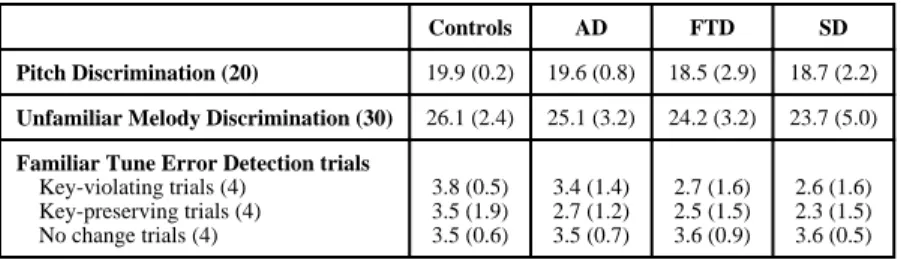
Results on Music Cognition Tasks.
Controls AD FTD SD
Pitch Discrimination (20) 19.9 (0.2) 19.6 (0.8) 18.5 (2.9) 18.7 (2.2) Unfamiliar Melody Discrimination (30) 26.1 (2.4) 25.1 (3.2) 24.2 (3.2) 23.7 (5.0) Familiar Tune Error Detection trials
Key-violating trials (4) Key-preserving trials (4) No change trials (4) 3.8 (0.5) 3.5 (1.9) 3.5 (0.6) 3.4 (1.4) 2.7 (1.2) 3.5 (0.7) 2.7 (1.6) 2.5 (1.5) 3.6 (0.9) 2.6 (1.6) 2.3 (1.5) 3.6 (0.5) AD = Alzheimer disease, FTD = frontotemporal dementia, SD = semantic dementia
NIH-PA Author Manuscript
NIH-PA Author Manuscript
NIH-PA Author Manuscript
Table 4
Correlation between gray matter regions and music cognition tasks.
Unfamiliar melody discrimination
Anatomical region (BA)
SPM space coordinates (x, y, z) T value Z score
right orbito frontal region (11) 25, 59, −19 3.81 3.41
inferior temporal gyrus (20) 29, 6, −50 4.19 3.69
temporal pole (20) 35, 18, −41 3.70 3.33
temporal pole (20) 33, 14, −45 3.63 3.28
left orbito frontal region (11) −25, 62, −17 4.24 3.72 Familiar melody pitch error
detection
right inferior temporal gyrus (20) 36, 1, −50 4.90 4.17
inferior temporal gyrus (36) 26, 8, −42 4.52 3.91
temporal pole (20) 38, 14, −44 4.21 3.71
superior temporal gyrus (22) 56, −28, 7 3.65 3.29
Familiar melody title recall
left inferior temporal gyrus (20) −40, −8, −38 5.75 4.69
temporal pole (38) −33, 18, −28 5.67 4.64
middle temporal gyrus (20) −45, −28, −13 5.33 4.44
hippocampus −25, 2, −29 5.26 4.39
thalamus −3, 17, 7 4.82 4.12
caudate −6, 21, 5 4.62 3.99
right inferior frontal gyrus – pars triangularis (48)
37, 22, 14 4.98 4.22
inferior temporal gyrus (20) 42, −1, −44 4.52 3.92
hippocampus 31. −6, −26 3.47 3.16