Université de Montréal
EVALUATING DNA DAMAGE RESPONSE (DDR)
ACTIVATION IN HUMAN PROSTATE CANCER
By Guila Delouya
Programme de Sciences Biomédicales Faculté de Médecine
Memoir presented to the Faculty of Medicine to obtain the degree of Masters (M.Sc) in Biomedical Sciences, General option.
APRIL 2014
© Guila Delouya, 2014 Université de Montréal
This memoir entitled:
EVALUATING DNA DAMAGE RESPONSE (DDR) ACTIVATION IN HUMAN PROSTATE CANCER
Presented by: Guila Delouya
Was evaluated by a jury composed of the following people:
Dr Elliot Drobetsky Reporting President
Dr Francis Rodier Research Director
Dre Anne-Marie Mes-Masson Research Co-director
Dr Thierry Muanza Jury Member
Summary
Background: Prostate cancer is the most frequently diagnosed cancer in Canadian men and is the third deadliest after lung and colon cancers. Currently, prostate cancer treatments are based on results obtained of digital rectal exam, Gleason scores from biopsy specimens and serum PSA (Prostatic Specific Antigen) levels. The identification of specific biomarkers for diagnosis and prognosis, as well as new therapeutic targets, is quickly paving the way for personalized medicine. Ideally, in the future, patient care will include molecular signature of a patient's disease to guide for a more efficient treatment. In this thesis, we evaluated the DNA damage response (DDR) as a potential biomarker in prostate cancer. DNA lesions in mammalian cells trigger the DDR signalling cascade that orchestrates DNA repair and activate cell cycle checkpoints to preserve genome integrity. Loss of genome stability is usually associated with cancer development, and activated DDR signalling in cells with genomic instability act as a cancer barrier in several pre-neoplastic human lesions, including prostate cancer. Thus, the DDR is an important cancer suppression mechanism. The DDR is also activated in response to anti-cancer agents including radiation therapy (RT) and DNA-damaging chemotherapies. Pre-existing DDR levels in prostate cancer cells may influence the outcome of these cancer treatments. DDR signalling has been detected during human prostate cancer progression from low levels in normal prostate cells to high levels in high-grade prostatic intraepithelial neoplasia (HG-PIN). However, DDR signalling variations detected from HG-PIN to adenocarcinoma remain unclear, and no correlations were performed with
patient clinical outcome data. Our hypothesis is that the levels of persistent DDR signalling activity will be variable with different grades and aggressiveness of prostate cancer. The levels of this activity could be correlated with the clinical responses to treatments and could even predict this process. We believe that having new biomarkers will help personalizing cancer treatment and certainly increase treatments’ efficiency. Our objectives are to characterize the occurrence of DDR activation in prostate carcinoma and to correlate it with patients’ survival and responsiveness to treatment.
Methods: We used tissue microarrays (TMAs) from human radical prostatectomy specimens of 300 men with prostate cancer and estimated the level of DDR protein expression in the stromal and epithelial compartments of normal and aggressive cancer tissues. The expression level of the DDR markers p53 binding protein-1 (53BP1), phosphorylated H2AX (p-H2AX), p65 (p65 subunit of Nuclear Factor (NF-κB) and phosphorylated checkpoint kinase-2 (p-CHK2) was quantified using immunofluorescence (IF) coupled to high-content automated imaging. The quantification of our DDR markers was first validated on an experimental TMA (TMA-cell) including normal and irradiated (to induce DDR signalling) cultured human fibroblasts. The data was quantified using binary layers commonly used to classify pixels in an image so areas could be analysed independently allowing the segregation of specific compartments including nuclei, epithelia and stroma. Arithmetic operations were performed to render values corresponding to DDR activation that were then correlated with clinical outcomes such as biochemical recurrence and occurrence of bone metastasis.
Results: We found that low levels of p65 protein expression in the nuclear epithelial compartments of normal prostate tissue were associated with a reduced probability of biochemical failure (which corresponds to a rise in the serum level of PSA in prostate cancer patients following treatment, surgery in this cohort of patients). Moreover, we also observed that low levels of 53BP1 protein expression in the nuclear epithelial compartments of normal and cancerous prostate tissue were associated with a lower incidence of bone metastasis.
Conclusion: These results confirm that p65 has prognostic value in patients with prostate adenocarcinoma. Based on our results, we suggest that 53BP1 marker may have a prognostic value as well. The validation of other markers and particularly DDR markers may correlate with patients’ outcome. With longer follow-up, it may translate into correlation with survival. Levels of DDR activity in cancer tissue could be used in daily clinic as part of the patient’s diagnostic profile as much as his prostatic specific antigen (PSA) or Gleason score in order to predict response and personalize the treatment in order to guide the patients towards the most appropriate treatment amongst all those available for their prostate cancer.
Résumé
Introduction: Au Canada, le cancer de la prostate est le cancer le plus fréquemment diagnostiqué chez les hommes et le plus mortel après les cancers du poumon et du côlon. Il y a place à optimiser le traitement du cancer de la prostate de manière à mettre en œuvre une médecine personnalisée qui s’adapte aux caractéristiques de la maladie de chaque patient de façon individuelle.
Dans ce mémoire, nous avons évalué la réponse aux dommages de l’ADN (RDA) comme biomarqueur potentiel du cancer de la prostate. Les lésions potentiellement oncogènes de l'ADN déclenche une cascade de signalisation favorisant la réparation de l'ADN et l’activation des points de contrôle du cycle cellulaire pour préserver l’intégrité du génome. La RDA est un mécanisme central de suppression tumorale chez l’homme. La RDA joue un rôle important dans l’arrêt de la prolifération des cellules dont les génomes sont compromis, et donc, prévient la progression du cancer en agissant comme une barrière. Cette réponse cellulaire détermine également comment les cellules normales et cancéreuses réagissent aux agents utilisés pour endommager l'ADN lors du traitement du cancer comme la radiothérapie ou la chimiothérapie, en plus la présence d,un certain niveau de RDA dans les cellules du cancer de la prostate peuvent également influer sur l'issue de ces traitements. L’activation des signaux de la RDA peut agir comme un frein au cancer dans plusieurs lésions pré-néoplasiques de l'homme, y compris le cancer de la prostate. Il a été démontré que la RDA est augmentée dans les cellules de néoplasie intra-épithéliale (PIN) comparativement aux cellules prostatiques normales. Toutefois, le devient de la RDA entre le PIN et l’adénocarcinome est encore mal documenté et aucune corrélation n'a été réalisée avec les données cliniques des patients. Notre hypothèse est que les niveaux d’activation de la RDA seront variables selon les différents grades et
agressivité du cancer de la prostate. Ces niveaux pourront être corrélés et possiblement prédire les réponses cliniques aux traitements des patients et aider à définir une stratégie plus efficace et de nouveaux biomarqueurs pour prédire les résultats du traitement et personnaliser les traitements en conséquence. Nos objectifs sont de caractériser l'activation de la RDA dans le carcinome de la prostate et corréler ses données avec les résultats cliniques.
Méthodes : Nous avons utilisé des micro-étalages de tissus (tissue microarrays-TMAs) de 300 patients ayant subi une prostatectomie radicale pour un cancer de la prostate et déterminé le niveau d’expression de protéines de RDA dans le compartiment stromal et épithélial des tissus normaux et cancéreux. Les niveaux d’expression de 53BP1, p-H2AX, p65 et p-CHK2 ont été quantifiés par immunofluorescence (IF) et par un logiciel automatisé. Ces marqueurs de RDA ont d’abord été validés sur des TMAs-cellule constitués de cellules de fibroblastes normales ou irradiées (pour induire une activation du RDA). Les données ont été quantifiées à l'aide de couches binaires couramment utilisées pour classer les pixels d'une image pour que l’analyse se fasse de manière indépendante permettant la détection de plusieurs régions morphologiques tels que le noyau, l'épithélium et le stroma. Des opérations arithmétiques ont ensuite été réalisées pour obtenir des valeurs correspondant à l'activation de la RDA qui ont ensuite été corrélées à la récidive biochimique et l'apparition de métastases osseuses.
Résultats : De faibles niveaux d'expression de la protéine p65 dans le compartiment nucléaire épithélial du tissu normal de la prostate sont associés à un faible risque de récidive biochimique. Par ailleurs, nous avons aussi observé que de faibles niveaux d'expression de la protéine 53BP1 dans le compartiment nucléaire épithéliale du tissu prostatique normal et cancéreux ont été associés à une plus faible incidence de métastases
osseuses.
Conclusion: Ces résultats confirment que p65 a une valeur pronostique chez les patients présentant un adénocarcinome de la prostate. Ces résultats suggèrent également que le marqueur 53BP1 peut aussi avoir une valeur pronostique chez les patients avec le cancer de la prostate. La validation d'autres marqueurs de RDA pourront également être corrélés aux résultats cliniques. De plus, avec un suivi des patients plus long, il se peut que ces résultats se traduisent par une corrélation avec la survie. Les niveaux d'activité de la RDA pourront éventuellement être utilisés en clinique dans le cadre du profil du patient comme le sont actuellement l’antigène prostatique spécifique (APS) ou le Gleason afin de personnaliser le traitement.
Mots clés: Réponse aux dommages de l’ADN (RDA), Cancer de la prostate, Marqueur
TABLE OF CONTENTS
Summary ………...i Keywords ……...iii Résumé ………...iv Mots-Clés ……….……...vi List of figures ...xList of tables ...xi
List of Acronyms and Abbreviations...xii
Acknowledgements ...xvii
CHAPTER 1: INTRODUCTION
...11.0 Prostate Cancer ...1
1.1 Overview of prostate Cancer ...1
1.2 Cellular effects of radiation therapy...6
2.0 The DNA damage response (DDR) ...8
2.1 DNA damage ...8
2.2 Cellular detection of DNA damage ...11
2.2.1 ATM-MRN ...12
2.2.2 DNA-PKcs-KU ...13
2.3 Signalling to effector pathways...14
2.4 DDR and ionizing radiation ...17
2.5 DNA repair ...19
2.6 Nuclear factor-kappa B (NF-κB) transcription factors ...20
2.7 DDR as a barrier to cancer progression ...26
3.0 Biomarkers ...31
3.1 Prognostic vs. Predictive markers ...31
3.2 DDR as a tissue marker ...32
4.0 The Tissue Microarrays (TMAs) ...34
HYPOTHESIS AND OBJECTIVES
...381. Premiss ...38
2. Hypothesis and objectives ...38
CHAPTER 2: EXPERIMENTAL RESULTS
...41Novel streamlined DNA damage response signalling quantification in human prostate cancer tissue samples reveals a prognostic role 53BP1 ...41
Abstract ...42
1.0 Introduction ...44
2.0 Materials and methods...48
3.0 Results ...54
4.0 Discussion ...59
Figure Legends ...72
Tables Legends ...78
Figures ...80
Tables ...92
CHAPTER 3: DISCUSSION
...961.0 The DDR as a cancer barrier ...96
1.1 DDR activity: a cancer barrier in prostate cancer ...97
2.0 Repair Mechanisms ...98
2.1 53BP1 and homologous recombination ...100
3.0 Why do we need biomarkers? ...101
3.1 DDR activity as a biomarker ...102
4.0 DDR activity in our cohort of patients ...104
5.0 Limitations of our study ...106
5.1 Validation study on TMA-cell and TMA-tissue ...106
5.2 Data quality control ...106
5.3 Immunofluorescence technique ...107
Conclusion ...109
Perspectives ...110
LIST OF FIGURES
CHAPTER 1:
Figure 1: Gleason grading system diagram ...6
Figure 2: Major DDR Activation pathways ...16
Figure 3: A model depicting distinct and common steps between NF-κB signaling induced by TNFα and DNA damaging agents...25
Figure 4: ATM activation in normal prostatic gland, PIN, and carcinoma ...27
Figure 5: Tissu Micro Arrays (TMAs) ...36
Figure 6: Hypothesis ...39
CHAPTER 2:
Figure 1: Detection of DDR activity and DDR foci in paraffin embedded cells using multicolour immunofluorescence ...80Figure 2: Multicolor immunofluorescence and mask segmentation analysis in prostate cancer TMA core ...81
Figure 3: Software-based quantification of immunofluorescence-detected DDR activity in paraffin embedded cells ...82
Figure 4: Quality Control of DDR data on duplicate samples for prostate cancer TMA...83
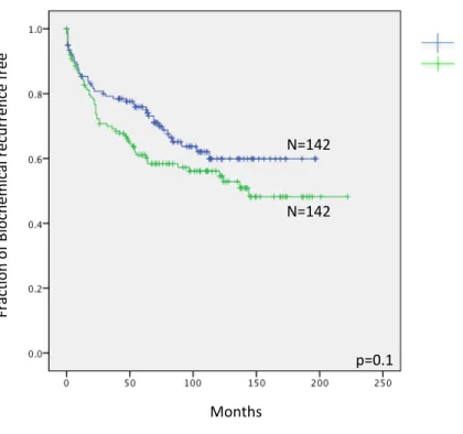
Figure 5: Kaplan–Meier PSA recurrence-free survival and bone metastasis free curves in patients with prostate cancer for specific DDR signals ……....86
Figure S1: A-Schematic representation of the TMA-cell approach for irradiated cells ...89 B-Map of the TMA-cell and representative full-core
immunofluorescence images ...89 C- Software mediated entire core detection and nucleus identification...89 Figure S2: Multicolor immunofluorescence staining in whole prostate cancer TMA …..90
Figure S3: Software quantification of immunofluorescence-detected DDR activity in paraffin embedded cells ...91
LIST OF TABLES
CHAPTER 1:
Table 1: D’Amico risk stratification for clinically localized prostate cancer ...3
CHAPTER 2:
Table 1: Terry Fox Research Institute TMA (TMA-TFRI) of prostate cancer patient cohort ...92 Table 2: Univariate and multivariate cox regression analysis for
A) Biochemical failure and B) Bone metastasis ...93 Table 3: Pearson Correlation (2-tailed) between DDR activity and clinico-
pathological parameters ...94
LIST OF ACRONYMS AND ABBREVIATIONS
53BP1 p53 binding protein-1ADT Androgen deprivation therapy ATM Ataxia Telangiectasia Mutated ATR
ATRIP
ATM and Rad 3-related ATR interacting protein Bax Bcl-2-associated x protein Bcl-2 B-cell lymphoma 2
BCR Biochemical recurrence
BMI-1 Polycomb complex protein B lymphoma Mo-MLV insertion region 1 homolog BPH Benign prostatic hyperplasia
BRCA1 Breast cancer 1
BRCT Breast cancer 1 (BRCA1) carboxy-terminal CAG Cytosine, adenine, and guanine
CAK CDK activating kinase CDC25 Cell division cycle 25 CDK1 Cyclin-dependent kinase 1 Cox-2 Cyclooxygenase-2
CRPC Castrate resistant prostate cancer Cy5 Rhodamine cyanine dyes
DAPI 4',6-diamidino-2-phenylindole
DDR DNA damage response
DNA Deoxyribonucleic acid DRE Digital rectal exam DSBs Double strand breaks EBRT External beam radiation ECE Extra-capsular extension
FFPE Formalin fixed paraffin embedded FISH Fluorescence hybridization
FIT-‐C Fluorescein isothiocyanate
GS Gleason score
GUROC Genito-Urinary Radiation Oncologists of Canada
Gy Gray
H/E Hematoxylin/eosin
HDR High dose rate
HG-PIN High-grade prostatic intraepithelial neoplasia HMCK High molecular cytokeratin
HR Homologous recombination
IF Immunofluorescence
IHC Immunohistochemistry
LDR Low dose rate
LNI Lymph node invasion
MDC1 Mediator of DNA damage checkpoint 1 MDM2 Mouse double minute 2 homolog
MFI Mean fluorescent intensity
MRN Mre11-Rad50-Nbs1
NCCN National Comprehensive Cancer Network NHEJ Non-homologous end-joining
p-CHK2 Phosphorylated checkpoint kinase-2 p-H2AX Phosphorylated H2AX
p65 p65 subunit of Nuclear Factor kappa B (NF-κB) PARPs Poly ADP ribose polymerase
PCa Prostate cancer
PIKK Phosphoinositide 3-kinase related kinase PIN Prostatic intraepithelial neoplasia
PKA Protein kinase A RI-alpha PML Promyelocytic leukemia protein ProCaRS Prostate Cancer Risk Stratification PSA Prostate specific antigen
pTNM Pathological TNM staging (Tumor Nodes Metastasis)
RNF2 Ring finger protein 2 ROC
ROS
Receiver operative characteristic Reactive oxygen species
RP Radical prostatectomy
RT Radiation therapy
RTOG Radiation Therapy Oncology Group S/T-Q
SSB
Ser/Thr-Gln
Single strand break
Stat-3 Signal transducer and activator of transcription 3 SVI Seminal vesicles invasion
TFRI Terry Fox Research Institute TMAs Tissue microarrays
TRIT-C UV
Tetramethyl rhodamine isothiocyanate Ultraviolet
To my husband Yohann To my 4 beautiful sons: Noah, Nathan, David, Liam
To my mother Ida To my father watching me over the sky Thank you for your support over the long years, I know I can always count on you
Acknowledgements
My interest in fundamental research began in 2008 during my second year of residency in radiation oncology when I was first introduced to Dre Anne-Marie Mes-Masson, now my co-director. It is through her welcome in her laboratory during an entire summer where I was paired to Véronique Barrès, my dear friend and now co-worker that I had the desire to continue in fundamental research and do my Master's degree in this big family of our laboratory. I was later introduced to Dr Francis Rodier, my director who had a particular interest in radiobiology and accepted to supervise me during those past three years. The confidence you have always given me has allowed me to evolve throughout my exciting project and grown out of this experience. You are always available and a pleasure to talk to. I am looking forward to our future collaborations.
Thank you to all the wonderful members of both laboratories, without which my experience would not have been so pleasant. Thank you all for your support, your smiles, your help and for all the discussions that have helped me to advance my project. I sincerely thank you all very much. In particular, I thank Véronique Barrès, Guillaume Chouinard and Véronique Ouellet for their valuable advice, their precious time and for answering all of my emails 24/7. Thank you to Guillaume Cardin who helped me find my way in the lab during the first months and guiding me through my first experiments. Thank you to Ingrid Labouba for her insight and experience on immunofluorescence. Thank you to Audrey Carrier-Leclerc, with whom I attended courses and conferences and whose great heart is always open to discussions and for giving piano lessons to my children after our day at the
lab. I would also like to thank Nawel Mechtouf, Amel Derdour, Stéphanie Nadeau, Shuofei Cheng, Sabrina Ghadaouia, Nicolas Malaquin, Aurélie Martinez, Llilians Calvo Gonzalez, Katia Caceres and all those that have passed through our lab throughout the years for their support, in all its forms. In particular, I wish to emphasize the friendship of Véronique, which was, is and always will be very precious. Our discussions helped me over the years. Thank you to professor A. Zini for reviewing this thesis.
I cannot help say a big thank you to all the other members of the ICM, the CR-CHUM and mostly to my great and special department of radiation-oncology that made my project possible. My colleagues have allowed me to spend many hours, days, weeks in the laboratory while they would see patients and particularly my patients when I was not around. I cannot thank you enough.
Finally, I wish to conclude this section by thanking with all my heart the people who have been supporting me throughout my 20 years at l’Université de Montréal. My husband, my children, my mother, my brothers and sisters, my amazing cousins, my in laws, my friends who were always around especially when I wasn’t. You always pushed me to challenge myself, and encourage me to pursue my dreams. You have always believed in me and knew how to be there to encourage me. You were all always there to support me in difficult times and to congratulate me in happier ones.
CHAPTER 1: INTRODUCTION
1.0 Prostate Cancer
1.1 Overview of prostate cancer
Prostate cancer is the most frequently diagnosed cancer in Canadian men and is the third deadliest cancer in males after lung and colon cancer 1.
According to the Canadian Cancer Society, Canadian males are most likely to develop prostate cancer, with one in eight males expected to be diagnosed with prostate cancer in their lifetime (statistics Canada website: http://www.cancer.ca/en/cancer-information/cancer-101/canadian-cancer-statistics-publication/?region=qc).
More than 23 000 new cases of prostate cancer were diagnosed in Canada in 2013 with almost 5000 of these being in the province of Quebec. High-grade prostatic intraepithelial neoplasia (HG-PIN) is considered the precursor of prostate carcinoma. It is associated with progressive abnormalities of phenotype and genotype, which are midway between normal epithelial cells and cancer cells. This indicates impairment of cell differentiation and regulatory control with advancing stages of prostatic carcinogenesis 2.
Management of localized non-metastatic prostate cancer remains very complex because there are multiple issues to consider including risk stratification, efficacy and
toxicity of the different treatments, relative risk of death from diseases other than the cancer itself and lastly, patient preferences 3. The National Comprehensive Cancer Network (NCCN), an alliance of the world's leading cancer centers, is an authoritative
source of comprehensive cancer, which develops guidelines to improve the quality,
effectiveness and efficiency of cancer care. Both the National Comprehensive Cancer Network (NCCN) risk classification and the D’Amico classification, which are very similar, are the most commonly used categorical strategies for pre-treatment risk estimation in prostate cancer. Originally developed in 1998 by a medical researcher named D’amico, this classification system is designed to evaluate the risk of recurrence following localized treatment of prostate cancer. It stratifies patients in three categories based on three parameters:
a) Patient’s diagnostic serum prostatic specific antigen (PSA): The PSA is a protein made by the cells of the prostate gland. It is mostly found in semen, but it is also normal to find small amounts of PSA in the blood of healthy men. In case of prostate cancer, PSA levels are often above normal as it is secreted in excess by the prostate cancer cells.
b) Patient’s highest biopsy Gleason score: The Gleason score is given to prostate cancer based upon its microscopic appearance. Lower grades are associated with small, closely packed glands. Cells spread out and lose glandular architecture as grade increases. Gleason score usually varies between 6-10 with 10 being the highest
possible grade.
c) Patient’s clinical stage: The clinical stage varies from T1 to T4 and is determined by the physician’s digital rectal exam of the prostate gland. Indeed, the prostate can be palpated through the rectum. T1 represents the absence of a palpable tumor whereas T4 represents a tumor invading the bladder or rectum.
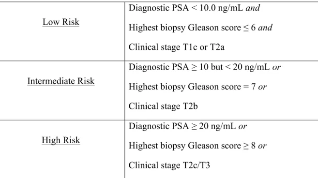
Low Risk
Diagnostic PSA < 10.0 ng/mL and Highest biopsy Gleason score ≤ 6 and Clinical stage T1c or T2a
Intermediate Risk
Diagnostic PSA ≥ 10 but < 20 ng/mL or Highest biopsy Gleason score = 7 or Clinical stage T2b
High Risk
Diagnostic PSA ≥ 20 ng/mL or Highest biopsy Gleason score ≥ 8 or Clinical stage T2c/T3
Table 1: D’Amico risk stratification for clinically localized prostate cancer
This risk stratification is based on clinical characteristics such as the diagnostic serum PSA, the Gleason score as determined by the pathologist from prostate biopsy and the clinical stage as determined by the physician’s digital rectal exam.
Nomograms are online tools that have been developed to guide and help clinicians decide which treatment approach (ie: surgery, radiation therapy (RT) or androgen deprivation therapy - ADT) can be offered to a patient. The nomogram analysis is based on multiple factors such as PSA values and number of positive biopsy cores.
Nomograms are often complex, time-consuming (making routine use difficult), and are not necessarily applicable to individual treating centers 4. An important advantage of risk grouping systems (ie: D’Amico or NCCN classifications) compared to nomograms is their simplicity 5. On the other hand, risk groups have the disadvantage of assuming that patients within the same group are homogeneous. Moreover, risk groups do not consider factors such as the number of positive biopsies or whether there are single or multiple intermediate-risk or high-risk factors present when considering the treatment decisions. Nomograms do incorporate this information, but more time is required to calculate the score and this information may not be applicable for use in daily clinical practice. Regardless of the scoring system, which is used by the clinician in his/her practice, neither the risk classification nor the nomograms incorporate the tumor’s biology. The most important criterion for a prediction tool is its ability to discriminate between patients with or without a given outcome with high accuracy 6. Predictive models for patients treated by RT for localized prostate cancer have been established 7. Recently, the Genito-Urinary Radiation Oncologists of Canada (GUROC) developed a pan-Canadian Prostate Cancer Risk Stratification (ProCaRS) database based on 7974
categories (i.e., very low risk and very high risk) may further improve patient risk categorization8. A recent trial (PIVOT trial) where patients with clinically localized prostate cancer were randomized between radical prostatectomy and active surveillance (ie: no treatment) showed that active surveillance remains an excellent option for 70% of patients diagnosed with low risk prostate cancer9. However, 90% of diagnosed patients still favour treatment because of the anxiety associated with an untreated cancer. Prostate cancer treatments include surgery to remove the prostate gland (radical prostatectomy (RP)), external beam radiation (EBRT), interstitial brachytherapy (low dose rate - LDR and high dose rate - HDR), ADT or a combination of these therapies. Each treatment comes with its own set of side effects, which are not negligible. Nevertheless, to date, clinicians do not have the necessary tools (markers) to identify men with slow growing cancers (that can be managed by active surveillance) from men with aggressive cancers (that require treatment). As such, until we establish better predictive models, clinicians will continue to rely on basic risk classification systems that typically incorporate information on DRE (digital rectal exam), serum PSA and pathology from biopsy specimens (Gleason score-GS).
1.2 Cellular effects of radiation therapy
Radiation therapy (RT) is administered by one of several methods. It can be given via an external source using a linear accelerator, which is directed toward the tumor (almost always by intensity modulated radiation therapy (IMRT) in order to protect healthy tissues). Alternatively, RT can be given by an internal source (e.g. brachytherapy where radioactive sources such as Iodine 125 are placed and decay
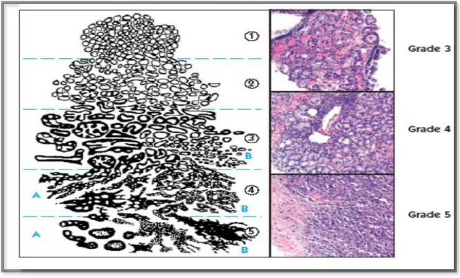
Figure 1: Gleason grading system diagram
This figure illustrates a cartoon (left panel) as well as histological (right panel) representation of cells according to the Gleason grade (1-5). The number is assigned to two of the areas of the prostate that have the most cancer on the biopsy cores taken. Those two numbers are added together to come up with the Gleason score, which ranges from 2 to 10.
within the tumor). Radiation interacts with matter by the photoelectric effect, the coherent scattering effect, the pair production effect, the photodisintegration or the Compton effect. The latter is the most relevant for the range of energies used in clinical RT. In the Compton effect, the observed biologic effect results from photons creating multiple ionizations by ejection of electrons from the target biomolecule 10. In this regard, the extent of biologic effects in cells after exposure to ionized radiation is largely due to oxygen with the subsequent production of free radicals. These free radicals can break cellular deoxyribonucleic acid (DNA), which is the critical target for the biologic effects of ionizing radiation. The extent of DNA damage will eventually determine cell death 11; 12. Cells that cannot effectively repair their DNA damage are therefore more sensitive to ionizing radiation 13. It is also known that when radiation is focused on the nucleus rather than on the cytoplasm, cell death occurs at a higher rate 14. Interference to DNA can occur directly or indirectly, but mostly via indirect means in that photons of radiation being more likely to ionize the molecules surrounding the DNA creating free radicals, which subsequently destabilize nucleic acids 15; 16. Cells that are unable to generate free radicals (cells under hypoxic conditions) are much less sensitive to ionizing radiation compared to those that are well oxygenated. This explains why the effect of radiation is related with blood flow and oxygen concentration of the target tissue 17.
2.0 The DNA damage response (DDR)
2.1 DNA damage
In human cells, both normal metabolic activities and environmental factors such as ultraviolet (UV) light and radiation can cause DNA damage, resulting in as many as 1 million individual molecular lesions per cell per day. Many of these lesions cause structural damage to the DNA molecule and can alter or eliminate the cell's ability to transcribe the gene that the affected DNA encodes. Other lesions induce potentially harmful mutations in the cell's genome, which affect the survival of its daughter cells after it undergoes mitosis. Therefore, if these lesions are not or are incorrectly repaired, they lead to mutations or aberrations that threaten viability. DNA damage leading to either single strand break (SSB), impair base pairing and/or DNA replication or transcription errors may be produced by 18:
a) Physiological processes (ex: DNA mismatches introduced during DNA replication)
b) Hydrolytic reactions and non-enzymatic methylations c) Abortive topoisomerase I and II activity
d) Reactive-oxygen compounds (ex: arising either from oxidative respiration or produced by macrophages and neutrophils at sites of infections and inflammation).
Double-strand breaks (DSB), although not as frequent as SSB, impair base pairing and DNA replication or transcription errors, are difficult to repair and are very lethal. DSBs can arise from a) two single strand breaks that are in close proximity b) ionizing radiation or c) from treatment using chemical drugs such as certain chemotherapy agents. Because of the importance of DNA, cells have developed a complex series of processes and pathways to ensure that the DNA remains undamaged and unaltered despite the continuous attack from the inside (ex: oxidation and alkylation owing to metabolism) as well as from the outside (ex: ingested chemicals, ultraviolet (UV) and ionizing radiation) 19. These include multiple types of DNA repair aimed at repairing various types of DNA damage caused by a variety of agents.
Specialized repair systems have therefore evolved for detecting and repairing damage: a) to bases: The Base Excision Repair (BER), where most of the damaged bases in the DNA will be detected and removed by specialized proteins called glycosylases. They cut out the damaged base without cutting the DNA backbone, resulting in an abasic site. Another class of enzyme (endonucleases) will recognize this and will cut the DNA backbone leaving a SSB. The resulting SSB can then be processed by either short-patch (where a single nucleotide is replaced) or long-short-patch BER (where 2-10 new nucleotides are synthesized) 20. Ligases will then seal the break.
b) To single-strand breaks: The Single strand break repair (SSBR) which is similar to BER but since radiation itself causes the break rather than being a repair intermediate,
the ends are not recognized by ligases. There is therefore an extra end-processing step, mainly by the enzyme polynucleotide kinase (PNK). Once a clean nick is produced, short or long patch repair can then follow as for BER.
c) To double-strand breaks (Homologous recombination (HR) and Non-homologous end joining (NHEJ)). These two repair mechanisms are quite different in the genes involved, the position in the cell cycle where they primarily act and in the speed and accuracy of repair. These processes are described in more detail below.
All these lesions are produced by ionizing radiation. There are also other DNA repair pathways, such as those for correcting mismatches of bases in DNA which can occur during replication, such as mismatch repair (MMR), and for repairing bulky lesions or DNA adducts such as those formed by UV light and some drugs such as the chemotherapeutic agent cisplatin (NER-Nucleotide Excision Repair).
The DNA damage response (DDR) is a highly complex and coordinated system that determines the cellular outcome of DNA damage. This system can be divided in two parts, the sensors of DNA damage and the effectors of the damage response. The sensors consist of a group of proteins that actively examine the genome for the presence of the damage. These proteins then signal this damage to three main effector pathways that together determine the outcome for the cell. These effector pathways include (1) programmed cell death pathways that destroy damaged cells, (2) DNA repair pathways that physically repair DNA breaks and (3) pathways that cause
temporary or permanent halts in the progress of cells through the cell cycle--the damage checkpoints.
Failure of the DDR or associated events causes genomic instability, an underlying cause of several human syndromes and diseases, particularly cancer.
2.2 Cellular detection of DNA damage
Shortly after exposure to ionizing radiation, a signal is transmitted to the regulators of the cell cycle machinery and the sensors of DNA damage. Cells with damaged DNA mostly undergo G1 or G2/M cell cycle arrest. During this cell cycle arrest, normal cells can either 1) repair and continue through the cell cycle, 2) not repair and stay arrested, or 3) not repair and undergo apoptosis 21. Cells have developed mechanisms that sense the presence of the DSBs and initiate the DDR. The DDR is essential to stop the proliferation of cells with genomic instability, and therefore, prevent events that can contribute to cancer initiation and progression.
The first cellular response to DSBs is characterized by the recruitment of many different proteins to the sites of DNA damage. This clustering (known as foci) can be observed microscopically as small regions or dots in the nucleus following staining with antibodies to these proteins (See Chapter 2, figure 1). One of the earliest events to occur in the DDR (occurring within 5-30 minutes after induction of a DSB) is the phosphorylation of the protein called histone H2AX 22. This phosphorylated form
(known as γ-H2AX or p-H2AX) is required for the recruitment of many of the other proteins involved in the DDR. The early phosphorylation of H2AX indicates that one or more kinases are activated at the sites of DSBs. Three related kinases have been shown to be able to phosphorylate H2AX at sites of DSBs 23:
(1) ATM-MRN (2) DNA-PKcs-KU (3) ATR-ATRIP
2.2.1 ATM-MRN
The phosphorylation of H2AX occurs primarily by the protein ATM (Ataxia Telangiectasia Mutated). ATM is normally present in the cell, but in an inactive form. Activation of ATM occurs once it becomes associated with a DSB resulting in phosphorylation of H2AX at the site of the DSB. However, in order to detect and locate the DSB and be activated, ATM requires at least one additional protein complex. This complex is known as MRN. The MRN complex consists of the proteins Mre11, Rad50, and Nbs1 24, 25, 26 . This MRN complex is able to recognize damaged DNA and bind to or near the site and transmit DDR signals downstream to the transducers, which is important for the ‘’processing’’ of the DSB. These transducers are members of the phosphoinositide 3-kinase related kinase (PIKK) family such as ATM and ATR (ATM and Rad 3-related) 24, respectively. Moreover, one of the key functions of Nbs1 is to
directly bind to ATM, and bring it to the site of damage and Rad50 directly binds to DNA.
2.2.2 DNA-PKcs-KU
DNA-dependent protein kinase (DNA-PKcs) is a kinase that is structurally related to ATM and which is very important in the NHEJ DNA repair. The mechanism through which it finds DSBs is very similar to that of ATM. Like ATM, DNA-PKcs is unable to act as a sensor of damage itself. This sensor function is carried out by the Ku70-Ku80 complex, which directly binds to the ends of DSBs. This binding then recruits DNA-PKcs allowing phosphorylation of H2AX.
2.2.3 ATR-ATRIP
AT-related kinase (ATR) does not a prominent role in the initial recognition of the DSBs but is important for the types of damage that occur during normal DNA replication. ATR phosphorylates H2AX in response to other types of DNA damage and abnormalities such as stalled or broken replication forks and single-stranded DNA. Just like ATM and DNA-PKcs, ATR is recruited to sites of damage by ATRIP (ATR interacting protein) that acts as the sensor of the damage. As described above, the ATM-MRN complex leads to processing of the DNA at sites of DSBs. This can create
stretches of single-stranded DNA, which will then activate ATR. Thus, ATR can be activated downstream of ATM which then phosphorylates a distinct set of proteins that participate in the DDR.
ATM and ATR are key components of these initial sensors of DNA damage 27; 28. While ATR is only activated when DNA is being replicated in S phase, ATM can be activated throughout the cell cycle.
Finally, although ATM and ATR have some overlapping activities, they are activated by separate signals and by different types of DNA damage 29; 30.
2.3 Signalling to effector pathways
Activation of ATM, DNA-PKcs and ATR leads to the phosphorylation not only of H2AX, but also of many other cellular proteins. It has been shown that as many as 700 proteins are substrates for the ATM and ATR kinases in response to DNA damage 31. They relay the signal to various downstream effectors that mediate cell cycle arrest, DNA repair, and apoptosis.
ATM substrates include H2AX and 53BP1, which facilitates checkpoint activation and repair, both essential for their efficient repair of DNA damage. Phosphorylated H2AX and 53BP1 rapidly localize to DSBs, forming characteristic foci. ATM also phosphorylates the kinase CHK2 (p-CHK2), which promotes growth arrest and p53, a
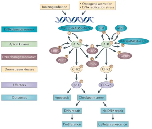
tumor suppressor and transcriptional regulator that coordinates repair and cell cycle arrest 32. This DDR leads to transient cell cycle arrest and DNA repair, cell death, or permanent cell cycle arrest (cellular senescence) preventing cells to replicate with accumulated mutations thereby protecting cellular integrity and avoiding the development of cancers 33; 34. Figure 2 35 illustrates in a cartoon fashion the major DDR activation pathways. DNA damage sensors (MRN and ATRIP complexes) detect DNA damage and then recruit ATM and ATR, respectively. These, in turn, phosphorylate (P) the histone variant H2AX on Serine 139 (γ-H2AX) in the region proximal to the DNA lesion.
Figure 2: Major DDR Activation pathways
MRN and ATRIP complexes detect DNA damage, which then recruit ATM and ATR, respectively. These in turn phosphorylate (P) the histone variant H2AX on Serine 139 (γ-H2AX) in the region proximal to the DNA lesion. γ-H2AX is required to recruit mediator of DNA damage checkpoint 1 (MDC1) that further maintains and amplifies DDR signalling by enforcing additional accumulation of the MRN complex and activation of ATM.
Sulli G, Di Micco R, d’Adda di Fagagna F. Crosstalk between chromatin state and DNA damage response in cellular senescence and cancer. Nature reviews. Cancer. Oct 2012;12(10):709-720. Written authorization obtained by Dr Fabrizion d’Adda di Fagagna, Principal Investigator IFOM Foundation – The FIRC Institute of Molecular Oncology
As mentioned previously, 53BP1 is also involved in sustaining DDR signalling by enhancing ATM activation. DDR signalling relies on additional mechanisms that are based on ubiquitination (a post-translational modification where ubiquitin is attached to a substrate protein). This post-translational modification affects proteins in many ways. It can be a signal for (1) protein degradation (via the proteasome), (2) change in cellular location, (3) change in protein activity and (4) change in protein-protein interactions. In DDR, γ-H2AX is ubiquitylated by ring finger protein 2 (RNF2), which causes the recruitment of Polycomb complex protein BMI-1 (B lymphoma Mo-MLV insertion region 1 homolog) to sites of DNA lesions. BMI1 is an oncogene which blocks transcriptional elongation at the DNA damage site and promotes DNA repair 35; 36.
Eventually, DDR signalling spreads away from the region of the damaged DNA and triggers the diffusible kinases CHK2 (which is mainly phosphorylated by ATM) and CHK1 (which is mainly phosphorylated by ATR) with signalling converging on downstream effectors such as p53 and the cell division cycle 25 (CDC25) phosphatases.
2.4 DDR and ionizing radiation
In order for a cell to synthesize its DNA and go through division, it needs to pass by the multiple cell cycle checkpoints. Blocks at these checkpoints can prevent important cell cycle transitions to ensure the integrity of the DNA 37; 38; 39; 40. All movement
through the cell cycle, be it in the G1, S, G2 or M phases, is driven by cyclin-dependent kinases (CDKs). The CDKs phosphorylate other proteins to initiate the processes required for progression through the cell cycle.
Depending on when the cell is exposed to radiation in its cell cycle, it may respond differently. Cells are extremely sensitive to radiation during mitosis (during which there is little DNA repair) 41; 42; 43; 44. DNA damage can activate multiple pathways that eventually lead to G1 arrest. When ATM is activated, it stabilizes p53 by phosphorylating its serine-15 and also adds a phosphate group on serine 395 of mouse double minute 2 homolog (MDM2). Phosphorylation of MDM2 prevents p53-MDM2 nuclear export and degradation of p53 45. As mentioned in the previous section, ATM is also known to phosphorylate Chk2, which subsequently phosphorylates p53 on serine 20 46. This further prevents interaction of p53 and MDM2 and hence increases levels of available nuclear p53, which is free to transcriptionally activate p21, a major inhibitor of the cyclin E- Cyclin-dependent kinase 2 (CDK2) complex 47.
ATM also controls a p53/p21 independent G1 arrest pathway. When ATM activates Chk2, it phosphorylates cdc25A, which is primed for ubiquination and subsequent degradation 48. Cdc25A is a phosphatase that removes inhibitory phosphates from CDK2 and CDK4, both of which are important G1 phase progression molecules 24. Cells have been shown to be radioresistant during the G1 phase, but their radiosensitivity increases at the end of this phase 49 . Most commonly, irradiated cells can be blocked in the G2/M phase, which is the next most sensitive phase in the cell
cycle post replication. Although the G2/M checkpoint remains complex and is not fully understood, there are multiple known pathways involved in this arrest 24. The final step in this pathway is deactivation of the cyclin B- Cyclin-dependent kinase 1 (CDK1) complex, which orchestrates the G2/M transition 50. The specific site of phosphorylation determines activation or deactivation of the CDK1 complex. The ATM/CDC25A pathway is also important because cdc25A is an activator of the cyclin B-CDK1 complex. ATM activates p21 (through p53), which is an inhibitor of an activator of CDK1, namely CAK (CDK activating kinase) 51.
2.5 DNA repair
As seen above, once cells are irradiated, they sense the DNA damage and eventually activate the mechanism for DNA repair. Various repair processes are activated according to the lesion types, with DSBs being the most lethal lesion to the cell compared to single-strand breaks. These lesions can be repaired either through homologous recombination (HR) or non-homologous end-joining (NHEJ) 52. In the former, either the intact chromosome or the sister chromatid serve as a template to reconstruct the missing DNA. HR is most effective in late S or G2 phase, when the sister chromatids have replicated but are still attached 53. NHEJ is more important in G1 and early S phase, but can essentially occur throughout the cell cycle 52. Once cells are irradiated, ATM phosphorylates histone H2AX resulting in quick localized
accumulation of the protein 53BP1. This protein is involved in enhancing phosphorylation of the tumor suppressor molecule p53, activating proteins essential for DNA repair, and inducing G2 checkpoint block 54; 55; 56. Thus, G2 checkpoint induced by radiation, possibly via 53BP1, provide more time for repair and increases the probability of escaping cell death.
2.6 Nuclear factor-kappa B (NF-
κ
B) transcription factorsOur group has already addressed the prognostic value of the p65 subunit of NF-κB in prostate cancer (p65) where it was observed that elevated amounts of nuclear p65 in tumors is associated with more aggressive prostate cancer 57; 58; 59.
The NF-κB family is composed of five transcription factors characterized by their Rel-homology domain responsible for DNA binding, dimerization and interaction with inhibitor of κB (IkB) proteins. The family is subdivided into two groups, members of the first group named RelA (p65), RelB, and c-Rel, carry a transactivation domain responsible for NF-κB potent activity as a transcription factor. The second group contain NFκB1 (p50 and its precursor p105) and NFκB2 (p52 and its precursor p100). The carboxy-terminal domain of p105 and p100 contains ankyrin repeats that must be degraded to create transcriptionally active p50 and p52 proteins respectively. All these transcription factors function as homo and heterodimers, the dimer most known and studied is composed of subunits p50 and RelA (p65). In most normal cells, the dimer
p50-p65 is kept inactive in the cytoplasm by association with IκB family of inhibitors (α and β) 60;61.
Research from the past 20 years has revealed that there are three major NF-κB pathways can be distinguished 62;63;64. The first is the classical or canonical pathway in which p65/p50, the main active dimer, is rendered inactive by IκB inhibitors (IκBα and IκBβ). This pathway may be activated by signals such as the proinflammatory cytokine tumor necrosis factor α (TNFα). The activation of NF-κB also often requires the activation of the IKB-Kinase complex (IKKα, IKKβ and IKKγ) via multiple signals including (but not limited to) cytokine binding to cell surface receptors, DNA damage, hypoxic conditions, and oxidative stress 65. Canonical NF-κB signaling is induced rapidly within minutes of stimulation without the need for de novo protein synthesis. The alternative or non-canonical pathway is activated by a smaller number of inducers, such as lymphotoxin β and B cell activating factor, and plays an important role in B-cell maturation and the formation of secondary lymphoid tissues 66;67. This pathway uses RelB/p52 as the active dimer.
In the canonical pathway (IKKβ-dependent), the IKK complex phosphorylates IKBs that are then ubiquitinated and degraded by proteasomes. In the non-canonical pathway (IKKα-dependent), the IKK complex regulates the processing of the p100 precursor. Subsequently, the NF-κB complex translocates to the nucleus and activates the expression of specific target genes 68;69. Although p65/p50 represents the main functional unit of the classical NF-κB pathway, p65 also forms transcriptionally active
dimers with p52. In the same way, RelB/p52 constitutes the main alternative functional unit, however, RelB can also dimerize with p50 resulting in another alternative NF-κB functional unit 70.
Finally, and particularly relevant to our study, the third NF-κB pathway, the so-called ‘atypical pathways’, refer to those pathways that do not fall in the abovementioned categories. Originally, all signaling cascades activated by atypical stimuli, such as DNA damage or oxygen stress, were classified as ‘atypical’ NF-κB activators, as they all induce a slow and weak NF-κB signal (with peak activities reached after 2–4 h). Later studies revealed that these stimuli could not be categorized in one class as they induced completely unrelated pathways. Thus, ultraviolet (UV)-induced NF-κB signaling appears to occur in an IKK-independent way, while most other genotoxic stress agents activate a pathway that follows more or less the classical NF-κB activation scheme. For example, ionizing radiation (IR)-induced NF-κB activation has been reported following both low and high doses of irradiation71;72. This NF-
κ
B activation results in the induction of anti-apoptotic genes, inhibiting apoptosis induced by many chemotherapeutic drugs and irradiation 73;74;75;76. Similarly, NF-κ
B activation impact numerous other molecular and biological functions that could be relevant for responses to chemo-radiation such as inflammation 77;78, angiogenesis 79, survival, migration, and invasion 80 are associated with the activity of NF-κ
B nuclear factors.ATM and NF-
κ
BGiven the nature of the biological processes modulated by NF-
κ
B, it is not surprising to see an overactivation of NF-κ
B in many cancers 81. Regarding solid tumors, aber-cant nuclear localization of subunits of NF-κ
B has been observed in various cancers, including pancreatic 82, breast 83, endometrium 84, renal 85 and melanoma 86. Moreover, a large number of cell lines exhibiting high activity of the dimer p50-p65 are resistant to various chemotherapeutic agents 76; 87.Furthermore, genotoxic agents used in cancer treatment (ex: IR, chemotherapy such as topoisomerase inhibitors, replication stress inducers such as hydroxyurea or DNA photolesions induced by UV-C) can activate p50/RelA NF-κB complexes through the atypical pathway or through activation of CK2 (formally known as casein kinase II). CK2 is a highly conserved and ubiquitous serine/threonine kinase that may participate in the transduction of survival signals 88;89 and CK2-mediated I
κBα phosphorylation has an important UV-protective function 90. Recently, several studies have shown that
ATM is essential for NF-κB activation following DNA damage 91;92;93;94;95.
The ATM-NEMO (Nf-κB essential modulator) pathway activates p50/RELA Nf-κB complexes via the induction of IKKβ following DNA damage 96;95. Following cellular stresses, the nuclear translocation of the RIP1/NEMO death domain complex occurs. This nuclear NEMO is then post-transcriptionally modified by the small ubiquitin-like
modifier (SUMO)1, and phosphorylated by ATM, which is simultaneously activated in case of DNA damage. Ubiquitination of the ATM–NEMO complex targets these proteins for nuclear export, enabling the complex to interact with and activate the IKK-β subunit and initiate IkB-α phosphorylation 97;95;91. This is substantiated by the discovery that the ATM inhibitor KU55933 block both constitutive and DNA damage-activated NF-κB in breast cancer cell lines with mutant p53 and the downstream inhibition of NF-κB activation is a major mechanism accounting for the radio-sensitizing effect of this ATM inhibitor98.
Unlike NF-κB signaling induced by cell surface receptors where the signal initiation event can be precisely defined, the necessary and sufficient molecular signal initiation events induced by various genotoxic agents are still being addressed. Highlighting the complexity of this issue, ultraviolet (UV) irradiation-induced NF-κB activation led to the conceptualization of nuclear-to-cytoplasmic signalling99. Accumulated evidence over the last two decades demonstrates that nuclear DNA damage is probably not the signal initiation event for immediate activation of NF-κB in this case 100;101;102. Importantly, several lines of evidence support the requirement of nuclear DSB in initiating the canonical IKK-NF-κB signaling pathway in response to many different genotoxic agents 96;103.
NF-κB activation by DNA damage 122
npg
Cell Research | Vol 21 No 1 | January 2011
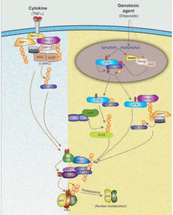
Figure 2 A model depicting distinct and common steps between NF-κB signaling induced by TNFα and DNA damaging
agents. TNFα stimulation of TNFR1 engages receptor adaptor proteins (TRADD and RIP1) and results in the recruitment of ubiquitin conjugating enzymes (Ubc13/Uev1A and UbcH5) and E3 ligases (cIAP1/2, TRAF2/5 and HOIL/HOIP) to promote K63-linked, mixed and linear polyubiquitination of multiple target proteins. These polyubiquitin chains form the scaffold on which TAK1/TAB2/3 and IKK/NEMO complexes are formed and TAK1-dependent activation of IKKβ is induced. DNA damaging agents, such as etoposide, cause ATM activation via induction of DSB and SUMOylation of NEMO through a mechanism dependent on PARP1, PIASy and Ubc9. PIASy dependent SUMOylation of NEMO may also be induced by ad-ditional stress conditions. ATM then phosphorylates NEMO, which results in cIAP1-dependent monoubiquitination of NEMO. ATM and NEMO are exported to the cytoplasm where K63-linked polyubiquitination of ELKS and TRAF6 via ATM-dependent mechanism, as well as monoubiquitination of NEMO on lysine 285 via cIAP1, are induced. The polyubiquitin scaffolds then activate IKK via TAK1, similar to the mechanism induced by TNFα. Active IKK then phosphorylates IκBα, which then causes K48-linked polyubiquitination and degradation of IκBα by the proteasome to liberate active NF-κB (p50/p65) dimer. Polyubiquitin is represented in repeated yellow units, phosphate is shown in orange oval with “P” and SUMOylation is shown in purple circle with “S”. LUBAC, linear ubiquitin assembling complex.
Miyamoto S., Nuclear initiated NF-κB signaling: NEMO and ATM take center stage, Cell Res, 21, 116-30, 2011. Written authorization obtained by
Dr Miyamoto and the journal through Copyright Clearance Center's RightsLink service
Figure 3: A model depicting distinct and common steps between NF-κB signalling induced by TNFα and DNA damaging agents
In this figure 3, one can see that TNFα stimulation of TNFR1 engages receptor adaptor proteins (TRADD and RIP1) and results in the recruitment of ubiquitin conjugating enzymes (Ubc13/Uev1A and UbcH5) and E3 ligases (cIAP1/2, TRAF2/5 and HOIL/HOIP) to promote K63-linked, mixed and linear polyubiquitination of multiple target proteins. These polyubiquitin chains form the scaffold on which TAK1/TAB2/3 and IKK/NEMO complexes are formed and TAK1-dependent activation of IKKβ is induced. DNA damaging agents, such as etoposide, a widely used chemotherapeutic drug, cause ATM activation via induction of DSB and SUMOylation of NEMO through a mechanism dependent on PARP1, PIASy and Ubc9. PIASy dependent SUMOylation of NEMO may also be induced by additional stress conditions. ATM then phosphorylates NEMO, which results in cIAP1-dependent monoubiquitination of NEMO. ATM and NEMO are exported to the cytoplasm where K63-linked polyubiquitination of ELKS and TRAF6 via ATM-dependent mechanism, as well as monoubiquitination of NEMO on lysine 285 via cIAP1, are induced. The polyubiquitin scaffolds then activate IKK via TAK1, similar to the mechanism induced by TNFα. Active IKK then phosphorylates IκBα, which then causes K48-linked polyubiquitination and degradation of IκBα by the proteasome to liberate active NF-κB (p50/p65) dimer. Polyubiquitin is represented in repeated yellow units, phosphate is shown in orange oval with “P” and SUMOylation is shown in purple circle with “S”. (LUBAC, linear ubiquitin assembling complex).
2.7 DDR as a barrier to cancer progression
The DDR acts as a barrier against the progression of cancer beyond its early stages. Previous research in animal models has shown that activation of numerous oncogenes or loss of tumor suppressors result in DNA replication stress or DNA damage. DNA damage triggers the DDR, which leads to cellular senescence or death of oncogene-transformed cells and results in delay or prevention of tumor formation. Recent discoveries reinforce this dogma and demonstrate that activated DDR signalling act as a cancer barrier in several pre-neoplastic human lesions, including prostate cancer 104; 105.
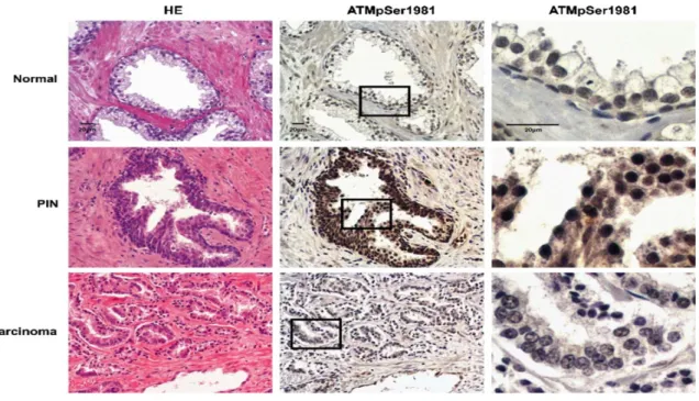
A Canadian group has analyzed 35 primary prostate cancer specimens for ATM activation, p-CHK2, p-H2AX and p53 by immunohistochemistry in normal prostate glands, PINs (the precursors to carcinomas) and carcinomas104. They had showed an increased intensity of p-ATM, p-CHK2 and p-H2AX in PINs, compared to normal prostatic glands or carcinomas. Figure 4 depicts representative images of the detected levels of ATM activation in normal prostatic glands and carcinoma compared to PINs.
These findings suggest that early oncogenic events result in ATM activation, that are attenuated by events occurring in the later stages of prostate tumorigenesis, perhaps via genome re-stabilization in advanced cancer cells. The authors mention that the activation of ATM at the early stages of prostate tumorigenesis prevents tumor progression and its desensitization to oncogenic signals promotes tumor development. However, little is known about DDR activity beyond the PIN stage. Of note this study
Figure 4: ATM activation in normal prostatic gland, PIN, and carcinoma
ATM activation in normal prostatic gland, PIN, and carcinoma: Morphologically normal prostatic gland, PIN, and carcinoma from the same patient (or slide) were H&E stained and immunohistochemically stained for Ser1981 phosphorylated ATM using an anti-pSer1981-ATM antibody. The inset areas are enlarged (right column).
Fan C, Quan R, Feng X, et al. ATM activation is accompanied with earlier stages of prostate tumorigenesis.. Biochimica
did not detect any significant DDR signaling differences between normal and adenocarcinoma prostate tissues.
Separately, and in support of a DDR cancer barrier in prostate cancer, investigators have established that a DDR-mediated tumor suppression activity restricts early-stage prostate cancer progression in mouse models 104; 105. This senescence-mediated DDR barrier occurs at a stage associated with persistent DDR signalling and is analogous to human high-grade prostatic intraepithelial neoplasia (HG-PIN)106;107. Further supporting this DDR-mediated cancer barrier, short telomeres (known to directly trigger the DDR) have been observed in human HG-PIN 108. Together, these lines of evidence strongly suggest that the DDR-mediated cancer barrier during HG-PIN is at least partially driven by DDR signalling. However, at the same time, the fact that DDR is persistently activated favours the outgrowth of malignant cells having defects in the DDR such as aberrations in the ATM cascade. Cancer cells, including prostate cancer cells, acquire an intrinsic capacity to tolerate DNA damage during cancer progression. This can happen through the loss of redundant DDR signalling pathways such as the p53 pathway, allowing HG-PIN to move to invasive carcinoma in mouse models. DDR and DNA repair are essential for genome stability and prolonged cell survival, therefore, cancer cells must maintain other redundant DDR pathways functional. It is thus proposed that inactivation of these remaining DDR pathways could greatly sensitize cancer cells to DNA damage 109; 110. The identification of these remaining pathways in prostate cancer could represent new therapeutic opportunities. On their
own, these potential therapeutic avenues warrant a better characterization of active DDR pathways during prostate cancer progression104; 111.
As described above, the DDR acts as a barrier against cancer progression. Moreover in prostate cancer, it has also been shown that promyelocytic leukemia protein (PML) body formation is defective in prostate tumor cells but is active in benign prostatic hyperplasia (BPH)112. PML was identified in the early 90’s as a gene target for translocations with the retinoic acid receptor gene in acute promyelocytic leukemia113. The expression of this protein in primary cells leads to cellular senescence. PML acts as a tumor suppressor and is often lost in human cancers114. It has been shown that PML represses genes involved in DNA replication, repair and checkpoints. Indeed, PML represses E2F target genes and induces p53 and the DDR. The decrease in the expression of genes required for DNA repair and checkpoints promptly after PML expression suggests a mechanism by which PML could contribute to the senescence cell cycle arrest, which we know involves DNA damage signals 115; 116; 117. Vernier and colleagues investigated whether the PML/senescence pathway is important in human cancers, and to do so, they measured PML expression by immunohistochemistry in TMAs with samples from patients with benign or malignant prostate tumors112. BPH is a benign prostate lesion characterized by the presence of senescence markers118 and low E2F target gene expression119. In their study, PML staining in the normal prostate was very weak, but a few PML bodies could be distinguished. In contrast, PML staining was stronger in BPH samples where many PML bodies were easily
distinguished. Inversely, PML bodies were rarely distinguished in prostate cancer samples, including PIN, although they did detect some homogenous expression in the nucleus or cytoplasm. Their results indicate that cells from BPH contain more PML bodies than cells from normal tissues or cells from the few cases of prostate carcinomas in their TMAs where PML bodies were visualized. Taken together, they suggest that PML bodies may suppress malignant transformation in the prostate by promoting senescence, and that PML staining could be used to distinguish benign from malignant lesions.
It is known that the retinoblastoma (Rb) pathway controls the cell cycle at the transcriptional level via repression of E2F target genes120; 121; 122. Many E2F target genes mediate DNA repair and checkpoints, and, in their absence, cells accumulate DNA damage signals that are essential for activation of p53 and the senescence process115;116; 117. According to this same group, the lack of PML bodies in tumor cells can explain why prostate tumors exhibit high levels of expression of E2F target genes such as EZH2123 and BRCA1124; 125 which is also one of the genes most efficiently downregulated in PML–senescent fibroblasts.
Furthermore, DDR determines how normal and cancer cells react to cancer therapy such as DNA damaging RT or chemotherapies. Indeed, DDR activity has been detected in prostate cancer tumors following chemotherapy 126.
cells/tumors would mount against therapy could highly improve treatment selection and successes. Currently, the exact regulation and outcomes of DDR signalling in cancer cells, particularly in prostate cancer, remain relatively unknown.
3.0 Biomarkers
3.1 Prognostic vs. Predictive markers
Prognostic markers may help clinicians guide their patients in their decision-making and avoid treatment and toxicities for men with slow growing cancer while promoting the initiation of treatment in the others. Although ‘’predictive’’ and ‘’prognostic’’ markers are often used interchangeably, they are different127. Prognosis refers to the ability to distinguish clinically important variation and reliably project the course, the progression, the pattern and the end of disease. Prognostic markers are associated with prognosis, unrelated to the treatment received. They predict the ‘’natural’’ outcome of the disease before a treatment is given or regardless of it. As such, the treatment can change the prognosis in addition to the end point (local control, PSA control, overall survival or preservation of the prostate). As for ‘’predictive’’ factors, they are those that foretell the response to a treatment. In summary, predictive markers suggest the outcome of a treatment, thus allowing the identification of patients who will benefit from particular therapies, whereas a prognostic factor is a marker for gravity of a disease and outcome that is independent of treatment.
3.2 DDR as a tissue marker
To our knowledge, the protein expression of DDR factors in tissues has not been studied as biomarkers for cancer biology. Few studies have looked at tissue markers predictive of radiosensitivity in prostate cancer because the analyses are done on very small specimens obtained during prostate biopsies. Moreover, there are no studies on post radiation prostatectomy specimens. The Radiation Therapy Oncology Group (RTOG) has identified several biomarkers, from patients treated by RT under phase 3 randomized trials. Those markers are mainly involved in the cell cycle or apoptosis. The first biomarker studied by the RTOG was p53. They found a statistically significant correlation between abnormal p53 protein expression and an increased risk of distant metastases (p=0.04), a decreased probability of progression-free survival (p=0.03), and a reduction in overall survival (p=0.02). Furthermore, patients with tumors exhibiting abnormal p53 expression who received RT with androgen deprivation therapy (ADT) who had tumors that exhibited abnormal p53 expression developed metastases faster (p=0.001) but this was not observed in patients treated by RT alone without ADT128. The RTOG also evaluated patients in a different randomized phase 3 study. Of the 777 patient cohort, 22% had abnormal p53 expression defined as ≥20% cells with positive nuclear staining by immunohistochemistry (IHC) and this was associated with a decreased survival (p=0.014) and an increased risk of distant metastasis (p=0.013). For patients treated with ADT, there was a correlation between the p53 status and cause-specific survival