Université de Montréal
A systematic analysis of the detrimental effect of
hormonotherapy on skeletal and non-skeletal
morbidities in male with prostate cancer
par Claudio Jeldres
Sciences biomédicales
Faculté des études supérieures et postdoctorales
Mémoire présenté à la Faculté des études supérieures et postdoctorales en vue de l’obtention du grade de Maître ès sciences biomédicales
Mars 2012
Faculté des études supérieures et postdoctorales
Ce mémoire intitulé :
A systematic analysis of the detrimental effect of hormonotherapy on skeletal and non-skeletal morbidities in male with prostate cancer
Présenté par : Claudio Jeldres
a été évalué par un jury composé des personnes suivantes :
Diego Barrieras, MD, FRCSC, président-rapporteur
Pierre I. Karakiewicz, MD, MPH, FRCSC, directeur de recherche Julie Franc-Guimond MD, FRCSC, membre du jury
i
Résumé
Introduction: Plusieurs patients atteints d’un cancer de la prostate (CaP) se verront prescrire l'hormonothérapie (HT) en raison du stade avancé ou en cas d’une récidive de la maladie, pour freiner la progression de la maladie et améliorer leur survie. Cependant, l’HT présente des effets pouvant nuire significativement à la santé du patient.
Objectif : Cerner l'impact de l’HT sur l’apparition de complications squelettiques et non-squelettiques.
Méthodes : Nous avons identifié 15 842 québécois atteints du CaP. Nous avons quantifié les complications squelettiques et non-squelettiques. Les effets de l’HT ont été quantifiés à l'aide de modèles statistiques de régression tenant compte des risques compétitifs, tels que les comorbidités pré-existantes ou la mort prématurée.
Résultats : À 8 ans, l’incidence d’ostéoporose est de 4.4% chez les hommes sans HT, 10.8% chez ceux avec HT≤1.8 ans, 19.7% HT>1.8 ans et 11.4% pour les orchiectomisés. Pour la même durée, l’incidence de fracture vertébrale est de 1.9%, 7.5%, 7.5% et 8.1% pour les même groupes. Pour la fracture de hanche les taux sont de 1.9%, 6.4%, 8.0% et 9.3%, respectivement. Dans les modèles mutlivariés, l’HT >1.8 ans et l’orchiectomie augmentent significativement le risque d’évènements squelettiques. Une exposition prolongée à l’HT et l’orchiectomie augmentent le risque de détérioration cognitive. Finalement, l’HT prolongée augmente significativement le risque de maladies vasculaires périphériques et cérébrovasculaires.
Conclusions : L’hormonothérapie prolongée et l’orchiectomie augmentent significativement le risque de complications squelettiques chez les hommes québécois. Nos données confirment également l’effet de l’hormonothérapie sur le risque cardiovasculaire et sur la détérioration de la fonction cognitive.
Mots-clés : cancer, prostate, hormonothérapie, fracture osseuse, ostéoporose, démence, maladies vasculaires
iii
Abstract
Purpose: In men with prostate cancer (PCa) androgen deprivation therapy (ADT) predisposes to skeletal-related events (SREs) defined as osteoporosis or bone fractures and non-skeletal-related (NSREs) events, such as diabetes mellitus and myocardial infarction. We tested whether this effect is also detectable within a large Canadian cohort.
Material and Methods: Within the Quebec Health Plan database we identified 15 842 men diagnosed with PCa who were treated between 1992 and 2000 with either luteinizing hormone-releasing hormone agonists [LHRHa] or orchiectomy. Separate competing-risks regression models tested the effect of orchiectomy vs. LHRHa vs. no ADT on the incident rate of three SREs and twelve NSREs.
Results: At 8 years, the incidence rates of osteoporosis were 4.4% in patients unexposed to ADT, 10.8% in LHRHa ≤1.8 years, 19.7% in LHRHa >1.8 years and 11.4% in orchiectomized patients. Similarly, the rates of spinal fracture were 1,9%, 7.5%, 7.25% and 8.1% for the same groups. Finally, the rates of hip fracture were respectively 1.9%, 6.4%, 8.0% and 9.3%. In multivariable competing-risks analyses, exposure to LHRHa >1.8 years and orchiectomy significantly increased all three SREs. Conversely, the independent predictor status was not confirmed for patients exposed to LHRHa ≤1.8 years. Moreover, prolonged LHRHa therapy increases the incidence rates of three NSREs, namely, dementia, peripheral and cerebrovascular disease (all, p≤0.02). Orchiectomy only increases the rate of dementia.
Conclusions: Exposure to prolonged LHRHa therapy or orchiectomy significantly increases the risk of SREs in Canadian men. Our results also confirm the effect of ADT on cardiovascular risks and cognitive function.
Keywords : prostate cancer, androgen deprivation therapy, skeletal fractures, metabolic complications
v
Table des matières
Introduction ... 1
Material and Methods ... 7
Results ... 10 Skeletal-Related Events: ... 12 Non-Skeletal-Related Events: ... 15 Discussion ... 24 Conclusion ... 28 References ... 29
Liste des figures
Figure 1. Percentage Distribution of Estimated New Cases and Deaths for Selected Cancers, Males, Canada, 2011.1……….1
Figure 2. Side effects of androgen deprivation therapy.13………..5 Figure 3. The type of androgen deprivation therapy used during the study period
vii
Liste des tables
Table 1. TNM staging system for prostate cancer………. 2 Table 2. Descriptive characteristics of the study population (n=15842)…………...10 Table 3. Cumulative incidence rates for skeletal-related events defined as osteoporosis, spinal fracture or hip fracture, according to type and duration of ADT (orchiectomy vs. LHRHa for ≤1.8 years vs. LHRHa for ≤1.8 years). The incidence rates account for other-cause mortality………13 Table 4. Multivariable competing-risks regression models testing the effect of
LHRHa or orchiectomy vs. no ADT on three separate endpoints: osteoporosis, spinal fractures and hip fractures. Each endpoint was analyzed in a separate model. Moreover, LHRHa exposure was tabulated between ≤1.8 years (median) and >1.8 years. The covariates consisted of patient age, anti-androgen exposure, postal code, region of residence, year of treatment, as well as of twelve non-skeletal morbidities that comprise the Charlson comorbidity index. Each of the Charlson comorbidities was coded as a time dependent covariate………..15 Table 5. Cumulative incidence rates for twelve non-skeletal-related events according to type and duration of ADT (orchiectomy vs. LHRHa for ≤1.8 years vs. LHRHa for ≤1.8 years). The incidence rates account for other-cause mortality………...16 Table 6. Multivariable competing-risks regression models testing the effect of LHRHa or orchiectomy vs. no ADT on twelve NSREs. Each endpoint was analyzed in a separate model. Moreover, LHRHa exposure was tabulated between ≤1.8 years (median) and >1.8 years. The covariates consisted of patient age, anti-androgen exposure, postal code, region of residence, year of treatment and other comorbidities. Each comorbidity was coded as a time dependent covariate………..23
Remerciements
Toute ma gratitude au Docteur Pierre I. Karakiewicz, à qui je dois davantage que ce qu’il m’est possible d’exprimer ici.
Introduction
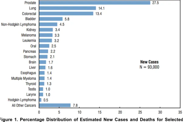
According to Statistics Canada, in 2011, prostate cancer (PCa) represented the most common cancer diagnosed in men (27.5% of new cancer cases, Fig. 1) and raked third in terms of mortality, after lung and colon cancers.1
Figure 1. Percentage Distribution of Estimated New Cases and Deaths for Selected Cancers, Males, Canada, 2011.1
In Canada, 25500 new cases were expected in 2011. In Quebec, at least 5100 new PCa cases were estimated. The natural history of PCa is more protracted that than for most other solid tumors. Its treated natural history usually spans between one or two decades, exposing cancer survivors to long period of treatments and their potential side effects.
Currently, PCa his staged both clinically and pathologically with the TNM (Tumor, Node, Metastasis) staging system (Table 1). Regular updated of this system are made to improve the classification and categorization of the patients, for clinical and research purpose. The most update system was published in 2010
by the American Joint Committee on Cancer (AJCC), in the AJCC Cancer Staging Manual, Seventh Edition, by Springer.
Table 1. TNM staging system for prostate cancer.
In it’s locally and advanced stage, the treatment of PCa requires the use of androgen deprivation therapy (ADT). The rationale of ADT in the field of PCa stems from the observations of a Canadian scientist Charles Huggins (Nobel Prize laureate 1966), who demonstrated that the male hormone testosterone is
3
necessary to stimulate PCa growth and its progression.2 The latter resulted in the use of ADT, initially in the form of surgical castration (bilateral orchiectomy) and later in the form of chemical castration (luteinizing hormone-releasing hormone agonists [LHRHa]), as the mainstay therapy for locally advanced or metastatic PCa. The beneficial effects of ADT in men with locally advanced or metastatic PCa consist of delayed disease progression and palliation of disease symptoms.3, 4 Approximately one third of all the newly diagnosed PCa cases are too advanced locally to benefit from definitive therapy or show frank evidence of metastatic disease.5 Additionally, ADT represents a well-recognized and widely used form of therapy for localized PCa, especially in individuals with restricted life expectancy due to important comorbidities. Some 10% of newly diagnosed PCa cases fulfill the characteristics of this category of individuals.
Recently, a second formal indication for ADT was coined, when ADT efficacy was confirmed in the setting of localized PCa. Specifically, its co-administration with radiation therapy improved survival in men with unfavourable disease features.6-9 Approximately, 25% of patients with newly diagnosed localized
PCa harbour unfavourable PCa characteristics and receive ADT in adjuvant setting for between 3 and 24 months.10 The third indication for ADT applies to patients that
demonstrate disease relapse (commonly elevated and rising serum prostate-specific antigen [PSA]) after either radical prostatectomy or external beam radiotherapy for initially localized disease.11 Five years after radical prostatectomy or radiotherapy, at least 30% of men fall into this category. Taken together, at least one in two individuals diagnosed with PCa will temporarily (in adjuvant setting) or permanently exposed to ADT. This implies that annually at least 12750 men will receive ADT throughout Canada. Of these, more than 2500 individuals will be treated with ADT in the province of Quebec. In consequence, a large proportion of men may be exposed to ADT.
Hormonal therapy is usually administered for several years and not infrequently for decades. Therefore, the burden related to the use of ADT is not only significant epidemiologically, but has important clinical (potential side-effects) and health economics (cost of ADT: average 4500$/patient/ syear) implications.12 The cost of ADT may actually be higher due to its side effects. Recently, the medical literature reported an exponential rise in the type and rates of such complications. Androgen deprivation therapy for PCa in the form of orchiectomy, luteinizing hormone-releasing hormone agonists (LHRHa) and/or antiandrogens has been associated with several side effects (Fig. 2).13 Beside the well-established side effects (loss of libido, erectile dysfunction, fatigue, hot flashes, altered body composition, etc.), recent report linked ADT to a broader range of adverse outcomes.3, 13, 14 These consist of skeletal complications (osteopenia, osteoporosis and skeletal fractures), as well as of metabolic changes (hyperglycemia and insulin resistance, frank diabetes mellitus, hypertension, atherosclerosis, cardiovascular disease and cognitive decline).15-22 Moreover, ADT-related side effects may undermine patient’s quality-of-life and may predispose to lower life expectancy.23-25 Even more worrisome, ADT has been associated with
increased cardiovascular morbidity and mortality. To date, no study addressed the rate of skeletal and metabolic (non-skeletal) complications in a Canadian population. Currently, virtually exclusively data from the United States are used to substantiate the association between ADT and various skeletal and metabolic detriments.
5
Figure 2. Side effects of androgen deprivation therapy.13
The link between ADT and fractures was addressed in one large-scale population-based analysis of the Surveillance, Epidemiology and End Results (SEER) database after linkage with Medicare records and restricted to United States male 65 years old or older (n= 50000).15 It demonstrated a 7% absolute
increase in bone fractures in PCa patients exposed to ADT. Unfortunately, no other contemporary, large-scale, population-based studies validated the effect of ADT on fracture risk or other, non-skeletal morbidities that may potentially stem from ADT. Of studies that addressed skeletal ADT morbidities, virtually all relied form the United States. Therefore, there is need for large-scale, population-based assessment of the association between ADT and fracture risk, to corroborate or refute that the suggested 7% absolute risk in skeletal complications, is of same magnitude in Canadian men.
An even grater greater paucity of data applies to several other potential ADT side effects. For example, the association between ADT and various manifestations of the metabolic syndrome were studied in only 40 patients from the United States and these data were used to propose a causal effect.26 Based on
these sample and generalizability limitations, it is possible that other variables predispose men from the United States to many putative side effects of ADT.
Clinical observations, suggest that the rates in Quebec men might not be as elevated, as is reported in predominantly literature from the United States (for example, virtually complete absence of clinically meaningful fracture). Under this premise, several of preventive measures (bone density scans, diabetes screening, etc.) might be unnecessary. However, no reports provide factual complication rates. Based on these observations, we hypothesized that the rates of skeletal and non-skeletal side effects that are associated with ADT are not significantly higher in ADT-exposed individuals than in ADT-unexposed PCa controls. Differences in genetic, environmental, life style and dietary risk factors that distinguish Quebec men from their United States counterparts may account for these clinical observations.27, 28 For example, fat-rich diet and higher rate of obesity, as know to affect many men from the US, may predispose to higher rates of bone loss.29, 30 Therefore, studies from the US may exaggerate the detrimental effect of ADT on SREs. Specifically, we examined the rate of skeletal and metabolic events in patients exposed to ADT administered in the context of PCa and compared these rates to those recorded in men treated with definitive therapy (radiation or radical prostatectomy) without any ADT exposure. The practical and clinical implications of our findings may result in more specific and more selective use of ADT. This may in turn prevent side effects and save cost.
Material and Methods
The Quebec Health Plan represents the exclusive insurer in the Province of Quebec. Its database allows ascertainment of all health services covered by the Plan. These include ADT (LHRHa or bilateral orchiectomy) for patients diagnosed with PCa. The Health Plan database is used for reimbursement purposes and analyses of health services utilization. The Health Plan relies on the 9th version of the International Classification of Diseases (ICD-9) diagnostic codes and their respective dates. The Health Plan allowed us to retrieve the date of the initial diagnosis of each of the specific morbidities, namely, skeletal morbidities: osteoporosis (ICD-9 733.0), hip fracture (ICD-9 820) and spinal fracture (ICD-9 805) and non-skeletal morbidities: myocardial infarction (ICD-9 410, 411), congestive heart failure (ICD-9 398, 402, 428), peripheral vascular disease (ICD-9 440-447), dementia (ICD-9 290, 291, 294), cerebrovascular disease (ICD-9 430-433, 435), chronic pulmonary disease (ICD-9 491-493), connective tissue disease (ICD-9 710, 714, 725), ulcer disease (ICD-9 531-534), moderate to severe renal disease (ICD-9 403, 404, 580-586), diabetes (ICD-9 250), mild liver disease (ICD-9 571, 573), and moderate to severe liver disease (ICD-9 070, 570, 572).
The database provides diagnostic codes from as early on as January 1st, 1983. Follow-up information was available until July 1st, 2004. Based on stage migration, which occurred in the PSA-era, only prostate cancer cases treated after January 1st, 1992 were included. Patients diagnosed and treated after December 31st, 2000 were excluded based on length of follow-up considerations.
To assess the effect of ADT on the incidence rate of the three SREs and twelve NSREs, we queried the database for PCa patients treated with LHRHa or bilateral orchiectomy, who did not undergo any form of definitive therapy, such as radical prostatectomy (RP) or external beam radiotherapy (XRT). We also queried
for patients treated with RP and/or XRT, who were not exposed to any form of ADT. This group represented the controls.
Due to the use of reimbursement records, the database allowed exact ascertainment of LHRHa exposure, orchiectomy, RP, and definitive XRT. For orchiectomy patients, the starting point of follow-up was defined at the date of bilateral orchiectomy. For LHRHa patients, only those who filled their prescription more than 90% of the time were considered. LHRHa patients were divided according to the median duration of LHRHa exposure (LHRHa ≤1.8 years and LHRHa >1.8 years). For RP and XRT patients (controls), the starting point of follow-up was defined at RP or first XRT session.
We examined fifteen separate endpoints. Each endpoint consisted of a new diagnosis of any SREs or NSREs. Each event was assessed in separate univariable and multivariable competing-risks regression models. The analyses tested the effect of ADT exposure (LHRHa ≤1.8 years vs. LHRHa >1.8 years vs. orchiectomy vs. no ADT) on these endpoints. All analyses accounted for other-cause mortality, as described by Fine and Gray.31 First, the incidence rates at 1, 5 and 8 years of follow-up were defined using cumulative incidence analyses that account for other-cause mortality. Subsequently, we relied on multivariable competing-risks regression models to account for the effect of other variables.
Age differences may confound more the relationship between ADT exposure and the diagnosis of SREs or NSREs. We adjusted the confounding effect of age in all multivariable competing-risks regression models. Moreover, the effect of socioeconomic status was adjusted for using the postal code and the geographic region of residence. We also adjusted for the year of treatment, since temporal trends may affect the rate of ADT associated morbidities. Finally, since other
pre-9
existing health conditions may predispose to additional health problems, we also adjusted for any morbidity (concomitant SRE or NSRE) that was diagnosed either before or after the beginning of follow-up. In multivariable competing-risks regression models, the dates of diagnoses of any pre-morbid conditions, if applicable, was coded as time-dependent covariates
All statistical tests were performed using S-Plus Professional, version 1.0 (MathSoft Inc., Seattle, Washington). Moreover, all tests were two-sided with a significance level set at 0.05.
Results
The descriptives of the 15842 assessable patients are shown in Table 2. The median age of the patients treated with LHRHa ≤1.8 years, LHRHa >1.8 years and orchiectomy was 7.7 to 9.4 years higher than that of their counterparts not treated with any form of ADT. The underlying comorbidities at the start of follow-up did not differ between the four groups. The effect of these and other potential differences were addressed in multivariable analyses.
Table 2. Descriptive characteristics of the study population (n=15842).
Legend
LHRHa: Luteinizing hormone-releasing hormone agonists ADT: androgen deprivation therapy
Overall population Patients unexposed to ADT Patients exposed to LHRHa for ≤1.8 years Patients exposed to LHRHa For>1.8 years
Patients treated with orchiectomy Number of patients 15842 (100%) 10072 (63.6%) 2037 (12.9%) 1877 (11.8%) 1856 (11.7%) Age (years) Mean (median) Range 69.1 (68.7) 29.5-100.9 66.5 (66.3) 41.2-92.86 75.2 (75.4) 39.9-100.9 75.0 (75.7) 46.9-96.0 70.2 (74.0) 29.5-96.5
Charlson Comorbidity Index at the start of follow-up
Mean (median) Range 2.1 (2.0) 0-13 2.0 (2.0) 0-12 2.3 (2.0) 0-13 2.1 (2.0) 0-10 2.2 (2.0) 0-12
Antiandrogen therapy use 3385 0 1686 (82.8) 1699 (90.5) 0
Prevalence of morbidities
Myocardial infarction Congestive heart failure Peripheral vascular disease Dementia
Cerebrovascular disease Chronic pulmonary disease Connective tissue disease Ulcer disease
Moderate to severe renal disease
Diabetes Mellitus Mild liver disease
Moderate to severe liver disease
3628 (22.9) 3226 (21.6) 4074 (25.7) 1319 (8.4) 2039 (12.9) 4749 (29.9) 1048 (6.6) 2551 (16.1) 1926 (12.2) 4014 (25.3) 831 (5.3) 206 (1.3) 2979 (20.7) 1725 (17.2) 2431 (24.2) 541 (5.4) 1176 (11.7) 2960 (29.4) 659 (6.5) 1642 (16.3) 983 (9.7) 2506 (24.9) 561 (5.6) 145 (1.5) 556 (27.3) 601 (29.5) 599 (29.4) 266 (12.0) 303 (14.9) 637 (31.3) 131 (6.4) 327 (16.1) 326 (16.0) 494 (24.3) 102 (5.0) 28 (1.4) 529 (28.2) 574 (30.6) 557 (29.7) 231 (12.3) 309 (16.5) 576 (30.7) 152 (8.1) 288 (15.4) 310 (16.5) 565 (30.1) 66 (3.6) 13 (0.7) 464 (25.0) 526 (28.4) 487 (26.2) 281 (15.2) 251 (13.5) 576 (31.0) 106 (5.7) 294 (15.9) 307 (16.5) 449 (24.2) 102 (5.5) 20 (1.1)
11
Figure 3 shows ADT use and type through the study period. From 1992 to 2000, almost half of patients received a form of ADT. Orchiectomy use declined significantly during this period of time, mostly due to introduction of chemical castration since 1995. For example in 1992, 39.7% were treated with orchiectomy, but in year 2000 only 6.3%.
Figure 3. The type of androgen deprivation therapy used during the study period (1992-2000).
Legend
ADT: androgen deprivation therapy
LHRH: luteinizing hormone-releasing hormone agonists
60.3 62.0 64.8 44.7 53.1 44.0 49.5 52.2 59.0 39.7 38.0 35.2 21.9 26.0 16.5 10.5 7.6 6.3 0.0 0.0 0.0 33.4 20.9 39.5 40.0 40.2 34.7
0%
10%
20%
30%
40%
50%
60%
70%
80%
90%
100%
1992 1993 1994 1995 1996 1997 1998 1999 2000
Skeletal-Related Events:
Table 3 shows incidence rates of newly diagnosed osteoporosis, hip fracture and spinal fracture that were obtained from cumulative incidence models that account for the effect of other-cause mortality. For example at 8 years of follow-up, the incident rate of osteoporosis was 10.8% (95% confidence interval [CI] 8.4-13.9) in patients treated with LHRHa ≤1.8 years, 19.7% (95%CI 17.0-22.8) for those treated with LHRHa >1.8 years and 11.4% (95%CI 9.3-13.9) in patients treated with orchiectomy vs. 4.4% (95%CI 3.8-5.0) in patients unexposed to any form of ADT. For the same time-points and for the same patients groups, incident rates of spinal fracture were 7.5% (95%CI 5.6-8.2), 7.5% (4.9-8.3), 8.1% (95%CI 6.5-8.8) and 1.9% (95%CI 1.1-2.3). Similarly, the corresponding incident rates of hip fractures were 6.4% (95%CI 4.7-7.8), 8.0% (95%CI 5.3-9.9), 9.3% (95%CI 6.7-11.1) and 1.9% (95%CI 2.2-4.7) for the same four groups of patients.
13
Table 3. Cumulative incidence rates for skeletal-related events defined as osteoporosis, spinal fracture or hip fracture, according to type and duration of ADT (orchiectomy vs. LHRHa for ≤1.8 years vs. LHRHa for ≤1.8 years). The incidence rates account for other-cause mortality.
The results of the multivariable competing-risks regression models are shown in Table 4. In addition to adjustment for other-cause mortality, competing-risks regression models controlled for the potential confounding effect of other covariates. In models that targeted the incident rate of osteoporosis, exposure to LHRHa >1.8 years (HR: 2.3; p<0.001) and exposure to orchiectomy (HR: 2.3; p<0.001) reached independent predictor status. In models that targeted spinal fracture rate, exposure to LHRHa >1.8 years (HR: 1.9; p=0.018) and exposure to orchiectomy (HR: 2.2; p<0.001) reached independent predictor status. Finally, when hip fracture was assessed exposure to LHRHa >1.8 years (HR: 1.7; p=0.04) and exposure to orchiectomy (HR: 2.2; p<0.001) reached independent predictors status.
15
Table 4. Multivariable competing-risks regression models testing the effect of LHRHa or orchiectomy vs. no ADT on three separate endpoints: osteoporosis, spinal fractures and hip fractures. Each endpoint was analyzed in a separate model. Moreover, LHRHa exposure was tabulated between ≤1.8 years (median) and >1.8 years. The covariates consisted of patient age, anti-androgen exposure, postal code, region of residence, year of treatment, as well as of twelve non-skeletal morbidities that comprise the Charlson comorbidity index. Each of the Charlson comorbidities was coded as a time dependent covariate.
SREs Osteoporosis Spinal
fracture Hip fracture
ADT exposure HR; p-value HR; p-value HR; p-value
Patients exposed to LHRHa for ≤1.8
years 1.1; 0.8 1.6; 0.06 1.3; 0.3
Patients exposed to LHRHa for>1.8 years 2.3; <0.001* 1.9; 0.018* 1.7; 0.04*
Orchiectomy 2.3; <0.001* 2.2; <0.001* 2.2; <0.001*
Legend
ADT: androgen deprivation therapy SREs: skeletal-related events
LHRHa: luteinizing hormone-releasing hormone agonists HR: Hazard ratio
*: Statistically significant
Non-Skeletal-Related Events:
Table 5 shows incidence rates of newly diagnosed twelve NSREs that were obtained from cumulative incidence models that account for the effect of other-cause mortality. For example at 8 years of follow-up, the incident rate of dementia was 8.5% (95% confidence interval [CI] 5.9-11.2) in patients treated with LHRHa ≤1.8 years, 11.3% (95%CI 8.2-14.4) for those treated with LHRHa >1.8 years and 10.2% (95%CI 8.3-12.1) in patients treated with orchiectomy vs. 3.8% (95%CI 3.2-4.3) in patients unexposed to any form of ADT. For the same time-points and for the same patients groups, incident rates of cerebrovascular disease were 4.8%
(95%CI 3.0-6.6), 7.6% (5.3-10.0), 4.4% (95%CI 3.1-5.7) and 6.2% (95%CI 5.5-6.8). Similarly, the corresponding incident rates of peripheral vascular disease were 10.5% (95%CI 6.8-14.1), 13.9% (95%CI 9.9-15.0), 8.8% (95%CI 6.9-10.7) and 8.4% (95%CI 7.6-10.3) for the same four groups of patients. For the remaining, NSREs reported on table 4, cumulative incidences throughout the study period were similar between the 4 patients groups.
Table 5. Cumulative incidence rates for twelve non-skeletal-related events according to type and duration of ADT (orchiectomy vs. LHRHa for ≤1.8 years vs. LHRHa for ≤1.8 years). The incidence rates account for other-cause mortality.
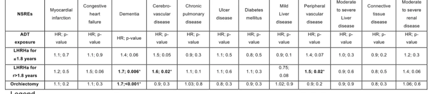
The results of the multivariable competing-risks regression models are shown in Table 6. In models that targeted the incident rate of dementia, exposure to LHRHa >1.8 years (HR: 1.7; p=0.006) and exposure to orchiectomy (HR: 1.7; p≤0.001) reached independent predictor status. In models that targeted cerebrovascular disease rate, exposure to LHRHa for >1.8 years reached independent predictor status (HR: 1.6; p=0.02). Finally, when peripheral disease was assessed, exposure to LHRHa for >1.8 years reached independent predictors status (HR: 1.5; p=0.02).
23
Table 6. Multivariable competing-risks regression models testing the effect of LHRHa or orchiectomy vs. no ADT on twelve NSREs. Each endpoint was analyzed in a separate model. Moreover, LHRHa exposure was tabulated between ≤1.8 years (median) and >1.8 years. The covariates consisted of patient age, anti-androgen exposure, postal code, region of residence, year of treatment and other comorbidities. Each comorbidity was coded as a time dependent covariate.
NSREs Myocardial infarction Congestive heart failure Dementia Cerebro-vascular disease Chronic pulmonary disease Ulcer disease Diabetes mellitus Mild Liver disease Peripheral vascular disease Moderate to severe Liver disease Connective tissue disease Moderate to severe renal disease ADT exposure HR; p-value HR; p-value HR; p-value HR; p-value HR; p-value HR; p-value HR; p-value HR; p-value HR; p-value HR; p-value HR; p-value HR; p-value LHRHa for ≤ 1.8 years 1.1; 0.7 1.1; 0.9 1.4; 0.06 1.5; 0.05 0.9; 0.3 1.1; 0.5 0.8; 0.5 0.9; 0.1 1.4; 0.07 1.0; 0.3 0.9; 0.2 1.2; 0.3 LHRHa for r>1.8 years 1.2; 0.5 1.5; 0.06 1.7; 0.006* 1.6; 0.02* 1.1; 0.1 1.1; 0.6 1.1; 0.3 0.75; 0.08 1.5; 0.02* 0.9; 0.6 0.8; 0.5 1.4; 0.06 Orchiectomy 1.1; 0.2 1.1; 0.3 1.7;<0.001* 0.9; 0.3 1.03; 0.8 0.8; 0.3 0.9; 0.3 1.02; 0.9 0.9; 0.2 0.9; 0.9 0.8; 0.3 1.06; 0.6 Legend
ADT: androgen deprivation therapy NSREs: non-skeletal-related events
LHRHa: luteinizing hormone-releasing hormone agonists HR: Hazard ratio
Discussion
Prostate cancer (PCa) is the most commonly male malignancy and is the second leading cause of cancer related death in North American men. However, rates of non-cancer deaths in men with prostate cancer are greater than in the general population and ADT treatment may, in part, be responsible for this increase.32 Moreover, a recent communication found that long-term LHRHa exposure was associated with greater non-cancer mortality than short-term LHRHa therapy.33 Furthermore, Keating et al. recently demonstrated that ADT increases the risk of incident diabetes, coronary heart disease, acute myocardial infarction, and sudden cardiac death.18 Moreover, several studies have demonstrated the deleterious effect of hormonal suppression on bone-health.34, 35 36 37 Virtually all large studies that addressed this topic were performed in the United States of America, where genetic, environmental and dietary risk factors may substantially differ from other part of the world. For example, obesity and PCa may exert a synergic effect on the rate of SREs.38, 39 Since obesity rate is higher in the US than
in Europe or Canada, SREs may be more frequent in the US.40 Finally, recent studies highlighted the increasing trend in ADT prescriptions for localized PCa, even in absence of any evidence-based benefit.41 Based on these observations, we tested the hypothesis that ADT may increase the risk of developing new non-cancer morbidities in a large population based cohort of patients treated for PCa with or without ADT. We tested this hypothesis by quantifying the rate of these established SREs and NSREs in Canadian men.
Our results indicated that LHRHa exposure in excess of the median duration (1.8 years) significantly predisposes to higher rates of SREs. At 8 years of follow-up, men exposed to LHRHa duration >1.8 years demonstrated a near 4-fold higher rate of SREs relative to men unexposed to ADT. Similarly, we found a near 2-fold increase in SREs rates in men exposed to orchiectomy. Interestingly, highest incident rates of SREs were recorded men treated with orchiectomy and those
25
exposed to LHRHa >1.8 years. Conversely, the SREs recorded in patients exposed with LHRHa ≤ 1.8 years (median) failed to demonstrate statistically significant differences from those recorded in patients unexposed to ADT. Taken together, our findings imply that LHRHa duration >1.8 years and orchiectomy treatment had the most detrimental effects on SREs rates.
In the analyses of spinal and hip fracture rates, orchiectomy was either more or equally detrimental to LHRHa exposure duration >1.8 years. Conversely, ADT exposure of ≤1.8 years duration was invariably less detrimental than >1.8 years of exposure or orchiectomy treatment. This observation is important from a clinical perspective as it suggests that intermittent ADT might be less deleterious than continuous ADT. Although this hypothesis is attractive it remains to be proven in randomized clinical trials.
Our results also show that for most of the NSREs, no significant differences in incidences rates were recorded after 8 years of follow up between men exposed compared to those unexposed to any type of ADT. This questions the detrimental effect of ADT on several metabolic morbidities, such as cardiovascular disease and diabetes mellitus. However, our data showed that patients exposed to LHRH for more than 1.8 years had near 2-fold higher risk of for peripheral and cerebrovascular disease then those unexposed. Multivariable models targeting the effect of ADT on vascular disease also confirm the detrimental effect of ADT on the vascular health of men exposed to ADT. Moreover, our results also confirmed the effect of ADT on cognitive decline, where men exposed to both prolong LHRH exposure and orchiectomy had more than 2-fold higher rates of dementia then men unexposed.
The novelty of our findings is several-fold. Our analysis is the first to control for cause mortality. Lack of accounting for mortality due to PCa or
other-causes may artificially overestimate the effect of ADT on SREs. Cumulative incidence and competing-risks regression analyses control for the confounding effect of mortality. This is particularly important in elderly patients, as patients who die prematurely have no chance of experiencing the adverse outcome of interest i.e., one or several SREs.
Elderly patients may also be affected by comorbidities. These may in turn lead to discontinuation of LHRHa. Such practice may also confound the true effect of ADT on SREs, as patients with multiple comorbidities may not be given an equal chance of being exposed to ADT and an equal chance of being diagnosed with one or several SREs. Our analysis controlled for comorbidities.
Additionally, our analyses also controlled for other known confounders, such as age, anti-androgen exposure, postal code, region of residence, and year of treatment. Interestingly, although we considered exposure to anti-androgen, this variable failed to affect the rates of SREs. It could have been postulated that anti-androgens might protect from SREs.
Our study is not devoid of limitations. First, its design is not prospective. Therefore, the ascertainment of diagnoses may be subject to underreporting. Not all conditions might have been identified using diagnostic codes. However, it is unlikely that there are differences in the rate of reporting of diagnostic codes according to orchiectomy exposure status. Therefore, it is also unlikely that the relative measures of risk have been affected by differential misclassification bias. Similarly, the ascertainment of exposure (LHRHa vs. orchiectomy vs. no ADT) is unlikely biased, as LHRHa medication codes and orchiectomy reimbursement codes were used for exposure definition. It is unlikely that reimbursement would not be sought in orchiectomized patients or that dispensed LHRHa injections would not be used.
27
Universal access to health care represents a weakness, as well as an advantage of the current design. Access to orchiectomy in the Province of Quebec is not dictated by health insurance status. Nonetheless, the socioeconomic status (education) and temporal trends may have affected the rate of orchiectomy use 42. In consequence, we adjusted for both variables. The adjustment for year of treatment likely decreased or eliminated the confounding effect of temporal trends. However, adjustment for socioeconomic status by proxy of region of residence and postal code may not have completely eliminated the effect of socioeconomic status. Moreover, our analyses were limited by lack of consideration of several pharmacological agents aimed at SRE prevention, such as calcium, vitamin D or bisphosphonates.
Finally, the administrative nature of our database precluded the consideration of disease stage. In consequence, it cannot be entirely excluded that orchiectomy patients by virtue of more advanced disease stage are not predisposed to higher rates of myocardial infarction and/or dementia. However, there is no evidence for such association and there is no biologic rationale for its existence.
Despite these limitations, our findings provide valuable information about potential detriments of ADT exposure. They should be used in treatment decision-making when ADT exposure is considered.
Conclusion
In conclusion, exposure to prolonged LHRHa therapy significantly increases the incidence rates of osteoporosis, spinal and hip fractures in Canadian men. Moreover, of twelve morbidities, exposure to prolonged LHRHa therapy increases the incidence rates of only three of them, namely: dementia, peripheral vascular disease and cerebrovascular disease. These findings confirm the detrimental effect of LHRHa on fracture rates in men from outside of the US. These results also confirm the effect of ADT on cardiovascular risks and cognitive function. Although the effect is weak, it still warrants caution in the setting of pre-existing morbidities when ADT is considered. Orchiectomy is associated with higher incidence rates of osteoporosis, spinal and hip fractures. Preventive or therapeutic measures should be considered if this treatment modality is chosen. However, of twelve morbidities, orchiectomy was associated only with higher incidence rates of dementia.
29
References
1. Canadian Cancer Society’s Steering Committee on Cancer Statistics. Canadian Cancer Statistics 2011. Toronto OCCS.
2. Huggins C, Hodges CV. Studies on prostatic cancer: I. The effect of castration, of estrogen and of androgen injection on serum phosphatases in metastatic carcinoma of the prostate. 1941. J Urol 2002;168(1): 9-12.
3. Sharifi N, Gulley JL, Dahut WL. Androgen deprivation therapy for prostate cancer. JAMA 2005;294(2): 238-44.
4. Messing EM, Manola J, Sarosdy M, Wilding G, Crawford ED, Trump D. Immediate hormonal therapy compared with observation after radical prostatectomy and pelvic lymphadenectomy in men with node-positive prostate cancer. N Engl J Med 1999;341(24): 1781-8.
5. Crawford ED, Eisenberger MA, McLeod DG, et al. A controlled trial of leuprolide with and without flutamide in prostatic carcinoma. N Engl J Med 1989;321(7): 419-24.
6. Bolla M, Collette L, Blank L, et al. Long-term results with immediate androgen suppression and external irradiation in patients with locally advanced prostate cancer (an EORTC study): a phase III randomised trial. Lancet 2002;360(9327): 103-6.
7. Kumar S, Shelley M, Harrison C, Coles B, Wilt TJ, Mason MD. Neo-adjuvant and adjuvant hormone therapy for localised and locally advanced prostate cancer. Cochrane Database Syst Rev 2006(4): CD006019.
8. Shelley MD, Kumar S, Wilt T, Staffurth J, Coles B, Mason MD. A systematic review and meta-analysis of randomised trials of neo-adjuvant hormone therapy for localised and locally advanced prostate carcinoma. Cancer Treat Rev 2009;35(1): 9-17.
9. Bolla M, Gonzalez D, Warde P, et al. Improved survival in patients with locally advanced prostate cancer treated with radiotherapy and goserelin. N Engl J Med 1997;337(5): 295-300.
10. Denham JW, Steigler A, Lamb DS, et al. Short-term androgen deprivation and radiotherapy for locally advanced prostate cancer: results from the Trans-Tasman Radiation Oncology Group 96.01 randomised controlled trial. Lancet Oncol 2005;6(11): 841-50.
sensitive metastatic, recurrent, or progressive prostate cancer: 2006 update of an American Society of Clinical Oncology practice guideline. J Clin Oncol 2007;25(12): 1596-605.
12. Shahinian VB, Kuo YF, Gilbert SM. Reimbursement policy and androgen-deprivation therapy for prostate cancer. N Engl J Med 2010;363(19): 1822-32. 13. Isbarn H, Boccon-Gibod L, Carroll PR, et al. Androgen deprivation therapy for the treatment of prostate cancer: consider both benefits and risks. Eur Urol 2009;55(1): 62-75.
14. Ebeling PR. Clinical practice. Osteoporosis in men. N Engl J Med 2008;358(14): 1474-82.
15. Shahinian VB, Kuo YF, Freeman JL, Goodwin JS. Risk of fracture after androgen deprivation for prostate cancer. N Engl J Med 2005;352(2): 154-64. 16. Smith MR, Boyce SP, Moyneur E, Duh MS, Raut MK, Brandman J. Risk of clinical fractures after gonadotropin-releasing hormone agonist therapy for prostate cancer. J Urol 2006;175(1): 136-9; discussion 39.
17. Smith MR, Lee WC, Brandman J, Wang Q, Botteman M, Pashos CL. Gonadotropin-releasing hormone agonists and fracture risk: a claims-based cohort study of men with nonmetastatic prostate cancer. J Clin Oncol 2005;23(31): 7897-903.
18. Keating NL, O'Malley AJ, Smith MR. Diabetes and cardiovascular disease during androgen deprivation therapy for prostate cancer. J Clin Oncol 2006;24(27): 4448-56.
19. Tsai HK, D'Amico AV, Sadetsky N, Chen MH, Carroll PR. Androgen deprivation therapy for localized prostate cancer and the risk of cardiovascular mortality. J Natl Cancer Inst 2007;99(20): 1516-24.
20. Basaria S, Muller DC, Carducci MA, Egan J, Dobs AS. Hyperglycemia and insulin resistance in men with prostate carcinoma who receive androgen-deprivation therapy. Cancer 2006;106(3): 581-8.
21. Jenkins VA, Bloomfield DJ, Shilling VM, Edginton TL. Does neoadjuvant hormone therapy for early prostate cancer affect cognition? Results from a pilot study. BJU Int 2005;96(1): 48-53.
22. Joly F, Alibhai SM, Galica J, et al. Impact of androgen deprivation therapy on physical and cognitive function, as well as quality of life of patients with nonmetastatic prostate cancer. J Urol 2006;176(6 Pt 1): 2443-7.
31 23. Gilbert SM, McKiernan JM. Epidemiology of male osteoporosis and prostate cancer. Current opinion in urology 2005;15(1): 23-7.
24. Groot MT, Boeken Kruger CG, Pelger RC, Uyl-de Groot CA. Costs of prostate cancer, metastatic to the bone, in the Netherlands. European Urology 2003;43(3): 226-32.
25. Krupski TL, Foley KA, Baser O, Long S, Macarios D, Litwin M. Health care cost associated with prostate cancer, androgen deprivation therapy and bone complications. The Journal of Urology 2007;178(4 Pt 1): 1423-8.
26. Braga-Basaria M, Dobs AS, Muller DC, et al. Metabolic syndrome in men with prostate cancer undergoing long-term androgen-deprivation therapy. J Clin Oncol 2006;24(24): 3979-83.
27. Moreau C, Vezina H, Yotova V, et al. Genetic heterogeneity in regional populations of Quebec--parental lineages in the Gaspe Peninsula. Am J Phys Anthropol 2009;139(4): 512-22.
28. Bherer C, Labuda D, Roy-Gagnon MH, Houde L, Tremblay M, Vezina H. Admixed ancestry and stratification of Quebec regional populations. Am J Phys Anthropol 2011;144(3): 432-41.
29. Nunez NP, Carpenter CL, Perkins SN, et al. Extreme obesity reduces bone mineral density: complementary evidence from mice and women. Obesity (Silver Spring) 2007;15(8): 1980-7.
30. von Muhlen D, Safii S, Jassal SK, Svartberg J, Barrett-Connor E. Associations between the metabolic syndrome and bone health in older men and women: the Rancho Bernardo Study. Osteoporos Int 2007;18(10): 1337-44.
31. Fine JP, Gray RJ. A proportional hazards model for the subdistribution of a competing risk. Journal of the American Statistical Association 1999;94(446): 496-509.
32. Brown BW, Brauner C, Minnotte MC. Noncancer deaths in white adult cancer patients. J Natl Cancer Inst 1993;85(12): 979-87.
33. Hanks GE, Pajak TF, Porter A, et al. Phase III trial of long-term adjuvant androgen deprivation after neoadjuvant hormonal cytoreduction and radiotherapy in locally advanced carcinoma of the prostate: the Radiation Therapy Oncology Group Protocol 92-02. J Clin Oncol 2003;21(21): 3972-8.
34. Saad F, Adachi JD, Brown JP, et al. Cancer treatment-induced bone loss in breast and prostate cancer. J Clin Oncol 2008;26(33): 5465-76.
the Treatment of Prostate Cancer: Consider Both Benefits and Risks. Eur Urol 2008.
36. Diamond T, Campbell J, Bryant C, Lynch W. The effect of combined androgen blockade on bone turnover and bone mineral densities in men treated for prostate carcinoma: longitudinal evaluation and response to intermittent cyclic etidronate therapy. Cancer 1998;83(8): 1561-6.
37. Maillefert JF, Sibilia J, Michel F, Saussine C, Javier RM, Tavernier C. Bone mineral density in men treated with synthetic gonadotropin-releasing hormone agonists for prostatic carcinoma. J Urol 1999;161(4): 1219-22.
38. Parekh DJ, Ankerst DP, Thompson IM. Prostate-specific antigen levels, prostate-specific antigen kinetics, and prostate cancer prognosis: a tocsin calling for prospective studies. J Natl Cancer Inst 2007;99(7): 496-7.
39. Greenwald P. Cancer prevention clinical trials. J Clin Oncol 2002;20(18 Suppl): 14S-22S.
40. Ford ES, Mokdad AH. Epidemiology of obesity in the Western Hemisphere. J Clin Endocrinol Metab 2008;93(11 Suppl 1): S1-8.
41. Cooperberg MR, Grossfeld GD, Lubeck DP, Carroll PR. National practice patterns and time trends in androgen ablation for localized prostate cancer. J Natl Cancer Inst 2003;95(13): 981-9.
42. Weight CJ, Klein EA, Jones JS. Androgen deprivation falls as orchiectomy rates rise after changes in reimbursement in the U.S. Medicare population. Cancer 2008;112(10): 2195-201.