Université de Montréal
3
A Study of the Role Bmp Growth Factors Play in Pituitary POMC Gene Expression
par Maria NUDI
Département de biologie moléculaire Faculté des études supérieures
Mémoire présenté à la Faculté des études supérieures en vue de l’obtention du grade de Maître ès sciences (M.Sc.)
en biologie moléculaire
Avril, 2002 © Maria MiDI, 2002
-de Montréal
Direction des bibliothèques
AVIS
L’auteur a autorisé l’Université de Montréal à reproduire et diffuser, en totalité ou en partie, par quelque moyen que ce soit et sur quelque support que ce soit, et exclusivement à des fins non lucratives d’enseignement et de recherche, des copies de ce mémoire ou de cette thèse.
L’auteur et les coauteurs le cas échéant conservent la propriété du droit d’auteur et des droits moraux qui protègent ce document. Ni la thèse ou le mémoire, ni des extraits substantiels de ce document, ne doivent être imprimés ou autrement reproduits sans l’autorisation de l’auteur.
Afin de se conformer à la Loi canadienne sur la protection des renseignements personnels, quelques formulaires secondaires, coordonnées ou signatures intégrées au texte ont pu être enlevés de ce document. Bien que cela ait pu affecter la pagination, il n’y a aucun contenu manquant.
NOTICE
The author of this thesis or dissertation has granted a nonexclusive license allowing Université de Montréal to reproduce and publish the document, in part or in whole, and in any format, solely for noncommercial educational and research purposes.
The author and co-authors if applicable retain copyright ownership and moral rights in this document. Neither the whole thesis or dissertation, nor substantial extracts from it, may be printed or otherwise reproduced without the author’s permission.
In compliance with the Canadian Privacy Act some supporting forms, contact information or signatures may have been removed from the document. While this may affect the document page count, it does not represent any loss of content from the document.
Identification du jury
Université de Montréal Faculté des études supérieures
Ce mémoire intitulé:
A Study of the Role Bmp Growth Factors Play in Pituitary POMC Gene Expression
Présenté par:
Maria NUDI
A été évalué par un jury composé des personnes suivantes:
Muriel AUBRY (président rapporteur)
Jacques DROUIN (directeur de recherche)
RÉSUMÉ
Les corticotropes sont les premières cellules à se différencier dans l’hypophyse embryonaire. Des expériences d’explants suggèrent un role négatif pour les signaux Brnps dans la différenciation corticotropique, tandis que des études de gain-de-function suggèrent le contraire. POMC étant un marqueur corticotropique, mon projet de maîtrise a porté sur le rôle des signaux Bmp dans l’expression de POMC. Je démontre que l’expression de POMC décroit dans les cellules AtT-20, suite à des traitements avec des protéines Bmp-4 recombinantes ou à la surexpression de composantes de la voie des Bmp/Smad, soient les récepteurs Alk-3/-6 et les facteurs de transcription $madl/4. La surexpression des inhibiteurs Smad6 et Smad7 renversent cette répression. L’effet négatif de Bmp sur le promoter POMC nécessite les éléments de réponse Pitx et Tpit, et interfère avec l’activité synergique des deux protéines. Des interactions in vitro entre Pitxl, Tpit et Srnadl appuient un mécanisme d’action des Smads qui passerait directement par Pitx et Tpit. Mots clés : différenciation cellulaire, signalisation, répression, transcription, hormone et Tgf-f3.
Corticotrophs constitute the first hormone-producing ceil type to emerge in the developing pituitary. Tissue expiant experiments had suggested a negative role for Bone morphogenic-protein (Bmp) signais in corticotroph differentiation, but transgenic studies had argued against this. Seeing that proopiornelanocortin (POMC) expression is a hallmark of corticotrophs, my Master’s project consisted in studying the roie of Brnp signaiing on POMC transcription. I found that POMC expression was downreguiated in AtT-20 corticotroph celis that either underwent recombinant (r)Bmp-4 treatments or were transientiy transfected with Alk-3/-6 constitutiveiy activated Bmp-type I receptors or Smadl/4 effector proteins. Overexpression of Smad6 or Smad7 counteracted this inhibitory Bmp signaling. Corticotroph-specific functions had previously been assigned to Pitx, Tpit and NeuroD 1 transcription factors. I show that Bmp action on POMC prornoter requires Pitx and Tpit reguiatory eiements, and appears to be exerted by directiy repressing Pitx/Tpit synergistic activities. In vitro interactions between Pitxl, Tpit and Smadl support the latter mechanisnt
Key words: celi differentiation, signalisation, repression, transcription, hormone and Tgf-.
TABLE 0F CONTENTS Titie page Identification ofjury ii Résumé iii Summary iv Table of Content y ListofFigures ix
List of Acronyms and Abbreviations x
Acknowledgements xi
CHAPTER 1-INTRODUCTION 1
1.1 Corticotrophs in the Mature Pituitary 1
1.2 Pituitary Organogenesis 2
1.2.1 Developmental Origin ofthe Pituitary 2
1.2.2 Pituitary Primordium Induction 4
1.2.3 Rathke’s Pouch Fonnation 4
1.3 Pituitary Cell Expansion and Differentiation 6
1.3.1 Somatotrophs and Lactotrophs $
1.3.1.1 Somatomammotropic Pit 1- and Prop 1- Dependent Differentiation
Pathways 8
1.3.1.2 Cooperation between Pit-1 and Nuclear Receptors 9 1.3.1.3 Pitx Homeobox Proteins Collaborate with Pit-1 10
1.3.2 Thyrotropli and Gonadotroph Ceils 11
Lhx factors 12
1.3.3 Corticotroph and Melanotrophs 13
1.3.3.1 Contribution ofPitx Factors to POMC Lineages 15 1.3.3.2 NeuroDi, an Exclusive bHLH Factor for Corticotropli 15 1.3.3.3 Conversion ofPOMC Celis into POMC Ceils by T-box Factor Tpit..16
1.3.3.3.1 Isolated ACTH Deficiency 17
1.3.3.4 Growth factor Signalling 19
1.4 POMC Transcription 20
1.4.1 Corticotroph-Specific Regulation ofPOMC Expression 20
1.4.1.1 POMC Promoter 20
1.4.1.2 NeuroDl and Pitx Synergistic literactions 21 1.4.1.3 Tpit, an Obligate Partner of Pitx Factors 24
1.4.2. Hormonal Regulation ofPOMC Expression 24
1.4.2.1 CRH 26
1.4.2.2 GR 27
1.4.3. Growth factor Regulation ofPOMC Expression 29
1.4.3.1 UF 29
1.4.3.2 Tgf-3 30
1.5 BMP signalling 31
1.5.1 Tgf-3 Superfamily Functions in Embryos 31
1.5.2 Ligand and Receptor Families 33
1.5.3 Smad Signalling 35
1.5.3.2 Specificity in Recruitrnent and Activation ofR-Srnads by
Receptor/Ligand Complexes 37
1.5.3.3 R-SmadlCo-Smad complex formation and transiocation
to the nucleus 39
1.5.4 Smads as Transcription factors 40
1.5.4.1 Smad Binding to DNA 40
1.5.4.2 Tgf3-Responsive Genes 42
1.5.4.3 Brnp-Responsive Genes 43
1.5.4.4 Co-Activators and Co-Repressors 45
1.5.5 Regulation ofthe Tgf- Signalling Pathway 47
1.5.5.1 Ligand/Receptor Interactions 47 1.5.5.2 Smad signalling 49 1.5.5.2.1 R-Smad availability 49 1.5.5.2.2 Ubiquitin-Proteosome Ubiquitination 49 1.5.5.2.3 hihibitorySmads 50 1.5.5.2.4 Crosstalk 51
1.5.5.3 Aberrant Regulation ofTgf-f3 Signalling Leading to Cancer and Human
Diseases 52
CHAPTER2- PROJECT DESCRIPTION 54
CHAPTER3- ARTICLE: “Bmp (Smad)-Mediated Repression of POMC
Transcription by Interference wïth PitxlTpit Activity”...55
3.3 Experimental Procedures 61 3.4 Resuits 66 3.5 Discussion 73 3.6 References 78 3.7 Figure Legends 84 3.8 Figures 87
CHAPTER4- DEFINING THE MECHANISM 0F ACTION 0F BMP-SPECIFIC
SMADS ON TIIEPOMCPROMOTER 95
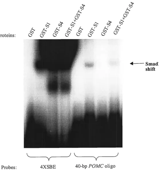
4.1 DNA-Binding Activity of Smad Proteins 95
4.2 SBEs in die POMC Promoter: Preliminary Resuits 96
4.3 SBEs in the POMC Promoter: Perspectives for the Future 102
CHAPTER5- CONCLUSION 106
LIST 0F FIGURES CHAPTER 1- INTRODUCTION Figure 1.1 3 Figure 1.2 7 Figure 1.3 14 Figure 1.4 18 Figure 1.5 23 Figure 1.6 25 Figure 1.7 32 Figure 1.8 34 Figure 1.9 38 CHAPTER 3- ARTICLE Figure3.1 87 Figure 3.2 88 Figure 3.3 89 Figure 3.4 90 Figure 3.5 91 Figure 3.6 93 Figure 3.7 94
CHAPTER 4- DEFINING TUE MECHANISM 0F ACTION 0F BMP-SPECIFIC SMADS ON TUE POMC PROMOTER
Figure 4.1 98
Figure 4.2 99
Figure 4.3 101
DNA: deoxyribonucleic acid RNA: ribonucleic acid POMC: proopiomelanocortin
ACTH: adrenocorticotropin hormone e: embryonary day RP: Rathke’s pouch Œ-MSH: a-melanocyte-stimulating hormone PRL prolactin LH: luteinizing hormone GH: growth hormone TSH: thyroid-stimulating hormone fSH: follicule-stimulating hormone Bmp: Bone morphogenic protein Fgf: fibroblast growth factor SHH: Sonic hedgehog
a-GSU: a-glycoprotein subunit PC: protein convertase
CRH: corticotropin-releasing hormone GR: glucocorticoid receptor
GRE: glucocorticoid response elernent cAMP: cyclic AMP
PKC: protein kinase C
HPA: hypothalamus-pituitary-adrenal NBRE: Nur-binding response element HP: leukemia inhibitory factor AIk: Activin-like kinase
Tgf-f3: transforming growth factor MH: Mad-homology
SBE: Smad-binding element RAT: histone acetylase transferase HDAC: histone deacetylase CBP: Creb-binding protein LAP: latency-associated protein
SARA: Smad anchor for receptor activation MAPK: mitogen-activated protein kinase INK: Jun N-terminal kinase
I would like to thank every member of Drouin’s and Nemer’s laboratory, past and present, who contributed ideas, advice and support to what lias resulted in the very finest experience as a graduate student.
In no special order, I am grateftil to:
Frédérique
For taking me under your wing, and introducing me to tissue culture teclmiques-à ta façon.
Eric
For that talk we had on our first day, and the discussions that would follow. Gino
For your very opininated view on science, working habits, and life aitogether.
Christine
For your generosity, your genuineness and your cornplicity against “les salops du labo” Ann e-Marie
for the energy you bring into the Iab, and for your companionship at meets we aftended. Marïo
Forbeing Rio, the man who was bom ready to take on the world! Orjust a cup ofcoffee.
Georges
For your sound advice, weather predictions and your general good sense. Bonne Année! Gwendal
for taking life at your owil particular beat, and making the world a better place to do yoga. Bruno L.
For your advice and ideas, and for keeping me aiways on alert of that chair of yours. Steve
For getting me started in the morning with your high-pitched laugh, once I’m off the walls. Michet, Yves, Lynda, Chantai
For the very best assistance and answers to ail of my questions (the good and the bad).
Emilie
for tarning those crazy Europeans, and taking on those primary cell cultures when you did. Pépijn
For ail those serious and then flot so serious discussions about research and the Dutch.
Jun
For your multiple HI!s on any single day, and for that contagious smile ofyours. Lise
For your work, your devotion and simply being the very best secretary in the universe. Jean-f rançois
For your work iast summer and your enthusiasm for the project.
Aurélio, Alain, Vincent, Mélanie and others collegues that I might be forgeting
For all your contributions, to my project and my experience in the lab.
Everyone in Mona’s mb (in general due to tack of space)
For your help, your kindliness and putting up with my intrusions at the 524. A very special thanks to my professor,
Jacques
For taking me on to your team, and providing me with the tools to do my research but rnostly for instilling in me the know hows ofcritical thinking that should serve me in manyjourneys.
I am also indebted to Dr. Hoang Trang and Viviaime Jodoin, from the Department of Molecular Biology, for their continued guidance during the course ofmy Master’s, and also to the members of the jury for taking on the task of correcting this thesis. And of course, none of this could have been possible without the support of those that are dearest to my heart.
1.1 Corticotrophs in the Mature Pituitary
The pituitary gland, also known as the hypophysis, is a specialized neuroendocrine organ that coordinates the control of peripheral physiology in response to stimuli derived from the brain and other endocrine glands. It can be divided rnorphologically and functionally into an anterior and intermediate lobe, which together constitute the adenohypophysis, and a posterior lobe known as the neurohypophysis. Corticotroph celis are found in the anterior lobe, and they principally produce adrenocorticotropin honnone (ACTH) by proteolytic processing of proopiomelanocortin (POMC). Adrenocortin (ACTH) is well known for its role in the regulation of the stress response by increasing the production of cortisol from the adrenal gland (108). Meianotroph celis, located in the intermediate lobe, also express the FOMC precursor gene, which is processed in a different manner to generate a-melanocyte-stimulating hormone (u-MSH). The intermediate pituitary is well-defined in rodents, but degenerates after birth in higher animais, inciuding humans wherein Œ-MSH is essentially produced by extrapituitary cells (115). Melanocortin (Œ-MSH) was initially characterized as a regulator of skin pigmentation by inducing the production of melanin from keratinocytes. It is now known to be a general modulator of skin biology and pathology (151)and (150).
four other hormone-producing cells are present in the pituitary anterior lobe; nameiy, prolactin (PRL)-secreting lactotrophs, thyroid-stimulating hormone (TSH) secreting thyrotrophs, luteinizing hormone (LH)- and follicle-stimulating hormone (F$H) secreting gonadotroplis, and growth hormone (GH)-secreting somatotrophs. These hormones regulate such functions as body growth (GH), rnamrnary growth and
developrnent (PRL), as well as thyroid gland (TSH) and gonad (LH and FSH) functions (121). The neuropituitary hormones oxytocin and vasopressin, secreted from nerve endings in the neurohypophysis, serve homeostatic functions in water balance and repi-oduction, respectively(25).
1.2 Pituïtary Organogenesis
1.2.1 Developmental Origin ofthe Pituitary
The pituitary gland is composed of tissues of two embryologicaly distinct origins: the adenohypophysis consisting of epithelial or glandular celis derives from ectodermal tissue, and the neurohypophysis is of neuroectodermal origin (217). Pituitaiy developrnent in the mouse occurs in a midiine region of oral ectoderrn or stomodeum that contacts neuroectoderm (ventral diencephalon) destined to become the floor of the forebrain (Figure 1.1). By embryonic day 9 (e9.O), the oral ectoderm involutes to form the pituitary rudiment, Rathke’s pouch. In what is probably the first complete anatomical account of early pituitary development (217), Schwind describes Rathke’s pouch of rat as a structure that arises from an invagination of the stomodeal ectoderm. The idea tliat Rathke’s pouch actively folds in to meet up with the neuroepithelium dorsally lias since been challenged. Through studies performed in quail/chick chimeras (41), Bufo albino/wild-typc chimeras (111) and mouse embryos (117), it was dernonstrated that contact between oral ectoderm and ventral diencephalon layers in the developing brain is sustained only at the level of the pituitary rudiment. Rather tlian invaginating, Rathke’s pouch merely appears to take an inward fold, because of rnaintained contact at pituitary level when everywhere else surface ectoderm and neuroepithelium get separated by invading mesenchyrne of pre-chordal plate and neural crest origin. At the same time, the overlaying ventral diencephalon grows downward to generate the infundibulum destined to become the neurohypophysis.
Ventral D iencephalon BMP4 Pituitary Primordium (Oral Ectoderm) e9 .0 Rathke ‘s Pouch (RP) elO.5 Anterior Pituitary e12.5
Figure 1.1 Expression Pattem of BMP and FGF Signaling Molecules During Early Pituitaiy Development (Adapted from Ericson, J. et al., 1998; Treier, M. et al., 1998 and 2001)
FGF8I1O FGF8I1O
j’.
t
1.2.2 Pituitary Primordium Induction
Rathke’s pouch formation requires that the pituitary prirnordium receive and correctly process inductive signais coming from the ventral diencephalon (44). Expression pattem studies have demonstrated Bone morphogenic protein (Bmp)-4, Fibroblast growth factor (Fgf)-$, and Fgf-1O to be expressed in a restncted region of the ventral diencephalon that is in direct contact with the pituitary primordium (244) (67). Bmp-4 expression is detectable in the ventral diencephalon by e8.5, prior to the appearance of Fgf-8 (Figure 1.1), suggestive of a role for Bmp-4 in the earliest phases of pituitary development. The compiete lack of pituitary rudiment in a smaii population of Bmp-4 mice that survived to elO, a time at which Rathke’s pouch formation is normally weil under way, support a role for Brnp-4 signaling inthe induction of the pituitary primordiurn (237). The possibility that conveyance of another inductive signal might have been affected in these Bmp-4 gene deleted mice is unlikely since contact between ventral diencephalon and oral ectoderm was rnaintained in the absence of Bmp-4. Such a penetrating phenotype was however not obsewed in mice upon ectopic expression of Noggin, a Bmp-specific antagonist (174), in Rathke’s pouch and oral ectoderm using the Fitxl promoter. Pituitary development in these
Fitxl-Noggin transgenic mice was abrogated only afier the rudimentary pouch had formed (244). A partial blockade of Brnp-4 function by Noggin could be to blame for the discrepancy with Bmp-4’ embryos.
1.2.3 Rathke’s Pouch Formation
Formation ofRathke’s pouch from the oral ectoderm also appears to be controlled by Fgf and Sonic hedgehog (Shh) signals. Fgf-8 and Fgf-1O have been shown to be expressed in the ventral diencephalon at the time (e9.5) ofpouch formation (22). Infgf 10
Fgf-10 and that has moreover been detected in Rathke’s pouch (237), is thought to mediate Fgf signaling within the oral ectoderm. Indeed, Fgfr-2(IIIb) gene deleted mice exhibit an arrest in pituitary development that is similar to that observed in fgf-]0’ mice (50). Targeted disruption of the homeobox gene T/ebp, expressed during early development in
the ventral diencephalon, also resulted in disruption of pituitary development subsequent to a loss of the neuroectodermal region of Fgf-8 expression (237). However, dysmorphogenesis of the vental diencephalon in these T/ebp nuli mice poses a problem in
interpreting a direct role for Fgf-$ in pituitary organogenesis.
Two reiated transcription factors are postulated to mediate fgf early signais, the
Lim horneobox proteins Lhx-3 and Lhx-4 which are both expressed specifically in the pituitary rudiment by the time Rathke’s pouch formation begins (e8.5). hi vitro experiments have shown that Fgf-$ lias the ability to maintain Lhx-3 expression (67). li the absence of both Lhx-3 and Lhx-4, mutant mice show no more than a rudiment of Rathke’s pouch (219)as do fgf]OE’ and fgfr(2111b7’ mice. Yet, neither Lhx-3 (220)or Lhx-4 (219) single mutant mice exhibit such a pronounced phenotype, each single mutant pituitary developing into a glandular structure. Hence, the presence of either Lhx-3 or Lhx 4 seems to be required for the progression ofpituitary developrnent beyond the rudimentary stage.
Shh is expressed in surrounding oral ectoderm, mesenchyme and diencephalon, but is specificaïly excluded ftom Rathke’s pouch (244) (245). Gene deleted mice for Shh have not been usefui to understand the role of Shh in pituitary development since total ioss of hedgehog function altogether interferes witli the establishment of ventral dienceplialic contacts with the pituitary primordium (245). The significance of Shh signaling was studied using Fitx]-Hip transgenic mice; nameiy, Pitx] promoter sequences known to
target gene expression to the oral ectoderm and Rathke’s pouch were used to ectopicaly express the hedgehog-specific inhibitor Hip (245). This block in pituitary hedgehog signaling interfered with progression beyond rudimentary pouch development. A direct role for Shh in Rathke’s pouch formation was nonetheless suggested by the Pitxl-Hip experiment since the expression domain ofFgf-8 was flot disrupted in presence of an intact ventral diencephalon.
1.3 Pituitary Ceil Expansion amiDifferentiation
The six celi types of the anterior pituitary exhibit an ontogenic pattem of hormone gene expression that follows a defined temporal, and sornewhat spatial sequence of appearance (100). The first sign ofpituitary ceil fate commitment cornes at elL5 with the appearance of a-GSU transcripts on the ventral side of the expanding anterior pituitary (Figure 1.2). Corticotroph celi specification, marked by the appearance of POMC/ACTH at e12.5 in the mouse anterior pituitary, appears nonetheless to precede the specification of any other celi lineage in the developing pituitary. Melanotrophs, which constitute the other lineage of POMC-expressing ceils in the pituitary, differentiate later as POMC/MSH are detected only at e14.5 in the intermediate pituitary. A transient population of thyrotroplis, identified by expression of the TSH-3 subunit, appear on e13 in the rostral end of the anterior pituitary (138). They will disappear after birth. A second cluster of TSH3-expressing thyrotrophs appears on e14.5, and represents the terrninally differentiated thyrotrophs, followed by the contemporaneous expression of GH (somatotrophs) and PRL (lactotrophs) on e16. Gonadotroph-specific LH-13 and FSH-13 transcripts finally appear at e16.5 and e17.5, respectively.
The specification and expansion of ceil fates from a common pituitary primordium has been proposed to be the consequence of overlapping expression pattems of specific sets
Exfrmnsic Factors BMP-2 SHET Fgf? Intrinsic Factors Pit-1 Pitxl/2 Lhx3/4
etc. NeuroDl TPit Pjtx-1/2
Pït-1 Gata-2
%iNt Pitx-1/2
El 6 Pitx-1!2 Pit-1
ER Pitx-1/2 Pit-1 RAR]
TR
Pitx
Gata-2 Spi Egr-1
1/2 Sf-1
ACTU (POMC) TSHf3
PRL GU
Figure 1.2 Ontogenic Pattem ofPituitary Hormone Expression. (R) rostral, (C) caudal, (D) dorsal, (V) ventral, (IP) intermediate pituitary, (AP) anterior pituitary.
1.5 Lhx-3 Pitx-1/2 E12.5 E14.5 lŒ-GSU D E14.5 E16 MSH-a (POMC) R4÷C F1 6.5 E17.5 Lll3 Zn-1 fFSllp Pitx Gata-2 1/2 SF-i
of extrinsic growth and intrinsic transcription factors, expressed in a precise spatiotemporal manner during organogenesis. One approach to understanding the molecular mechanisms that mediate the emergence of pituitary ceil types is to study promoter elernents implicated in the regulation of hormone marker gene expression (Figure 1.2), as transcription factors conferring cell-specificity are also often implicated in celi differentiation.
Pituitary celis can be classified into three groups according to similarities between components mediating their distinct differentiation pathways. One group is comprised of somatotrophs and lactotrophs, another of thyrotrphs and gonadotrophs, and yet another of corticotrophs and melanotrophs. A paraïlel can be made between group members with regards to their hormone structure and developrnental origin.
1.3.1 Somatotroplis and Lactotroplis
On the basis of primary structure and biological function similarities, GH and PRL
have been grouped together with the related placental lactogen (PL) in the PRL/GH/PL family. GH, FRL and FL genes are thought to have arisen from a common ancester by gene duplication and evolutionary divergence (23,33). CelIs that secrete both GH and PRL, known as mammosomatotrophs, have been identified in neonate and aduit rats by reverse hemolytic plaque assays (27) and inirnunocytochemistry (29,29). Gene ablation techniques specifically targeting GH-expressing celis in the developing pituitary have shown an almost complete absence of both somatotrophs and lactotrophs in transgenic mice, reinforcing the concept of a stem-somatotroph as common precursor to somatotroph and lactotroph ccli populations. Also, sornatomaminotropic pituitary tumors are quite common (33).
1.3.1.1 Somatomammotropic Piti- and Propi-dependent differentiation pathway
The molecular basis of somatotroph and iactotroph ccli differentiation implicates the activities of the pituitary-specific horneodomain protein Pit-1. Pit-1 expression is
arise (100). Experiments have shown that Pit-1 mRNA transcripts are actually expressed at the same level in ail pituitary ceil types, but translated to significant protein levels only in somatotrophs, lactotrophs and thyrotrophs (230). Initiaily identified as Growth Hormone Factor- 1 (GHF- 1), Pit- 1 was cloned as a transactivator of FR1 and GH gene promoters (97,240).
The importance of Pit-1 as a regulator of somatotroph and lactotropli differentiation was demonstrated by the absence of these two celi types in the pituitary glands of $nell (dw) and Jackson (dw) dwarf strains of mice in which the Fit-1 gene is mutated (6,35,135). Acting upstream in the differentiation pathway ofPit-1 is the pituitary specific paired-like homeodomain factor Prophet of Pit-1 (PROP-1), detected in Rathke’s pouch from elO.5 to e 16.0 (225). Prop-] mutations were identified in the Ames (df) dwarf mouse, which moreover exhibits defective Fit-1 gene expression and shares phenotypic defects with dw and dwJ FUi-mutant mice. Pituitary hypoplasia in dw, dw and df dwarf strains of mice is in support of a role for PROP-1 and Pit-1 not oniy in the establishement and maintenance of differentiated phenotypes, but aiso in the proliferation of precursor cells (135,225). How ceil proliferation signais are coupied to celi commitment and differentiation signais during pituitary development remains to be eiucidated. In hurnans, PROF-1 mutations have been characterized in and related to an absence or low leveis of GH, PRL, T$H, LH, FSH and recently ACTH (183) in human combined pituitary hormone deficiency (CPHD) (64).
1.3.1.2 Cooperation between Pit-1 and Nuclear Receptors
Extensive analysis of rat and human GH gene reguiatory elements have implicated Pit-l binding and ensuing cooperation with retinoic acid receptor (RAR), thyroid hormone nuclear receptor (TR), and the zinc finger Zn- 15 protein in effective sornatotroph-specific
expression (40,140). Pit-1 is believed to direct lactotropli-specific PRL gene expression by collaborating with estrogen nuclear receptor (ER), as weÏl as Ets and Pitx factors (42,48,248).
1.3.1.3 Pitx Homeobox Proteins Collaborate with Piti
The homeobox Pitx 1 and Pitx2 transcription factors are other general regulators of pituitary-specific transcription, including GH and PRL gene expression (Figure 1.2). Pitx 1 was identified as a transcriptional regulator of POMC gene expression (12$), and also as a factor interacting with Pit-1 (235). Fitx2 was isolated as the causative gene by haploinsufficiency for Rieger’s syndrome (21$). Pitxl and Pitx2 expression defines the oral ectoderm as early as e$.0, and is rnaintained in derivative structures throughout pituitary development (129,137). In addition to their pan-pituitary expression, Pitxl and Pitx2 are expressed in distinct regions of the embryo. Their complex pattern of expression is consistent with the roles of Pitxl in such developmental processes as craniofacial and limb development, and the roles of Pitx2 in establishment of laterality, as well as heart, Iting, and craniofacial development (129,137,234). These roles will not be discussed further. The last member of the Pitx subfamily, Pitx3, is flot expressed in the pituitary (132) which suggests that it does flot play a role in pituitary functions.
Pitxl and Pitx2 have sirnilar transcriptional activities on POMC, a-GSU, LH-/3,
FSH-/3, TSH-fi, PRL, and GH pituitary-specific promoters (247). Their pan-pituitary expression, and contribution to ceil-specific transcription of many pituitary specific genes may reflect the common origin of pituitary celis. Like Pit- 1, Pitx factors are thought to confer promoter-specific expression through synergistic interactions with cell-restricted factors. With respect to sommatolactotroph ceil differentiation, Pitxl was shown to cooperate not only physically, but also transcriptionally with Pit-1 on the GH and PRL
expression levels of either GH or PRL (130,234) as Pitx2 is thought to have compensated for pituitary ceil differentiation functions. Such a role for Pitx2 in the differentiation of the sornrnatolactotroph ceil lineage couli flot be studied in Pitx2 mice because of premature pituitary developmentaÏ arrest (137).
1.3.2 Ihyrotrophs ami Gonadotrophs
The glycoprotein hormones LH, FSH and TSH are heterodirners cornposed of a common Œ-glycoprotein subunit (a-GSU) noncovaÏently assernbled with respective hormone-specific 13-subunits LH-13, FSH-13, or T$H-f3. The f3-subunit confers hormone specificity, while the Œ-subunit is homologous within a species. Phylogenetic studies using nucleotide and arnino acid sequence alignrnents predict that both Œ- and f3-subunits evolved
from a single ancestor through gene duplication (134). During pituitary development, thyrotrophs are thought to derive from a pool of precursor ceils that also give rise to sommatotrophs and lactotrophs, but not gonadotrophs. In a-GSU gene-deleted mice, hyperpiasia and hypertrophy of TSH-positive cells as a resuit of thyroid dysfunction was accompanied by a reduction of GH and PRL celis (49).
1.3.2.1 Interplay between Pit-1 and GATA-2 Activities
Although thyrotrophs and gonadotrophs do not seem to share a common precursor, reciprocal interactions between Pit-1 and the zinc finger protein GATA-2 would be implicated in the specification of both cell types (46). Differential GATA-2 function in these two celi types is defined by extrinsic Bmp2 and Shh signaling (46,245). Bmp2 signals are detected in the ventral juxtapituitary mesenchyrne (VJM) as well as in the ventral region of the committed pituitary around e 12.5, while Shh is expressed around but not in the pituitary (67,244). Transgenic studies have shown that gonadotroph- and
thyrotroph-specific GATA-2 expression is induced by BMP-2, itselfinduced by Shh (245). Elevated GATA-2 expression levels have been associated with the inhibition of endogenous Pit-1 gene expression in gonadotrophs (46,244). Expanding GATA-2 expression under the control ofPit-1 regulatory elements in transgenic mice is sufficient to convert ail Pit-1 dependent lineages to the gonadotroph fate (46). In thyrotrophs, lower levels of GAlA-2 are not believed to interfere with Pit-1 expression; in these ceils, Pit-1 and GAlA-2 were shown to interact and functionally cooperate to activate the TSH-fi promoter (73). In thyrotrophs, Pit-i is moreover thought to function to inhibit GATA-2 binding and activation ofLH and FSH regulatory elements that do not contain adjacent Pit 1 binding sites. The ability of Pit-1 to interfere with GAlA-2 function is lost in Snell dw mice that have a W48C mutation in the Pit- 1 POU homeodomain which dismpts Pit
1/GATA-2 interactions, causing thyrotrophs to assume a gonadotroph fate (46). 1.3.2.2 Celi-Specific Collaboration ofPïtx Factors with Egr-1, SF-, Spi and Lhx
Factors
In pituitaries of Pitxl-deleted mice, LH-, FSH-J3 and TSH-3 levels are severily reduced, suggesting that Pitxl is required for expression and/or maintenance of gonadotroph and thyrotroph lineages (130,234). The Pitxi binding site in the LH-/3 promoter was dernonstrated to be essential for its activity in vivo; transgenic mice harbouring a mutation of the Pitxl binding site in the LH-fl promoter lost basal as well as Gonadotropin-Realesing Hormone (GnRH)-stirnulated pituitary expression (201). GnRH is a critical hypothalamic peptide that is required for the production and secretion of gonadotropins LH and FSH (54). Along with Pitxl, the zinc finger protein Early response 1 (Egr- 1) and Spi proteins, as welI as nuclear receptor steroidogenic factor- 1 (SF- 1) have been shown to coordinate the complex control of basal andlor GnRH-stimulated LH-fi gene
compromised (98,153,243). Expression 0fFSH-13 was also absent in Sf-] pituitaries. It appears that LH and FSH deficiencies in $FF’ mice may be rnediated by defective GnRH expression in hypothalamus (98). The integration of Pitxl in previously described synergistic functions of GATA-2 and Pit-1 on the T$H-/i promoter is stili poorly understood.
Pitx and Lhx factors, via the actions of Lim-associated cofactor (CLIM), have been implicated in gonadotroph-specific activity of the bovine a-G$U promoter (9,109). Distinct cis-acting elements, stiil undefined, are thought to regulate a-GSU expression in thyrotrophs (49).
1.3.3 Corticotroplis and Melanotroplis
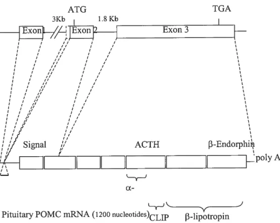
Both anterior lobe corticotrophs and intermediate lobe melanotrophs express and transcribe the POMC gene, beginning in mice at e12.5 in corticotroplis and at e14.5 in melanotrophs (100). A POMC cDNA was originally obtained from the intermediate lobe of bovine pituitaries (175), and since then the POMC gene has been cloned in rat (59), human (236), and mouse (185) among other species. Two introns and three exons make up the rat POMC gene (figure 1.3), which measures approximately 6.5 kilobases (kbs). A mature 1200 nucleotide POMC mRNA transcript is found in the pituitary as a result of gene spticing (62). It is essentially the same transcript that is translated in corticotrophs and melanotroplis, but the resulting POMC peptide prohormone is subsequently processed in a different manner in each celi type by distinct cohorts of protein convertases (PC). ACTH resuits from corticotroph PCi proteolytic activity, and the additional PC2 in rnelanotrophs processes ACTH further into Œ-MSH and CLIP (11). ACTH, -1ipotropin, and
Rat POMCgei 5Kb) ATG TGA 3Kb 18Kb I ___1iJl_/,L]Exon
J
Exon3 I II j / I I /f I / I I / I I I / I I I I 1 ,// j I il / I ‘ f? I / I II I / j I /1 I / j I il I i / I I I /1 I / , /1 I / j ,/ I / / / I / j ,/ I / I ,/ I / ,/ j f / / II / II ,/ , I, I / I j I /, j If j j j’ j’ I ,, j’// ACTH 3-Endorphii fII
[...
poly Â. ‘j cxPituitary POMC rnRNA(1200 nucleotïdes)CLIP J3-lipotropin
(152). Processing into ACTH depends on the PCi proteolytic activity; the additional activity ofPC2 in mclanotrophs processes ACTH ftirther into Œ-MSH and CLIP (11). 1.3.3.1 Contribution of Pitx Factors to POMC Lineages
In the end, single Pitx gene ablation experiments have not proven to be useful in assessing the actual need for Pitx activity for corticotroph and melanotroph ccli function. In neither Pitxi’ nor Pitx2 mice was POMC expression affected (130,137,234), suggesting that POMC expression in vivo would require either Pitx gene activity. Pitx]/Pit2 double knock-outs could have been more informative in this case. However, the premature block in pituitary development observed in the latter double nuil mice has made the study ofpituitary ceil differentiation impossible (229).
Clues on the contribution of Pitx factors to the specification of corticotroph, and possibly melanotroph ccli fates, have been provided by studies perfomied on the POMC promoter in AtT-20 corticotroph ccli model. An account of POMC gene transcription studies will be given below. It is worth mentioning at this point the central position that Pitx factors hold in mediating protein interactions with two major determinants of the corticotroph ccii fate; that is, the bHLH factor NeuroDiand the newly characterized T-box factor Tpit.
1.3.3.2 NeuroDi, an Exclusive bHLH Factor for Cortïcotrophs
The bHLH class of transcription factors lias been studied extensively for their role in myogenic and neurogenic processes, as tissue-specific regulators of ccii specification. NeuroDi is a neuro- and pancreatic-islet-specific bHLH transcription factor implicated in neuronal precursor differentiation and tissue-specific expression of the insutin gene, respectively (133,17$). The expression of NeuroDi is transient in the developing mouse
pituitary, present between e12 and e15, but not afler e16 (198); moreover, its expression is restricted to corticotrophs. NeuroDi mRNA but flot protein can be detected in the aduit mouse (199). NeuroDi transcripts have also been detected in normal hurnan pituitaries and in ACTH-secreting pituitary adenomas (191). NeuroDl expression preceedes that of POMC in differentiating corticotrophs, inferring a role for NeuroDi in the induction of corticotroph differentiation. NeitroDi nuil mice did not exhibit a significant loss of pituitary POMC expression when analyzed at e17.5. However, a delay in the appearance of POMC expression in the anterior pituitary of NeuroDT’ mice was recorded at earlier times (e14.5), suggesting that NeuroDi participates but is not essential for the onset of corticotroph differentiation (Lamolet B, unpublished). NeuroDl has been demonstrated to confer corticotroph-specific activity to thePOMCpromoter through synergistic interactions with Pitxl, the details ofwhich are discussed below.
1.3.3.3 Conversion of POMC Ceils into POMC1 Celis by the T-box Factor Tpit
T-box factors are defined by a conserved DNA-binding motif known as the T-box, named afier the first-discovered T-box gene Tor Brachytuy (255). Members of this farnily of transcription factors have been identified in both vertebrates and invertebrates, where they have been implicated in developrnental decisions concerning patteming (224)and recently celi differentiation (127,233). Tpit expression studies have shown that it is restricted to corticotroph and melanotroph POMC-expressing celis in the developing pituitary and precedes POMC expression in each cell type by 0.5 days, suggesting a role for this factor in corticotroph and melanotroph differentiation. Transgenic expression of Tpit under the control of the a-GSU promoter was sufficient to induce expression of POMC in ceils ofthe rostral tip ofthe developing pituitary (127). This particular structure contains a population oftransient, proliferating, uncornmitted cells (179) that do flot normally express
POMC transcription in Pitx-1-expressing uncomrnitted pituitary cells (Figure 1.4). The expression of the corticotroph marker NeuroDi was not induced in these ectopic POMC expressing celis, indicating that additional information is required for terminal differentiation into corticotrophs.
1.3.3.3.1. Isolated ACTH Deficiency
In humans, POMC is expressed in three tissues: in the anterior pituitary to
stimulate cortisol production by the adrenal gland (the intermediate pituitary degenerates afier birth in humans), in the hypothalamus to regulate appetite via the leptin pathway, and in skin where it plays a role in pigmentation and cutaneous inflammation. Genetic defects in the POMC gene have been identified (122). Consistent with the expression pattem of POMC, these patients suffer from adrenal insufficiency, early onset obesity and red hair pigmentation. The lack of a-MSH peptide in POMC-deficient patients is likely to blarne for the weight and pigmentation abnorrnalities. Hypocorticolism and hypoglycemia on the other hand are thought to reflect ACTH insufficiency, a phenotype that has moreover been documented separately inpatients with an isolated deficiency of pituitary ACTH (26). Our findings on Tpit, and its particular expression and role in ACTH-expressing corticotrophs
in mouse, led us to investigate whether mutations in the TRIT human gene should produce an isolated deficiency of pituitary ACTH.
Two out of three analyzed cases of children bom with an isolated deficiency in ACTH tumed up with mutations in the TFIT gene (127). In one case, a homozygous point mutation was identified that introduces a premature translation termination codon in the open reading frame of TPIT. As a result, the TPIT protein is either translated in a truncated
D
C. R
V
1.4 Ectopic Expression of Tpit in aGSU-Tpit Rostral Ceils Induces the Expression ofPOMC (adapted from Larnolet B, 2001). (R) rostral, (C) caudal, (D) dorsal, (V) ventral.
E14.5
WT
a-GSU
POMC and Tpit non-expressing ceils • POMC and Tpit expressing ceils
mechanism known as non-sense mediated mRNA decay (NMD) ($6). This chiid’s parents, and one grandmother, were found to be heterozygotes for the mutation but free of the ACTH deficiency, making inheritance recessive. The second child studied was found to be heterozygous for a point mutation that changes serine residue 12$, a residue conserved in the T-box of ail known famiiy members, to phenylalanine. The ensuing mutant TPIT protein in this heterozygote patient might be acting in a dominant negative mairner to inhibit normal TPIT function coming from the TFJTallele that is not mutated.
1.3.3.4 Growth Signaling Factors
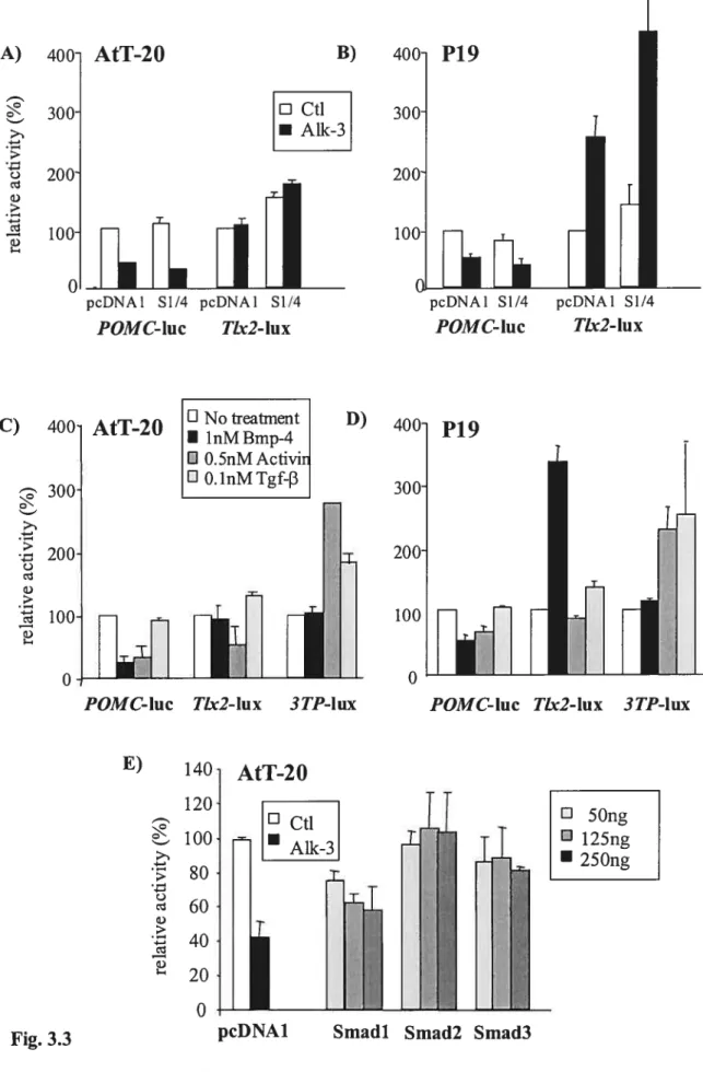
How the activities of Pitx, NeuroD I and Tpit transcription factors are regulated by outside growth signaling factors is stiii misunderstood. Signaling factors intrinsicaily expressed in Rathke’s pouch have been shown to suffice for corticotroph ce!! specification from e9.5 and on (67,244). Prior to this, Shh signais seem to be required either for induction or maintenance of ACTH expression, which is lacking in Fitx]-Hip transgenic mice (245). No other gene deieted or mutant animais for pituitary restricted signaling or transcription factors have shown a compiete absence of POMC/ACTH expression. The early arrest of pituitary development caused by the absence of such pleiotropic molecules as Bmps and Fgfs, or the possible incomplete penetrance of transgenic experirnentai methods render the in vivo study of the specific roies of signaling factors in the differentiation of POMC-expressing corticotrophs and melanotophs difficult. My Master’s project stems from contradictory resuits that had previously been obtained by two groups of investigators in their attempt to define the role of Bmp-4 signaling on embryonic corticotroph specification. Indeed, ACTH expression was downregulated in ex vivo e9.5 Rathke’s poucli expiants cultured with Bmp-coated beads (67), whiie ACTH expression did
flot appear to be affected in gain-of-function aGSU-BMP4 transgenic embryos (244). By means of the extensive characterization of the rat POMC gene promoter in Dr. Drouin’s laboratory and given that POMC expression is a marker of corticotroph ceils in the anterior pituitary, my approach in elucidating the contribution of Bmp factors to corticotroph celi differentiation has consisted in studying the role that Bmp signaling plays in the control of FOMC promoter activity.
1.4 POMC Transcription
POMC promoter studies in our laboratory have been executed in the mouse POMC/ACTH-expressing AtT-20 celi une, which has its origins in pituitary turnorigenic tissues (24). Seeing that these ceils express such early protein rnarkers as NeuroDi, Pitx and Tpit, AtT-20 celis are used as a model of differentiating corticotrophs (127,128,199). The same rnechanisrns driving POMC promoter activity in AtT-20 cells would hence be expected to induce POMC expression in corticotrophs, but not necesarily in rnelanotrophs which seem to differ. NeuroDi for example is not expressed in melanotrophs (199). POMC promoter studies in our laboratory have typically lcd to supporting in vivo evidence in mouse models, and hence constitute an important tool to study the molecular basis of corticotroph differentiation.
1.4.1 Corticotroph-Specfflc Regulation of PO1’IC Expression 1.4.1.1 POMCPromoter
FOMCpromoter sequences from —480 to +63 bp were shown to have corticotroph
specific activity in AtT-20 ceils, as well as in transgenic mice (141,249). The —4801+63 bp promoter, hereafier referred to as the full length promoter, wasonly expressed in transgenic
anterior and intermediate pituitary but not in hypothalamic regions also known to express POMC. Other regulatory sequences would be involved in hypothalamic expression of POMC (272). Deletion studies that made use of different regions of the pituitary-specific
reporter gene and transfected into AtT-20 celis, were performed to determine the contribution to promoter activity made by the distal (-480/-324), central (-323/-166) and proximal regions (-165/-34) (241). In doing so, the distal together with the central dornains of the promoter were demonstrated to confer specificity to FOMC prornoter activity. Out of the three latter regions, only the central one was shown to possess some activity on its own.
Through footprint and mutation analyses, it was shown that individual regulatory elements within the FOMC promoter contribute to its transcriptional activity (241). Indeed, the loss of any of these elements decreased POMC prornoter activity significantly. Among the many transcription factors that have been identified to mediate transcriptional control of POMC at such binding sites (f igure 1.5), only a few have been retained as having corticotroph-specific roles. Namely, the bHLH factor NeuroDi acting in the distal region, and the homeodomain-containing transcription factor Pitx 1 with its obligate T-box partner, Tpit, both acting in the central region ofthe promoter. Not only would these two regulatory elements, namely the distal NeuroDi Ebox (EbOXneuo) and central Pitx/Tpit binding sites, be responsible for corticotropli specificity but they would moreover be mediating transcriptional synergy between the distal and central domains ofthe FOMC promoter.
1.4.1.2 NeuroDi and Pitx Synergistic Interactions
Analysis of the regulatory elements contributing to AtT-20 cell-specific transcription within the central domain of the POMC promoter led to the cloning of the homeobox transcription factor Pitxl (Ptxl) (12$). Drosophilct bicoid-related Pitxl is a member of a subfamily that also includes mammalian Pitx2, Pitx3, Otxl, Otx2, and goosecoid (60). The Pitx subfamily of transcription factors is characterized by a paired-like DNA-binding homeodomain in which residue 50 is a lysine. Pitx factors bind as monomers
to a single site in the POMC promoter, where they act as transcriptional activators tbrough their C-terminal transactivation domain (128). Besides its pituitary actions, Pitxl has also been implicated in POMC expression in human small ceil lung carcinomas (SCLC) (180).
Pitx factors have been shown to be the basis for synergistic activities between thc distal and central domains of the FOMC promoter (Figure 1.5), Pitx bound to the central domain interacting with NeuroD I -containing 5HLH heterodimers bound to the distal domain (198,199,242). NeuroDl, and related tissue-specific bHLH factors like MyoD in muscle, dimerize through their HLH motif with ubiquitously expressed bHLH factors E12 and E47 (72). Only in the heterorneric form may NeuroDi, through its basic motif bind to EbOXneuro and moreover collaborate with Pitx factors. ftideed, in vitro binding as well as in vivo co-immunoprecipitation and transfection studies have shown that it is the ubiquitous bHLH partner ofNeuroDl that directly interacts with Pitx factors (19$).
In the context of the full length promoter, both Pitx and NeuroD 1 binding sites are required for transcriptional synergism (198). In transfection assays however, transcriptional synergism can be reconstituted in the absence ofPitxl DNA binding activity but not independently of DNA binding by bHLH factors, highlighting the importance of protein:protein interaction for synergism (19$). Deletion ofthe distal domain resuÏted in a thousand-fold loss of POMC prornoter activity in transgenic mice (198), and mutation of Eboxueuro significantly compromised the activity of the full length FOMC prornoter in transfected AtT-20 ceils (199). Although the Eboxneuro binding site for NeuroDi bHLH transcription factors seems to be at the foundations of corticotroph-specific synergistic activation of the POMC promoter, NeuroD 1 itself is flot required sensu strictu for FOMC transcriptional activation. Indeed, substituting the Eboxneuro for an Ebox that binds ubiquitous bHLH homodimers (Eboxb) restored promoter activity (199). $till, the EbOXneuroofthe distal region of the POMC prornoter is incapable ofbinding ubiquitous
Regulatory factors: Regulatory POMC elements: Base pairs: Corticotroph-specific synergistic activity
Nurr77/ NeuroD 1 /E47 TpitPitx (E12/E47) GR N
fos/Ju
-480
distal central proximal minimal
-166 -34 +63
bHLH hornodimers, hence the requirement for NeuroDi in corticotrophs for sequence specific recognition and activation of the EbOXneuro.
1.4.1.3 Tpit, an Obligate Partner of Pitx Factors
Pitx activity on the central domain of the POIiC promoter is dependent on
synergistic interactions with Tpit. from a mutational analysis of the regulatory elements surrounding the Pitx binding site, Tpit was recently cloned in our laboratory as an obligate Pitx partner (127). The Tpit binding site in the POMC promoter actually conesponds to a haif site of the palindrornic Brachyury (T)-binding element (170). Tpit activity on POMC can actually be replaced by T, to which it is the most reiated, but not by Tbxl which is another T-box factor that is expressed in AtT-20 celis (127).
Pitx and Tpit can each weakly activate POMC transcription through their respective binding sites, which are 5 bp away fiom each other. Together, they collaborate in a synergistic interaction that would originate from their ability to cooperativeiy bind DNA, as was evidenced in in vitro binding studies (127). The loss of either binding site obliterates the synergistic activity between Pitx and Tpit. Protein:protein interactions between Pitx 1 and Tpit factors are thought to be mediated by the Pitx 1 horneodomain. Synergistic interactions between T-box factors and Pitx family members has been shown in celi culture for Pitxl, Pitx2 and Pitx3, but flot for the ciosely related subfamily of Otx transcription factors (127).
1.4.2 Hormonal Regulation of POMCExpression
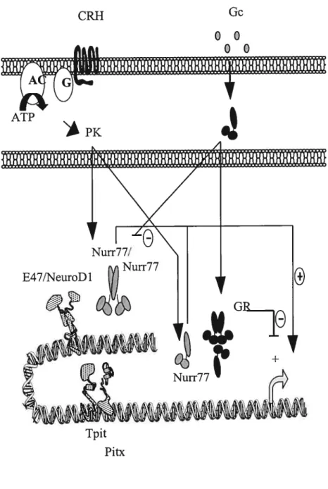
Once the terminal differentiation of corticotrophs is cornpleted, the regulation of FOMC expression mainly falis under a balance of stimulatory signais such as hypothalamic corticotropin-releasing-hormone (CRH) and negative feedback signais such as glucocorticoids (Gc) from the adrenal gland (Figure 1.6). These two modes ofregulation
CRH Gc 00 00 ATP Tpit Pitx
converge in corticotrophs to mainly control the secretion of pre-stored ACTH hormone in an immediate response, or in a long-tenn response to adjust the transcription rate of the FOMC gene.
1.4.2.1 CR11
The biological actions of CRH are mediated through the G-protein coupÏed transmembrane CRI-I receptor (193). Receptor activation subsequently turns on the adenylate cyclase second messenger system, leading to the production of cyclic AMP (cAMP) and activation of cAMP-dependent protein kinase A (PKA). PKÀ is known to control cellular functions through the phosphorylation of such transcription modulators as the cAMP response element binding protein (CREB), cAMP response element modulator (CREM), activation transcription factor (ATF)1/2 and general coactivators CBP/p300 (166). No such regulatory eÏements are present with the —4$0/+63 FOMC prornoter, yet CRH stimulation ofAtT-20 ceils does resuit in the increase of FOMC transcription (70,71). These CRI-1 effects are mimicked by cAMP analogs or by forskolin, and do flot appear to require de novo protein syrithesis since POMC levels increase even in the presence of cyclohexamide, an inhibitor of translation.
Several pituitary-specific POMC promoter sequences have been implicated in CRH responses (8,15,102,136), but flot much is known of their mechanism of action. We and others have shown that CRH inductive regulation of POMC expression in AtT-20 corticotroph ceils seerns to be the function ofprornoter regulatory elements for members of the Nur77 subfamily of orphan nuclear receptors (154,172,194). Ibis subfamily of transcription factors includes Nur77, also known as NGFI-B (164), Nur-related factor 1 (Nurrl) (131)and neuron-derived orphan receptor 1 (NOR-1) (186). Evidence that Nur77-reÏated activity is the principal mediator of CRH inductive effects on POMC comes from experirnents wherein the overexpression of a dominant negative Nur77 mutant blocks both
in the distal region (-395bp) of the FOMC promoter (194). This Nur response element (NurRE) is a palindrome that binds Nur factor homodimers and heterodimers (154). The two half sites share a partial homology with the monomeric Nur-binding response element (NBRE) first characterized in yeast (256). Such a NBRE is moreover found in the proximal region (-63bp) of the POMC promoter, but as the NurRE has been shown to be much more responsive to CRH treatments, corticotroph-specific CRH actions on the POMC prornoter would appear to be primarily mediated through the NurRE rather than the NBRE. Unfortunately, functional redundancy between members of the Nur subfamily have interferred with in vivo analysis, through gene deletion teclmiques, of their individual pituitary or hypothalamo-pituitary functions (38,43). (38)
1.4.2.2 GR
Glucocorticoid (Gc) negative regulation of ACTH release and FOMC transcription has emerged as a concept of feedback regulation operating to adjust activity of the hypothalamo-pituitary-adrenal (HPA) axis with physiological homeostasis. Gc acts at two levels in the HPA, basicalÏy on CRH neurons in the hypothalamus and on corticotrophs in the pituitary. Gc effects are mediated through the glucocorticoid receptor (GR) (155). To date, two different mechanisms have been proposed to account for GR-mediated repression
of POMC prornoter activity. Whether one or both of these mechanisms interfere with FOMC expression during development, HPA homeostasis or pathology is not well understood.
In one case, GR would mediate Gc-induced repression of POMC through direct DNA contacts with a negative Gc response element (nGRE) located in the proximal domain of the promoter (63). Deletion analysis has dernonstrated that the nGRE is required for Gc repression of POMC in AtT-20 cells (63,173) and in transgenic mice (79,249). In vitro,
three molecules of GR bind to the nGRE (61); however, the exact molecular basis of transcriptional repression by the GR complex at this site is stili elusive. In vivo experiments are moreover in support of a nGRE-mediated mechanisrn for GR repression of POMC expression. GR-deficient mice are characterized by an upregulation of CRH and POMC at both levels in the HPA axis (37). Mice that express, using knock-in technologies, a mutant GR deficient in dimerization do flot exhibit the same phenotype. GR molecules that no longer dimerize would stili be able to participate in transrepression mechanisrns of gene control, but flot in GRE/nGRE binding. Mice expressing this forrn of GR showed upregulated POMC expression in the pituitary, while their CRH levels in the hypothalamus were under negative control presumably by monomers of GR (205). GR dimerization hence seems to be required for the negative regulation of pituitary POMC levels. Nevertheless, other mutant mice models are in support of alternative mechanisms of negative Gc feedback on the FOMC promoter (168,169).
Centered at —63bp, the nGRE overlaps with the NERE, which suggests that GR may likewise participate in a competitive mode of action to interfere with CRH/Nur mediated activation of the POMC promoter. However, through AtT-20 transfection experiments, the distal NurRE appeared to be a target for GR. Using lue reporters containing three copies of the NurRE, GR was shown to be able to block Nur-dependent activity (195). Direct protein interactions between GR and Nur77 are possible in vivo (156), suggesting that the mechanism of GR transrepression of FOMC would be one that is similar to the one characterized between AP-1 and GR on the collagenase gene promoter. Control of transcription on this latter promoter, which is devoid of GR-binding sites, appears to rest on antagonistic protein:protein interactions between AP-1 activators and GR (10,103,269).
1.4.3.1 LIF
Leukemia Inhibitory Factor (LIF) is a member of the interleukin (IL)-6 family of cytokines and is expressed in normal fetal and aduit corticotrophs, as well as in ACTH secreting adenomas (1). In vivo, LW has been shown to favor the differentiation of the POMC-expressing celis at the expense of other pituitary celis. That is, the pituitary glands of aGSU-LIF transgenic mice exhibited a hyperpiasia of ACTH-expressing ceils, which accounted for 65% ofthe population of anterior pituitary ceils in comparison to 13% in the wild-type (270). Although a potential role in the development and maintenance of corticotroph ceil biology is suggested by these resuits, an understanding of the precise actions of LfF on either proliferation or differentiation processes in the pituitary is lacking. While LJF has been attributed a role in the regulation of corticotropli differentiation in the pituitary, it has also been shown to inhibit proliferation of AtT-20 ceils (227). LTF does not seem to be required for the establishment of the corticotroph celi lineage seeing that the pituitaries of LIF knockout mice exhibit reduced, but stiil easily detectable POMC mRNA levels (34).
In the context ofPOMC promoter activity, LIT has been demonstrated to enhance CRH effects (204). LIF signaling is mediated by STAT transcription factors, and in particular by STAT3 in pituitary celis. A few LIF/STAT3 response elements have been identified on the POMC promoter (19), including one that overlaps the NurRE (20). Not much more is known of the LTT-induced STAT3 mechanisrn of action on the POMC promoter.
1.4.3.2
IGF-Studies had previously looked at the direct modulation of FOMC expression by Transforming growth factor (Tgf)-t3 and Activin members of the Tgf- superfamily. Activin-A has been reported to suppress basal ACTH secretion and POMC mRNA accumulation from AtT-20 cells (14). Activins were initially isolated and characterized based on their ability to prornote fSH secretion from pituitary gonadotropes (139). In Bilezikjian’s work, recombinant (r) human Activin-A (13Af3A) treatments ofAtT-20 ceils for 4$ lus were shown to suppress ACTH secretion and POMC rnRNA expression by about 50%; this same treatment was however shown to inhibit by 25% the growth rate of AtT-20 cells (14). While the latter study reported that rTgf-f31 had no effect on POMC transcript levels in AtT-20 ceils; more recent work suggests otherwise (1$). Tgf-13 is secreted by hypothalamic astrocytes and the presence of TgfR-I receptors transcripts in POMC expressing neurons had suggested that Tgf-J3 might 5e implicated in the modulation of POMC neuronal activity. Indeed, rTgf-f31 treatrnents of mediobasal hypothalamic fragments lead to an average 50% decrease of POMC mRNA levels detected by in situ
hybridization (1$).
A relationship between members of the Tgf-3 superfamiÏy of signaling molecules and FOMC expression had already been established in the aforernentioned studies. The basis of this reîationship seems to rest on negative modulation of POMC expression by members of the Tgf-3 family. Whether Bmp regulatory pathways also lead to the decline ofPOMC expression lias yet to be determined.
1.5.1 Tgf-3 Superfamily Functïons in Embryos
Bmps are a subfamily ofthe large Tgf- superfarnily ofpolypeptide growth factors characterized by three conserved pairs of disulfide bonds and that moreover includes Tgf
r3s, Activins, hihibins, Growth differentiation factors (GDfs) Nodals and Mullerian
inhibiting substance or MIS (158). TgfJ3-related factors are secreted factors that mediate a diverse set of cellular responses in species ranging from worms to mammals. Contributing to our current knowledge of the developmental aspects of Tgf-3 signaling are three general types of genetic experiments: genetic loss-of-function experirnents in Drosophila, ectopic expression (mRNA injection into embryos or factor addition to tissues) in Xenopus, and genetic loss of function in mice by homologous recombination. Through such work, the Bmp homologue Decapentaplegic (Dpp) in Drosophila, initially identified as a dorsalizing agent, has been shown to mediate among other developrnental events the dorsoventral patteming of the embryonic ectoderm and midgut morphogenesis (113). Xenopus Brnp2/4 and Activin have been implicated in the formation of ventral and dorsal rnesoderm, respectively (45,77); whule mammalian Activin and Tgf-3 have been found to be modulators of ceil-cycle arrest, adhesion, and death (213). Bmp factors have been designated as regulators of embryonal celÏ specification and morphogenic processes, such as bone formation for which they were first identified (87).
The basic Tgf- signaling system involves ligand-induced assembly of a transmembrane receptor complex, direct activation of Smad receptor substrates, nuclear translocation and formation of a Srnad muÏtisubunit tra;;scriptional complex (Figure 1.7). An important way in which diversity is achieved in Tgf- responses is through specific ligand-receptor and receptor-Smad interactions.
I
BMP2/4/7 ActivinlNodal TGf-Ligand Type II Receptors Type I Receptors I I TI3R-II BMPR-II ActR-II ActR-IIB ALK-3 ALK-6 ALK-2 Srnadl/5/80’
ActR-II ActR-IIB ALK-4 Smad2/3*
ALK-5 Smad2/3 Smad4/413Ail Tgf]3-related ligands are synthesized as inactive precursors that are proteolytically activated by cleavage to yield mature C-terminal peptides that subsequently assemble into dimers. This processing event is thought to occur within the trans-Golgi network and would regulate the rate of Tgf-3 peptide secretion and hence signal production during embryonic development. Subtilisin-like proprotein convertases (SPCs) have been implicated in the latter maturation process. The function of these SPCs has been evaluated
in vivo and determined to be essential for Tgf- activity since SPC-deficient mice (39,209)develop defects as severe as the ones observed in mice deficient in ligand, receptor or Smad constituents ofthe Tgf-3 signaling pathway.
Tgf-3 ligands bind to two different types, termed typel and II, of ceil membrane receptors with intrinsic serine/threonine kinase activity. Ligand binding experirnents suggest that the receptor complex is a heterotetramer of two type I and type II molecules (190). The type I receptor is inactive because a wedge-shaped GS (glycine-serine rich) region is inserted into the kinase domain, disrupting the catalytic center (92). This GS region is subject to phosphorylation by the ligand-activated typeil receptor, which is itself constitutively active but requires ligand binding to trigger receptor complex formation and activation of receptor kinase I (7). In the activated receptor complex, it is the type I receptor that is the primary transducer for specific intracellular responses. Mutating GS domain serines and threonines arrests both phosphorylation and signal propagation (7). Mutations within the GS domain can also lead to the constitutive activation of type I receptors. Replacing glutamine
(Q)
residues 233 in the type I Activin-like kinase (Alk)-3 receptor (BmpR-IÀ) and 203 in Alk-6 (BmpR-IB) by an aspartic acid residue generates constitutively activated Alk-3 (Q223D) and Alk-6 (Q203D), which activate the Bmp0
Type II GS Type I Signal Transductioniigands are thought to first bind to type II receptors to then recruit type I receptors (Figure 1.7). However, Bmp ligand binding ability does flot appear to be restricted to receptor type and impiicates a cooperative model (Figure 1.8) of receptor interactions with Bmp iigands (87,145). Bmps can directly bind to type I receptors overexpressed in COS ceils (239), but require type II receptor phosphorylation for activation of the signaling pathway. The latter requirement can be bypassed through the overexpression of constitutivety active fonns of Bmp-specific type I receptors Alk-3 (Q223D) and Alk-6 (Q203D), which exert Bmp effects even in the absence of ligand stimulation and type II receptors (2,238).
1.5.3 Smad Sïgnaling
The first mediator of Tgf-13 signais to be characterized was the DrosophiÏa Mothers Against Decapentapiegic (Mad) protein, identified in a genetic screen for genes required to maximize Dpp signaling (124,203). Mad homologues Srna-2, Srna-3 and Srna 4 were then identified in C. elegans (215), while the first reported vertebrate homologue was the tumor supressor DPC4/Smad4 (78). The general tenn Srnad, derived from Mad and Sma, has been adopted to designate members of this famiiy of proteins which can be classified into three groups according to structural and functional similarities. That is; receptor-regulated Smads (R-Smads) Smadl, -2, -3, -5, and -8 invoived in ligand-specific signaling; co-mediator Smads (Co-Smads) Smad4 and Smad4Ç3 participating in signaling by diverse Tgf-f3 famiiy members; and inhibitory Smads (I-Srnads) Smad 6 and Srnad7 that negativeiy regulate these pathways by preventing R-Smad phosphoryiation or formation of R-/Co-Srnad functional complexes (36).
Smad proteins are the only known intraceiiular mediators of Tgf-f3 responses with an estabiished capacity to transmit signal directly from the ceil membrane into the nticleus.
Overexpression of R-Smads with Co-Smads in celi culture systems rnirnicks Tgf-j3 effects, which are counteracted by specific Srnad signaling inhibitors ($5). Be that as it may, Bmp signaling lias also been shown to take cffect in a Smad-independent fashion. The TgfJ3-mediated decrease in IGFBP-5 transcript and protein syrnthesis, ultimately resulting in a bÏock of muscle differentiation, are thought to implicate the c-Jun N-terrninalinal kinase (JNK) signaling pathway rather than the Smad pathway (210). Inhibitors of MAP (Mitogen-activated protein) kinase kinase-4 (MKK4), an upstream JNK activator, but flot of Smad signaling blocked Tgf- effects on IGRBF-5 expression. It is however unlikely that JNK is the primary mediator of Tgf-J3 signais seeing that the JNK-kinase takes several hours to respond. The activity of a particular member of the Extracellular signal-regulated kinase (Erk) subfarnily of MAPKK kinases (MAPKKKs), known as TGff3-activated kinase I (TAK1), had been shown to be rapidly increased by Tgf- and Bmp-4 (267). The indispensable role that Bmp factors seem to have in cardiomyocyte differentiation for example, as inducers of cardiac transcription factors, appears to be rnediated by TAK1 (167). Tgf-3 activation of TAXi lias been described to occur through Bmp receptors as well as the upstream reguiator TAB1 (TAK1 binding protein) (266). TAK1 in tum has been shown to stirnulate the stress activated kinase p38 pathway, which subsequently induces the nuclear activity of Activating transcription factor 2 (ATF2) (80). Smad and the TAK1/p38 pathways were moreover found to act together in synergistically enhancing the activity of the ATF-2 (214). Such work undelines the important role that Srnad independent pathways play as mediators of Tgf-3 signaiing.
1.5.3.1 Smad Structure
Smad proteins (figure 1.9), about 400-500 amino acids in length, are made up of
(MH1) domain typicalÏy corresponds to the DNA-binding domain of Smads (223). Once recruited to DNA, Smads canindependently regulate transcriptional processes through their MH2 C-terminal (C-terminal) transactivation domain. Indeed, a construct composed of the Smad MH2 domain fused to the heterologous GAL4 DNA-binding domain was able to transactivate transcription of a reporter construct containing a GAL4-binding site (143). A construct containing full length Smad was however inactive in this assay. This speaks of the tight regulation of MH2 transactivating capacity by the MH 1 (transactivation repressor) domain in the absence of ligand. The MH2 domain furthermore functions in receptor interactions, $mad oligomer formation, and negative regulation of MH1 DNA-binding activity (161).
1.5.3.2 Specificity in Recruitment and Acfivatïon ofR-Smads by Receptor/Ligand Complexes
In the absence of ligand stimulation, R-Smads as well as Co-smads are mainly found in the cytoplasm as oligomers in a closed conformation. Upon ligand-induced TgfJ3-receptor complex formation, cytoplasmic R-Smads are recruited and phosphorylated by activated type I receptors thereby alleviating the mutually inhibitory interactions between the MH1 and MH2 domains (161). Receptor phosphorylation of R-Smads occurs in a ligand-specific manner on a serine-rich SSXS motif found exclusively in the C-terminal domain ofR-Smads (120): thus, the Tgf-f3 and Activin type I receptors activate Smad2 and Smad3, whereas the Bmp type I receptors target Smadl,-5, and -8. Smad-receptor interactions are prompted by a basic pocket in the C-terminal domain of R-Smads that docks to the phosphorylated GS receptor region. Specificity in the latter interaction is mediated by loop 3 and a-helix 1 (aH-l) ofR-Smads (Figure 1.9) and loop 45 ofthe type I
N-rMH1domaln__Unker
C
MH2domaïn
s$xs
Fïgure 1.9 Crystallographic Representation of Smad3 MH1 Domain Bound to 5’ -AGAC-3’ DNA Seauence and Smad2 MH2 Domain (Massaué J. 2OOO
ŒH-1
ail-2
L3 Ioop
into complexes with Co-Smad4, subsequently transiocating into the nucicus where they either activate or repress gene transcription in collaboration with specific DNA-binding and co-regulator proteins.
1.5.3.3 R-SmadlCo-Smad Complex Formation and Transiocation into the Nucleus Transcriptional activity of R-Smads requires the participation of Smad4 in the activated Srnad nuclear complex, as dernonstratcd for Ga14-Smadl and -Srnad2 fusion proteins in Smad4-deficient celis (144). Stnicturally, Srnad4 is very simila;- to R-Smads minus the C-terminal phosphorylation SSXS motif (15$). Smad4 was identified as the tumor supressor product ofthe deleted in pancreatic carcinoma (DPC) locus 4 (5$); (126), mutated in nearly haif of pancreatic cancers (216). The requirement for Smad4 in Tgf-3 signaling is suggested by its partnership with TgfÏ3-, as well as Activin- and Bmp-activated R-Smads (126,276,277). Interactions between R- and Co-Srnads are mediated by respective MH2 domains; Smad-mediated transcription is suppressed by Smad4 MH2 mutations disnipting Smad hetero-oligomerization (161). Smad4 is not required for nuclear transiocation of the Smad complex, nor for the association of R-Smads with DNA-binding partners. Rather, Smad4 promotes binding to DNA and stability to the transcriptional Smad complex through its MHldomain, while its MH2 domain provides Smad4 with the capacity to act as a transcription co-activator ($2). Smad transcriptional activity is abrogated in Smad4-defective cells (144). In vivo, interactions between Smad and general co-activators on promoters actually requires intact Smad4 activity, as is the case for efficient co-activation of Srnad3 by CREB-binding protein (CBP) (6$).
Tgf-mediated regulation of gene responses depends on the transiocation of the activated Srnad complex to the nucleus. One set of data suggests that this nuclear import