Université de Montréal
Cell lines and animal model in the analysis of
pharmacogenomics markers in childhood acute
lymphoblastic leukemia
par
BAHRAM SHARIFASKARI
Département de Pharmacologie
Faculté de médecine
Thèse présentée à la Faculté de médecine en
vue de lʼobtention du grade de doctorat
en Pharmacologie
Mai 2014
Université de Montréal Faculté de médecine
Cette thèse intitulée
:Cell lines and animal model in the analysis of
pharmacogenomics markers in childhood acute
lymphoblastic leukemia
présentée par:
BAHRAM SHARIFASKARI
a été évaluée par un jury composé des personnes suivantes:
Dr. Guy Rousseau
président-rapporteur
Dr. Maja Krajinovic
directeur de recherche
Dr. Elie Haddad
co-director de recherche
Dr. Noël J-M Raynal
membre du jury
Dr. Francine Durocher
examinateur externe
Dr. Devendra Amre
représentant du doyen
i
Abstract
Acute lymphoblastic leukemia (ALL) is the most frequent malignancy of childhood. It is the principal cause of cancer–related mortality in children due to a persistent group of patients who does not respond to standard anti-cancer treatment. Susceptible patients may also suffer from number of toxicities associated with intensive chemotherapy treatment. Pharmacogenetic studies of our group, showed that particular genetic variants of the folate dependent enzymes, particularly, dihydrofolate reductase (DHFR) and thymidylate synthase (TS), major targets of methotrexate (MTX), correlate both individually and combined with increased risk of relapse in patients with childhood ALL. Furthermore, variations of ATF5 gene involved in asparagine synthetase (ASNS) regulation and of
ASNS gene were associated with higher risk of ALL relapse or with ASNase related
toxicity in patients who received E.coli asparaginase (ASNase). The major goal of my doctoral research project was to further understand from the functional point of view the role of genetic variations underlying therapeutic responses of childhood ALL, by focusing on two major components of ALL treatment, MTX and ASNase.
My specific goal was to analyze associations found in clinical setting using cellular proliferation assay in lymphoblastoid cell lines (LCLs, n=93) and xenograft mice model of ALL. Genetic variation in TS polymorphism (homozygosity for triple repeat allele, 3R) and of DFHR haplotype *1b (defined by particular allelic combination derived from six polymorphic sites in the major and minor promoter of DHFR), on MTX sensitivity was assessed using cellular proliferation assay. Similar in vitro assay in response to E.coli ASNase was used to access the T1562C variation in the ATF5 5’UTR and particular haplotypes of ASNS gene (defined by two genetic variation and arbitrarily named
ii
haplotype *1). Xenograft mouse model was used to access the effect of TS 3R3R genotype.
Analysis of additional polymorphisms in ASNS gene revealed diversification of haplotype
*1 of ASNS gene in 5 subtypes, two polymorphisms (rs10486009 and rs6971012,)
defining particular subtypes correlated with in vitro sensitivity to ASNase and one of them (rs10486009) seems particularly important for reducing sensitivity to ASNase in
vitro, possibly providing mechanistic explanation for lower sensitivity of haplotype *1
observed in clinical setting. No association between ATF5 T1562C variation and cellular proliferation assay in response to E.coli ASNase was found.
We did not observe genotype-related association when in vitro sensitivity to MTX in LCLs was analyzed. In contrast, in vivo results using xenograft mouse model demonstrated the relationship between the TS 3R/3R genotype and the resistance to MTX treatment in dose-dependent manner. Obtained results provided function explanation for the significantly higher risk of relapse seen in 3R/3R ALL patients and suggest that these patients might benefit from increase dose of MTX. Through these experiments we also showed that xenogeneic mice model can serve as a preclinical tool to explore individualized treatment options.
In conclusion, the knowledge acquired through my doctoral work confirmed and/or identified some functional variants in MTX and ASNase action pathway which may facilitate dose individualization strategies, allowing for optimal treatment selection or tailoring childhood ALL therapy based on individual genetics.
iii
Keywords: Pharmacogenomics, dihydrofolate reductase (DHFR), thymidylate synthase (TS), polymorphisms, methotrexate (MTX), asparaginase (ASNase), asparagine synthetase (ASNS), childhood leukemia, animal model.
iv
Résumé
La leucémie aiguë lymphoblastique (LAL) est le cancer pédiatrique le plus fréquent. Elle est la cause principale de mortalité liée au cancer chez les enfants due à un groupe de patient ne répondant pas au traitement. Les patients peuvent aussi souffrir de plusieurs toxicités associées à un traitement intensif de chimiothérapie. Les études en pharmacogénétique de notre groupe ont montré une corrélation tant individuelle que combinée entre les variants génétiques particuliers d’enzymes dépendantes du folate, particulièrement la dihydrofolate réductase (DHFR) ainsi que la thymidylate synthase (TS), principales cibles du méthotrexate (MTX) et le risque élevé de rechute chez les patients atteints de la LAL. En outre, des variations dans le gène ATF5 impliqué dans la régulation de l’asparagine synthetase (ASNS) sont associées à un risque plus élevé de rechute ou à une toxicité ASNase dépendante chez les patients ayant reçu de l’asparaginase d’E.coli (ASNase). Le but principal de mon projet de thèse est de comprendre davantage d’un point de vue fonctionnel, le rôle de variations génétiques dans la réponse thérapeutique chez les patients atteints de la LAL, en se concentrant sur deux composants majeurs du traitement de la LAL soit le MTX ainsi que l’ASNase. Mon objectif spécifique était d’analyser une association trouvée dans des paramètres cliniques par le biais d’essais de prolifération cellulaire de lignées cellulaires lymphoblastoïdes (LCLs, n=93) et d’un modèle murin de xénogreffe de la LAL. Une variation génétique dans le polymorphisme TS (homozygosité de l’allèle de la répétition triple 3R) ainsi que l’haplotype *1b de DHFR (défini par une combinaison particulière d’allèle dérivé de six sites polymorphiques dans le promoteur majeur et mineur de
v
prolifération cellulaire. Des essais in vitro similaires sur la réponse à l’ASNase de E. Coli ont permis d’évaluer l’effet de la variation T1562C de la région 5’UTR de ATF5 ainsi que des haplotypes particuliers du gène ASNS (définis par deux variations génétiques et arbitrairement appelés haplotype *1). Le modèle murin de xénogreffe ont été utilisé pour évaluer l’effet du génotype 3R3R du gène TS.
L’analyse de polymorphismes additionnels dans le gène ASNS a révélé une diversification de l’haplotype *1 en 5 sous-types définis par deux polymorphismes (rs10486009 et rs6971012,) et corrélé avec la sensibilité in vitro à l’ASNase et l’un d’eux (rs10486009) semble particulièrement important dans la réduction de la sensibilité in
vitro à l’ASNase, pouvant expliquer une sensibilité réduite de l’haplotype *1 dans des
paramètres cliniques. Aucune association entre ATF5 T1562C et des essais de prolifération cellulaire en réponse à ASNase de E.Coli n’a été détectée.
Nous n’avons pas détecté une association liée au génotype lors d’analyse in vitro de sensibilité au MTX. Par contre, des résultats in vivo issus de modèle murin de xénogreffe ont montré une relation entre le génotype TS 3R/3R et la résistance de manière dose-dépendante au traitement par MTX. Les résultats obtenus ont permis de fournir une explication concernant un haut risque significatif de rechute rencontré chez les patients au génotype TS 3R/3R et suggèrent que ces patients pourraient recevoir une augmentation de leur dose de MTX. À travers ces expériences, nous avons aussi démontré que les modèles murins de xénogreffe peuvent servir comme outil préclinique afin d’explorer l’option d’un traitement individualisé.
En conclusion, la connaissance acquise à travers mon projet de thèse a permis de confirmer et/ou d’identifier quelques variants dans la voix d’action du MTX et de
vi
l’ASNase qui pourraient faciliter la mise en place de stratégies d’individualisation de la dose, permettant la sélection d’un traitement optimum ou moduler la thérapie basé sur la génétique individuelle.
Mots clés: Pharmacogénomique, dihydrofolate reductase (DHFR), thymidilate synthase (TS), polymorphismes, méthotrexate (MTX), asparaginase (ASNase), asparagine synthétase (ASNS), leucémie pédiatrique, modèle animal.
vii
Table of Contents
ABSTRACT
IRÉSUMÉ
IVTABLE OF CONTENTS V
IILIST OF FIGURES
XIILIST OF TABLES
XVABBREVIATIONS AND ACRONYMS
XVIIACKNOWLEDGMENTS
XXIICHAPTER I:
Part I INTRODUCTION ……….………..…………..1
Acute lymphoblastic leukaemia ………... 1
Etiology and epidemiology of childhood leukemia ………... 2
Genetic alterations in ALL ……….. 4
Genetic profile and immunophenotypic features of B-ALL ……….……... 7
Ploidy within a leukemia blast cell population ……….……... 7
Molecular Monitoring and Response to Therapy ………..……... 8
Treatment ……….…... 9
Drugs used in ALL ………. 10
viii
Part II ………..… 18
Methotrexate (MTX) as a major antineoplastic component in ALL treatment protocol ………...… 18
Pharmacogenetics of MTX and MTX-related toxicity and resistance……….…..… 23
Pharmacogenetic markers in MTX pathway of interest for this study ……… 25
1. DHFR gene polymorphisms ……….. 26
2. TS gene polymorphisms ………..…….…. 28
3. Combined effect of DHFR and TS genes; impact of MTX metabolic pathway.... 35
Part III ……….…….……... 37
L-Asparagines (ASNase) as a standard component in the childhood ALL Treatment ... 37
ASNase-related toxicity and resistance……… 38
Pharmacogenetic markers in ASNase pathway of interest for this Study………..……….……. 41
References ……….…………. 45
CHAPTER II
………. 52Hypothesis and Objectives ……….………….. 53
Materials and Methods ………...……….. 56
A. In vitro proliferation assay in response to MTX and ASNase ……….……. 56
A.1. Cell viability test ……… 56
A.2. Genotyping ………. 58
B. in vivo xenograft mouse model of human ALL ……….. 60
ix
B.2. Xenograft mouse model of human ALL ……….……….. 60
B.3. MTX treatment ………...……… 61
B.4. Flow cytometry analysis ………...….. 62
B.5. Statistical analysis ………..………… 62
CHAPTER III
……… 64Article I………... 65
Dihydrofolate Reductase Gene Variations in Susceptibility to Disease and Treatment Outcomes ………...……….. 65
Authors’ contribution ……….... 68
Abstract ……….……….. 69
Introduction ……….... 70
DHFR Polymorphisms and disease susceptibility ……….….. 71
DHFR gene polymorphisms and response to treatment ……… 75
Conclusion ………..………… 80
References ………...… 81
CHAPTER IV
...………... 92Article II………... 93
Thymidylate synthase polymorphism is associated with a resistance of leukemic blasts to methotrexate in an in vivo xenogeneic mouse model ………...… 93
Authors’ contribution ……….…………... 95
x
Introduction ……….... 97
Methods ……….. 98
Results and discussion ………... 99
Acknowledgements ……….. 102 References ………. 103 Supplemental Material ……….... 110 Supplemental Methods ……….…... 111 Patient samples ……….….…... 111 Genotyping ………... 111
Xenograft mouse model of human ALL and MTX chemotherapy ……….… 111
Flow cytometry analysis ……….. 113
Statistical analysis ……….…... 113
References ……….….... 114
CHAPTER V
...……….….… 117Article III ………... 118
Polymorphisms of asparaginase pathway and asparaginase-related complications in children with acute lymphoblastic leukemia ………. 118
Abstract ………... 123
Introduction ……….. 125
Patients and methods ……….….. 126
Study population and end points in the analysis ……….…….. 126
xi Results ……….……….. 128 Discussion ………. 130 Acknowledgement ……….………... 133 References ……….………….... 134
CHAPTER VI
……….……….….…... 145 Unpublished results ………..……... 146CHAPTER VII
……….……... 149 Discussion ………... 149Importance of pharmacogenomics approaches for childhood ALL leukemia ….. 150
ALL as a model cancer disease ………...………..…….. 151
Lymphoblastoid cell lines (LCLs) and their utility ……….……….. 152
Xenograft mouse model of ALL ………. 154
Importance of polymorphisms in DHFR gene ……….….… 156
Importance of polymorphisms in TS gene ………. 158
ASNase-related polymorphisms in childhood ALL and personalized ASNase therapy ………...…. 163
Perspective of personalized therapy in ALL ……….…… 167
Impact of ALL pharmacogenomics ……… 169
Limitations ……….…... 170
Conclusions ……….…….. 171
References ………. 172
xii
List of Figures
CHAPTER I
Figure 1. Different causes of childhood ALL………. 3 Figure 2. Morphologic and immunohistochemical (IHC) pictures of
B-ALL patients ……… 3 Figure 3. Genetic subtypes in childhood B-ALL and distribution of acute
lymphoblastic leukemia subtypes by age………... 6 Figure 4. MTX as an important antifolate drug ………. 21 Figure 5. Summary of the intracellular metabolism and targets of MTX ………... 22 Figure 6. DHFR risk haplotypes in major promoters ……… 28 Figure 7. TS gene important polymorphisms ……….. 31 Figure 8. Event-free survival (EFS), for patients with and without TS 3R/3R genotype ……….. 33 Figure 9. Effect of 28-bp repeat variation genotypes following stratification of 3R allele on 3RC and 3RG: Impact of EFS for ALL patients ……….……… 34 Figure 10. EFS for ALL patients following stratification of 3R3R genotypes by 6 bp+/- polymorphism ……….………. 34 Figure 11. Combined effect of DHFR haplotype *1b and TS 3R/3R genotypes on EFS in patients with ALL…….……….… 36 Figure 12. Protection of leukemic lymphoblasts with bone marrow MSCs; Impact of ASNS role in resistance to ASNase ……….………….…. 40
xiii
Figure 13. ATF5 gene polymorphisms and EFS for patients with ALL according to
ATF5 genotypes ………. 43
Figure 14. Tandem repeat polymorphism of ASNS gene in relation to allergies and pancreatitis in ALL patients of discovery cohort ……… 44
CHAPTER II
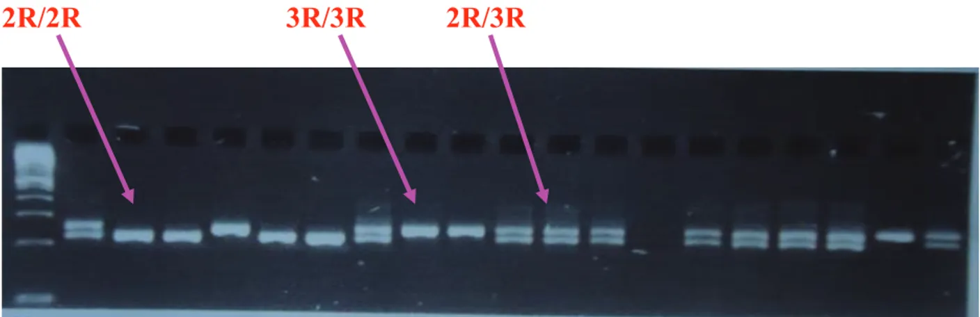
Figure 1: Lymphoblastoid cell lines treated with MTX and ASNase ………….….. 57 Figure 2: TS triple repeat (3R) 28 bp genotyping ……….. 59 Figure 3: MTX efficacy assessment in NSG mice, optimization experiments ……. 63
CHAPTER IV
Figure 1. Response to MTX in NSG mice injected with patients’ leukemia cells without and with TS 3R/3R genotype ……… 108 Figure 2. Efficacy of MTX treatment per dose………. 109 Figure S1. TS genotypes before and after leukemic cell engraftment in
NSG mice………... 116
CHAPTER V
Figure 1. Asparaginase-related acute complications in childhood ALL in relation to tandem repeat polymorphism and resulting haplotypes in asparaginase synthase (ASNS) gene……….……….. 140 Figure 2. ASNS polymorphisms, haplotype *1 sub-classification and cellular proliferation assay……….……… 141
CHAPTER VI
Figure 1. Frequency of sensitive, resistant and intermediate LCLs against E.coli ASNase ………..………… 148
xiv
CHAPTER VII
Figure 1. MTX increase dose to overcome resistance in NSG mice injected with blasts from TS+ ALL patients ……… 162
xv
List of Tables
CHAPTER I
Table 1. ALL treatment protocol; DFCI 1985–2000……….. 13 Table 2. Risk Group Classification on DFCI ALL Consortium Protocols
(1985–2000) ………... 14 Table 3. TS polymorphisms characteristics ……….………..….…… 32
CHAPTER III
Table 1. Summary of the studies associating DHFR polymorphisms with disease susceptibility or response to treatment………. 90
CHAPTER IV
Table S1. Patient samples characteristics ………..… 115
CHAPTER V
Table 1. Characteristic of ALL patients in the test (QcALL) and validation
(DFCI) cohort ……….. 137 Supplemental Table 1. Polymorphisms in ATF5, ASNS and ASS1 genes
Analyzed in the relation to ASNase related complications ………...……. 142 Supplemental Table 2. Identity of polymorphisms genotyped only in controls….. 143 Supplemental Table 3. In silico prediction of transcription factor binding sites affected by ATF5 polymorphisms……… 144
CHAPTER VI
Table 1: Association analysis between sensitivity status of LCLs and
xvi
Table 2: Results of association of IC50 distribution across ATF5 T1562C
Variation .……… 148
xvii
Abbreviations and Acronyms
ALL: Acute Lymphoblastic Leukemia ABC: ATP-binding cassette transporters
ABCG2: ATP-binding cassette superfamily G member 2 ATF5: Activating transcription factor 5
AICART: Aminoimidazole Carboxamide Ribonucleotide Transformylase AML: Acute Myeloid Leukaemia
AML1: Acute myeloid leukemia 1 protein ASO: Allele-specific Oligonucleotide ASS1: Argininosuccinate Synthase 1 ASNase: Asparaginase
ASNS: Asparagine Synthetase AVN: Avascular necrosis
BCRP: Breast Cancer Resistance Protein BCR-ABL1: Breakpoint cluster region–ABL1 Bcl-XL: B-cell lymphoma-extra large
Bcl-2: B-cell lymphoma 2 BH3: Bcl-2 homology 3
BIM: BCL-2-interacting mediator of cell death BM: Bone Marrow
xviii CCND1: Cyclin D1
CD: Cluster of differentiation
CEPH: Centre d’Etude du Polymorphisme Humain CML: Chronic Myeloid Leukaemia
CRLF2: Cytokine Receptor-Like Factor 2 CRPC: Castration-resistant prostate cancer
DFCI: Dana-Farber Cancer Institute DHF: Dihydrofolate
DHFR: Dihydroflate reductase DFS: Disease Free Survival DNA: Deoxyribonucleicacid
dTMP: Deoxythymidine mono phosphate dUMP: Deoxyuridine mono phosphate EBV: Epstein-Barr virus
E. coli ASNase: Escherichia coli Asparaginase EFS: Event Free Survival
ERG: ETS-related gene ETP: Early T-cell precursor
ETV6 RUNX1: Ets Variant Gene 6 Runt-related transcription factor 1 FPGS: Folylpolyglutamate Synthetase
GARTF: Glycinamide ribonucleotide transformylase GC: Glucocorticoids
xix GVHD: Graft-Versus-Host Disease
GWAS: Genome Wide Association Study HDMTX: High dose Methotrexate
hIFN-α: Human interferon-alpha HR: High risk
iAMP21: intrachromosomal amplifi cation of chromosome 21 IBD: Inflammatory bowel disease
IC50: Half maximal inhibitory concentration
IG: Immunoglobulin IHC: Immunohistochemical IL-27: Interleukin (IL)-27
IKZF1:Ikaros family zinc finger protein 1 LCL: Lymphoblastoid Cell Line
LDMTX: Low dose Methotrexate LICs: Leukemia-Initiating Cells
LYL1: Lymphoblastic leukemia derived sequence 1 MTHFR: Methylenetetrahydrofolate reductase
MLL: Mixed Lineage Leukemia
MRD: Minimal Residual Disease
MRPs: Multidrug Resistance-related Proteins MSCs: Mesenchymal Cells
MTX: Methotrexate
xx NSG mice: NOD/LtSz-scid IL-2Rgc null mice NTD: Neural Tube Defects
OS: Overall survival
PBX1: Pre-B-cell leukemia homeobox 1 PCR: Polymerase Chain Reaction PDNS: Purines de novo synthesis
PEG-ASNase: Polyethylene glycol–conjugated asparaginase P-gp: P-glycoprotein
RFC1: Reduced Folate Carrier 1
RUNX1: Runt-related transcription factor 1
2R and 3R: Double and Triple repeat polymorphisms SLC19A1: Solute Carrier family 19 member 1 SNPs: Single Nucleotide Polymorphisms SR: Standard risk
TAL1 : T-cell acute leukemia 1
TCF3 PBX1: Transcription factor 3 Pre-B-cell leukemia homeobox 1 TCR: T-cell receptor
THF: Tetrahydrofolate
TLX1 : T-cell leukemia homeobox 1 TLX3 : T-cell leukemia homeobox 3 TMP: Trimethoprim
TMQ: Trimetrexate
xxi TRAIL: TNF-related apoptosis-inducing ligand TS: Thymidylate Synthase
3´UTR: 3´untranslated region 5´UTR: 5´untranslated region VHR: Very high risk
xxii
Acknowledgments
It is my great pleasure to acknowledge all of those who have contributed in many ways to make this thesis possible.
First of all, I wish to express my endless thanks to my advisor, Maja Krajinovic. I am grateful for her expert and valuable guidance and constant encouragement, for her enthusiasm and inspiring vision. I particularly want to thank her for giving me the opportunity to join her laboratory and work on diverse and exciting projects.
I wish to acknowledge Elie Haddad, my co- advisor for his valuable comments and suggestions during my Ph.D program, especially regarding NSG immunodeficient humanized mice platform, in order to improve my in vivo experiments.
I would like also deeply thanks to Guy Rousseau, Noël J-M Raynal and Francine Durocher for taking the time to review this thesis, being on my thesis committees and for providing useful suggestions and comments.
I am also glad to thanks Dr. Sinnett, who is established ALL tissue bank at CHUSJ, which leukemic cells from this bank used for this project. I also special thank Caroline Alfieri for her valuable help with in vitro experiments and François Fontaine, for his great help with in vivo experimental works and technical support.
My sincere thanks go to all members of the Krajinovic’s lab, Margaret Labuda, Vincent Gagne, Mohamed Aziz Rezgui and Julie Rousseau for their valuable help and friendship. I would like to thank all the animal facility staff for caring the mice. I am also glad to acknowledge the help I received from the administrative staff at Ste--Justine and at the
xxiii
department of Pharmacology. Alida Hounyovi, Sandy Lalonde, Dominika Kozubska, Sylvie Caron and Carole David deserve special mention for their kind assistance.
Finally I would like to thanks the financial support of this project, Cancer Research Society and Funds de recherche Santé Québec (FRSQ) and Faculty of Medicine, Université de Montréal.
xxiv
I dedicate this thesis to my wife, Manzar,
whose love and support guided me along this journey,
and my beloved daughter Bahar, such a good girl
Chapter I
1
Introduction
Part I
Acute lymphoblastic leukaemia:
Acute lymphoblastic leukaemia (ALL) is the most common malignancy of childhood and the most common cause of cancer related mortality affecting children from 0 to 14 years. About approximately 57 000 cases reported worldwide, particularly, 6000 and 2500 new ALL cases are diagnosed every year only in the USA and Canada respectively [1-4]. B-cell precursor ALL is a main morphologic and histological subtype of childhood leukemia with approximately 75-80% of cases. It is one of the most common malignant disorders of lymphoid progenitor cells in children with peak prevalence of 3-5 years [5-8]. During last four decades introduction of multi-agent treatment protocols and risk-guided treatment changed outcome for children with ALL. Currently, 5-year event-free survival (EFS) rates for childhood ALL is more than 80-85% in the developed countries [9]. The other two important childhood leukemia include myelogenous leukemia (AML) with frequency of 20% to 25% and chronic myelogenous leukemia (CML) which account for 5% of leukemia cases [7]. Although ALL occurs in children and adults, children showed better prognosis compared to adults [6, 10]. It has been shown that 95% of children achieve complete remission after phase of treatment conducted with several intensive multi-agent chemotherapy and about 70% are long-term survivors. Nevertheless
2
Etiology and epidemiology of childhood leukemia:
Childhood ALL might arise from interactions between exogenous (e.g. infection, environmental exposure) or endogenous exposures (e.g. inflammation, oxidative stress) and genetic (inherited) susceptibility increasing the chance of a mutation in relevant oncogenes/tumor suppressor genes and chromosome alterations (Figure 1) [3]. There are several known risk factors including ionizing radiation and congenital or genetic syndromes (such as Down’s, neurofibromatosis, Shwachman syndrome, Bloom's syndrome, and ataxia telangiectasia, and Fanconi’s anaemia) which are associated with ALL (collectively <5% of ALL) [4, 7, 8]. Children with Down syndrome (trisomy 21) are 15 times more likely to develop leukemia compared with children without trisomy 21[4, 7]. Diet of the mother and child, parental smoking, pesticides and household chemicals, traffic fumes are environmental exposures suggested to contribute to ALL development. The incidence rate of disease increased by 1% during last two decades. Furthermore, immune modulators through for example infections and vaccinations, can as well play a role in onset of lymphoblastic leukemia in particular the pre-B cell childhood ALLs [4]. A large number of risk factors including genetic component of ALL remains still undefined.
3
Figure 1: Different causes of childhood ALL (Inaba 2013).
Figure 2: Morphologic and immunohistochemical (IHC) pictures of B-ALL patients: A, The blasts with azurophilic granules. B, Diffuse infiltrate replacing bone marrow medullary space. Wright-Giemsa–stained bone marrow (Zhou 2012).
Endogenous
Genetic Exogenous
4
Genetic alterations in ALL:
Based on evidences commonly mutated genes in ALL patients are those involved in the development and differentiation of various types of blood cells, and these genes vary according pre-B or T cells leukemia [12]. ALL also has been classified into B-acute lymphoblastic leukemia (B-ALL) and T-cell lineage ALL (T-ALL). Furthermore, ALL has been traditionally classified into precursor B, B-cell and precursor T (or T-cell) phenotypes. According karyotypic abnormalities, ploidy and translocations, they will be further subdivided [13]. B-ALL is derived from B-cell progenitors (Figure 2). B-ALL accounting for 80%, while T-ALL comprises 12%-15% of all newly diagnosed paediatric childhood ALL. T-cell ALL is associated with a poor prognosis with only 7–23% of patients surviving for 3 to 5 years [10, 14]. Patients with B-ALL can be classified to
ETV6-RUNX1 (TEL-AML1) B-ALL, TCF3-PBX1 (E2A-PBX1), BCR–ABL1 ALL, MLL–
rearranged B-ALL according to specific genetic abnormality [15, 16] (Figure 3).
ETV6-RUNX1 involves ETV6, previously known as TEL, on chromosome 12p13 and ETV6-RUNX1,
located on chromosome 21q22, previously known as AML1 which is a transcription factor required for hematopoietic development and differentiation in embryonic and adult stages. TEL-AML1 translocation occurs in 20% to 25% of childhood ALL cases and confers a favorable prognosis [10, 12, 15, 17]. Another most common genetic abnormality is t(1;19) (q23;p13) which results in the fusion of the TCF3 (formerly E2A) gene with the PBX1 gene which occurs in 3-5% of B-ALL patients and is usually associated with favorable outcome, but in adult ALL the prognosis of t(1;19) patients with French and Italian studies reporting poor outcomes [10, 18]. High risk, includes
5
rearrangements of the mixed lineage leukemia (MLL) gene; and very high risk includes translocation t(9:22) which generate the BCR-ABL1 fusion gene, known as the Philadelphia chromosome, and hypodiploidy [15, 16, 19, 20]. The frequency of Philadelphia chromosome is about 3% to 6% among childhood ALL, but occurs more frequently (around 25%) among adults [15]. Based on recent studies of genome-wide profiling using microarrays, candidate gene, and second-generation sequencing helped to identify other genetic alterations that define new ALL subtypes. Deletion of the IKZF1 (IKAROS) is common in BCR-ABL1 lymphoid leukemia but is rarely present in CML at chronic phase. IKAROS is a zinc finger transcription factor required for the development of all lymphoid lineages. IKZF1 alteration cooperate with BCR-ABL1 in the induction of lymphoblastic leukemia and subsequent resistance to therapy in recent experimental models of BCR-ABL1 ALL [15]. ALL patients carrier IKZF1 deletion had lower 5 years EFS (30%) comparing those without this genotype (51%) and they showed very poor outcome in spite of haemopoietic stem-cell transplantation [14]. Deletion in IKZF1 gene which involved in regulating B-cell differentiation at relapse, commonly occur in the
BCR-ABL1-like ALL subtype, BCR-ABL1-like ALL include more than 15% of childhood
B-ALL cases which exhibit a gene expression profile similar to that of BCR-ABL1– positive ALL, often have deletion/mutation of IZKF1, and associated with a very poor outcome [14, 15].
6
A)
B)
Figure 3. A) Genetic subtypes in childhood B-ALL and B) Distribution of acute lymphoblastic leukemia subtypes by age. (Inaba 2013 and Wiemels 2012).
BCR –ABL1 2% Others (T -cel l dise ase ) 2 % ETV6–RUNX1 t(12;21) Hyperdiploid (>50 chromosomes) 20% 22% MLL rearrangements e.g., t(4;22), t(11;19), t(9;11) 6-8% TL X 1 0· 3% TLX3 2·3%ETP 2%
Others (B-cell disease)
9% ERG 3% BCR-ABL1-like 9% (Other) 5·5% (CRLF2) 3·5% LYL1 1·4% TAL1 7% CRLF2 4% iAMP21 2% Hypodiploid (<44 chromosomes) 1% D icen tric 3% TCF3 –PBX1 4% BCR –ABL1 2% Others (T -cel l dise ase ) 2 % ETV6–RUNX1 t(12;21) Hyperdiploid (>50 chromosomes) 20% 22% MLL rearrangements e.g., t(4;22), t(11;19), t(9;11) 6-8% TL X 1 0· 3% TLX3 2·3%ETP 2%
Others (B-cell disease)
9% ERG 3% BCR-ABL1-like 9% (Other) 5·5% (CRLF2) 3·5% LYL1 1·4% TAL1 7% CRLF2 4% iAMP21 2% Hypodiploid (<44 chromosomes) 1% D icen tric 3% TCF3 –PBX1 4% Children 1-15 Adults 16+ Infants< 1 year
7
Genetic profile and immunophenotypic features of B-ALL:
To differentiate cancer arises from precursors of B or T cell, the bone marrow smear undergoes cytochemical staining for distinguishing between lymphoid and myeloid blasts. To further classify the leukemia, the blasts undergo immunophenotypic classification that measures the expression of cytoplasmic and surface antigens, which can discriminate B-cell, early pre-B-cell, pre-B-cell, or T-cell leukemia [10, 20]. In this regard, early pre–B lymphoblasts express CD19 (100% of patients), cytoplasmic CD22, and CD34 and later pre–B cells are positive for CD10, CD45 and CD20. Likewise T-cell leukemia is positive for CD3, CD5, CD7, CD10 and CD34 [10, 21].
Ploidy within a leukemia blast cell population:
Determination of the modal number of the leukemia chromosomes is one of the prognostic factors for B-cell precursor ALL patients. Chromosome number (ploidy) within a leukemia blast cell population includes hyperdiploidy with 51–68 chromosomes, low hyperdiploidy with 47-50 chromosomes, pseudodiploidy with 46 chromosomes, diploid or normal with no cytogenetic abnormalities and hypodiploidy with ≤45 chromosomes which occur respectively 25% to 30%, 11%, 28%, 30% and 7%–8% of childhood ALL cases [15, 20, 22]. Hyperdiploid leukemia confers a favourable prognosis, but hypodiploid leukemia with a chromosome number less than 46 is associated with poor prognosis [15, 20].
8
Molecular Monitoring and Response to Therapy:
The significance of the morphological examination is now being replaced by the detection of minimal residual disease (MRD), through flow cytometric detection of leukemia-associated immunophenotypes or PCR amplification of fusion transcripts, immunoglobulin (IG) and T-cell receptor (TCR) gene [23-25]. MRD is the single most powerful prognostic marker for patients in all risk groups, which can determine the response to treatment in ALL patients more precisely than morphological screening of bone marrow smears. Likewise, MRD now become the new definition for leukemia remission and is measured concurrently with the morphological examination. The other advantages and applications of MRD detection in patients with ALL are determining remission status before and after transplantation, and detecting early relapse. Accordingly, measuring MRD levels during remission induction therapy provide important prognostic information [25]. MRD level inversely correlates with 5 years event free survival (EFS) rate. Based on obtained results, the EFS of 88% is correlates to MRD level less than 0.01% and, EFS of 59%, 49% and 30% are correlates with MRD level more than 0.01%, 0.1%, and 1% respectively. Now, cytogenetic and MRD statuses are the 2 most important prognostic factors both in childhood B-ALL and adult B-ALL[10]. Based on Dana-Farber Cancer Institute (DFCI) protocol and investigation, end-induction MRD level can be considered as a significant independent predictor of outcome [26]. According to DFCI treatment protocol, children with high end-induction MRD (≥0.001) had a 10.5-fold greater risk of relapse than those with low MRD [26]. Considering MRD levels in the BM at day 15 were well correlated with risk of relapse, determining MRD levels at day 15 is an informative checkpoint to test the in vivo sensitivity of the leukemia
9
in the individual precursor B-cell ALL [27]. Likewise, patients presenting high levels of MRD at day 33 (at the end of induction phase) and no MRD detectable at day 78 (at the end of intensification phase) had a favourable outcome, but those with detectable MRD at day 78, showed significant increase risk of relapse [28].
Treatment:
Treatment of paediatric ALL has greatly improved due to the introduction of effective combination risk-adapted therapies. DFCI Consortium treatment protocol and their risk group classification for childhood ALL, is one of the referent protocol currently used for childhood ALL management and treatment (Table 1, II) [26]. DFCI ALL Consortium has been conducting multi institutional clinical trials in the United States and Canada in childhood ALL since 1981. DFCI Consortium treatment protocol assigned as a research protocols and in term of clinical research always addressing particular research questions through randomization. The major goal of DFCI ALL Consortium Protocol 91-01 conducting randomized clinical trials in childhood ALL and continuing focusing on improving efficacy and outcome of newly diagnosed ALL children while minimizing acute and late toxicities [26, 29]. Based on DFCI results, 1457 children aged 0–18 years were treated on four consecutive protocols among 1985–2000 [26]. The 2 year DFCI treatment program consisted of four phases; multi-agent remission induction therapy (4 weeks); CNS-directed treatment (3 weeks); intensification (20-30 weeks) and therapy will be continued until the completion of 24 months of complete remission [26, 29]. Treatment protocols vary based on risk group classification determined at the time of diagnosis, include; standard risk (SR), high risk (HR) and very high risk (VHR). CNS treatment for SR and HR patients began on the day that complete remission was attained.
10
The goal of remission induction treatment (also called induction therapy) for ALL is to restore normal hematopoiesis in 96–99% of children and 78–92% of adults with ALL and bring about a complete remission with as few toxic adverse effects as possible [3, 30, 31]. The speed rate and depth of the early response to remission induction therapy recognise as a major index of subsequent therapy in many protocols worldwide [32]. Remission induction is 4 weeks duration phase and patients received a vincristine, corticosteroids such as prednisone and dexamethasone, doxorubicin, cytarabin, ASNase and MTX. The goal of central nervous system (CNS) directed therapy is critical to maximize and improved survival rates. CNS therapy which elongated three weeks includes all remission induction phase drugs plus 6-mercaptopurine [26, 33]. CNS treatment for infants was delayed until the age of 12 months [33]. Intensification therapy is a critical phase of lymphoblastic leukemia treatment protocol to eradicate residual leukaemic cells [3, 26, 33]. Although about two-thirds of lymphoblastic leukemia cases can be treated successfully only with 12 months of treatment. Patients with childhood leukaemia need continuation treatment in order to prevent possible relapse [34]. Thus continuation therapy is beginning after intensification, and continued until the completion of 24 months of complete remission [26, 33].
Drugs used in ALL:
Protocol phases and related drugs based on risk group presented in table 1 and 2 [26, 33]. Cytosine arabinoside (Cytarabine, Ara-C) is an antimetabolite widely used in acute leukemia. Cytarabine must be phosphorylated to cytotoxic metabolite Ara-CTP (Ara-C three phosphates) and exert its cytotoxicity during or after incorporation into the DNA where they interfere with DNA strand elongation, replication, or repair processes [35].
11
Cytarabine therapy is associated with several adverse side effects, including myelosuppression, infections, mucositis, neurotoxicity, and acute pulmonary syndrome [36].
Doxorubicin is an anthracycline antibiotics commonly used in the treatment of leukemias. The major doxorubicin related concern is well-known dose-related cardiotoxicity [36]. Doxorubicin attack cancer cells by multiple mechanisms; they work primarily by DNA intercalation and inhibiting replication which lead to DNA damage and cell death. Also doxorubicin binds to mitochondrial DNA, inhibiting other cellular functions. It can inhibit the action of topoisomerase II, an enzyme that unzips the DNA molecule for replication [37, 38].
6-Mercaptopurine (6-MP) is an antimetabolite and an analog of purine base. It is important component of ALL treatment administered during intensification and maintenance treatment phases. Upon entry into the leukemic cells it is metabolized to 6-thioguanine nucleotides (6-TGN) by thiopurine methyltransferase (TPMT). 6-MP exerts its cytotoxic effect by inhibition of DNA synthesis and by interfering with the activity of nucleic acid-processing enzymes. More precisely, TGN incorporation into the DNA and RNA results in DNA strand-breaks, chromatid damage and subsequent cell cycle arrest and apoptosis. 6-TGN is more potent than 6-MP; based on in vitro and in vivo data, lymphoblasts are more sensitive to 6-TGN and exert more toxicity compared to 6-MP. 6-TGN is no longer used for continuation treatment, because it has been associated with thrombocytopenia, an increased risk of death, and hepatic veno-occlusive disease upon prolong use of thioguanine at a dose more than 40 mg/m2 [13, 19]. 6-TGN is active
12
metabolite of 6-MP which has cytotoxic and immunosuppressive properties [39, 40]. TPMT genotyping may be useful to predict this risk prior to drug administration [36, 40]. Vincristine is important vinca alkaloids known as antimicrotubule agent. The most frequent and clinically important side-effect of vincristine is dose-limiting autonomic and peripheral sensory-motor neuropathy [36, 41]. Vincristine induces cytotoxicity by interacting with and disrupting microtubules polymerization, especially those comprising the mitotic spindle apparatus so microtubules do not form. Vincristine also binds to neuronal tubulin, lead to disrupting axonal microtubules and causing neurotoxicity [42]. Corticosteroids induce apoptosis in the malignant lymphoid cells and are critical component of combination therapy for ALL. Glucocorticoids exert their cytotoxicity effects by binding with glucocorticoid receptors (GR). After binding, GR can homodimerize and translocate to the nucleus, and interact with glucocorticoid response elements to transactivate gene expression, resulting expression alteration in various oncogenes, cell cycle arrest and apoptosis [43][44]. Dexamethasone and prednisone may cause avascular necrosis (AVN) or osteonecrosis as a chronic complication of leukaemia treatment [45]. However, dexamethasone has a longer half-life and better central nervous system (CNS) penetration, appears to yield better CNS control [43].
Mechanism of action and pharmacogenomics of MTX and ASNase as two major components of DFCI protocol precisely presented in future sections.
13
Table 1: ALL treatment protocol; DFCI 1985–2000; SR: standard risk, HR: high risk, IV: intravenous, IM: intramuscular (Modified from LeClerc. 2002 and Silverman 2010). Induction (4 weeks) CNS therapy (3 weeks) Intensification (20–30 weeks) Every 3 week cycles Continuation (78 weeks) Every 3 week cycles prednisone daily, 40mg/ m2 (days 0–28) IV (SR) Vincristine 2 mg/m2 IV (day 1) (SR) Prednisone daily 40 mg/m2 orally days 1–5 or (SR) Dexamethasone daily 6 mg/m2 for 5 days (SR) Prednisone daily 40mg/ m2
for 5 days within 3- weeks cycle (SR) or Dexamethasone 6-18 mg/m2 (SR & HR) Vincristine 1.5 mg/m2 (days 0, 7, 14, 21) IV (SR) Hydrocortisone 9-15 mg (SR) Doxorubicin 30 mg/m2 day 1 of each cycle (HR) Vincristine 2.0 mg/m2 IV day 1 (SR) Cytarabine intrathecal 1 dose according to age (day 0) (SR) Cytarabine intrathecal twice weekly × 4 doses (SR) Vincristine 2.0 mg/m2 IV day 1 (SR) Methotrexate weekly 30 mg/m2 (SR) Doxorubicin 30 mg/m2/dose (days 0 and 1) IV (SR) Methotrexate intrathecal 4 doses of 6-12 mg Methotrexate 30 mg/m2 IV or IM days 1, 8, 15 (SR) 6-Mercaptopurine daily 50 mg/m2 orally days 1–15 (SR) Methotrexate one dose of 4g/ m2 with leucovorin (day 3) IV (SR) 6-Mercaptopurine daily 50 mg/m2 orally days 1–15 (SR) Asparaginase E.coli ASP 25,000 IU/m2 weekly or PEG ASP 2500 IU/m2 every 2-weeks (SR) Asparaginase E.coli or Erwinia ASP 25,000 IU/m2 × 1 dose (day 4) (SR) Doxorubicin 30 mg/m2 day 1 of each cycle (HR) 6-Mercaptopurine daily 50 mg/m2 orally days 1–15 for 14 days (SR)
14
Table 2: Risk Group Classification on DFCI ALL Consortium Protocols (1985–2000) (Modified from Silverman 2010).
Standard Risk: (All of the following)
High Risk: (Any of the following)
Very High Risk: (Any of the following)
Age 1985–95: 2 to < 9 years 1995–2000: 1 to < 10 years 1985–95: ≥ 9 years 1995–2000: ≥ 10 years 1985–2000: < 12 months WBC (× 109/L) 1985–95: < 20 1995–2000: < 50 1985–91: 20 to < 1001991–5: ≥ 20 1995–2000: ≥ 50 1985–91: ≥ 100
Cell type B-precursor T-cell
Clinical CNS disease Absent Present
TEL-AML1 (ETV6-RUNX1) - t(12:21)
Present Absent
Mixed Lineage Leukemia
(MLL) Absent Present
BCR-ABL1-t(9;22) Philadelphia chromosome
15
Pharmacogenomics and importance of ALL:
Pharmacogenomics is the study of genetic variation of drug-metabolizing enzymes,
receptors, transporters and targets, and talk about how these genetic variations (either inherited, acquired or both) affects drug-related phenotypes, such as drug response or toxicity. The major goal of pharmacogenomics is providing strategies to individualize therapy based on human genetic variability and its influence on drug response. Although pharmacogenetics and pharmacogenomics terms are often used interchangeably, the pharmacogenetics is the study of genetically determined variation in how individuals respond to drugs, but pharmacogenomics used to describe the study of the entire spectrum of genes that determine drug responses in relation to the diversity of the human genome sequence and its clinical consequences [46, 47]. Progress in pharmacogenomics studies provide significant potential for further clinical application in order to improve clinical outcomes in individual patients [46]. The treatment of paediatric ALL has greatly improved due to the introduction of effective combination risk-adapted therapies. However, therapy resistance is still a major obstacle to successful treatment, whereas intensive treatment has also important short-term side effects and long-term consequences. Unfavourable clinical pharmacokinetics and pharmacodynamic as well as cellular drug resistance play a major role in ALL treatment failure [48]. In this regard, treatment related toxicity is one of the major reasons for withdrawal or discontinuation, which might be increase risk of relapse despite of standard treatment approaches. On the other hand intensive treatment has short and long term consequences, such as myelosuppression, infection, and thrombosis, alteration in glucose metabolism, secondary malignancies, growth retardation, bone abnormalities, neurotoxicity or cognitive
16
impairments [36, 49-51]. Then, most important concern of oncologist is to recognize ALL patients before relapse due to fail in current treatment protocols [9]. Numerous pharmacogenetic studies have shown that polymorphisms in genes that encode drug metabolising enzymes and targets can affect enzymatic activity and modulate drug action [52]. Drug resistance and drug-related side effects can be due to polymorphisms in genes that encode enzymes involved in drug pharmacokinetics and pharmacodynamic. Polymorphisms in such genes often referred to as candidate genes are analyzed in pharmacogenetic studies for their capability to predict unfavourable treatment responses. Identification of such polymorphisms may lead to different treatment schedules, improving the efficacy of treatment and allowing for a reduction in drug side effects [50]. ALL was widely addressed in number of pharmacogenetic studies including candidate gene approach and Genome Wide Association Studies (GWAS). These studies increased our knowledge on genetic contribution to specific drug metabolism and signalling pathways [19]. One of the best examples of pharmacogenomics studies in ALL is 6-MP dosing based on TPMT genotypes and subsequent association of dose adjustment with better outcomes and less toxicity [19] TPMT enzyme is affected by functional polymorphisms which define the most prevalent mutant alleles associated with loss of catalytic activity and play an important role in metabolites pharmacokinetics variability affecting the balance between 6-MP efficacy and toxicity. These polymorphisms include three SNPs in the TPMT gene; TPMT*2 (238G > C), TMPT*3A (G460A), and TPMT*3C (719A > G) alleles, which account for 90% of the enzymatic deficiency in most populations. Due to higher TGN accumulation these polymorphisms have been associated with increased risk of myelosuppression [19, 36, 78] in ALL patients treated
17
with standard 6-MP doses. However in a case of treatment adjusted according to the TPMT genotypes. Based on TPMT genotyping, about 90% of the individuals have two wild-type TPMT alleles (TPMT*1), thus they have normal enzyme activity whereas individuals with two nonfunctional alleles (1 in 300) have very low or lack of TPMT activity, resulting high levels of TGN and acute hematopoietic toxic effects. Patients who carry two nonfunctional TPMT alleles experience severe myelosuppression, if treated with standard doses of thiopurines. These patients require a reduction of more than 90% of the standard doses. Likewise administration of 50% of the standard dose is recommended for about 5-10% of individuals who carrier heterozygous alleles and have intermediate levels of enzyme activity. TPMT-deficient patients, upon thiopurine dose adjustment can tolerate full doses of their other ALL chemotherapeutic agents which associated with greater therapeutic efficacy [36, 78].In addition to the analysis in clinical setting which addresses an association between polymorphisms and therapeutic responses, understanding functional effect of identified polymorphism makes also the part of pharmacogenetic studies spectrum and may as well give an indication for drug dose adjustments. This is of particular interest when treatment in composed of multiple therapeutics as in case of ALL.
18
Part II
Methotrexate (MTX) as a major anti-neoplastic component in ALL
treatment protocol:
MTX is an important antifolate drug, designed to mimic a folate molecule which bind in the active site of folate-dependent enzymes and block its action (Figure 4). MTX is an essential drug used in the treatment’s protocol of several malignant and non-malignant diseases including ALL, choriocarcinoma, osteogenic sarcoma, seminoma, as well as lung, mammary gland and ovarian cancer [53-57].Apart of antitumor effects, MTX play a major role in treatment autoimmune diseases, such as rheumatoid arthritis (RA) and moderate to sever psoriasis [58, 59], lupus erythematosis and inflammatory bowel disease (IBD) [60] primary billiary cirrhosis and intrinsic asthma, as well as prevention of graft-versus-host disease (GVHD) [49]. MTX is used in induction, CNS relapse prevention, and maintenance therapy in ALL treatment protocol [26, 29]. Also MTX as a broad spectrum antitumor component used in treatment’s protocol of several malignancies such as accordingly, high-dose MTX (HDMTX) is commonly given in induction phase, and low-dose oral MTX (LDMTX) is given in maintenance therapy in ALL treatment’s protocols[61-63]. MTX uses the reduced folate carrier 1 (RFC1)or solute carrier family 19 member 1 (SLC19A1), for entering the cells [28, 36, 50, 57, 64], ABC proteins (specifically the ABCC1-4 and breast cancer resistant, ABCG2 proteins) for the transport out of the cell (Figure 5) [28, 50, 57, 63]. Overexpression of ABCC2 [(multidrug resistance-associated protein (MRP) 2] may lead to lower cellular MTX accumulation and subsequent moderate to high level of resistance [65, 66]. Impaired function of RFC1 has been recognized as a frequent mechanism of anti-folate resistance
19
[49]. Variations in this gene have been also showed to contribute to MTX response. RFC gene is located on human chromosome 21, Mostly investigated polymorphism is RFC1 80G>A. Patients with AA genotype showed higher MTX plasma level of MTX in their plasma due to impaired MTX influx [50, 56, 67, 68]. Some of chromosomal translocations that are typical for ALL cells may have altered level of MTX influx and efflux proteins. For example, E2A-PBX1 ALL patients showed significantly lower expression of RFC1 and TEL-AML1 ALL patients showed higher expression of ABCG2. Both conditions lead to decrease in MTX polyglutamates (MTXPG) level in the cells and subsequent resistance to MTX [56]. The solute carrier organic anion transporter 1B1 (SLCO1B1), which is mainly expressed in the liver, is important for hepatic uptake and thus for the pharmacokinetics of MTX [68]. It has been shown that variants in the
SLCO1B1 gene are associated with MTX clearance and gastrointestinal (GI) toxicity in
ALL patients [68]. After entering the cells (especially in hepatocytes, erythrocytes, and tumour cells), MTX undergoes polyglutamylation to MTX polyglutamates via folylpolyglutamate synthetase (FPGS) (Figure 5). Polyglutamation increases intracellular retention of MTX and subsequent affinity for the MTX-sensitive enzymes within the cell [55, 57, 63, 69]. In the other words, MTX gains its antileukemic effects through an accumulation of long-chain MTXPG [56, 57, 67] the level and time-course of MTXPG formation in tumour cells is a key determinant of the cytotoxic activity exerted by MTX [65]. Pre B-ALL have more efficient accumulation of (long chain) MTXPGs compared to T-ALL and acute myeloid leukemia [69]. Reversed process is directly dependant on active transport of MTXPG into lysosome and further hydrolysis by γ-glutamyl hydrolase (GGH), which allows efflux of the drug out of the cell [50, 55, 57, 68]. Moreover,
20
MTXPG can inhibit glycinamide ribonucleotide transformylase (GARTF) and aminoimidazole carboxamide ribonucleotide transformylase (AICART), the two enzymes essential for de novo biosynthesis of purines [55, 68]. MTX and MTXPG acts as competitive inhibitors of several key enzymes in folate pathway such as dihydrofolate reductase (DHFR), thymidylate synthase (TS), 5,10-methylenetetrahydrofolate reductase (MTHFR) [28, 36, 64], and enzyme of purine synthesis resulting in DNA synthesis impairment and cell death [50, 55, 67]. In contrast to classic antifolates, nonclassical or novel antifolates, defined as antifolates that do not require active transport or polyglutamation, can passively diffuse into cells and do not depend on FPGS or the RFC. The most important nonclassical antifolates are trimethoprim (TMP), trimetrexate (TMQ) and piritrexim (PTX) [70-72].
21 Figure 4: MTX as an important antifolate drug;
MTX is designed to mimic a folate molecule, binds to the active site of the folate-dependent enzymes and blocks its action. Methotrexate has the same size and chemical composition as folate molecule. The similar structure of MTX and folate, suggests their competitive binding to same molecules (2014 David Goodsell & RCSB Protein Data Bank, http://www.rcsb.org/pdb/101/motm.do?momID=34).
22
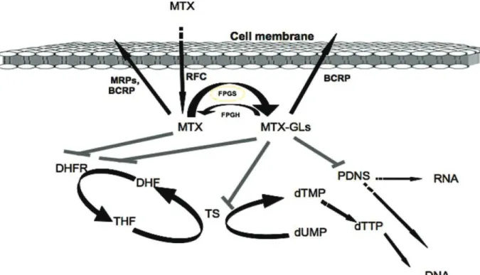
Figure 5: Summary of the intracellular metabolism and targets of MTX:
MTX is transported actively into the cell by RFC, where it undergoes polyglutamylation and exerts its toxic effects by inhibiting DHFR, TS and de novo biosynthesis of purines (PDNS). DHFR, dihydrofolate reductase; DHF, dihydrofolate; THF, tetrahydrofolate; TS, thymidylate synthase; dTMP, deoxythymidine monophosphate; dUMP, deoxyuridine monophosphate; FPGS, folypolyglutamate synthetase; FPGH, folypolyglutamate hydrolase; MRPs, multidrug resistance-associated proteins; BCRP, breast cancer resistant protein; MTXGLs, MTX polyglutamates (Fotoohi 2008).
23
Pharmacogenetics of MTX and MTX-related toxicity and resistance:
MTX dosing has traditionally been adjusted based on several prognostic factors such as age, weight, body surface or side effects. However, despite dose adjustments of MTX, some ALL patients do not respond well to MTX treatment [68]. Based on evidences, MTX causes severe dose-limiting adverse events, including ulcerative stomatitis, nausea, abdominal distress, fever and myelosuppression [28, 64]. Treatment-related toxicity is not only life threatening but also is one of the main reasons for early drug withdrawal, and subsequent increase in relapse risk [11, 55, 63]. In vitro or in vivo studies have shown that polymorphisms in genes encoding folate dependent enzymes may affect response to MTX [52]. Two important groups of genes are involved in MTX resistance. First group are genes that encode drug metabolizing enzymes and transporters which control pharmacokinetic properties and the second group includes genes that encode drug target and affects pharmacodynamic properties of MTX [52]. In regard to the latter, increase in DHFR and TS activity, due to overexpression of DHFR and TS gene is a common mechanism underlying resistance to MTX [28, 55, 73, 74]. Other important determinants of MTX resistance include decreased intracellular retention (caused by decreased polyglutamylation and decreased affinity of DHFR for MTX), qualitative and/or quantitative alterations in influx and/or efflux transporters, and expansion of the tetrahydrofolate (THF)- pool size in cells [55, 65, 74]. The cellular THF- pool is an important index of polyglutamation; cells with low level of THF pool are more sensitive to antifolates and large THF pool is associated with resistance to MTX and other antifolates, which require polyglutamylation for their activity within the cell [55, 70, 74]. DHFR is a member of the reductase enzyme family, is ubiquitously expressed in all24
organisms. One of the major and critical roles of DHFR is maintaining intracellular THF-cofactor pools at normal levels irrespective of the rate of synthesis of thymidylate [74]. Reduction of DHFR enzymatic activity diminishes the THF pool inside the cell affecting the level of folate coenzymes and thus purine and pyrimidine synthesis [53, 75, 76]. MTHFR is another folate dependent enzyme, is not directly inhibited by MTX and inhibited by metabolites of MTX. This enzyme is influenced by MTX due to effects of MTX on the intracellular folate pool. MTHFR by converting 5-10 methylene-THF into 5- methyl-THF, provide a methylene group for homocysteine methylation resulting maintenance of homocysteine level in plasma [48, 49, 68, 77]. Moreover, MTHFR, as a major enzyme in folate and homocysteine cycles, has two famous known single nucleotide polymorphisms (SNPs), 677C>T and 1298A>C, associated with reduced enzyme activity. Both have been shown to alter sensitivity to MTX. In the case of
677C>T polymorphism, it has been shown that the heterozygous 677CT have 65% of
their enzyme activity whereas homozygotes 677TT have 30% of activity compared with the homozygous CC individuals (677CC). Likewise, homozygous CC1298 cells exhibit reduced enzyme activity [55]. Homozygosity for the 677T allele was associated with an increased risk of hepatotoxicity or myelosuppression adult ALL [78]. Based on association studies conducted in childhood ALL patients, there is no association of 677T allele with MTX-related toxicity. However based on recent studies, children with MTHFR polymorphisms developed more frequently myelosuppression and had higher creatinine levels [48, 79]. Beside folate dependent enzymes some other genes, like those involved in cell cycle regulation and apoptotic cascade, are also correlated with resistance to MTX [55] such as mutation in P53 gene [80] or apoptosis regulators Bcl-XL and Bcl-2
25
[81]. Cyclin D1 (CCND1) and retinoblastoma genes are key proteins involved in cell cycle regulation. They may as well play a role in MTX resistance, through increase in
DHFR transcription. For example, CCND1 A870G polymorphism can modulate the ratio
of CCND1 mRNA isoforms, favoring isoforms with longer half-life [82, 83]. The A870G polymorphism was associated with a significant increase in cancer risk including ALL and also affected response to treatment in ALL patients [84].Overall, the molecular basis of resistance to antifolates, such as MTX, has been associated with target enzyme overexpression, change in the expression of a number of transporters and polymorphisms of genes affecting the activity of these proteins [65].
Pharmacogenetic markers in MTX pathway of interest for this study:
DHFR catalyzes the reduction of dihydrofolate (DHF) to THF which are needed for the action of folate dependent enzymes and thus are essential for DNA synthesis and methylation. DHFR inhibition by MTX diminishes the THF pool inside the cell affecting the level of folate coenzymes and thus purine and pyrimidine synthesis and finally causing cell death [36, 53, 68]. TS is a key enzyme in the nucleotide biosynthetic pathway that catalyzes conversion of deoxyuridine monophosphate (dUMP) to deoxythymidine monophosphate (dTMP) (Figure 5). TS is inhibited by MTXPGs, which leads to inhibition of purine and pyrimidine synthesis and inhibition of cell proliferation [36, 55, 65].
26
1. DHFR gene
polymorphisms:
The functional gene is mapped at chromosome 5q11.2-13.2. Multiple intronless pseudogenes or dihydrofolate reductase-like genes have been identified on separate chromosomes [53]. The changes in DHFR expression or activity can be partly due to the functional polymorphisms in the DHFR gene, thereby influencing a risk of folate-dependent diseases or affect response to antifolates. Association analysis performed in our group, identified particular DHFR haplotype that may affect the response to treatment in childhood ALL patients. The initial analysis of 15 DHFR minor promoter polymorphisms showed an association between homozygosity for A-317 and C-1610 alleles with lower event free survival (EFS) [85, 86]. Haplotype analysis identified an association with haplotype *1 harbouring both the A-317 and C-1610 alleles. Same haplotype was associated with higher DHFR expression, Further analysis performed in our lab, on the adjacent ~400-bp major DHFR promoter (located between minor and major transcript initiation sites) (Figure 6A) identified 6 polymorphisms, including 5 single nucleotide polymorphisms (SNPs) and one length polymorphism composed of variable number of 9-bp elements and 9-bp insertion/deletion [85]. The haplotype analysis revealed diversification of haplotype *1 into five subtypes, (*1a to *1e) (Figure 6B). Only *1b, conferred high transcriptional activity and was associated with lower EFS (Figure 6C). DFHR haplotype *1b is defined by particular allelic combination derived from three tag SNPs (C-1610G/T, C-680A and A-317G) in the minor promoter and three tag polymorphisms (C35T, G308A and compound length polymorphisms) in the major promoter [68, 85].
27 A B C
coding RNA (major) non-coding RNA
(minor)
Major promoter and minor transcript region Minor promoter ~ 2 kb ~ 400 bp *1b - 209 (35) *1b+ 47 (14) p=0.01 EFS Time (month)
28
Figure 6: DHFR risk haplotypes in major promoters: A. Structure of the human
DHFR gene. The region of interest (~400-bp) is located between the minor and major
transcript initiation sites indicated by arrows. B. Haplotype *1b includes six polymorphisms in the major promoter. Tagging polymorphisms of the major promoter are at positions 35, 63/91, and 308. Haplotype *1b is composed of three polymorphisms (C35T, G308A and compound length polymorphisms composed of 9-base pair (bp) insertion at position 63 and triple 9bp element at position 91) in the major promoter region. In major promoter lower event-free survival was associated with an A allele of
G308A polymorphism and with haplotype *1b. C. The carriers of haplotype *1b showed
reduced EFS (Dulucq 2008 and Al-Shakfa 2009).
2. TS gene
polymorphisms:
Thymidylate synthase (TS) is an essential enzyme in folate metabolism, and is encoded by the TS gene located on chromosome 18p11.32. It plays a vital role in maintaining a balanced supply of deoxynucleotides required for DNA synthesis and repair by catalyzes conversion of dUMP to dTMP in the presence of 5, 10-methylenetetrahydrofolate (methylene-THF) [36, 65, 87]. TS is an important target for various chemotherapeutic and anticancer drugs, such as MTX and 5-fluorouracil. Inhibition of TS by methotrexate polyglutamates leads to inhibition of pyrimidine synthesis, deoxythymidine triphosphate depletion, cytotoxicity through thymineless death, uracil misincorporation into DNA, chromosome breaks and consequently to an inhibition of cell proliferation and cell death [74, 88]. TS also is a key enzyme in the nucleotide biosynthetic pathway that folate metabolism alteration due to functional polymorphisms in TS, were associated with
29
increased risk of hematological malignancies [55, 89]. Likewise polymorphisms in TS gene may affect response to cytotoxic drugs targeting this enzyme, such as MTX and 5-FU [82, 88]. Three important polymorphisms have been well defined in TS gene; the variable number of 28-bp tandem repeats (VNTR) 2R or 3R polymorphism located on the 5´-UTR enhancer region of the TS promoter (thymidylate synthase enhancer region [TSER]), the G>C substitution at the 12th nucleotide in the second repeat of the 3R allele (3RG>3RC) and the 6-bp deletion in the 3´untranslated region (3´UTR) (Figure 7) [36, 73, 80, 90]. According molecular epidemiologic studies the majority of population harbour either a 2R or a 3R allele; however they have been shown four, five or nine repeats in African and Asian populations (Table 3). It was shown that increasing number of repeats would increase the amount of several transcription factors mainly the upstream stimulating factors (USF) which stimulate transcription of TS [82]. Based on several studies, each of these polymorphisms can alter TS expression and consequently drug resistance and toxicity [87, 91]. The presence of a triple versus double 28-bp repeat in the enhancer region of 5'-UTR, has been associated with an increased TS expression both in
vivo and in vitro studies [80-82]. Based on several studies, the 3R/3R genotype has been
identified as a predictor of poor clinical outcome to MTX-based chemotherapy in childhood ALL patients (Figure 8) [73, 92]. It might be hypothesized that inhibition of 3R/3R genotype, requires higher concentrations of MTX in comparison to other genotyping groups to efficient target inhibition [68]. In addition to tandem repeat sequence G to C polymorphism in the second repeat element of the 3R allele (3RC vs. 3RG) of TS gene, can influence gene expression and TS mRNA transcriptional and translational efficiency [73, 87, 93]. This SNP can further diversify the 3R allele into