Experimental Hematology 29 (2001) 515–524
0301-472X/00 $–see front matter. Copyright © 2001 International Society for Experimental Hematology. Published by Elsevier Science Inc. P I I S 0 3 0 1 - 4 7 2 X ( 0 0 ) 0 0 6 8 2 - 2
Cell cycle activation of hematopoietic progenitor cells
increases very late antigen-5–mediated adhesion to fibronectin
Olivier Giet
a, Sandra Huygen
a, Yves Beguin
a, and André Gothot
baDepartment of Medicine, Division of Hematology, and bDepartment of Laboratory Medicine, Laboratory Hematology, University of Liège, Liège, Belgium (Received 5 July 2000; revised 6 November 2000; accepted 29 November 2000)
Objective. Recent studies suggested that trafficking of hematopoietic progenitor cells is related to cell cycle status. We studied whether adhesion of progenitor cells to extracellular matrix proteins was modulated by cell cycle transit.
Materials and Methods. Mobilized peripheral blood CD34⫹ cells were stimulated ex vivo for 48 hours with stem cell factor, flt-3 ligand, and thrombopoietin and fractionated by adhesion to fibronectin or vascular cell adhesion molecule-1 (VCAM-1). Adherent and nonadherent cells were assayed for cell cycle status, long-term culture-initiating cell frequency, and integrin function.
Results. Binding to fibronectin, but not to VCAM-1, displayed a cell cycle selectivity as the ad-herent fraction to fibronectin was enriched in cycling CD34⫹ cells and in cycling long-term culture-initiating cells compared to the nonadherent fraction. Combined cell cycle and pheno-typic analysis showed that the expression of VLA-5 was upregulated during S/G2⫹M but that
of VLA-4 remained constant. The selective binding of cycling CD34⫹ cells to fibronectin was reverted by anti–VLA-5 but not by anti–VLA-4 blocking antibodies. Also, cycling CD34⫹ cells preferentially adhered to the VLA-5 binding domain but not to the VLA-4 binding domain of fibronectin. Adhesion of cycling CD34⫹ cells to fibronectin was a reversible process modulated by cell cycle progression, because adherent cells could exit the cell cycle and return to a nonad-hesive state within an additional 48-hour culture period.
Conclusion. The results indicate that the enhanced binding capacity of cycling progenitor cells to fibronectin is mediated by VLA-5. © 2001 International Society for Experimental Hematology. Published by Elsevier Science Inc.
Introduction
During cytokine or chemotherapy-induced mobilization, he-matopoietic progenitor cell levels increase up to 50-fold in the peripheral blood (PB). It has been demonstrated that mobilized cells include stem cells with repopulating ability [1]. Intriguing differences in cell cycle status of CD34⫹ cells isolated from mobilized PB compared to bone marrow (BM) were reported by several investigators. In contrast to BM CD34⫹ cells, PB CD34⫹ cells are blocked in the G0/G1
phase of the cell cycle [2]. In particular, primitive long-term culture-initiating cells (LTC-IC) isolated from PB are quies-cent, whereas significant numbers of BM LTC-IC are ac-tively cycling [3]. Furthermore, using DNA/RNA fraction-ation, we observed that PB CD34⫹ cells are depleted of cells in late G1 phase, compared to BM CD34⫹ cells [4].
The quiescence of PB progenitor cells is not due to the pres-ence of proliferation inhibitors in the blood [5]. It still is not known whether mobilization occurs only in noncycling cells or whether cycling cells are effectively mobilized and rapidly cleared from the circulation. Most strikingly, en-graftment of transplanted hematopoietic cells displays a similar selectivity for quiescent cells. It was first demon-strated by Ramshaw et al. [6] that murine stem cells ac-quired an engraftment defect when induced to proliferate by in vivo 5-fluorouracil treatment. Subsequently, Habibian and colleagues [7] demonstrated that this negative effect on engraftment was related to transit through S and G2 phases
of the cell cycle and was reverted after the completion of mitosis. As for human stem cells, we also observed that cell cycle progression was associated with a decrease of engraft-ment capacity in nonobese diabetic/severe combined immu-nodeficient recipient mice [8]. In light of these findings, it can be envisioned that both mobilization and engraftment are regulated by a cell cycle-based mechanism, the exact
na-Offprint requests to: André Gothot, M.D., University of Liège, Labora-tory Hematology, CHU Sart Tilman B35, 13, avenue de l’Hôpital, B-4000 Liège, Belgium; E-mail: agothot@ulg.ac.be
516 O. Giet et al./Experimental Hematology 29 (2001) 515–524
ture of which is not known at present. We thus postulated that the adhesive properties of hematopoietic cells to the BM stroma were modulated during cell cycle transit.
Within the BM, stem/progenitor cells are located in tight contact with stromal cells and extracellular matrix (ECM) proteins, most notably fibronectin (Fn). Very late antigen (VLA)-4 (␣41 integrin) was first recognized to mediate in-teraction between mouse repopulating cells [9] or human primitive progenitor cells [10] and Fn. More recently, it was shown that human and mouse stem cells bind to Fn through VLA-5 (␣51 integrin) as well [11]. VLA-4 also interacts with vascular cell adhesion molecule (VCAM)-1 expressed by stromal cells [12]. It has been reported that the expres-sion of VLA-4 and VLA-5 by human hematopoietic pro-genitor cells is modulated during continuous cytokine ex-posure in ex vivo cultures [13]. However, the relationship between cell cycle transit and integrin expression and/or func-tion was not directly established.
In the present study, we investigated whether adhesion of progenitor cells to defined ECM proteins was dependent on cell cycle status. Freshly isolated PB CD34⫹ cells were in-duced to cycle in suspension cultures in response to the combination of stem cell factor (SCF), flt-3 ligand (FL), and thrombopoietin (TPO), which has been shown to stimulate the proliferation of primitive progenitor cells [14,15]. We present evidence that adhesion of CD34⫹ cells and LTC-IC to Fn is enhanced during cell cycle transit, whereas adhe-sion to VCAM-1 is not modified. Furthermore, our data show that VLA-5–mediated but not VLA-4–mediated adhe-sion to Fn is increased during cell cycle transit. Finally, our results suggest that adhesion of replicating cells to Fn is re-versible after completion of the cell cycle.
Materials and methods
Cells
After obtaining informed consent, mobilized PB samples were ob-tained from normal adult volunteers according to the guidelines es-tablished by the Ethical Committee of the University of Liège. Mobilization was achieved by daily granulocyte colony-stimulat-ing factor (G-CSF) administration at 10 g/kg for 5 consecutive days. Cells were collected by apheresis on day 5. PB CD34⫹ cells were isolated by immunomagnetic selection using a large-scale Isolex 300i system (Baxter Healthcare, Newbury, United King-dom) as per manufacturer’s instructions. Cells were cryopreserved in small aliquots in 90% fetal calf serum (FCS; Life technologies, Merelbeke, Belgium) and 10% dimethylsulfoxide (Sigma Chemi-cal Corp., St. Louis, MO, USA). In some instances, fresh PB sam-ples were obtained from normal donors providing CD34-selected progenitor cells for related recipients. Thawed or fresh CD34⫹ cells were purified further using MACS CD34 isolation kits (Miltenyi Biotech, Auburn, CA, USA). In some experiments, cells recovered from the Isolex system were labeled with anti–CD34-flu-orescein isothiocyanate (FITC; Becton Dickinson Immunocytome-try Systems [BD], San Jose, CA, USA), anti–CD38-phycoerythrin (PE; BD), and propidium iodide (PI; Sigma). CD34⫹CD38lowPIneg
cells were sorted on a FACStar Plus flow cytometer (BD). CD34⫹ cell purity in the final product always exceeded 95%.
Short-term suspension cultures
CD34⫹ cells were plated in a serum-free medium consisting of Is-cove’s medium supplemented with 10 g/mL bovine serum albu-min (BSA), 10 g/mL bovine insulin, 200 g/mL transferrin (all from Stem Cell Technologies, Vancouver, BC, Canada), 2 mM alanyl-glutamine, 1% (v/v) lipids cholesterol-rich, 1 mM sodium pyruvate (all from Sigma), 100 U/mL penicillin, 100 g/mL strep-tomycin, and 5 ⫻ 10⫺2 mM 2-mercaptoethanol (all from Biowhit-taker, Petit-Rechain, Belgium). Cells were stimulated by a combi-nation of 100 ng/mL SCF (Amgen, Thousand Oaks, CA, USA), 50 ng/mL TPO (Amgen), and 100 ng/mL FL (R&D Systems, Abing-don, United Kingdom) and maintained at 37⬚C in a 100% humidi-fied atmosphere with 5% CO2. The percentage of CD34⫹ cells up to 96 hours in culture was always greater than 90%.
Cell cycle fractionation with Hoechst 33342
Cultured CD34⫹ cells were resuspended at 1 ⫻ 106 cells/mL in a 1
g/mL solution of Hoechst 33342 (Hst; Molecular Probes, Eu-gene, OR, USA) in Hst Buffer. Hst buffer consisted of Hank’s bal-anced salt solution (HBSS; Biowhittaker), 20 mM HEPES (Bio-whittaker), 1 g/L glucose, and 10% FCS. After incubation at 37⬚C for 45 minutes, cells were washed once, resuspended in Hst buffer, and sorted on a FACS Vantage (BD) equipped with a multiline ul-traviolet laser (351–364 nm) providing excitation for Hst. Hst sig-nal was detected with a 424 ⫾ 22-nm bandpass filter.
Adhesion assays
Fn (Sigma) or 40-kDa and 120-kDa Fn fragments (Life Technolo-gies) were adsorbed at 4⬚C to wells of nontissue culture-treated 24-well plates (BD) at 20 g/cm2 in phosphate-buffered saline (PBS; Biowhittaker). VCAM-1 (R&D) was coated at 0.15 g/cm2 in tissue culture-treated 24-well plates (BD). Coating was done at 4⬚C for 12 to 16 hours. Control plates were coated with 1% BSA (Life Tech-nologies). The coating solution was removed by aspiration and plates were incubated with RPMI 1640 (Biowhittaker) containing 1% fraction V BSA (Life Technologies, Paisley, United Kingdom) at 37⬚C for 30 minutes to block nonspecific binding sites. After two washes in RPMI 1640, 1 to 2 ⫻ 105 unmanipulated or cultured CD34⫹ cells were plated in RPMI 1640 containing 0.1% BSA at 37⬚C. After 1-hour incubation, nonadherent cells were harvested by two standardized washes using warm PBS. Adherent cells were recovered after a 2-minute incubation in an enzyme-free cell dissoci-ation buffer (Life Technologies) at 37⬚C followed by vigorous pipet-ting. Adherent and nonadherent cells were counted in Fuchs-Rosenthal hemocytometers (volume 3.2 L). Percent adhesion was calculated as (number of adherent cells)/(number of adherent cells ⫹ number of nonadherent cells). Adhesion on BSA-coated control plates was less than 1%. In some experiments, CD34⫹ cells were preincubated with anti-human ␣4 integrin (clone P4C2; Life Tech-nologies) at 1:400 dilution, anti-human integrin ␣5 (clone P1D6, Life Technologies) at 1:100 dilution or control mouse IgG3 (Phar-mingen, San Diego, CA, USA) for 30 minutes before the adhesion assay, as described by Prosper et al. [13].
LTC-IC and colony-forming cell assays
Absolute frequencies of LTC-IC were determined by limiting dilu-tion analysis over M2-10B4 fibroblast feeder cells [16] (American Type Culture Collection Rockville, MD, USA). Briefly, M2-10B4
O. Giet et al./Experimental Hematology 29 (2001) 515–524 517
cells harvested from large-scale cultures in RPMI 1640 with 10% FCS were irradiated at 7,500 cGy and plated in 96-well plates at 15,000 cells/well in 100 L Myelocult (Stem Cell Technologies). Within 1 week, human test cells were plated in limiting dilution at 24 wells/cell dose in another 100 L Myelocult and maintained at 37⬚C in 100% humidified atmosphere containing 5% CO2, with weekly half-medium change. After 5 to 6 weeks, medium was carefully aspirated from each well followed by the addition of 200
L of Methocult H4435 (Stem Cell Technologies). After an addi-tional 2 weeks, wells were scored for the presence or absence of hematopoietic colonies and the frequency of LTC-IC was calcu-lated using the maximum likelihood estimator [17]. Colony-form-ing cells (CFC) were assayed in Methocult H4435 as per manufac-turer’s instructions.
Cell cycle analysis of LTC-IC was performed by hydroxyurea (HU; Sigma) suicide assay as described by Cen and Levin [18], with minor modifications. In preliminary experiments with HU doses ranging from 0.1 to 10 mg/mL, we established the optimal dose allowing discrimination between quiescent and cycling pro-genitor cells, using PB and BM, respectively. At HU concentration of 2 mg/mL, killing of BM CFC was 25.4% ⫾ 6.0% (n ⫽ 7), whereas killing of PB CFC was 4.6% ⫾ 4.3% (n ⫽ 4). These data are consistent with previously published results [19]. In LTC-IC killing assays, 2 to 10 ⫻ 104 CD34⫹ cells were treated with 2 mg/
mL HU in Iscove’s medium and 1% BSA for 1 hour at 37⬚C. Con-trol cells were incubated in medium only. After two washes in Is-cove’s medium with 1% BSA, cells were replated in limiting dilu-tion in LTC-IC assays. The percentage of cycling LTC-IC (% kill) was estimated using the following calculation: % LTC-IC after HU treatment/% LTC-IC after incubation in control medium. Flow cytometric analysis of integrin expression
FITC-conjugated anti–VLA-4 (CD49d) or anti–VLA-5 (CD49e) (both from Coulter Immunotech, Marseille, France) were used in combination with anti–CD34-PE (BD) to analyze integrin expres-sion on CD34⫹ cells. As more than 90% of either fresh or cultured cells expressed CD34, anti–CD49e-FITC and anti–CD49d-PE (Pharmingen) also were analyzed, together without simultaneous CD34 staining. Cells were incubated with antibodies or isotype-matched control immunoglobulin G (IgG) for 20 minutes on ice in the dark. Cells then were washed in PBS 1% calf serum (Biowhit-taker) and fixed in PBS 1% paraformaldehyde (Sigma). Data were acquired on a FACSort (BD) flow cytometer and analyzed using Cellquest software (BD). Positive cells were identified as those displaying a fluorescence greater than 99% of the isotypic control. Integrin density was expressed as the mean channel fluorescence ratio (MCFR) defined as MCF of integrin expression divided by MCF of fluorescence-matched isotypic control.
Flow cytometric analysis of cell cycle status
Conventional cell cycle analysis by DNA staining with PI was per-formed by incubating cells in PBS 0.6% IGEPAL CA-630 (Sigma) containing 50 g/mL PI and 1 mg/mL RNAse (Boehringer, Mann-heim, Germany). After a 30-minute incubation on ice in the dark, cells were analyzed on a FACSort flow cytometer using FL-2 channel. The proportion of cells in the different phases of the cell cycle was determined using ModFit software (Verity Software, Topsham, ME, USA). Alternatively, cells were analyzed for Ki-67 expression and DNA content as described by Jordan et al. [20], with minor modifications. Cells were washed and resuspended in 1
mL PBS 0.4% paraformaldehyde. After 30 minutes at 4⬚C, 1 mL of PBS 0.2% Triton X-100 (Sigma) was added and cells were left overnight at 4⬚ C. Cells then were washed twice in PBS 1% BSA and stained with FITC-conjugated anti–Ki-67 (clone MIB-1; Coulter Immunotech) for 60 minutes at 4⬚ C. Isotype controls were stained in parallel. Finally, cells were washed and resuspended in PBS 1% BSA containing 5 g/mL 7-aminoactinomycin-D (7-AAD; Sigma). After a 3-hour incubation on ice, samples were run on a FACSort flow cytometer using FL-1 and FL-3 channels for Ki-67 and 7-AAD, respectively.
Combined cell cycle analysis and cell surface staining was per-formed according to the following procedure. Cells were stained with FITC-conjugated anti–VLA-5 (CD49e, Coulter Immunotech), PE-anti–VLA-4 (CD49d, Pharmingen), or nonspecific isotype-matched IgG for 20 minutes at 4⬚C in the dark. After washing, cells were fixed and permeabilized with PBS containing 0.5% paraform-aldehyde and 0.5% saponin (Sigma) at 4⬚C for 5 minutes. Cells were washed twice in PBS 1% BSA and stained with PBS 1% BSA containing 5 g/mL 7-AAD and 1mg/mL RNAse for 3 hours on ice. Samples were run on a FACSort flow cytometer. Isotypic limits were set to include 99% of the cells.
Statistical analysis
Results are reported as mean ⫾ SEM. Statistical analysis was per-formed using SigmaStat software (Jandel, San Rafael, CA, USA). All P values are two-sided.
Results
Adhesion of PB CD34⫹ cells
to Fn is increased after ex vivo culture
To analyze the consequences of mitotic activation on adhe-sion to ECM proteins, for these experiments we used PB CD34⫹ cells as a “baseline” population of nonadherent and mitotically inactive hematopoietic cells [13,21]. PB CD34⫹ cells were stimulated in suspension culture for 48 hours in a serum-free medium supplemented with SCF (100 ng/mL), TPO (50 ng/mL), and FL (100 ng/mL), a cytokine cocktail conducive for the onset of primitive progenitor cell replica-tion [14,15]. In these condireplica-tions, both CD34 expression (97.9% ⫾ 2.1% CD34⫹ cells at initiation vs 98.2% ⫾ 1.4% after 48 hours; n ⫽ 12) and CFC frequency (40.5% ⫾ 11.9% at initiation vs 53.0% ⫾ 7.9% after 48 hours; n ⫽ 12) remained constant during culture.
Before and after stimulation, adhesion was determined by plating the cells onto ECM protein-coated dishes for 1 hour at 37⬚C and collecting adherent and nonadherent frac-tions. Adhesion of CD34⫹ cells to VCAM-1 (44.7% ⫾ 6.3% of input for fresh cells) was not modified after ex vivo culture (39.9% ⫾ 9.6% for cultured cells; n ⫽ 4). In con-trast, there was a two-fold increase in adhesion of CD34⫹ cells to Fn after culture, from 7.0% ⫾ 3.5% of input for un-manipulated cells up to 14.4% ⫾ 4.5% of input for cultured cells (n ⫽ 12; p⬍ 0.05). Similarly, adhesion of CFC to Fn rose from 5.2% ⫾ 0.5% at culture initiation to 9.5% ⫾ 1.8% after 48 hours ex vivo.
518 O. Giet et al./Experimental Hematology 29 (2001) 515–524
Actively cycling CD34⫹ cells
adhere preferentially to Fn but not to VCAM-1
The cell cycle status of PB CD34⫹ cells binding to Fn or VCAM-1 was assessed by PI staining and FACS analysis, before and after cytokine stimulation. More than 98% of freshly isolated PB CD34⫹ cells were blocked in the G0/G1
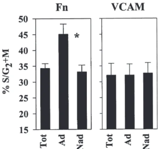
phase of the cell cycle. There was no difference in cell cycle status between adherent and nonadherent cells, whether ad-hesion was assessed on Fn or on VCAM-1 (data not shown). After 48 hours in culture, 34.3% ⫾ 1.5% of the cells were engaged in S/G2⫹M (n ⫽ 18). When adherent and
nonad-herent cells to Fn were analyzed separately, the adnonad-herent fraction was enriched in cycling cells: 45.1% ⫾ 3.2% of ad-herent cells were in S/G2⫹M vs 33.2% ⫾ 2.1% of
nonad-herent cells (n ⫽ 18; p ⬍ 0.00001) (Fig. 1). Thus, transit into the replicative phase of the cell cycle was associated with increased adhesion to Fn. In contrast, when adhesion of cultured CD34⫹ cells was assessed on VCAM-1, no asso-ciation between cycle activation and adhesion was ob-served. The proportion of cells in S/G2⫹M was similar in
adherent (32.1% ⫾ 3.5%) and nonadherent (32.7% ⫾ 3.3%) fractions (n ⫽ 6) (Fig. 1).
We next determined the proportion of CD34⫹ cells in G0
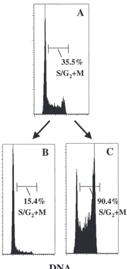
in adherent and nonadherent fractions by using high-resolu-tion cell cycle analysis by Ki-67/7-AAD staining. In this pro-cedure, a DNA histogram is generated by 7-AAD and plotted against the expression of the nuclear antigen Ki-67, which is present in cycling cells but not in G0 cells. To enrich the
start-ing cell population in G0 cells, PB CD34⫹CD38low/⫺ cells
were isolated (instead of total CD34⫹ cells), cultured for 48 hours in the same conditions, and fractionated by adhesion to Fn. The proportion of cells remaining in G0 was 15.0% in
adherent cells compared to 24.6% in nonadherent cells, in a
representative experiment (of three) depicted in Figure 2. Therefore, not only the transit in S/G2⫹M but also the G0–G1
transition was associated with increased cell binding to Fn. To rule out the possibility that adhesion to Fn by itself was responsible for the higher cycling status of the adherent fraction, cultured CD34⫹ cells were first fractionated into cycling and noncycling cells after staining with Hoechst 33342 (Fig. 3) and then assayed for Fn binding capacity (Table 1). Adhesion was significantly increased in cycling cells (25.1% ⫾ 2.6%) compared to noncycling cells (8.0% ⫾ 1.1%, n ⫽ 3; p ⬍ 0.05).
LTC-IC are stimulated to
bind to Fn during cell cycle transit
Binding capacity and cell cycle status of primitive progeni-tor cells were assayed using LTC-IC assays by limiting di-lution analysis. We first determined the absolute numbers of LTC-IC at days 0, 2, and 6 of ex vivo culture. We estab-lished that after 48 hours of suspension culture in SCF, FL, and TPO, there was an initial 51% ⫾ 22% loss of LTC-IC; 4 days later, LTC-IC numbers had returned to 99% ⫾ 32% of day 0 levels (n ⫽ 4). This suggests the existence of at least two subsets of LTC-IC: a first fraction that was not sup-ported by the ex vivo conditions used (e.g., for lack of a par-ticular cytokine and/or stromal accessory cells), and another
Figure 1. Binding to Fn, but not to VCAM-1, is increased in cycling cells.
PB CD34⫹ cells were stimulated ex vivo during 48 hours and replated onto Fn (n ⫽ 18) or VCAM-1 (n ⫽ 6) coated plates and fractionated into adher-ent and nonadheradher-ent fractions. The percadher-entage of cells in S/G2⫹M was determined by PI staining in total (tot), adherent (Ad), and nonadherent (Nad) cells. *p ⬍ 0.00001 compared to nonadherent cells.
Figure 2. Dual-parameter cell cycle analysis of adherent and nonadherent
CD34⫹ cells to Fn after 48 hours of ex vivo culture. PB CD34⫹CD38⫺ cells were stimulated ex vivo for 48 hours and then allowed to bind to Fn. The cell cycle status of cells recovered in the adherent and nonadherent fractions was assessed by simultaneous Ki-67 and 7-AAD staining. The percentages of cells in G0, G1, and S/G2⫹M are shown in the lower right quadrant of both panels. A representative experiment is shown.
O. Giet et al./Experimental Hematology 29 (2001) 515–524 519
more responsive subset that eventually provided the recov-ery of initial LTC-IC numbers after several cell divisions. Thus, although no LTC-IC expansion could be observed in these experiments, LTC-IC replication occurred between days 2 and 6.
Adherent and nonadherent cells to Fn harvested either from freshly isolated CD34⫹ cells or from cells cultured for 48 hours were replated in LTC-IC assays. In unmanipulated cells (n ⫽ 4), the frequency of LTC-IC was similar in the adherent (5.4% ⫾ 1.4%) and the nonadherent fractions (4.3% ⫾ 1.2%). After 48 hours in culture (n ⫽ 10), the per-centage of LTC-IC was higher in the adherent fraction (4.8% ⫾ 0.6%) compared to the nonadherent fraction (2.7% ⫾ 0.4%; p ⫽ 0.0019). LTC-IC assays also were performed af-ter incubating the cells for 1 hour at 37⬚C on Fn-coated plates and pooling adherent and nonadherent cells. No dif-ference in LTC-IC frequency could be demonstrated be-tween cells exposed to Fn and control cells (data not
shown), indicating that, in our conditions, the increase in LTC-IC frequency in adherent cells was not the result of a growth-supporting effect mediated by Fn, as reported else-where [22,23].
We next examined the cell cycle status of LTC-IC col-lected in the adherent and nonadherent fractions by suicide assay. Cells were treated with HU for 1 hour at 37⬚C in or-der to kill cells during S-phase transit. Control cells were in-cubated for 1 hour in medium only. Cells then were replated in limiting dilution in LTC-IC assays and the proportion of cycling LTC-IC (% kill) determined by calculating the ratio between the LTC-IC frequency of HU-treated cells and the LTC-IC frequency of control cells. In freshly isolated PB CD34⫹ cells, no loss of LTC-IC was observed after HU treatment, confirming these to be a quiescent population [3] (Fig 4). After 48 hours in culture, the adherent fraction was significantly enriched in cycling LTC-IC (% kill ⫽ 58.7% ⫾ 6.7%) compared to the nonadherent fraction (% kill ⫽ 19.7% ⫾ 6.5%, n ⫽ 4; p ⬍ 0.05) (Fig. 4). These results in-dicate that, after a 2-day ex vivo culture, not only was the adherent cell population enriched in LTC-IC, but, more im-portantly, the Fn binding capacity of cycling LTC-IC was higher than that of quiescent LTC-IC.
Enhanced adhesion of replicating
progenitor cells to Fn is mediated by VLA-5
The expression of VLA-4 and VLA-5 integrins at the sur-face of PB CD34⫹ cells was analyzed before and after 48 hours of cytokine stimulation. The vast majority (99.3% ⫾ 0.5%, n ⫽ 5) of freshly isolated CD34⫹ cells were VLA-4⫹ and after ex vivo culture there was a slight, but not statisti-cally significant, reduction in VLA-4 expression (93.6% ⫾ 3.6% VLA-4⫹ cells). VLA-4 density, expressed as MCFR, increased from 14.8 ⫾ 2.2 in fresh cells to 30.5 ⫾ 10.9 in cultured cells, but the difference did not reach statistical sig-nificance (n ⫽ 5; p ⬎ 0.05). On the contrary, VLA-5 ex-pression, which was detectable in only 1.8% ⫾ 2.4% of
un-Figure 3. Cell cycle fractionation by Hoechst staining. Cultured CD34⫹
cells were stained with Hoechst 33342 and fractionated into noncycling (G0/G1) and cycling (S/G2⫹M) fractions. The purity of sorted cells was assessed by PI staining of input (A) and sorted cells [(B) noncycling; (C) cycling]. A representative experiment is shown.
Table 1. Adhesion of FACS-sorted cycling and noncycling CD34⫹ cells Cell fraction
Experiment no. Total CD34⫹ G0/G1 CD34⫹ S/G2⫹M CD34⫹
1 12.1 6.0 21.1
2 17.3 8.3 24.3
3 14.5 9.7 29.9
Mean 14.6 8.0 25.1*
SEM 1.5 1.1 2.6
Cultured CD34⫹ cells were fractionated into noncycling (G0/G1) and cy-cling (S/G2⫹M) fractions by Hoechst staining and cell sorting. Adhesion to fibronectin was measured as described in the Methods section. Data are from three independent experiments and are given as percent adherent cells.
*p ⬍ 0.05 compared to G0/G1 CD34⫹ and total CD34⫹ cell populations
(one-way ANOVA followed by Student-Newman-Keul’s tests for multiple pairwise comparisons).
520 O. Giet et al./Experimental Hematology 29 (2001) 515–524
manipulated CD34⫹ cells, increased after 48 hours of cytokine stimulation (32.9% ⫾ 2.2% VLA-5⫹ cells, n ⫽ 9;
p ⬍ 0.001) (Fig. 5).
We next analyzed in more details whether adhesion of replicating CD34⫹ cells to Fn was mediated by VLA-4 or by VLA-5. VLA-4 and VLA-5 expression in cultured CD34⫹ cells was measured separately in G0/G1 and in S/G2⫹M.
VLA-4 was expressed by more than 90% of the cells in either phase of the cell cycle and with similar density (MCFR 22.9 ⫾ 2.7 in noncycling cells vs 23.4 ⫾ 4.4 in cycling cells, n ⫽ 3;
p ⬎ 0.05). VLA-5 was expressed at higher levels in actively
cycling cells: 59.8% ⫾ 9.6% of CD34⫹ cells in S/G2⫹M
were VLA-5⫹ vs only 23.0 ⫾ 5.2% of cells in G0/G1 phase
(n ⫽ 3; p ⫽ 0.02) (Fig. 6). These data document a modula-tion of VLA-5 expression, but not of VLA-4, by cell cycle progression.
To test directly VLA-4 and VLA-5 function in stimu-lated CD34⫹ cells, cells were preincubated with blocking antibodies against ␣4 (clone P4C2) or ␣5 (clone P1D6) in-tegrins and then assayed for adhesion to Fn. The selectivity of both blocking antibodies was confirmed with two non-overlapping Fn fragments. The 40-kDa chymotryptic Fn fragment contains the CS-1 binding site for VLA-4 but lacks the RGDS binding sequence for VLA-5 [24]. The 120-kDa Fn fragment contains the RGDS binding site but
Figure 4. Enhanced binding of cycling LTC-IC to Fn. PB CD34⫹ cells were cultured ex vivo for 48 hours and then replated on Fn-coated dishes. The cell cycle status of LTC-IC was determined by HU suicide assay in total CD34⫹ cells before culture (Tot) and in cultured CD34⫹ cells recov-ered in the adherent (Ad) and in the nonadherent (Nad) fractions to Fn (n ⫽ 4). The percentage of cycling LTC-IC was estimated by the proportion of LTC-IC killed by HU treatment. *p ⬍ 0.05 compared to nonadherent cells.
Figure 5. Changes of VLA-4 and VLA-5 expression in CD34⫹ cells dur-ing ex vivo culture. VLA-4 and VLA-5 expression was determined in freshly isolated (0 h) and in cultured (48 h) CD34⫹ cells. The percentage of cells in each quadrant is indicated. A representative experiment is shown.
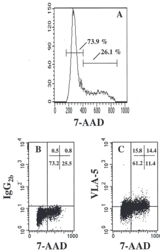
Figure 6. VLA-5 expression in cultured CD34⫹ cells is upregulated dur-ing S/G2⫹M. VLA-5 expression was analyzed in combination with DNA staining by 7-AAD in cultured CD34⫹ cells. A representative experiment is shown. (A) DNA histogram showing the distribution of cells in G0/G1 (73.9%) and in S/G2⫹M (26.1%) phases of the cell cycle. (B) Same sample showing simultaneous staining with control IgG. The horizontal line indi-cates the limit for nonspecific staining. The vertical line separates cells in G0/G1 and those in S/G2⫹M. (C) Combined VLA-5 and 7-AAD staining. The proportion of cells expressing VLA-5 in G0/G1 is 20.5% [15.8/(15.8 ⫹ 61.2) ⫻ 100] compared to 55.8% [14.4/(14.4 ⫹ 11.4) ⫻ 100] in S/G2⫹M.
O. Giet et al./Experimental Hematology 29 (2001) 515–524 521
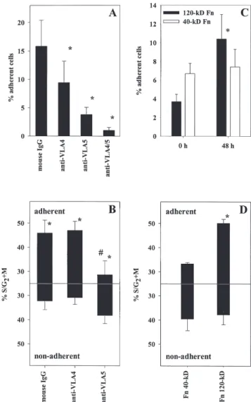
not the CS1 sequence [25]. Anti–VLA-4 blocked CD34⫹ cell binding to the 40-kDa Fn fragment but not to the 120-kDa Fn fragment, whereas the reverse was observed for anti–VLA-5 antibody (data not shown). Compared to non-specific IgG, adhesion of cultured CD34⫹ cells to intact Fn was predominantly inhibited by anti–VLA-5 and to a lesser extent by anti–VLA-4 (Fig. 7A), indicating that VLA-4, al-though present, was not functionally active on a high proportion (⬎50%) of the cells. When cells were treated simultaneously with both anti–␣4 and anti–␣5, adhesion to Fn was completely abolished, which excludes the possibility that another receptor played a significant role in our experimental conditions.
To analyze separately the contribution of VLA-4 and VLA-5 in mediating adhesion of replicating cells to Fn, cul-tured CD34⫹ cells were incubated with a blocking antibody against either ␣4 or ␣5. Cells then were allowed to bind to Fn and the cell cycle status of adherent and nonadherent cells was assessed by PI staining (n ⫽ 4). When ␣4 was blocked, i.e., cells were bound via VLA-5 only, the frequency of cycling S/G2⫹M cells in the adherent fraction (47.0% ⫾
3.8%) was similar to that observed after incubation with mouse IgG (45.9% ⫾ 5.4%; p ⬎ 0.05) (Fig. 7B). In contrast, when ␣5 was blocked and cells were bound to Fn solely by VLA-4, there were only 28.7% ⫾ 5.7% cycling cells in the adherent fraction (p ⬍ 0.05 compared to adherent cells after IgG or anti–␣4 treatment) and cycling cells then were pre-dominantly collected in the nonadherent fraction (Fig. 7B).
Similar results were obtained when the contribution of VLA-4 and VLA-5 was assessed using Fn fragments. CD34⫹ cells, either fresh or stimulated for 48 hours, were allowed to adhere onto plates coated with 40-kDa or 120-kDa Fn fragments containing the CS-1 and RGDS binding sites, respectively. Binding of CD34⫹ cells to the 40-kDa fragment was similar before (6.7% ⫾ 1.1% adherent cells) and after cytokine stimulation (7.4% ⫾ 1.9% adherent cells;
p ⬎ 0.05), whereas adhesion to the 120-kDa Fn fragment
was significantly increased after ex vivo culture, from 3.7% ⫾ 0.8% to 10.4% ⫾ 2.6% adherent cells (p ⬍ 0.05) (Fig. 7C). In addition, cell cycle analysis of adherent and nonadherent CD34⫹ cells to each fragment revealed that there were more cycling cells in the adherent fraction to the 120-kDa frag-ment (50.1% ⫾ 1.7% S/G2⫹M cells) than in the adherent
fraction to the 40-kDa fragment (33.3% ⫾ 0.5% S/G2⫹M
cells; p ⬍ 0.05) (Fig. 7D). Taken together with the blocking antibodies experiments, these data demonstrate that the in-creased adhesion capacity of cycling CD34⫹ cells to Fn was primarily mediated by VLA-5.
Fn binding of replicating
CD34⫹ cells is a reversible process
Finally, we wanted to study whether the increase in VLA-5–mediated adhesion to Fn in cultured CD34⫹ cells could be reverted after completion of cell division. To this end, in a final series of experiments (n ⫽ 5), adherent cells were col-lected after a 2-day ex vivo culture and transferred to fresh
Figure 7. Enhanced binding of cycling CD34⫹ cells to Fn is mediated by VLA-5. (A) Cultured CD34⫹ cells were treated with isotype-matched mouse IgG or with blocking antibodies against VLA-4, VLA-5, or both, and replated on Fn-coated dishes. The percentage of cells recovered in the adherent fraction is indicated in each condition (n ⫽ 8). *p ⬍ 0.05 com-pared to mouse IgG. (B) Cultured CD34⫹ cells were treated with isotype-matched mouse IgG or with blocking antibodies against VLA-4 or VLA-5 and replated onto Fn-coated dishes for 1 hour, after which adherent and nonadherent cells were harvested. The percentage of cells in S/G2⫹M was determined in adherent and in nonadherent fractions by PI staining (n ⫽ 4).
#p ⬍ 0.05 compared to adherent cells recovered after mouse IgG or
anti-VLA-4 treatment; *p ⬍ 0.05 compared to nonadherent cells in the corre-sponding treatment group. (C) Fresh (0 h) and cultured (48 h) CD34⫹ cells were plated on the 40-kD or the 120-kD fragment of Fn, containing CS-1 and RGDS sites, respectively. The percentage of adherent cells was mea-sured in each condition. *Binding to 120-kD Fn increased after culture (p ⬍ 0.05 compared to adhesion at 0 h). (D) Cultured CD34⫹ cells were plated on the 40-kD or the 120-kD Fn fragment for 1 hour, after which adherent and nonadherent cells were collected. The percentage of cycling cells was determined in each fraction. *Percentage of S/G2⫹M cells was higher in cells binding to the 120-kD fragment compared to cells binding to the 40-kD Fn fragment (p ⬍ 0.05).
522 O. Giet et al./Experimental Hematology 29 (2001) 515–524
cultures for an additional 48 hours. The percentage of CD34⫹ cells up to 96 hours in culture was always greater than 90%. Progeny cells resulting from these secondary cul-tures again produced an adherent and a nonadherent popula-tion. Both cell fractions were assayed for cell cycle status. Most progeny cells (80.6% ⫾ 3.6%) had lost their binding capacity to Fn. These nonadherent cells were depleted in cy-cling cells (25.7% ⫾ 3.0% S/G2⫹M cells) compared to the
starting adherent population (39.6% ⫾ 4.1% in S/G2⫹M; p ⬍
0.05) and compared to cells that were still able to bind to Fn (34.6% ⫾ 2.6% in S/G2⫹M; p ⬍ 0.05). A representative
experiment is depicted in Figure 8. These results suggest that adhesion of CD34⫹ cells to Fn during ex vivo culture is transient and reversible after completion of the cell cycle.
Discussion
We examined whether the adhesive properties of progenitor cells to ECM proteins are modulated by cell cycle transit. PB CD34⫹ cells were used for these experiments, as they are blocked in G0/G1 and have very low adhesiveness to Fn
when freshly isolated. After a 2-day ex vivo culture, we ob-served the appearance of a population of adherent cells to Fn. In this adherent population, there was a significant en-richment in cycling (S/G2⫹M) CD34⫹ cells and, more
im-portantly, in cycling primitive progenitor cells assayed as LTC-IC. Moreover, quiescent CD34⫹ cells remaining in G0
after 48 hours of cytokine stimulation were more frequent in the nonadherent population. Thus, transition from a resting state to an activated state in the cell cycle is associated with a potentiation of adhesion to Fn. Both growth-supporting and growth-inhibiting effects of Fn have been reported [22,23,26]; thus, it may be argued that Fn binding by itself may alter the proportion of progenitors in the different phases of cell cycle. To rule out this possibility, we sorted cycling and noncycling CD34⫹ cells after ex vivo culture and showed that cycling cells had a three-fold higher binding capacity as compared to noncycling cells. This shows unambiguously selective ad-hesion of cycling cells. However, cell cycle activation does not directly cause adhesion to Fn, because there also were many cycling cells in the nonadherent fraction. Still, both cy-cling CD34⫹ cells and cycling LTC-IC have a higher ten-dency to bind to Fn than their noncycling counterparts. These observations are in line with a recent report of Yamaguchi et al. [27], who described preferential binding of proliferating CD34⫹ cells to MS-5 stromal layers. Our data further identify Fn, but not VCAM-1, as a preferential ligand for cycling cells. We did not observe any quantitative change in adhesion to VCAM-1 after cytokine stimulation, and binding to VCAM-1 was not enhanced in cycling CD34⫹ cells. Thus, enhanced adhesiveness of cycling progenitor cells is restricted to specific ligands. It remains to be determined whether other ECM proteins or stromal cell adhesion molecules also are involved in this cell cycle selectivity.
As for integrin receptors binding to Fn, VLA-4 and VLA-5
were affected differently by cell cycle activation. The pro-portion of cells expressing VLA-5 was increased during a 2-day ex vivo culture, specifically on the surface of cells traversing S/G2⫹M. VLA-4 was expressed on the vast ma-Figure 8. Binding of CD34⫹ cells to Fn during cell cycle transit is revers-ible. Freshly isolated PB CD34⫹ cells were stimulated ex vivo for 48 hours and plated over Fn. Adherent (Ad) and nonadherent (Nad) cells were har-vested separately. Adherent cells were replated in suspension culture for an additional 48 hours. Cells from these secondary cultures were fractionated further into adherent and nonadherent cells to Fn. The percentage of S/G2⫹M cells was determined in each fraction by PI staining. A representative experiment is shown.
O. Giet et al./Experimental Hematology 29 (2001) 515–524 523
jority of both unmanipulated and cultured CD34⫹ cells and with a higher density on cultured cells (although this was not statistically significant). However, VLA-4 density and the proportion of VLA-4 expressing cells were not different in cycling and noncycling cells. Prosper et al. [13] reported upregulation of VLA-4, but not of VLA-5, at the surface of PB CD34⫹ cells after a 4-day ex vivo culture. This discrep-ancy with our results may arise from differences in the cul-ture conditions used. The cocktail of SCF, FL, and TPO at high doses used in the present study was selected to induce maximal proliferation of primitive progenitor cells [14,15]. In contrast, a combination of granulocyte-macrophage col-ony-stimulating factor (GM-CSF), G-CSF, SCF, leukemia inhibitory factor, macrophage inhibitory prote1a, and in-terleukin-6 at low doses was used in the study by Prosper et al. to promote the survival of progenitor cells with minimal proliferation. Therefore, it should not be surprising that the increase of VLA-5–mediated adhesion to Fn was not ob-served by Prosper et al., because it occurs mainly in cycling progenitor cells. A formal comparison of integrin changes after progenitor cell stimulation with physiologic or phar-macologic cytokine doses should be undertaken to provide further support for this interpretation.
There has been controversies in the literature as to the extent of VLA-4 expression on PB CD34⫹ cells. In several reports, VLA-4 was found on a smaller proportion of PB CD34⫹ cells as compared to BM [13,28], whereas in other studies [27,29], including our own, it was reported that VLA-4 was expressed on the vast majority of PB CD34⫹ cells. These discrepancies are likely due to the fact that VLA-4 expression is quite homogeneous, with no clear sep-aration between negative and positive cells. Depending on the sensitivity of the method and/or the equipment used, PB CD34⫹ cells labeled with anti–VLA-4 may or may not overlap with isotypic controls, whereas BM CD34⫹ cells are brightly positive. Lichterfeld et al. [29] reported that VLA-4 is expressed by the majority of PB CD34⫹ cells but at a lower density (or MCFR) than in BM CD34⫹ cells.
VLA-4 and VLA-5–mediated adhesion to Fn was func-tionally assessed by blocking antibodies. When ␣4 integrin was blocked, there was a 50% reduction in adhesion of cul-tured CD34⫹ cells compared to nonspecific IgG, indicating that although expressed on more than 90% of the cells, VLA-4 was not fully active. In that case, binding to Fn oc-curred via VLA-5 only and was still increased in cycling cells. When ␣5 integrin was blocked, adhesion to Fn was re-duced by approximately 80%, demonstrating a predominant role of VLA-5 in Fn binding of cultured CD34⫹ cells. Most importantly, cycling cells were less able to attach to Fn and then were predominantly collected in the nonadherent frac-tion. These data demonstrate that the preferential binding of cycling CD34⫹ cells to Fn is more specifically mediated by VLA-5. This was further shown by using Fn fragments. Fol-lowing cytokine stimulation, adhesion of CD34⫹ cells was increased on the 120-kDa Fn fragment recognized by
VLA-5 but not on the 40-kDa fragment recognized by VLA-4. In addition, binding to the 120-kDa Fn fragment was increased in cycling cells compared to noncycling cells, whereas adhesion to the 40-kDa Fn fragment was not.
VLA-4 and VLA-5 are expressed by resting progenitor cells in a low-affinity state providing weak interaction with Fn. Cytokines such as interleukin-3, GM-CSF, SCF, and TPO [30–33] increase transiently the affinity of VLA-4 and VLA-5 for Fn without concomitant changes in the expres-sion of these receptors. In this study, we show that, in addi-tion to the regulaaddi-tion of integrin affinity, cytokine stimula-tion allows overexpression of activated VLA-5 once the cells are engaged in cell replication. Therefore, we propose that fast changes in the affinity of integrin receptors occur independently of cell cycle status, whereas slower modula-tion of integrin expression (particularly of VLA-5) takes place during cell proliferation.
Many reports suggest that trafficking of hematopoietic stem cells in and out of the BM is modulated by cell cycle status. First, only resting progenitor cells are found in PB [2,5]. Our results support the hypothesis that the absence of cycling hematopoietic cells in PB may result from their se-lective retention in the BM stroma or from their sese-lective uptake in extramedullary tissues onto Fn-coated sites. Sec-ond, engraftment of transplanted cells is impaired during cell replication [7,8,34]. When viewed in the context of ex vivo expansion and gene therapy, our results show that after 48 hours of culture in conditions promoting stem cell prolif-eration, adhesion of replicating cells to Fn is significantly augmented. Whether enhanced VLA-5–mediated adhesion to Fn could possibly modify migration and/or homing and engraftment of cultured cells remains to be tested in vivo. Interestingly, several reports now have established a recip-rocal relationship between adhesion and migration. When adhesion is maximal, the cell is immobilized and cannot migrate, whereas at intermediate adhesiveness, dynamic disruption of cell–substratum interactions may allow cell lo-comotion [35,36]. If the same applies to primitive hemato-poietic cells, higher expression of VLA-5 during cell repli-cation in ex vivo conditions subsequently may limit the intramedullary migrating ability and hence the capacity to lodge to specific niches in the BM microenvironment.
Acknowledgments
This work was supported by grants from the National Fund for Scien-tific Research (Brussels, Belgium), Télévie, and the “Centre Antican-céreux” at the University of Liège (Liège, Belgium). O.G. was sup-ported by a Télévie fellowship. Y.B. and A.G. are Research Director and Senior Research Assistant, respectively, of the National Fund for Scientific Research (Brussels, Belgium). We thank Amgen for the generous gift of recombinant cytokines. We also thank Dr. J. Siquet and Mrs. N. Wanten from the Transfusion Center at the University of Liège for performing cytaphereses and CD34⫹ cell selection. The au-thors gratefully acknowledge the assistance of Dr. Dirk Van Bocks-taele, University of Antwerp (Antwerp, Belgium) for Hoechst sorting.
524 O. Giet et al./Experimental Hematology 29 (2001) 515–524
References
1. Bensinger W, Singer J, Appelbaum F, et al. (1993) Autologous transplan-tation with peripheral blood mononuclear cells collected after administra-tion of recombinant granulocyte stimulating factor. Blood 81:3158 2. Uchida N, He D, Friera A, et al. (1997) The unexpected G0/G1 cell
cy-cle status of mobilized hematopoietic stem cells from the peripheral blood. Blood 89:465
3. Ponchio L, Conneally E, Eaves C (1995) Quantitation of the quiescent fraction of long-term culture-initiating cells in normal human blood and marrow and the kinetics of their growth factor-stimulated entry into S-phase in vitro. Blood 86:3314
4. Gothot A, Pyatt R, McMahel J, Rice S, Srour EF (1997) Functional heterogeneity of human CD34⫹ cells isolated in subcompartments of the G0/G1 phase of the cell cycle. Blood 90:4384
5. Roberts AW, Metcalf D (1995) Noncycling state of peripheral blood progenitor cells mobilized by granulocyte colony-stimulating factor and other cytokines. Blood 86:1600
6. Ramshaw HS, Rao SS, Crittenden RB, Peters SO, Weier HU, Quesen-berry PJ (1995) Engraftment of bone marrow cells into normal unpre-pared hosts: effects of 5-fluorouracil and cell cycle status. Blood 86:924 7. Habibian HK, Peters SO, Hsieh CC, et al. (1998) The fluctuating phe-notype of the lymphohematopoietic stem cell with cell cycle transit. J Exp Med 188:393
8. Gothot A, van der Loo JCM, Clapp DW, Srour EF (1998) Cell cycle-related changes in repopulating capacity of human mobilized periph-eral blood CD34⫹ cells in NOD/SCID mice. Blood 92:2641 9. Williams DA, Rios M, Stephens C, Patel VP (1991) Fibronectin and
VLA-4 in haematopoietic stem cell-microenvironment interactions. Nature 352:438
10. Verfaillie CM, McCarthy JB, McGlave PB (1991) Differentiation of primitive human multipotent hematopoietic progenitors into single leage clonogenic progenitors is accompanied by alterations in their in-teraction with fibronectin. J Exp Med 174:693
11. van der Loo JC, Xiao X, McMillin D, Hashino K, Kato I, Williams DA (1998) VLA-5 is expressed by mouse and human long-term repopulat-ing hematopoietic cells and mediates adhesion to extracellular matrix protein fibronectin. J Clin Invest 102:1051
12. Simmons PJ, Masinovsky B, Longenecker BM, Berenson R, Torok-Storb B, Gallatin WM (1992) Vascular cell adhesion molecule-1 ex-pressed by bone marrow stromal cells mediates the binding of hemato-poietic progenitor cells. Blood 80:388
13. Prosper F, Stroncek D, McCarthy JB, Verfaillie CM (1998) Mobiliza-tion and homing of peripheral blood progenitors is related to reversible downregulation of ␣41 integrin expression and function. J Clin In-vest 101:2456
14. Luens KM, Travis MA, Chen BP, Hill BL, Scollay R, Murray LJ (1998) Thrombopoietin, kit ligand, and flk2/flt3 ligand together induce increased numbers of primitive hematopoietic progenitors from human
CD34⫹Thy-1⫹Lin⫺ cells with preserved ability to engraft SCID-hu
bone. Blood 91:1206
15. Murray LJ, Young JC, Osborne LJ, Luens KM, Scollay R, Hill BL (1999) Thrombopoietin, flt3, and kit ligands together suppress apopto-sis of human mobilized CD34⫹ cells and recruit primitive CD34⫹ Thy-1⫹ cells into rapid division. Exp Hematol 27:1019
16. Sutherland HJ, Eaves CJ, Landsdorp PM, Thaker JD, Hogge DE (1991) Differential regulation of primitive human hematopoietic cells in long-term cultures maintained on genetically engineered murine stromal cells. Blood 78:666
17. Taswell C (1981) Limiting dilution assays for the determination of im-munocompetent cell frequencies. J Immunol 126:1614
18. Cen D, Levin J (1992) Cell cycle status of murine megakaryocyte and
granulocyte-macrophage colony-forming cells in bone marrow and spleen. Exp Hematol 20:1094
19. Lemoli RM, Tafuri A, Fortuna A, et al. (1997) Cycling status of CD34⫹ cells mobilized into peripheral blood of healthy donors by recombinant human granulocyte colony-stimulating factor. Blood 89:1189
20. Jordan CT, Yamasaki G, Minamoto D (1996) High-resolution cell cy-cle analysis of defined phenotypic subsets within primitive human he-matopoietic cell populations. Exp Hematol 24:1347
21. Leitner A, Strobl H, Fischmeister G, et al. (1996) Lack of DNA syn-thesis among CD34⫹ cells in cord blood and in cytokine-mobilized blood. Br J Haematol 92:255
22. Yokota T, Oritani K, Mitsui H, et al. (1998) Growth-supporting activi-ties of fibronectin on hematopoietic stem/progenitor cells in vitro and in vivo: structural requirement for fibronectin activities of CS1 and cell-binding domains. Blood 91:3263
23. Schofield KP, Humphries MJ, de Wynter E, Testa N, Gallagher JT (1998) The effect of ␣41-integrin binding sequences of fibronectin on growth of cells from human hematopoietic progenitors. Blood 91:3230 24. Wayner EA, Garcia-Pardo A, Humphries MJ, McDonald JA, Carter
WG (1989) Identification and characterization of the T lymphocyte ad-hesion receptor for an alternative cell attachment domain (CS-1) in plasma fibronectin. J Cell Biol 109:1321
25. Pierschbacher MD, Hayman EG, Ruoslahti E (1981) Location of the cell-attachment site in fibronectin with monoclonal antibodies and pro-teolytic fragments of the molecule. Cell 26:259
26. Jiang Y, Prosper F, Verfaillie CM (2000) Opposing effects of engage-ment of integrins and stimulation of cytokine receptors on cell cycle progression of normal human hematopoietic progenitors. Blood 95:846 27. Yamaguchi M, Ikebuchi K, Hirayama F, et al. (1998) Different adhe-sive characteristics and VLA-4 expression of CD34⫹ progenitors in G0/G1 versus S⫹G2/M phases of the cell cycle. Blood 92:842 28. Dercksen MW, Gerritsen WR, Rodenhuis S, et al. (1995) Expression
of adhesion molecules on CD34⫹ cells: CD34⫹ L-selectin⫹ cells predict a rapid platelet recovery after peripheral blood stem cell trans-plantation. Blood 85:3313
29. Lichterfeld M, Martin S, Burkly L, Haas R, Kronenwett R (2000) Mobili-zation of CD34⫹ haematopoietic stem cells is associated with a functional inactivation of the integrin very late antigen 4. Br J Haematol 110:71 30. Levesque JP, Leavesley DI, Niutta S, Vadas M, Simmons PJ (1995)
Cy-tokines increase human hematopoietic cell adhesiveness by activation of very late antigen (VLA)-4 and VLA-5 integrins. J Exp Med 181:1805 31. Levesque JP, Haylock DN, Simmons PJ (1996) Cytokine regulation of
proliferation and cell adhesion are correlated events in human CD34⫹ hemopoietic progenitors. Blood 88:1168
32. Kovach NL, Lin N, Yednock T, Harlan JM, Broudy VC (1995) Stem cell factor modulates avidity of ␣41 and ␣51 integrins expressed on hematopoietic cell lines. Blood 85:159
33. Cui L, Ramsfjell V, Borge OJ, Veiby OP, Lok S, Jacobsen SE (1997) Thrombopoietin promotes adhesion of primitive human hemopoietic cells to fibronectin and vascular cell adhesion molecule-1: role of acti-vation of very late antigen (VLA)-4 and VLA-5. J Immunol 159:1961 34. Szilvassy SJ, Meyerrose TE, Grimes B (2000) Effects of cell cycle
ac-tivation on the short-term engraftment properties of ex vivo expanded murine hematopoietic cells. Blood 95:2829
35. Palecek SP, Loftus JC, Ginsberg MH, Lauffenburger DA, Horwitz AF (1997) Integrin-ligand binding properties govern cell migration speed through cell-substratum adhesiveness. Nature 385:537
36. DiMilla PA, Stone JA, Quinn JA, Albelda SM, Lauffenburger DA (1993) Maximal migration of human smooth muscle cells on fibronec-tin and type IV collagen occurs at an intermediate attachment strength. J Cell Biol 122:729.