Université de Montréal
Antidiabetic activity of Vaccinium vitis-idaea, a
medicinal plant from the traditional pharmacopeia of
the James Bay Cree
parHoda Mohamed Mahmoud Eid
Département de Pharmacologie Faculté de Médicine
Thèse présentée à la Faculté des études supérieures en vue de l’obtention du grade de Doctorat
en pharmacologie
Avril, 2010
Université de Montréal
Faculté des études supérieures et postdoctorales
Cette thèse intitulée:
Antidiabetic activity of Vaccinium vitis-idaea, a medicinal plant from the traditional pharmacopeia of the James Bay Cree
Présentée par :
Hoda Mohamed Mahmoud Eid
a été évaluée par un jury composé des personnes suivantes :
Dre Audrey Claing, président-rapporteur et représentante du doyen
Dr Pierre Haddad, directeur de recherche Dre May Faraj, membre du jury Dre Amira Klip, examinateur externe
Résumé
L’incidence du diabète chez les premières nations du Canada est plus de trois fois celle du reste du pays, dû, en partie, aux traitements culturellement inappropriés. Notre projet vise à traiter le diabète chez ces populations à partir de leur pharmacopée de médicine traditionnelle afin d’améliorer l’acceptation des traitements. En utilisant une approche ethnobotanique, notre équipe a identifié 17 plantes médicinales utilisées pour traiter des symptômes du diabète par les Cris d'Eeyou Istchee (Baie James, Québec). Parmi eux, l'extrait éthanolique de baies de Vaccinium vitis-idaea a montré un effet stimulateur sur le transport du glucose dans les cellules musculaires squelettiques et les adipocytes en culture. Le but de cette thèse était d’élucider les mécanismes par lesquels cet extrait exerce ses effets anti-hyperglycémiants, d’identifier ses principes actifs et de confirmer in vivo, son efficacité. Les résultats démontrent que V.vitis a augmenté le transport du glucose dans les cellules musculaires en cultures, C2C12 et L6 et a stimulé la translocation des transporteurs GLUT4 dans les cellules L6. L'extrait a également inhibé la respiration dans les mitochondries isolées du foie du rat. Cet effet est semblable à celui de la metformine et en lien avec la production du stress métabolique et l'activation de l'AMPK. De plus, la voie de signalisation de l’insuline ne semble pas être impliquée dans le mécanisme d’action de V. vitis.
Le fractionnement guidé par la stimulation du transport du glucose a mené à l'isolation des principes actifs; la quercétine, la galactoside, et la quercétine-3-O-glucoside. Comparable à l'extrait brut, ses composés ont stimulé la voie AMPK. Cependant, la quércetine était la seule à inhiber la respiration mitochondriale.
Pour valider l'effet de V.vitis in vivo, l'extrait (1% dans l'eau de boisson) a été administré aux souris KKAy pendant 10 jours. La glycémie et le poids corporel ont été significativement réduits par V.vitis. Ces effets ont été associés à une diminution de la prise alimentaire, ce qui suggère que V.vitis diminue l'appétit. L'étude pair-fed a confirmé que les effets de V.vitis sont, majoritairement, dû à la réduction de l’appétit. De plus, V.vitis a augmenté la teneur en GLUT4 dans le muscle squelettique, a stimulé la
phosphorylation de l'ACC et a augmenté les niveaux de PPAR-α dans le foie des souris KKAy. Ces effets se voient être additifs à l’effet anorexigène de V. vitis.
Au cours du fractionnement bioguidé de l’extrait, l’ester méthylique de l'acide caféique (CAME), un produit formé lors de la procédure du fractionnement, a démontré un effet stimulateur puissant sur le transport du glucose dans les celules C2C12 et donc un potentiel anti-diabétique. Pour identifier d'autres acides caféique active (AC) et pour élucider leurs relations structure-activité et structure-toxicité, vingt dérivés AC ont été testés. Outre CAME, quatre composés ont stimulé le transport du glucose et ont activé l'AMPK suite au stress métabolique résultant d'un découplage de la phosphorylation oxydative mitochondriale. L’activité nécessite une fonction d’AC intacte dépourvu de groupements fortement ionisés et ceci était bien corrélée avec la lipophilicite et la toxicité. Les résultats de cette thèse soutiennent le potentiel thérapeutique de V. vitis, ses composés actifs ainsi que de la famille de l’AC et pour la prévention et le traitement du diabète.
Mots-clés : Vaccinium vitis, diabète de type 2, AMPK, ACC, PPAR-α, OPD, KKAy, GLUT4, produits de santé naturels, médecine traditionnelle, la forêt boréale canadienne, les Autochtones d’Amérique du nord.
Abstract
Type 2 diabetes in Canadian First Nations is three times higher than the national average. Poor prognosis is partly attributed to cultural inappropriateness of pharmaceutical products. Our project aims to develop culturally adapted diabetes treatment based on traditional medicine pharmacopoeia. Our team has identified 17 plants used to treat the symptoms of diabetes by the Cree of Eeyou Istchee (James Bay, Quebec). Among them, the ethanol extract of Vaccinium vitis-idaea berries was found to have an important stimulatory effect on glucose uptake in cultured skeletal muscle cells and adipocytes. The goal of this thesis was to elucidate the mechanisms of action of this plant product as well as to isolate and identify its active constituents using a bioassay-guided fractionation approach and finally to validate the antidiabetic activity in vivo. The extract of V.vitis enhanced glucose uptake in cultured C2C12 and L6 skeletal muscle cells and stimulated the translocation of GLUT4 transporters to the cell membrane of L6 cells. It mildly inhibited ADP-stimulated oxygen consumption in isolated rat liver mitochondria, an effect similar to that of metformin and consistent with metabolic stress and the consecutive activation of AMP-activated protein kinase (AMPK) pathway. The insulin pathway does not seem to be involved in V.vitis signaling.
Fractionation of this plant extract, guided by glucose uptake activity, resulted in the isolation of the active principles, O-galactoside, quercetin, and quercetin-3-O-glucoside. Similar to the crude extract, the quercetin glycosides and the aglycone stimulated the AMPK pathway. However, only the aglycone inhibited ATP synthase in isolated mitochondria.
To validate the effect of V.vitis in vivo, the extract (1% in drinking water) was administered to KKAy mice for 10 days. Glycemia and body weight were significantly reduced by V.vitis. These effects were associated with decrease of food intake, suggesting that V.vitis reduces the appetite. The pair-fed study confirmed that the previous effects of V.vitis are almost mediated by its appetite reducing action. In addition, V. vitis-treatment increased the content of GLUT4 protein in skeletal muscle, stimulated the phosphorylation of ACC and increased the levels of PPAR-α in the liver of KKAy mice. These effects could be additives to the apetite controling effect of V. vitis.
In the course of bioguided-fractionation, caffeic acid methyl ester (CAME), a by-product of fractionation procedure, has been shown to potently stimulate glucose uptake in cultured skeletal muscle cells and therefore to have anti-diabetic potential. To identify other active caffeic acid (CA) derivatives and to elucidate their structure–activity and structure-toxicity relationships, twenty CA derivatives were tested. In addition to CAME, four compounds were found to stimulate glucose uptake and activate AMPK. Uncoupling of mitochondrial oxidative phosphorylation by these compounds resulted in metabolic stress which could explain the activation of AMPK. The activity required an intact caffeic acid moiety devoid of strongly ionized groups and was well correlated with lipophilicity and toxicity.
The results of the present thesis support a therapeutic potential for V.vitis, and its active compounds, as well as the CA family of compounds for the prevention and treatment of diabetes.
Keywords: Vaccinium vitis, type 2 diabetes, AMPK, ACC, PPAR-α, OPD, KKAy, GLUT4, natural health products, traditional medicine, Canadian boreal forest, Aboriginal populations of North America.
Table of content
1. Introduction ... 1
1.1 Energy homeostasis ... 3
1.1.1 Glucose homeostasis ... 3
1.1.1.1 The insulin receptor: transduction through tyrosine kinase signaling ... 3
1.1.1.1.1 PI3-Kinase pathway and downstream targets ... 4
1.1.1.1.2 The Ras–MAPK cascade ... 4
1.1.1.1.3 Cbl/CAP/TC10 pathway in lipid rafts ... 5
1.1.1.1.4 Regulation and termination of IR signal ... 5
1.1.1.2 Glucose transporters and insulin action ... 6
1.1.1.3 Alternative pathway to glucose uptake: AMP-activated protein kinase pathway... 9
1.1.1.4 Regulation of hepatic glucose production ... 12
1.1.2 Regulation of lipogenesis and lipolysis ... 13
1.2 Diabetes mellitus ... 14
1.2.1 Definition and diagnosis ... 14
1.2.2 Diabetes Classification ... 14
1.2.2.1 Type 1 diabetes mellitus (T1DM) ... 15
1.2.2.2 Type 2 Diabetes mellitus (T2DM) ... 15
1.2.2.3 Gestational diabetes mellitus (GDM)... 16
1.2.2.4 Other Specific Types of diabetes... 16
1.2.3 Pathogenesis of type 2 diabetes ... 16
1.2.3.1 Insulin resistance ... 17
1.2.3.1.1 Body-Fat distribution and insulin resistance ... 17
1.2.3.1.2 Secretory function of adipose tissue ... 18
1.2.3.1.2.1 Leptin ... 18
1.2.3.1.2.2 Adiponectin ... 19
1.2.3.1.3 Inflammation in obesity ... 20 1.2.4 Diabetes complications ... 22 1.2.5 Treatment ... 22 1.2.5.1 Lifestyle interventions... 22 1.2.5.2 Pharmacological treatment ... 23 1.2.5.2.1 Insulin secretagogues ... 23 1.2.5.2.1.1 Sulfonylureas ... 23
1.2.5.2.1.2 Rapid-acting insulin secretagogues (the glitinides) ... 24
1.2.5.2.2 Insulin sensitizers ... 24
1.2.5.2.2.1 Biguanides ... 24
1.2.5.2.2.2 Thiazolidinediones (TZDs) ... 25
1.2.5.2.3 α-Glucosidase Inhibitors ... 25
1.2.5.2.4 Dipeptidyl peptidase 4 inhibitors and GLP-1 analogues ... 26
1.2.5.3 Herbal medicine and diabetes ... 26
1.3 Genus Vaccinium ... 27 1.3.1 Vaccinium vitis-idaea ... 28 1.3.1.1 Phytochemicals of V. vitis ... 29 1.3.1.1.1 Flavonoids ... 29 1.3.1.1.1.1 Anthocyanins ... 30 1.3.1.1.1.2 Flavonols ... 30 1.3.1.1.1.3 Flavan-3-ols... 31 1.3.1.1.2 Phenolic acids ... 31 1.3.1.1.3 Stilbenes ... 32 1.3.1.1.4 Tannins... 33
1.4 Scope and objectives of the study ... 34
2. Article 1 ... 36
3. Article 2 ... 77
4. Article 3 ... 120
5. General discussion ... 157
6. Conclusion and perspective ... 163
List of tables
Introduction
Table 1 Distribution of mammalian facilitative glucose transporters ... 8
Article 1
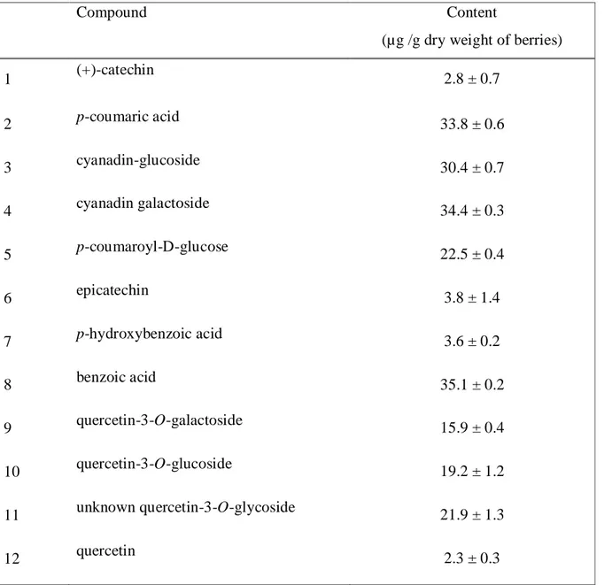
Table 1 Yield of V. vitis berry extract constituents ... 76
Article 2
Table 1 Physicochemical parameters and measured activities of test compounds ... 111
Article 3
Table 1 Blood parameters of KKAy mice from study #1... 149 Table 2 Histological scores of liver steatosis from control and V. vitis treated KKAy
diabetic mice. ... 150
List of figures
Introduction
Figure 1 Insulin signaling pathways ... 6
Figure 2 major effects of AMPK on glucose and fatty acid metabolism in liver and skeletal muscle ... 11
Figure 3 Activators of AMPK ... 12
Figure 4 Molecular mechanism underlying adipose tissue inflammation ... 21
Figure 5 Flowers and fruits of V. vitis-idaea... 29
Figure 6 The structures of anthocyanidins detected in V. vitis-idaea ... 30
Figure 7 The structures of quercetin and kaempherol... 31
Figure 8 The structure of catechin... 31
Figure 9 The structures of benzoic acid and hydroxycinnamic acid ... 32
Figure 10 The structure of resveratrol... 33
Figure 11 Structure of tannins isolated from V. vitis-idaea ... 34
Article 1 Figure 1 Phytochemical fractionation of V. vitis berry extract guided by muscle cell glucose-uptake activity ... 68
Figure 2 HPLC chromatograms of V. vitis berry crude extract ... 69
Figure 3 chemical structures of the 12 isolated constituents of V. vitis berry ethanol extract ... 70
Figure 4 V. vitis berry extract and its active principles stimulate the AMPK signaling pathway but not the insulin receptor pathway... 71
Figure 5 The V. vitis berry extract induces a mild instantaneous inhibition of respiration in isolated rat liver mitochondria, as illustrated by a representative oxygen consumption tracing. ... 72
Figure 6 Quercetin (A), but not quercetin-3-O-glycosides (B), induces an important instantaneous and dose-dependent inhibition of respiration in rat liver mitochondria, as illustrated by representative oxygen consumption tracings.. ... 73
Figure 7 Quercetin does not increase the rate of secretion of acid equivalents by C2C12 (A) or H4IIE (B) cells ... 74 Figure 8 Quercetin does not reduce intracellular ATP concentration in H4IIE hepatocytes.75
Article 2
Figure 1 Caffeic acid phenethyl ester (CAPE) increased non-insulin-stimulated (basal) 3H-deoxyglucose uptake in differentiated C2C12 skeletal muscle cells by more than 3-fold. ... 112 Figure 2 CAPE induced a powerful uncoupling effect in isolated rat liver mitochondria. 113 Figure 3 CAPE induced cytotoxicity in C2C12 myotubes. ... 114 Figure 4 Compounds selected to address specific structure–activity hypotheses. ... 115 Figure 5 The magnitude of stimulation of glucose uptake in C2C12 mytotubes induced
following an 18 h treatment with caffeic acid derivatives is correlated to the magnitude of mitochondrial uncoupling activity induced by these compounds in isolated liver mitochondria. ... 116 Figure 6 Active caffeic acid derivatives increased phosphorylation of ACC, an effector of
AMPK, in C2C12 myotubes. ... 117 Figure 7 The lipophilicity of active caffeic acid derivatives is a good predictor of their
activity. ... 118 Figure 8 Structural constraints for bioactive caffeic acid derivatives. ... 119
Article 3
Figure 1 V. vitis increased non-insulin-stimulated (basal) 3H-deoxyglucose uptake and GLUT4 translocation in L6 GLUT4myc myotubes. ... 151 Figure 2 V. vitis increased phosphorylation of AMPK and p-38MAPK but not of Akt in
L6 myotubes. ... 152 Figure 3 V. vitis reduced cumulative change in body weight, cumulative change in food intake, non-fasting blood glucose concentration, and cumulative change in fluid intake in KKAy mice of study #1. ... 153
Figure 4 Effect of 10 days V. vitis-treatment and pair-feeding on cumulative change in body weight, and non-fasting blood glucose concentration, and cumulative change in fluid intake in KKAy mice of study #2. ... 154 Figure 5 V. vitis has no effect on cumulative change in body weight, cumulative change
in food intake, non-fasting blood glucose concentration. It significantly reduced cumulative change in fluid intake in normal C57BL/6 mice of study #3. ... 155 Figure 6 Effect of V. vitis on GLUT4 levels in skeletal muscles, phosphorylation of ACC
and PPAR-α levels in livers from diabetic KKAy mice of study #1 (A) and study #2 (C) Quantification of GLUT4/β-actin (B), quantification of PPARα/β-Actin (D), and quantification of p-ACC/ β-actin (E).. ... 156
List of abbreviations
µg: MicrogramµL: Microliter
ACC: Acetyl-CoA carboxylase
AICAR: aminoimidazole carboxamide ribonucleotide AMP: adenosine monophosphate
AMPK: AMP-activated protein kinase ATP: Adenosine triphosphate
CA: Caffeic acid
CAM: Complementary and alternative medicine CAME: Caffeic acid methyl ester
CAP: Cbl-associated protein
CAPE: Caffeic acid phenethyl ester CEI: Cree of Eeyou Istchee
cm: Centimeter
DMSO: Dimethyl sulfoxide DPP-4: Dipeptidylpeptidase 4 FFAs: Free fatty acids
g: Gram
G-6-P: Glucose 6-phosphate G-6-Pase: Glucose 6-phosphatase GLUT: Glucose transporters GS: Glycogen synthase
GSK-3: Glycogen synthase kinase 3 h: Hour
HPLC: high performance liquid chromatography IGT: Impaired glucose tolerance
IL: Interlukin IR: Insulin receptor
Kg: Kilogram
LDL: Low density lipoprotein
MAPK: mitogen-activated protein kinase MC4-R: melancortin receptor 4
min: Minute mL: Milliliter
NMR: Nuclear magnetic resonance NPY: Neuropeptide Y
OGTT: Oral glucose tolerance test
OPD: O-phenylenediamine dihydrochloride
PDK1: 3-phosphoinositide-dependent protein kinase-1 PEPCK: Phosphoenolpyruvate carboxykinase
PGC-1α: Peroxisome proliferator-activated receptor gamma coactivator-1 PKC: Protein kinase C
PPAR: Peroxisome-proliferator–activated receptor PTB: Phosphotyrosine-binding
PTPases: Protein tyrosine phosphatases
RASOC: the rate of ADP-stimulated O2 consumption
RBOC: the rate of basal oxygen consumption SGLT: Sodium glucose co-transporters SOS: Son of Sevenless
SREBPs: Sterol regulatory element-binding proteins T1DM: Type 1 diabetes mellitus
T2DM: Type 2 diabetes mellitus TNF-α: Tumor necrosis factor TZDs: Thiazolidinediones UCPs: Uncoupling proteins UV: Ultraviolet
Acknowledgement
I owe my deepest gratitude to my supervisor, Dr Pierre Haddad, whose constant encouragement and guidance made this thesis possible. Throughout my thesis period, he provided valuable advice, scientific and emotional support during difficult moments. Thanks a lot for being my supervisor and for believing in me. I really have a lot of appreciation for you.
I am heartily thankful to Dr Thor John Arnason who gave me the opportunity to work in his laboratory in the Department of Biology, University of Ottawa and gave me untiring help during my study. I would like also to thank Dr Ammar Saleem and Dr Mohammed Asim who did not spare any effort to help me with the laborious phytochemical work.
I would like to express my appreciation for Dr Gary Sweeney who welcomed me in his laboratory in York University, Toronto. I warmly thank Dr. Farah Thong, his team leader, for her valuable advice and friendly help. I wish also to thank Riya and Tien for helping me out and for making my stay in Toronto an enjoyable one.
Special thanks are due to Diane Vallerand and Antoine Brault for the help and advice and for being always there for the students especially during difficult moments.
I wish to extend my sincere thank to all my colleagues especially Ali, Meriem, and Abir for helping me to get through the difficult times, and for all the support, and caring they gave.
I also owe heartful thanks to Dr Lina Musallam for assistance with thesis writing and for her suggestions and comments.
It is a pleasure to thank Sylvie Caron from the departments of Pharmacology, for sympathetic help in secretarial work. She is such a nice and open-minded person.
I also wish to express my love and gratitude to my beloved family; for their endless love and support through my life. I wish to especially thank my parents, my three sisters and my brother. To them I dedicate this thesis.
I do not forget the rest of people at the Department of Pharmacology, to whom I’m also very thankful.
I’m also grateful for the members of my committee for taking the time to review my thesis.
Lastly, and most importantly, thanks to God for granting me his gift of life and health and for his many blessing.
1. Introduction
The last few decades of the 20th century have witnessed the rise of worldwide epidemic obesity along with type 2 diabetes mellitus in adults, as well as in children and adolescents. Type 2 diabetes is a metabolic disorder characterized by hyperglycemia. Its prevalence varies among different ethnic groups. In Canada, the overall prevalence of diabetes among Canadian adults was approximately 5.5% in 2004-2005 according to National Diabetes Surveillance System (NDSS) data. The populations most affected are the aboriginal populations with a higher prevalence in women compared to men. The recent socio-cultural changes experienced by these populations including adoption of a sedentary lifestyle, the consumption of non-traditional foods, along with the genetic predisposition to the disease are the major causes of the epidemic (Boston et al. 1997); (Young et al., 2000); (Hegele, 2001). Aboriginals also suffer from diabetes complications, including end-stage renal failure, retinopathy and peripheral neuropathy, at a disproportionately high rate. It was reported that death from diabetes complications is 5-fold higher among aboriginal women as compared with Canadian women (Young et al., 2000).
The Cree represent the largest aboriginal group in Canada, with more than 72,000 registered individuals (Statistics Canada, 2002). Eeyou Istchee, which literally means ―land of the people‖, is the homeland of the Cree Nation of Eastern James Bay. The Cree of Eeyou Istchee (CEI) have a population of approximately 14,000 people who live in 9 communities spread across the northern part of the province of Quebec (Secrétariat aux affaires autochtones, 2004). Over the past decade, diabetes has reached unprecedented proportions among the CEI with a prevalence of 17.7% among adults aged over 20 years (Légaré, 2004). A high prevalence of gestational diabetes has been also observed in CEI, which ranks second among aboriginal groups worldwide (Rodrigues et al., 1999).
To address this serious aboriginal health issue, diabetes prevention and treatment projects need to be adapted to the cultural and social environment of these populations. Aboriginal peoples have a long tradition of using plants in their environment for healing purposes; through millennia of trial and errors they had developed a comprehensive
traditional pharmacopeia that was handed down from generation to generation largely as verbal teaching and as part of their cultural tradition (Young et al., 2000).
After the European colonization, some of their traditional knowledge was lost; fortunately much was documented by anthropologists and was recognized in the official Pharmacopeia of the United States and Canada. The first Pharmacopoeia of the United States, published in 1820, included 170 indigenous plant species. Similarly, the Canadian Pharmaceutical Journal contained more than twenty species prescribed by First Nations as medication (Erichsen-Brown, 1979; Vogel, 1990).
Despite the wealth of the Cree Nations’ traditional knowledge, no work has been done to examine the potential of their medicinal plants to treat the relatively recent diabetes epidemic. The approach that our research team has adopted was to develop culturally relevant diabetes treatment options in these communities. Taking into consideration the complexity of diabetes and its relatively recent evolution among the Cree population, the ethnobotanical approach adopted by our project was based on the the symptoms and complications of the disease. Hence a questionnaire that included 15 symptoms of type 2 diabetes, rather than diabetes per se was prepared. In collaboration with the CEI, we have conducted ethnobotanical surveys in four CEI communities: Mistissini, Whapmagoostui, Nemaska and Waskaganish and have identified seventeen medicinal plant species that are traditionally used to treat symptoms related to diabetes (Leduc et al., 2006), (Fraser et al., 2007), (Harbilas et al., 2009). Bioactivity screening projects for antidiabetic properties of these plant products have showed that over half of them possess significant antidiabetic activities (Spoor et al., 2006), (Harbilas et al., 2009). The present study focuses on the antidiabetic properties of the berries of Vaccinium vitis-idaea, a medicinal plant product used in the communities of Whapmagoostui and Mistissini to treat frequent urination and a number of other symptoms of diabetes (Leduc et al., 2006); (Fraser et al., 2007).
1.1 Energy homeostasis
1.1.1 Glucose homeostasis
Glucose is the primary metabolic fuel for all body tissues and the obligatory energy substrate for the brain. Despite the relatively small weight of the brain (2% of body weight), it uses 25% of the total body glucose (Dong et al., 2003). Therefore, a constant and adequate supply of glucose is necessary to maintain normal brain function (Tirone and Brunicardi, 2001). During fed and fasting states, healthy individuals are able to maintain plasma glucose in narrow ranges (fasting blood glucose of 3.3–5.6 mmol/L and post-prandial glucose of 4.40–6.94 mmol/L). To keep this tight control of blood glucose concentration, a balance should be achieved between glucose absorption by the intestine, glucose production by the liver and glucose disposal in peripheral tissues (Beardsall et al., 2008). Insulin is the master regulator of blood glucose level in the fed state and does so by controlling glucose uptake by muscle and fat cells and by suppressing hepatic glucose production. On the other hand, glucagon and other counter-regulatory hormones (catecholamines, cortisol and growth hormone) maintain blood glucose levels during fasting (Beardsall et al., 2008).
1.1.1.1 The insulin receptor: transduction through tyrosine kinase signaling
The insulin receptor (IR) is a heterotetrameric plasma membrane protein receptor that consists of two extracellular α- and two intracellular β-subunits linked by disulfide bonds. It belongs to a subfamily of receptor tyrosine kinases that also includes the insulin-like growth factor and an orphan receptor, known as the IR-related receptor (Ward, 1999). Before ligand binding, IR is inactive, although it is oligomerized, since the α subunit exhibits allosteric inhibition of the β subunit catalytic activity. Binding of the ligand (insulin) induces a conformational change which stimulates the catalytic activity and induces transphosphorylation of the receptor on specific tyrosine residues. Once phosphorylated, these tyrosine residues, along with the sequence of adjacent amino acids, create binding sites for docking proteins, which contain domains that bind phosphotyrosine such as Src homology 2 domain (SH2) and phosphotyrosine binding (PTB). Scafolds
(insulin receptor substrate (IRS), Gab, and Shc), adaptors (Grb2), kinases (phosphatidylinositol 3-kinase (PI3-K), Src), phosphatases (Shp2) and ubiquitinating proteins (c-Cbl) are recruited to the phosphorylated active IR through these domains to be phosphorylated by the catalytic activity of IR on tyrosine residues (Baron et al., 1992), (Saltiel and Kahn, 2001). This links IR to several major signaling pathways, one involves phosphatidylinositol 3-kinase (PI3-K) and the other involves Ras/mitogen-activated protein kinase (MAPK) cascade (Figure 1).
1.1.1.1.1 PI3-Kinase pathway and downstream targets
This pathway is responsible for the metabolic action of insulin. Recruitment of PI3-K to IRS through its regulatory subunit, p85, results in the phosphorylation of phosphatidylinositol (4,5) bisphosphate (PI(4,5)P2) by PI3-K catalytic subunit, p110, which generates phosphatidylinositol (PI(3,4,5)P3). PI(3,4,5)P3 serves as docking site for pleckstrin homology domain (PH) containing serine/threonine kinase Akt/PKB. Once correctly positioned at the membrane, Akt gets phosphorylated by its activating kinases, phosphoinositide-dependent protein kinase-1 (PDK1), at threonine 308 (Jacob et al., 2008). PDK1 also phosphorylates and activates the atypical forms of protein kinase C (PKC), including PKCξ and PKCλ. Both Akt and atypical PKC have been shown to mediate insulin dependent glucose transport (Czech and Corvera, 1999). This is discussed in detail further below.
1.1.1.1.2 The Ras–MAPK cascade
The Ras-MAPK pathway is mainly involved in mediating cell growth, survival and cellular differentiation. Phosphorylation of IRS-1 by IR induces the translocation of cytosolic adaptor protein, growth factor-bound protein 2 (Grb-2) through its SH2 domain. Grb2 also contains SH3 domain which allows the constitutive association with the proline rich region of the guanyl nucleotide exchange factor, son of sevenless (SOS). Alternatively, Grb2/SOS complex is recruited to IR through the assistance of another adaptor, Shc. The recruitment of Grb2 from the cytoplasm to the plasma membrane binds the small GTPase Ras. Through guanine exchange, SOS activates Ras, thus allowing its
interaction with downstream effectors and their activation. Ras initiates a kinase cascade via the stepwise activation of Raf, the MAP kinase-kinase MEK and the MAP kinases ERK1 and ERK2. Once activated, ERKs promote gene expression and protein synthesis by phosphorylating targets such as p90 ribosomal protein S6 kinase (p90RSK) and the transcription factor ELK1 (Avruch, 1998).
1.1.1.1.3 Cbl/CAP/TC10 pathway in lipid rafts
The insulin receptor also phosphorylates other substrates such as the Cbl-binding protein APS. Phosphorylation of APS is necessary for its binding with Cbl and the subsequent phosphorylation of Cbl by IR. Cbl interacts also with Cbl-associated protein (CAP), a protein that belongs to the Sorbin homology (SoHo) family of adaptor proteins. The phosphorylated APS-Cbl–CAP complex then translocates to lipid raft domains in the plasma membrane through the interaction of the SoHo domain of CAP with the lipid raft protein, flotillin. Once translocated into the lipid raft, the phosphorylated Cbl recruits the SH2/SH3 adaptor protein CrkII through its SH2 domain along with the guanyl nucleotideexchange protein C3G. C3G, in turn, catalyze the nucleotideexchange of GTP for GDP on the lipid -raft-associated protein TC10, a Rho family GTPase, resulting in its activation. Along with the PI3-K, TC10 stimulates the trafficking of Glut4 vesicles, their docking and their fusion with the plasma membrane (Saltiel and Kahn, 2001), (Kimura et al., 2002). This pathway was first described in 3T3-L1 adipocytes, but was later reported in cardiac muscle and adipose tissue in vivo (Gupte and Mora, 2006). Another group suggested that the Cbl/CAP/TC10 signaling cascade was present in skeletal muscle, activated by insulin, and impaired by high-fat feeding (Bernard et al., 2006).
1.1.1.1.4 Regulation and termination of IR signal
Given the important biological functions of IR, its signal must be tightly regulated. This occurs by terminating IR signaling through its internalization and dephosphorylation by protein tyrosine phosphatases (PTPases) such as PTP1B. IR is then ubiquitinated and degraded by the proteasome (Zinker et al., 2002), (Saltiel and Kahn, 2001).
Alternative regulation of IR could be the result of cross talk signaling from other receptors such as the epidermal growth factor receptor (EGFR), tumor necrosis factor alpha (TNF-α) and integrin receptors. IR has been shown to be phosphorylated at Ser/ Thr residues, which results in the attenuation of insulin signaling (Coba et al., 2004). Finally, IRS-1 is also the target of Ser/ Thr phosphorylation by PKC-β, which results in the inhibition of the catalytic activity of IR (Aguirre et al., 2002) (Liberman et al., 2008) (Ishizuka et al., 2004). IRS can also be negatively regulated by kinases mediators of insulin signalling upon prolonged insulin stimulation such as PKCξ,(Liu et al., 2001), mTOR/S6K1 (Tremblay et al., 2007) and certain MAPK (Engelman et al., 2000). Novel members of PKC family (nPKCs) such as PKC-ε, -η and –θ can directly phosphorylate IRS-1 on serine/therionine residues. nPKCs are involved in free fatty acids (FFAs)-induced insulin resistance. The metabolism of FFA increases the content of intramyocellular diacylglycerol (DAG), a potent allosteric activator of conventional and novel PKCs (Dey et al., 2006).
Figure 1 Insulin signaling pathways (Saltiel and Kahn, 2001)
1.1.1.2 Glucose transporters and insulin action
In the fed state, insulin promotes glucose disposal mainly through glucose uptake by peripheral tissues and its subsequent utilization/storage. Because glucose is a polar
molecule, it does not diffuse through the lipid bilayer of cell membranes and therefore glucose transporters are required (Olson and Pessin, 1996).Two different types of glucose transporters have been identified: the energy dependent sodium glucose co-transporters (SGLT) and the facilitative glucose transporters (GLUT). SGLT are expressed mainly in the intestine and kidney, where they actively transport glucose against its concentration gradient by using the energy derived from the co-transport of sodium down its electrochemical gradient (Shepherd and Kahn, 1999).
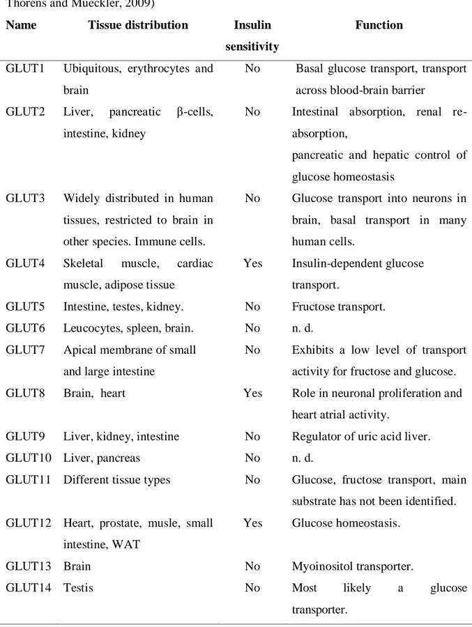
GLUT transporters are a family composed of 14 structurally related proteins that mediate facilitative transport of glucose along its concentration gradient. These proteins have 12 membrane spanning domains with both C- and N- terminal tails of the protein oriented on the cytoplasmic side (Augustin, 2010; Olson and Pessin, 1996; Thorens and Mueckler, 2009; Wood and Trayhurn, 2003). They are encoded by distinct genes and have distinct substrates and tissue distribution (Table 1). GLUT family can be grouped into three classes bases on their structural similarities, class I that includes GLUT1-4 and GLUT14, class II comprises GLUT5, 7, 9, 11, and class III which has GLUT6, 8, 10, 12 and the proton driven myoinositol transporter HMIT (or GLUT13). GLUT4 is the main insulin-responsive glucose transporter and is located primarily in skeletal muscle cells, cardiac muscle cells and adipocytes (James et al., 1989). In these tissues, GLUT4 is responsible for most of the glucose uptake, even though they also express the GLUT1 isoform (Giorgino et al., 2000). In the basal state, less than 5% of GLUT4 resides in the cell surface, the rest are present in the membrane of the vesicles. Upon insulin stimulation, GLUT4 is translocated to the cell surface and glucose uptake is increased (Cushman et al., 1998), (Suzuki and Kono, 1980), (Marette et al., 1992).
Table 1 Distribution of mammalian facilitative glucose transporters (Augustin, 2010; Thorens and Mueckler, 2009)
Name Tissue distribution Insulin
sensitivity
Function
GLUT1 Ubiquitous, erythrocytes and brain
No Basal glucose transport, transport across blood-brain barrier
GLUT2 Liver, pancreatic β-cells, intestine, kidney
No Intestinal absorption, renal re-absorption,
pancreatic and hepatic control of glucose homeostasis
GLUT3 Widely distributed in human tissues, restricted to brain in other species. Immune cells.
No Glucose transport into neurons in brain, basal transport in many human cells.
GLUT4 Skeletal muscle, cardiac muscle, adipose tissue
Yes Insulin-dependent glucose transport.
GLUT5 Intestine, testes, kidney. No Fructose transport. GLUT6 Leucocytes, spleen, brain. No n. d.
GLUT7 Apical membrane of small and large intestine
No Exhibits a low level of transport activity for fructose and glucose. GLUT8 Brain, heart Yes Role in neuronal proliferation and
heart atrial activity.
GLUT9 Liver, kidney, intestine No Regulator of uric acid liver.
GLUT10 Liver, pancreas No n. d.
GLUT11 Different tissue types No Glucose, fructose transport, main substrate has not been identified. GLUT12 Heart, prostate, musle, small
intestine, WAT
Yes Glucose homeostasis.
GLUT13 Brain No Myoinositol transporter.
GLUT14 Testis No Most likely a glucose
Once inside muscle, glucose is phosphorylated by hexokinase or glucokinase, yielding glucose 6-phosphate (G-6-P). G-6-P can be then used either for the synthesis of glycogen via the activity of glycogen synthase (GS) or metabolized in the glycolytic pathway via enzymes such as pyruvate kinase (Roach, 2002).
1.1.1.3 Alternative pathway to glucose uptake: AMP-activated protein kinase pathway
AMP-activated protein kinase (AMPK) is a ubiquitous heterotrimeric enzyme composed of α, β and γ subunits. The α subunit contains a serine/threonine protein kinase catalytic domain. It also holds Thr/172 which is the principal site of its phosphorylation by upstream kinases and thus its activation. The β subunit has glycogen-binding domains and appears to stabilize the interaction between α and γ subunits. The γ subunit binds AMP or ATP in a mutually exclusive manner (Wong and Lodish, 2006).
AMPK is a metabolic stress-sensing protein kinase that is activated in response to depletion of ATP content and an increase in the cellular AMP/ATP ratio under stress conditions such as hypoxia, physical exercise and inhibition of mitochondrial respiration (Chen et al., 1999). AMP is thought to allosterically activate the enzyme and prevent its dephosphorylation. On the other hand, the tumor suppressor LKB1 is the major kinase responsible for phosphorylation of AMPK on Thr172 in the α-subunit. However, LKB1 is not directly activated by AMPK, but the later induces conformational changes in AMPK rendering it more susceptible to phosphoylation by LKB1. Ca2+/calmodulin-dependent almodulin protein kinase kinase (CAMKK) can also phosphorylate AMPK on Thr172 and activate it (Shen et al., 2007). When AMPK is activated, energy-consuming anabolic pathways are shuts down (such as fatty acid, protein and cholesterol synthesis) and ATP-producing catabolic pathways are activated (such as fatty acid oxidation) (Viollet et al., 2003).
Within skeletal muscle, the activation of AMPK increases glucose uptake through the stimulation of GLUT4 translocation to the plasma membrane by a mechanism distinct
from PI3-K stimulated by insulin. P38 MAPK appears to be activated by several stimulators of AMPK, including AICAR and mitochondrial uncouplers, and is thought to mediate the effect of AMPK on activation of GLUT4 exposed at the cell surface (Lemieux et al., 2003; Pelletier et al., 2005; Ribe et al., 2005). In non-insulin sensitive tissues, AMPK stimulates glucose transport by activating GLUT1 at the plasma membrane. In addition, AMPK upregulates the expression of GLUT4 possibly through the direct phosphorylation of the transcriptional co-activator PPARγ coactivator-1 (PGC 1α) or through the derepression of the transcription factor myocyte enhancer factor-2 (MEF2). Moreover, activation of PGC-1α by AMPK also increases the mitochondrial biogenesis resulting in greater mitochondrial oxidative capacity. Thus, AMPK activation might protect against mitochondrial dysfunction thought to predispose to metabolic diseases such as obesity and type 2 diabetes (Lowell and Shulman, 2005).
On the other hand, AMPK phosphorylates and inhibits acetyl-CoA carboxylase (ACC). There are two isoforms of ACC in mammalian tissues: ACC1 and ACC2. ACC1 is a cytosolic protein involved in fatty acid synthesis, while ACC2 exists mainly in the mitochondria and regulates fatty acid oxidation. Collectively, inhibition of ACC leads to reduction of malonyl-CoA concentrations, an allosteric inhibitor of carnitine palmitoyl transferase (CPT-1). The later enzyme regulates the transport of long chain fatty acids to mitochondria for β-oxidation. Thus, the derepression of CPT-1 by ACC phosphorylation decreases intramyocyte accumulation of lipids and increases insulin sensitivity of muscle (Winder and Hardie, 1999), (Zhou et al., 2009) (Fogarty and Hardie, 2010).
In a similar manner, activation of AMPK in the liver stimulates fatty acid oxidation and inhibits expression of genes encoding lipogenic enzymes (fatty acid synthase and ACC) (Viollet et al., 2003). The action of AMPK on the lipogenic genes is mediated by reduction of transcription activators namely carbohydrate responsive element-binding protein (ChREB) and sterol regulatory element-binding protein 1c (SREPB-1c). In addition, AMPK inhibits the synthesis of cholesterol via the suppression of 3-hydroxy-methyl-glutaryl-CoA reductase (HMGR).
AMPK also decreases hepatic glucose production mainly by inhibiting the expression of gluconeogenic genes such as phosphoenolpyruvate carboxylase (PEPCK) and Glucose 6-phosphate (G-6-Pase). The action of AMPK on the expression of these genes involves regulation of transcription factors including cAMP-response element-binding protein (CREB), hepatocyte nuclear factor-4α (HNF4-α), Forkhead box O1( FOXO1), and the orphan nuclear receptor small heterodimer Partner (SHIP). The metabolic actions of AMPK are summarized in Figure 2.
Figure 2 major effects of AMPK on glucose and fatty acid metabolism in liver and skeletal muscle (Hwang et al., 2009).
It has been reported that the insulin-sensitizing drugs thiazolidinediones and biguanides, though chemically unrelated, exert part of their effects through regulation of the activity of AMPK (Fryer et al., 2002). Biguanides such as metformin and phenformin are reported to be transported by the organic cation transporter-1 (OCT1) which is highly expressed in liver. The detailed mechanisms of these agents will be discussed under treatment section. On the other hand, the synthetic nucleotide analogue nucleoside 5-aminoimidazole-4-carboxamide riboside (AICAR) has been widely used to study the effect
of AMPK in animals. Inside the cell, AICAR is metabolized to the monophosphorylated derivative ZMP, which mimics the effect of AMP on AMPK activation. Interestingly, direct activators of AMPK such as A-769662 and PT1 have been recently developed. The action of these agents does not seem to be mediated by AMP. Activation of AMPK by A-769662 appears involve the β-subunit (Fogarty and Hardie, 2010)
Figure 3 : Activators of AMPK (Fogarty and Hardie, 2010)
1.1.1.4 Regulation of hepatic glucose production
The liver is responsible for maintaining glucose homeostasis by achieving a balance between the uptake and conversion of glucose to glycogen (glycogenesis) on the one hand, and the release of glucose by breaking down glycogen (glycogenolysis) or by de novo synthesis of glucose (gluconeogenesis), on the other hand (Nordlie et al., 1999). In gluconeogenesis, glucose is synthesized from non-carbohydrate precursors such as lactate, glycerol, and alanine. The enzymes controlling these processes are mainly regulated at the
level of gene transcription by several hormones, principally glucagon and insulin (Shao et al., 2005).
In the fasting state, insulin levels drop while glucagon levels rise. Indeed, glucagon enhances glycogenolysis and gluconeogenesis in the liver, while inhibiting glycogenesis to increase glucose production and maintain blood glucose levels stable (Aronoff et al., 2004). Similarly, growth hormone and cortisol regulate blood glucose levels during fasting by stimulating lipolysis, which increases levels of circulating FFAs and glycerol. This glycerol, once transported into the liver is phosphorylated and converted into glucose (Beardsall et al., 2008).
In contrast, during fed state and in response to rising blood glucose levels, insulin stimulates glycolysis by increasing gene transcription of glucokinase and pyruvate kinase. In parallel, insulin decreases gene transcription of gluconeogenic enzymes, fructose 1,6-bisphosphatase and the rate limiting enzymes, PEPCK and G-6-Pase. In addition insulin exerts an indirect control on gluconeogenesis by decreasing substrate availability (Beardsall et al., 2008), (Barthel and Schmoll, 2003).
Finally, in the liver, as in the muscle, insulin induces glycogenesis. Indeed, insulin-stimulated activation of Akt results in the dephosphorylation and the activation of glycogen synthase (GS) through the inhibition of glycogen synthase kinase 3 (GSK-3) following its phosphorylation (Bouskila et al., 2008). Parallel to that, insulin inhibits liver glycogen phosphorylase activity, the enzyme catalzing glycogenolysis (Petersen et al., 2001).
1.1.2 Regulation of lipogenesis and lipolysis
As is the case with glucose metabolism, insulin is also a key hormone regulating lipid metabolism. It does so by controlling both lipogenesis and lipolysis in adipose tissue. It promotes the activity of lipoprotein lipase (LPL), the enzyme that hydrolyzes lipids in lipoproteins such as chylomicrons and very low density lipoprotein (VLDL), and its secretion by the liver (Pradines-Figueres et al., 1988). Simultaneously, insulin inhibits the activity of hormone-sensitive lipase (HSL), the key enzyme of lipolysis in the adipocytes, thereby limiting the release of fatty acids and glycerol and allowing the storage of
triglycerides (Kraemer and Shen, 2002). Moreover, insulin enhances the activity of enzymes involved in lipid synthesis including pyruvate dehydrogenase, fatty acid synthase and ACC (Moustaid et al., 1996). Sterol regulatory element-binding proteins (SREBPs) are transcription factors that regulate enzymes required for cholesterol and fatty acid synthesis. Recent studies showed that expression of SREBP-1 was enhanced by insulin in liver, fat, and skeletal muscle, the three major insulin-sensitive tissues (Nadeau et al., 2004).
1.2 Diabetes mellitus
1.2.1 Definition and diagnosis
Diabetes mellitus is a worldwide epidemic currently affecting 246 million people and that number is expected to rise to 380 million by 2025 (Levitt, 2008). It is characterized by increased circulating glucose concentration associated with abnormalities in the metabolism of carbohydrate, fat and protein (WHO 1999). Diabetes stems from inadequate insulin secretion, from the failure of the body to respond to insulin normally or from both (Cavaghan et al., 2000). Patients presenting symptoms of diabetes such as polyuria, thirst, unexplained weight loss, fatigue and in severe cases drowsiness or coma can be diagnosed for diabetes by monitoring their fasting blood glucose levels. In healthy subjects, fasting blood glucose levels should be less than 100 mg/ dL (5.5 mmol/ L). Subjects with fasting glucose levels above those values are very likely to have either impaired glucose tolerance (IGT) or diabetes and should undergo an oral glucose tolerance test (OGTT). OGTT should be preceded by 10 to 16 hours overnight fast and blood samples are drawn at fasting and during the 2 hours following a 75 g oral glucose load. A fasting glycemia of 126 mg/dl (7 mmol/L) or above as well as a glycemia of 200 mg/ dl (11.1 mmol/L) after 2 hours following an OGTT confirms diagnosis of diabetes (WHO 1999).
1.2.2 Diabetes Classification
The classification of diabetes has changed considerably over the past three decades. In 1980, the WHO proposed two major classes of diabetes mellitus and named them type 1
diabetes or insulin dependent diabetes mellitus (IDDM) and type 2 diabetes or non insulin-dependent diabetes mellitus (NIDDM). Both the 1980 and 1985 WHO reports recognized other classes of diabetes including Gestational Diabetes Mellitus (GDM) and Other Types Diabetes. As knowledge regarding the pathophysiology of diabetes evolved, a newer classification based on the etiology of the different forms of diabetes has been adopted. The etiological classification of diabetes mellitus currently recommended by WHO and the American Diabetes Association (ADA) recognize two broad etiopathogenetic categories, now called type 1 and type 2 diabetes. The terms IDDM and NIDD which classified patients based on treatment are no longer used (Gavin et al. 1997, WHO). While continuing to recognize gestational diabetes independently, other specific types of diabetes, secondary to or associated with specific diseases or with a distinct etiology, have been categorized under ―Other specific types‖ (WHO, 1999).
1.2.2.1 Type 1 diabetes mellitus (T1DM)
T1DM is responsible for 5-10% of all cases of diabetes. This form of disease encompasses the cases that are primarily due to β-cell destruction with autoimmune or idiopathic origin and are ketoacidosis-prone. It does not include forms of β-cell destruction with specific causes (e.g. cystic fibrosis) (WHO 1999). Patients who have T1DM are metabolically normal before the disease is clinically diagnosed (Kahn and Saltiel, 2005).
T1DM is usually characterized by the presence of auto-antibodies against islet cells or against insulin. Individuals who have one or more of these antibodies are classified under type 1A diabetes. On the other hand, some forms of type 1 diabetes have no known etiology or clinical evidence of autoimmune antibodies and are classified under the term type 1B or idiopathic diabetes (WHO, 1999, (Tanaka et al., 2000), (Kahn and Saltiel, 2005). A major characteristic of T1DM is the dependence on exogenous administration of insulin due to an absolute deficiency of insulin (WHO 1999).
1.2.2.2 Type 2 Diabetes mellitus (T2DM)
T2DM accounts for approximately 90-95% of cases diagnosed with diabetes worldwide. It results from insufficient insulin secretion, combined with insulin resistance
(WHO 2003). These individuals might not require insulin treatment, although many end up needing it to maintain blood glucose control. Obesity, family history and physical inactivity increase the risk of developing T2DM (Oguma et al., 2005). Until recently, T2DM was considered as adult-onset disease. However, evidence is accumulating that, as a consequence of the current epidemic of obesity, diabetes among children and adolescents is becoming increasingly apparent (Copeland et al., 2005).
1.2.2.3 Gestational diabetes mellitus (GDM)
This term refers to hyperglycemia first appearing or first being diagnosed during pregnancy whether or not insulin treatment is adopted or diabetes persists after pregnancy. In Canada, GDM affects 3.7% of non-Aboriginal women and 8–18% of Aboriginal pregnant women (Rodrigues et al., 1999). GDM is considered a risk factor for developing T2DM (Kim et al. 2005).
1.2.2.4 Other Specific Types of diabetes
Sometimes referred to as secondary diabetes, these types are less common causes of diabetes mellitus. This category comprises a variety of conditions, in which the molecular defects are well defined or the underlying disease process can be identified (WHO 1999). An example is β-cell dysfunction associated with specific monogenetic defects known as MODY, or maturity onset diabetes of the young, since the onset of hyperglycemia occurs at a young age (Hattersley, 1998).
1.2.3 Pathogenesis of type 2 diabetes
T2DM is a polygenic disease where genetic defects underlie insulin resistance and insulin insufficiency, the two major players in the pathogenesis of T2DM. For many years, β-cell dysfunction was thought to be secondary to increased secretory demands induced by insulin resistance. However, it is now well established that the two processes evolve in parallel, more or less independently of one another (Saltiel and Kahn, 2001). Indeed, environmental factors and life style including consumption of high-calorie foods, physical inactivity, and obesity contribute to the development of the disease (Hamman, 1992).
Toxicities from hyperglycemia (glucotoxicity) and FFAs (lipotoxicity) affect β-cell function and further aggravate insulin resistance. The net outcome is the impairment of basal and stimulated insulin secretion, the dysregulation of glucose production by the liver and of glucose uptake by muscle (Unger, 1995), (Weyer et al., 1999).
1.2.3.1 Insulin resistance
Insulin resistance is a condition in which normal levels of insulin do not generate a normal biological response. Because one of insulin’s major physiological actions is to maintain glucose homeostasis, a rise in the fasting plasma glucose level can eventually occur (Krentz, 1996). If the pancreatic islet is normal, it will compensate for the insensitivity to insulin by increasing β-cell insulin secretion, with ensuing hyperinsulinemia. During progression towards diabetes, pancreatic β-cells can no longer produce enough insulin and glucose levels rise relatively rapidly (Bonner-Weir, 2000).
The metabolic syndrome, or syndrome X, is a cluster of three or more abnormalities that includes obesity (notably, abdominal adiposity measured by waist circumference; see next section), dyslipidemia (hypertriglyceridemia and hypercholesterolemia), high fasting plasma glucose and hypertension (Reaven, 2002). Insulin resistance is the center of these abnormalities and is more common in obese subjects. Therefore, it is considered a risk factor for the development of both type 2 diabetes and cardiovascular diseases (Silfen et al., 2001). As a matter of fact, 90% of individuals with type 2 diabetes are either overweight or obese (Torgerson et al., 2004). As the association between obesity and insulin resistance has been reported in both normoglycemic and diabetic individuals, it is now believed that a strong connection exists between insulin resistance in T2DM and increased adiposity (Steinberger and Daniels, 2003).
1.2.3.1.1 Body-Fat distribution and insulin resistance
The distribution of fat rather than the degree of obesity appears to be an important indicator for the metabolic complications of obesity. Fat accumulated around abdominal organs, referred to as central obesity, is more metabolically active than subcutaneous fat. As a result, there is a greater influx of FFAs and increased accumulation of triglycerides in
non-adipose tissues (ectopic fat). This ectopic fat aggravates insulin resistance since intramyocellular triglycerides (IMTG) inhibit insulin-stimulated glucose uptake in the muscle (Shulman 2002). In the liver, increased delivery of FFAs leads to increased hepatic glucose output and increased hepatic insulin resistance (Gastaldelli et al., 2007). FFAs accumulation in the pancreas leads to accumulation of toxic fatty acid metabolites inducing apoptosis and β-cell dysfunction (Lencioni et al. 2008). Finally, intra-abdominal fat is also more active in producing adipocyte-secreted hormones (adipokines) and inflammatory cytokines than is subcutaneous fat (Xu, 2003).
1.2.3.1.2 Secretory function of adipose tissue
White adipose tissue (WAT) was once thought to be an inactive storage tissue. Over the past few years, however, it was found to be an active secretory organ producing many hormones. These are referred to as adipokines because they are secreted by the adipocytes; leptin, adiponectin and resistin are prominent examples. WAT also secretes a large variety of other cytokines such as interleukine-6 (IL-6) and its receptor antagonist (IL-1Ra) as well as TNF-α (Fain et al., 2004), (Juge-Aubry et al., 2005). Under normal physiological conditions adipokines play an important role in regulating inflammation, glucose and lipid metabolism, as well as contributing to the maintenance of energy homeostasis. In obesity, and particularly in central obesity, however, they contributed to related metabolic and vascular complications (Kusminski et al., 2005).
1.2.3.1.2.1 Leptin
Leptin is the product of ob/ob gene and is secreted mainly by WAT. It acts on receptors in the brain and other tissues to diminish food intake and increase energy expenditure thus decreasing body weight (Meli et al., 2004). Leptin action is mediated through AMPK pathway, which can be activated either directly by leptin or indirectly through the hypothalamic-sympathetic nervous system axis (Minokoshi et al., 2002). Leptin stimulates fatty acids oxidation and increases glucose uptake, and prevents ectopic lipids accumulation in non-adipose tissues, thereby enhancing insulin sensitivity. Although
circulating leptin levels increase in obesity, which should result in better insulin response, obese individuals were found to be resistant to leptin action (Zimmet et al., 1996).
1.2.3.1.2.2 Adiponectin
Adiponectin is a hormone with potent insulin sensitizing properties, whose expression was though to be restricted to adipose tissue (Ziemke and Mantzoros). Interestingly, human and murine cardiomyocyte were found to express adiponectin and its receptors (Ding et al., 2007; Guo et al., 2007; Pineiro et al., 2005). Recently, the human vascular smooth muscle cells (VSMC) of coronary artery were also reported to express and secrete adiponectin (Ding et al., 2010). In contrast to the other adipokines (e.g. leptin and resistin), circulating adiponectin levels are reduced in obese, insulin-resistant humans. Indeed, adiponectin expression correlates negatively with visceral adipose tissue in either lean or obese subjects (Lihn et al., 2004). In addition, experimental studies in animals and humans have shown that insulin-sensitizer such as thiazolidinediones substantially increase adiponectin concentrations (Lindsay et al., 2002). A similar action has recently been found with a fermented blueberry juice prepared in our laboratory (Vuong et al., 2009).
Similar to the action of leptin, the insulin-sensitizing properties of adiponectin can be attributed, at least in part, to activation of AMPK in skeletal muscles and adipocytes, thereby increasing fatty acids oxidation and increasing glucose uptake. Furthermore, adiponectin activates AMPK in the liver, resulting in reduced rate of hepatic glucose production (Kadowaki et al., 2008). Therefore, the decrease in adiponectin levels in insulin resistance further aggravates hyperglycemia.
1.2.3.1.2.3 Resistin
This hormone discovered in 2001 appears to be a pro-inflammatory cytokine that has a potential role in inflammation and immunity (Tilg and Moschen, 2006). Murine resistin consists of 114 amino acids, while human resistin is composed of 108 amino acids. However, although the expression of resistin in mice was originally detected in the adipocytes, it has also been detected in the pituitary glands, the hypothalamus and in the blood circulation. Conversely, in humans, resistin is highly expressed in the bone marrow
and to a much lesser extent in the adipose tissue where its expression is confined to non-fat-stroma-vascular fraction. It is also found in the placenta, pancreatic islets, synovial tissues and circulating blood Like many proinflammatoy cytokines such as TNF-α, resistin targets adipocytes and enhances inflammation in adipose tissue through a distinct signaling pathway despite that factor that both of them activates NF-κB. Several studies suggest that increasing levels of resistin is involved in insulin resistance and type 2 diabetes (Filkova et al., 2009; Kusminski et al., 2005). A recent study has shown that resistin induced β-cell apoptosis in rat RINm5F insulinoma cells (Gao et al., 2009). In several cell lines, Akt, a downstream target of PI3K, can be phosphorylated by resistin which leads to attenuation of insulin signaling (Gao et al., 2007a).
1.2.3.1.3 Inflammation in obesity
Obesity is associated with a state of chronic, low-grade systemic inflammation. There is now much evidence that a strong relation exists between distribution and metabolism of adipose tissue and inflammation. Visceral adipose tissue is reported to be more metabolically active and has a higher lipolytic rate than subcutaneous adipose tissue. Several studies have demonstrated the association between visceral adiposity and several inflammatory markers such as C-reactive protein (CRP), TNF-α, IL-6, macrophage migration inhibitory factor (MIF), monocyte chemoattractant protein-1 (MCP-1), and macrophage inflammatory protein (MIP). The mechanism of cytokine production by the adipose tissue has been demonstrated by several studies. In summary, the adipose tissue undergoes a constant remodeling that is accelerated in the case of obesity. Accumulation of fat in adipocytes induces hypertrophy and increases endoplasmic reticulum stress and number of dead adipocytes. This process is followed by infiltration of immune cells such as macrophages, T-lymphocytes and neutrophils. Throughout cross talk between macrophages and the adipocytes, inflammatory cytokine and leptin production is enhanced. On the other hand, the production of the anti-inflammatory adipokine adiponectin is downregulated (Lee et al., 2010). Leptin receptors have reported on the surface of various immune cells and have been associated with production of TNFα- and IL-6. The macrophage derived TNF-α enhances the release of FFAs via lipolysis. FFAs and
inflammatory cytokines in their turn drain into the portal circulation, where they can substantially affect glucose and lipid metabolism in the liver. Moreover, FFAs are a potent ligand of Toll-like receptors on the macrophages. These receptors mediate activation of NF-κB pathway; hence a vicious circle of inflammation that involves adipocyets and marcophages is initiated (Figure 4) (Mathieu et al.; Suganami and Ogawa, 2010).
Figure 4 Molecular mechanism underlying adipose tissue inflammation (Suganami and Ogawa, 2010)
It has been suggested that the inflammatory state of obesity and particularly the production of inflammatory adipokines, is associated with complications of obesity such as type 2 diabetes, as well as the other components of the metabolic syndrome (Clement and Langin, 2007). It is also noteworthy that insulin resistance is established, in part, through the phosphorylation of IRS on serine residues by inflammatory cytokines such as TNF-α (Hotamisligil, 1999).
1.2.3.2 Pancreatic Beta Cell dysfunction
Normal beta cells exhibit a biphasic insulin secretion in response to a rapid and sustained glucose challenge. The first phase of insulin response is a transient phase that peaks in 5-10 minutes, and decays toward baseline in 20 minutes. During this phase, the
liver responds quickly by switching off glucose production. A second sustained phase has a lower peak, is often oscillatory in nature and can last for several hours. A defect in the first-phase insulin response was detected in subjects with impaired glucose tolerance (IGT) and in the early stage of type 2 diabetes (Saltiel and Kahn, 2001). A defective second-phase insulin secretion detected in individuals with T2DM is the manifestation of complete β-cell failure and this necessitates insulin therapy (Gerich, 2002).
Several mechanisms underline the progressive loss of β-cell function. Some studies suggest that increased islet apoptosis and diminished proliferation are genetically programmed. Chronic hyperglycemia, hyperlipidemia and deposition of islet amyloid are thought to be involved in reduction of β-cells mass and ultimately β-cell failure (Steppel and Horton, 2004), (Hoppener et al., 2000).
1.2.4 Diabetes complications
Diabetes mellitus is clearly associated with a number of long-term microvascular and macrovascular complications. The etiology of these complications is likely multifactorial and closely related to chronic hyperglycemia. The microangiopathies of diabetes include retinopathy, nephropathy, and peripheral and autonomous nervous system damages. Although microangiopathies represent a serious problem in diabetic patients, mortality and morbidity rates in type 2 diabetes are often associated with macroangiopathic complications such as coronary artery disease, peripheral vascular disease and cerebrovascular disease (Singhania et al., 2008).
1.2.5 Treatment
1.2.5.1 Lifestyle interventions
The majority of cases of type 2 diabetes can be avoided by lifestyle modification. A combination of factors such as weight loss, regular exercise, eating a diet high in fiber and low in saturated and trans-fats, smoking cessation and alcohol reduction was associated with a reduced risk for type 2 diabetes (Klein et al., 2004).
1.2.5.2 Pharmacological treatment
Some patients with type 2 diabetes mellitus respond well to body weight reduction, diet and exercise. However, most patients eventually require drug therapy to maintain adequate glycemic control, in part because adherence to lifestyle changes is difficult to maintain or integrate. Several classes of oral antidiabetic drugs are currently available but due to the progressive nature of type 2 diabetes, a considerable proportion of patients will eventually resort to insulin therapy. In these patients, insulin is used either as monotherapy or as combined therapy with other oral antidiabetic agents, to maintain adequate glycemic control (Ambavane et al., 2002), (Krentz and Bailey, 2005).
The main classes of oral antidiabetic drugs include those that increase insulin secretion, known as insulin secretagogues (sulphonylureas and rapid-acting secretagogues), insulin sensitizers (biguanides and thiazolidinediones), inhibitors of intestinal carbohydrate digestion and absorption (α-glucosidase inhibitors) (Krentz and Bailey, 2005) and the novel class of incretin-based antidiabetic drugs (Ng et al., 2007).
1.2.5.2.1 Insulin secretagogues
1.2.5.2.1.1 Sulfonylureas
These drugs lower blood glucose concentrations by stimulating insulin secretion by binding to the β-cell’s sulphonylurea receptor (SUR-1), which is a member of the ATP-binding cassette or the traffic ATPase superfamily. This interaction inhibits the conductance of adenosine triphosphate (ATP)- dependent postassium (KATP) channels
leading membrane depolarisation and opening of voltage-dependent calcium channels. The rise in the intracellular calcium concentration leads to increased fusion of insulin containing granules and insulin exocytosis (Ducobu, 2003).
The main adverse effect of sulfonylureas is hypoglycaemia, which can be prolonged and life-threatening, although rarely. The first generation sulfonyureas such as chlorpropamide and glibenclamide are associated with a greater risk of hypoglycaemia than the second-generation sulfonylureas (gliclazide, glimepiride, glipizide) because they bind carrier proteins in the blood which makes them prone to drug interaction.
Sulfonylureas can induce significant weight gain, mainly as a result of increasing insulin levels (Krentz and Bailey, 2005).
1.2.5.2.1.2 Rapid-acting insulin secretagogues (the glitinides)
Glitinides stimulate rapid, but short-term insulin secretion, thus should be taken immediately before meals to improve postprandial glycemic control. Glitinides include the carbamoylmethyl benzoic acid derivative repaglinide and the d-phenylalanine derivative nateglinide (Ambavane et al., 2002). Like sulphonylureas, these agents bind to the SUR1 in the plasma membrane of the β-cell but at a different binding site. They provide superior glycemic control when used in combination therapy with metformin or thiazolidinediones. Due to their short metabolic half-life, hypoglycaemia, if ever occurs, is rarely prolonged (Schmitz et al., 2002), (Dailey, 2005).
1.2.5.2.2 Insulin sensitizers
1.2.5.2.2.1 Biguanides
Metformin is the only biguanide currently available on the market after the withdrawal of phenformin from the United States in 1975 due to induction of lactic acidosis. It ameliorates hyperglycemia without increasing insulin secretion, weight gain, or hypoglycaemia. This drug has been shown to have several metabolic effects. However, despite numerous studies on metformin, its complete cellular mechanisms of action have yet to be fully identified. The major mechanism of action of metformin is to suppress hepatic glucose production by improving hepatic sensitivity to insulin on the one hand and decreasing the availability of certain gluconeogenic substrates (e.g. lactate) on the other hand. Metformin also enhances peripheral insulin sensitivity; it increases insulin-dependent glucose uptake by skeletal muscle through a mechanism involving GLUT4 translocation to the cell membrane. Moreover, metformin acts in an insulin-independent manner to induce fatty acids oxidation, suppress lipogenic enzymes expression and reduce triglyceride levels in patients with hypertriglyceridaemia. Until now, these pleiotropic therapeutic benefits of this drug are almost all attributed to activation of AMPK (Zhou et al., 2001), (Krentz and Bailey, 2005).