ISSN: 1524-4636
Copyright © 2008 American Heart Association. All rights reserved. Print ISSN: 1079-5642. Online 7272 Greenville Avenue, Dallas, TX 72514
Arteriosclerosis, Thrombosis, and Vascular Biology is published by the American Heart Association.
DOI: 10.1161/ATVBAHA.107.158725
29, 2008;
2008;28;1614-1620; originally published online May
Arterioscler Thromb Vasc Biol
Alitalo, Ulf Eriksson, Peter Carmeliet and Lieve Moons
Kari
Matthew L. Springer, Georges von Degenfeld, Mieke Dewerchin, Helen M. Blau,
Hiromichi Wada, Maarten Vanwildemeersch, Agnes Noel, Jean-Michel Foidart,
Michael Jeltsch, Toni Diez Juan, Richard E. Sievers, Emmanuel Chorianopoulos,
Xuri Li, Marc Tjwa, Inge Van Hove, Berndt Enholm, Elke Neven, Karri Paavonen,
Revascularization of the Ischemic Myocardium
Reevaluation of the Role of VEGF-B Suggests a Restricted Role in the
http://atvb.ahajournals.org/cgi/content/full/ATVBAHA.107.158725/DC1
Data Supplement (unedited) at:
http://atvb.ahajournals.org/cgi/content/full/28/9/1614
located on the World Wide Web at:
The online version of this article, along with updated information and services, is
http://www.lww.com/reprints
Reprints: Information about reprints can be found online at
journalpermissions@lww.com
410-528-8550. E-mail:
Fax: Kluwer Health, 351 West Camden Street, Baltimore, MD 21202-2436. Phone: 410-528-4050. Permissions: Permissions & Rights Desk, Lippincott Williams & Wilkins, a division of Wolters
http://atvb.ahajournals.org/subscriptions/
Biology is online at
Reevaluation of the Role of VEGF-B Suggests a Restricted
Role in the Revascularization of the Ischemic Myocardium
Xuri Li, Marc Tjwa, Inge Van Hove, Berndt Enholm, Elke Neven, Karri Paavonen, Michael Jeltsch,Toni Diez Juan, Richard E. Sievers, Emmanuel Chorianopoulos, Hiromichi Wada, Maarten Vanwildemeersch, Agnes Noel, Jean-Michel Foidart, Matthew L. Springer,
Georges von Degenfeld, Mieke Dewerchin, Helen M. Blau, Kari Alitalo, Ulf Eriksson, Peter Carmeliet, Lieve Moons
Objective—The endogenous role of the VEGF family member vascular endothelial growth factor-B (VEGF-B) in
pathological angiogenesis remains unclear.
Methods and Results—We studied the role of VEGF-B in various models of pathological angiogenesis using mice lacking
VEGF-B (VEGF-B⫺/⫺) or overexpressing VEGF-B167. After occlusion of the left coronary artery, VEGF-B deficiency
impaired vessel growth in the ischemic myocardium whereas, in wild-type mice, VEGF-B167overexpression enhanced
revascularization of the infarct and ischemic border zone. By contrast, VEGF-B deficiency did not affect vessel growth in the wounded skin, hypoxic lung, ischemic retina, or ischemic limb. Moreover, VEGF-B167overexpression failed to
enhance vascular growth in the skin or ischemic limb.
Conclusion—VEGF-B appears to have a relatively restricted angiogenic activity in the ischemic heart. These insights
might offer novel therapeutic opportunities. (Arterioscler Thromb Vasc Biol. 2008;28:1614-1620)
Key Words: VEGF-B 䡲 angiogenesis 䡲 arteriogenesis 䡲 collateral growth 䡲 cardiac ischemia 䡲 limb ischemia
V
ascular endothelial grow factor (VEGF) is a key regu-lator of angiogenesis in health and disease by binding to VEGF receptor-2 (VEGFR-2),1but the angiogenic activity ofits homologue VEGF-B, which binds VEGFR-1 (Flt-1), is less defined.2,3 By contrast, genetic and inhibition studies
revealed that Flt-1, and its other specific ligand PlGF, stimulate pathological angiogenesis only.4,5
See accompanying article on page 1575 VEGF-B is produced as 2 different isoforms: a heparin-binding VEGF-B167 and diffusible VEGF-B186 isoform.3,6,7
VEGF-B is widely expressed in many tissues and cell types, including cardiac and skeletal myocytes and endothelial and mural cells.3,6,7In vitro, VEGF-B stimulates endothelial cell
growth and proliferation.8By contrast, loss-of-function
stud-ies failed to reveal a consistent role for VEGF-B in patho-logical angiogenesis. Indeed, in 2 lines, loss of VEGF-B did not cause vessel defects in the embryo or healthy adult mouse, whereas recovery of coronary flow was impaired after
transient occlusion ex vivo but, apparently, not because of reduced coronary vessel growth.9,10 Moreover, loss of
VEGF-B failed to reduce angiogenesis in the cornea9 or
ischemic retina.11One study proposed a role for VEGF-B in
pulmonary hypertension,12but this finding was contested by
others.13The effects of VEGF-B deficiency on
revasculariza-tion of the infarcted myocardium in vivo remain unknown. Gain-of-function studies also showed inconsistent effects. Indeed, overexpression of VEGF-B stimulates angiogenesis in skin wounds and ischemic limbs14 –16or promotes
hyper-trophy of remote myocardium after myocardial infarction,17
whereas VEGF-B had negligible effects after adenoviral gene transfer in rabbit carotid arteries or normoxic hindlimbs.18,19
The therapeutic potential of VEGF-B (VEGF-B167) to
pro-mote revascularization of ischemic myocardium remains unknown. Overall, the precise role of VEGF-B in patholog-ical angiogenesis remains to be established.
As these controversies might be attributable to differences in genetic background or phenotyping methodology, we
Original received October 30, 2007; final version accepted May 21, 2008.
From the Vesalius Research Center (X.L., M.T., I.V.H., E.N., T.D.J., E.C., H.W., M.D., P.C., L.M.), VIB, 3000 Leuven, Belgium; Vesalius Research Center (X.L., M.T., IV.H., E.N., T.D.J., E.C., H.W., M.D., P.C., L.M.), K.U. Leuven, 3000 Leuven, Belgium; Ludwig Institute for Cancer Research (X.L., M.V., U.E.), Karolinska Institute, Stockholm, Sweden; Molecular/Cancer Biology Laboratory (B.E., K.P., M.J., K.A.), Biomedicum Helsinki, University of Helsinki, Finland; Division of Cardiology (R.E.S., M.L.S.), University of California San Francisco; Laboratory of Biology of Tumors and Development (A.N., J.-M.F.), University of Lie`ge, Belgium; and Baxter Laboratory in Genetic Pharmacology (M.L.S., G.v.D., H.M.B.), Stanford University, Calif. X.L. was a visiting scientist from the Ludwig Institute for Cancer Research, Stockholm, at the CTG, Leuven while performing these studies. Current address for M.T.: Leibniz AG, Center for Molecular Medicine, University of Frankfurt, Germany.
X.L. and M.T. contributed equally to this study.
Correspondence to Peter Carmeliet, Vesalius Research Center (VRC), Flanders Institute for Biotechnology (VIB), K.U. Leuven, Campus Gasthuisberg, Herestraat 49, B-3000, Leuven, Belgium. E-mail peter.carmeliet@med.kuleuven.be
© 2008 American Heart Association, Inc.
Arterioscler Thromb Vasc Biol is available at http://atvb.ahajournals.org DOI: 10.1161/ATVBAHA.107.158725
characterized the angiogenic role of VEGF-B in different mouse models of disease. By using VEGF-B⫺/⫺ mice on a pure C57BL/6 inbred background, and various strategies to overexpress VEGF-B, we found that VEGF-B promotes vessel growth in the ischemic heart but not in other organs.
Methods
For detailed Methods, please see the supplemental materials (avail-able online at http://atvb.ahajournals.org).
Animals, Models, Histology, Immunohistochemistry, and Morphometric Analyses
VEGF-B⫺/⫺mice9were back-crossed onto a C57BL/6 background
for 8 generations. C57BL/6 and NMRI nu/nu mice (all 8 to 12 weeks old) were obtained from Charles River Laboratories (Les Ocins, France) and adult SCID mice were from Taconic M&B Europe (Ry, Denmark). LacZ-tagged Flt1 mice, expressing galactosidase (-Gal) under the control of the Flt-1 promotor were kindly provided by J. Rossant (Toronto, Ontario, Canada).20Animal experiments were
approved by local committees. All experimental approaches are described in detail in the supplemental methods.
Production and Administration of VEGF-B Protein, Plasmid, and Adenovirus
Recombinant human VEGF-B167 (rhVEGF-B167) protein was
ob-tained from Amrad Corporation and delivered systemically via osmotic minipumps. Adenoviruses, constructed by cloning the mu-rine PlGF-2, or the human VEGF-B167or VEGF-B186cDNA into the
pACCMVpLpA plasmid, were intradermally or intravenously in-jected. A plasmid expressing murine VEGF-B167
(pcDNA3.mVEGF-B167) or an empty pcDNA3 plasmid was administered via muscle
electroporation. These experimental methods are described in more detail in the supplemental methods.
Statistics
Data (mean⫾SEM) were analyzed using 2-tailed Student t test, with
P⬍0.05 considered statistically significant.
Results
Loss of VEGF-B Impairs Revascularization After Myocardial Infarction
To study the potential of VEGF-B to stimulate revasculariza-tion of the ischemic heart (infarct and border zone), we used a previously established model of acute myocardial infarction (MI) in wild-type (WT) and VEGF-B⫺/⫺ mice.21At 7 days
after MI, the density of both thrombomodulin positive (TM⫹) capillaries and smooth muscle ␣-actin positive (SMA⫹) covered vessels in the infarct area of VEGF-B⫺/⫺ mice was only 65% of that of WT mice (n⫽14; P⬍0.05 in all groups; Figure 1A and 1B). Revascularization of the ischemic border zone was also impaired in VEGF-B⫺/⫺ mice (TM⫹vessels: 340⫾39 vessels/mm2in WT versus 234⫾38 vessels/mm2in
VEGF-B⫺/⫺ mice; n⫽5; P⬍0.05). The number of macro-phages, infiltrating into the infarct area, was normal (Mac3⫹ area/infarct area: 0.22⫾0.06% in VEGF-B⫺/⫺ mice versus 0.22⫾0.05% in WT mice; n⫽12; P⫽NS). Thus, loss of VEGF-B impairs angiogenesis and vessel maturation in the ischemic heart.
We next analyzed whether administration of recombinant human (rh) VEGF-B167protein rescued the impaired
myocar-dial revascularization in VEGF-B⫺/⫺ mice, and therefore administered rhVEGF-B167, the predominant isoform in
car-diac and skeletal muscle.7Continuous systemic delivery of a
daily dose of 1.5g rhVEGF-B167for 1 week to VEGF-B⫺/⫺
mice normalized the impaired revascularization of the infarct (n⫽5; P⬍0.05; Figure 1A and 1B), and increased the growth of TM⫹ vessels in the ischemic border zone by 1.5-fold (P⬍0.05, n⫽5).
VEGF-B167Therapy Enhances Ischemic Myocardial Revascularization
We then used 3 different techniques to investigate whether delivery of VEGF-B stimulated revascularization of ischemic hearts in WT mice. First, we administered VEGF-B167
pro-tein. Pilot studies revealed that delivery of rVEGF167 via
osmotic minipumps significantly increased the VEGF-B167
blood plasma levels (Note I, please see supplemental mate-rials). VEGF-B167 protein therapy indeed increased the
den-sity of capillaries and arterioles in the infarct area (TM⫹ vessels/mm2: 206⫾10 after vehicle versus 285⫾33 after
rhVEGF-B167; SMA⫹vessels/mm2: 47⫾4 after vehicle versus
62⫾11 after rhVEGF-B167; n⫽5; P⬍0.05; Figure 1C through
1F), and stimulated vessel growth in the ischemic border by 1.5-fold and 1.3-fold, respectively (P⬍0.05; n⫽5).
Second, similar results were obtained after intravenous injection of an adenoviral vector encoding hVEGF-B167
(Ad.hVEGF-B167), known to transduce hepatocytes, which then
release the transgene product into the circulation for up to 21 days (Note II, please see supplemental materials). Compared to control Ad.RR5 virus, VEGF-B167gene transfer increased the
densities of TM⫹and SMA⫹vessels in the infarct and in the ischemic border (n⫽9; P⬍0.05 versus Ad.RR5; Figure 1G and 1H) at 7 days after MI. VEGF-B167 gene transfer did not
stimulate macrophage recruitment (Mac3⫹ area/infarct area:
Figure 1. A and B, Morphometric analysis revealed a reduced
number of TM⫹and SMA⫹vessels in the infarcted area of VEGF-B⫺/⫺mice, as compared to WT animals (*P⬍0.05) or to VEGF-B⫺/⫺mice, treated with rhVEGF-B167(#P⬍0.05 vs no
treat-ment). C–F, Immunostaining showed increased TM⫹and SMA⫹ vessels after delivery of rhVEGF-B167to WT mice. G and H,
Sys-temic injection of Ad.hVEGF-B167stimulates the growth of TM⫹
and SMA⫹vessels in the infarct area and border zone of WT mice (*P⬍0.05). Scale bars: 50m.
0.20⫾0.05% after Ad.hVEGF-B167 versus 0.19⫾0.06% after
Ad.RR5; n⫽9; P⫽NS).
Third, implantation of VEGF-B167-expressing myoblasts,
but not control LacZ⫹myoblasts, in the ischemic border zone induced vessel growth in the ischemic myocardium (supple-mental Figure I). Hence, using different techniques (delivery of protein or gene transfer) and routes of administration (locally or systemically), VEGF-B167therapy enhanced vessel
growth in ischemic hearts of WT mice.
VEGF-B Does Not Affect Vessel Growth in Skin, Lung, or Retina
To further study the role of VEGF-B in pathological angio-genesis, we analyzed skin wound healing, using a linear skin incision model. Daily measurements revealed no defects in the rate or extent of skin wound healing in VEGF-B⫺/⫺mice (Figure 2A through 2C). Consistent herewith, loss of VEGF-B failed to affect the number of endothelial cell–lined and mural cell– covered vessels in the granulation tissue at 5 days after wounding (CD31⫹ vessels/mm2: 270⫾16 in WT
mice versus 290⫾20 in VEGF-B⫺/⫺ mice; SMA⫹ vessels/ mm2: 67⫾9 in WT mice versus 62⫾7 in VEGF-B⫺/⫺mice;
n⫽5; P⫽NS). Macrophage infiltration was also normal
(F4/80⫹area as % of total granulation tissue area: 4.59⫾0.25 in WT mice versus 4.55⫾0.92 in VEGF-B⫺/⫺ mice; n⫽5; P⫽NS). Additional studies using adenoviral vectors to lo-cally overexpress VEGFB167 or VEGF-B186 in the skin
con-firmed that VEGF-B does not affect the skin vasculature (supplemental Figure II).
Furthermore, loss of VEGF-B failed to affect mural cell recruitment and vessel remodeling in hypoxic lungs (supple-mental Figure IIIA through IIIC) or neovascularization in ischemic retinas (supplemental Figure IIIE through IIIF). Together, VEGF-B plays a negligible role in vessel growth, maturation, and remodeling in normal or wounded skin, in hypoxic lungs, or in ischemic retinas.
Loss of VEGF-B Does Not Affect Revascularization of Ischemic Limbs
We next analyzed whether VEGF-B affects revascularization of ischemic limbs.21 In an established mouse model of
hind-limb ischemia, the ischemic gastrocnemius muscle is revascularized by capillary angiogenesis, whereas collateral vessel growth occurs in the adductor muscle.21In the
ische-mic gastrocnemius muscle, vessel densities in the regenerat-ing areas were comparable in VEGF-B⫺/⫺mice at 7 days after ischemia (Table). In addition, laser Doppler perfusion anal-ysis and endurance/graded treadmill exercise tests confirmed normal revascularization of ischemic limbs in VEGF-B⫺/⫺ mice after ischemia (Table; supplemental Figure IVA and IVB). Moreover, to exclude the possibility that VEGF-B would only act as a modifier of PlGF, we analyzed mice lacking both VEGF-B and PlGF. However, compared to WT or VEGF-B⫺/⫺ mice, limb reperfusion was comparably re-duced in mice lacking PlGF alone or in mice lacking both VEGF-B and PlGF (supplemental Figure IVC and IVD; Table), indicating that VEGF-B is redundant for limb reper-fusion even in conditions of genetically crippled limb
revas-Figure 2. A–C, Skin wound healing is comparable in WT and
VEGF-B⫺/⫺mice as analyzed by measurement of the wound width (A), and illustrated by macroscopic inspection (B and C).
Table. Negligible Role of VEGF-B in Revascularization After Limb Ischemia
WT Mice
WT Mice VEGF-B⫺/⫺Mice Empty Plasmid pmVEGF-B167 Ad.RR5 Ad.hVEGF-B167
Angiogenesis
Capillary-to-myocyte ratio 0.96⫾0.06 0.97⫾0.06 ND ND 1.16⫾0.06 1.19⫾0.10
Total limb perfusion
Laser Doppler (% of non-ligated) 90⫾5 90⫾11 86⫾9 82⫾6 92⫾5 94⫾4
Treadmill exercise test
Endurance (% of baseline) 56⫾6 50⫾7 ND ND ND ND
Grade exercise (% of baseline) 57⫾4 52⫾6 ND ND ND ND
Collateral growth
Lumen area,m2
Main collateral artery 2810⫾300 2480⫾150 2890⫾190 2630⫾280 2294⫾425 2415⫾511
2nd collateral branch 740⫾75 790⫾50 760⫾60 790⫾65 704⫾190 688⫾163
3rd collateral branch 120⫾10 140⫾7 96⫾3 98⫾6 105⫾13 92⫾13
Collateral side branches (n/mm2)
2nd collateral branches 3⫾0.8 3⫾0.7 2⫾0.3 3⫾0.4 4⫾1.8 3⫾1.4
3rd collateral branches 5⫾1.0 6⫾1.5 8⫾1.2 9⫾1.6 7⫾1.6 10⫾3.6
Total perfusion area,m2/mm2 2730⫾700 3000⫾800 3140⫾360 3460⫾670 3098⫾569 3020⫾524
Analysis was performed as described in methods. ND indicates not determined; P⫽NS for WT/control mice vs VEGF-B⫺/⫺/VEGF-B–treated animals.
cularization in PlGF⫺/⫺mice.22When analyzing in the
adduc-tor muscle the number and lumen size of preexisting collateral vessels, we found, again, no genotypic differences at 7 days after ischemia (Table; supplemental Figure IVE and IVF). Macrophage recruitment around the collaterals (supple-mental Figure IVG through IVI) as well as in vitro macro-phage activation were also unaffected by, respectively, loss or addition of VEGF-B (supplemental Figure IVG through IVI). Thus, endogenous VEGF-B is redundant for the revascular-ization of ischemic limbs.
VEGF-B167Therapy Does Not Enhance Revascularization in Ischemic Limbs
As our findings above do not exclude the possibility that overexpression of VEGF-B167 might enhance collateral
growth or capillary angiogenesis in ischemic limbs, as sug-gested by others,15,16we overexpressed VEGF-B
167in
ische-mic hindlimbs of WT ische-mice using various established meth-ods. To analyze the effect of VEGF-B167 overexpression on
both collateral growth and capillary angiogenesis in the ischemic limb, we intravenously injected the adenoviral vector Ad.hVEGF-B167, similar as done for our MI
experi-ments and resulting in increased VEGF-B plasma levels (Note II, please see supplemental materials). At 7 days after ligation, Ad.hVEGF-B167, gene transfer did not improve
angiogenesis in the gastrocnemius muscle (Table; Figure 3A and 3B) and failed to increase the number and size of preexisting collateral vessels in the adductor muscle (Table). VEGF-B167 therapy did not stimulate infiltration of
macro-phages (numbers around the main collateral vessel: 14⫾2 after Ad.RR5 versus 12⫾1 after Ad.hVEGF-B167gene
trans-fer, n⫽5; P⫽NS), and failed to improve limb perfusion (Table). Also, VEGF-B167therapy failed to stimulate ischemic
limb revascularization at later time points, thus excluding a delayed effect (Note III, please see supplemental materials). To further assess any possible effect of VEGF-B on ischemic limb revascularization (in particular capillary angio-genesis), we implanted VEGF-B167– expressing mouse
myo-blasts into the anterior tibialis muscles (supplemental Figure I). In healthy (nonligated) muscle, no vessel growth was induced at the engraftment site of VEGF-B167– expressing
myoblasts, as compared to control LacZ⫹myoblasts (Figure 3E). To assess whether VEGF-B167 affects vessel growth in
ischemic skeletal muscle, myoblasts were implanted imme-diately after ligation of the femoral artery. Capillary density was increased in the ischemic anterior tibialis muscle at 28 days postligation, but implantation of VEGF-B167– expressing
myoblasts did not further enhance angiogenesis (Figure 3C through 3E). To exclude a transient effect on vessel growth, legs were also harvested 7 days after ischemia induction and myoblast implantation, again not resulting in increased vessel densities (not shown).
In addition, in vivo electroporation of a plasmid expressing murine VEGF-B167 (pmVEGF-B167) in the adductor muscle
(to analyze collateral growth) was ineffective (Note IV, please see supplemental materials; Table). Overall, using various established strategies, VEGF-B167 therapy failed to
stimulate revascularization of ischemic limbs.
Upregulation of VEGF-B167, but not of Flt-1, in the Ischemic Myocardium
In an effort to obtain some insight in the cardio-restricted properties of VEGF-B, we analyzed, by ELISA, the levels of VEGF-B in cardiac and skeletal muscle of WT mice. Com-pared to skeletal muscle, the heart expressed higher levels of VEGF-B in baseline conditions (pg/mg protein: 272⫾8 in heart versus 174⫾6 in skeletal muscle; n⫽6; P⬍0.05). At 4 days after MI, levels of VEGF-B were increased by 1.8⫾0.3 -fold and 2.0⫾0.3-fold in the infarct and infarct border zone, respectively (n⫽6; P⬍0.05). At 7 days, the fold upregulation of VEGF-B was 1.4⫾0.1 and 1.5⫾0.1, in the respective areas (n⫽3; P⫽NS). By contrast, at 4 and 7 days after limb ischemia, VEGF-B protein expression did not increase in the ischemic skeletal muscle (fold increase: 0.9⫾0.1 at day 4 and 0.8⫾0.1 at day 7; n⫽3 to 6; P⫽NS). This cardio-restricted upregulation of VEGF-B expression was specific, as the PlGF levels were increased in both ischemic myocardium and limb (not shown). Thus, VEGF-B expression is upregulated in ischemic hearts only, consistent with the cardio-restricted phenotype of VEGF-B⫺/⫺mice (for discussion, see below).
To analyze the expression of Flt-1, we performed RT-PCR using specific primers and found more abundant Flt-1 tran-scripts in the heart than skeletal muscle in baseline conditions (trancript levels of Flt-1 per 1000 copies of GADPH: 1.29⫾0.11 in the myocardium versus 0.40⫾0.04 in skeletal muscle, n⫽5; P⬍0.05). However, Flt-1 mRNA expression did not increase in the heart or skeletal muscle at 4 and 7 days after induction of ischemia (not shown). To identify the cell types expressing Flt-1 in vivo, we subjected LacZ-tagged Flt-1 reporter mice to MI or limb ischemia. Immunostaining
Figure 3. A and B, Immunolabeling for CD31 showed
compara-ble vessel densities in regenerating gastrocnemius muscles of Ad.RR5 or Ad.hVEGF-B167injected mice. C and D, Double
stain-ing for-Gal (green cells, marked by an asterisk) and CD31 revealed an identical vascularization pattern in gastrocnemius muscles, injected with control or VEGF-B167overexpressing
myoblasts. E, Capillary-to-myocyte ratio is comparable in nor-mal or ischemic limbs, injected with control or VEGF-B167
over-expressing myoblasts. Scale bars: 50m.
for-Gal revealed a similar labeling pattern for Flt-1 in both heart and skeletal muscle in ischemic as well as in nonische-mic conditions. Indeed, double immuno-labeling studies showed expression of Flt-1 in SMA⫹SMCs and most CD31⫹ ECs of nonischemic (Figure 4A, 4C, 4E, and 4G) and ischemic heart or limb tissue at 4 (Figure 4B, 4D, 4F, and 4H) and 7 days (not shown) after induction of ischemia. In the ischemic heart and skeletal muscle, Flt-1 expression also colocalized with some of the CD45⫹infiltrating leukocytes (not shown). Thus, the cardio-restricted activity of VEGF-B is unlikely explained by differences in Flt-1 expression.
Discussion
We studied loss- and gain-of-function of VEGF-B in patho-logical angiogenesis by using various genetic and experimen-tal approaches. The principal finding is that VEGF-B has a relatively restricted role in pathological angiogenesis and, even more remarkably, predominantly in the ischemic myocardium.
The precise role and therapeutic potential of VEGF-B in the revascularization of the ischemic myocardium in vivo has not been studied thus far. By stimulating the ingrowth of new vessels in the ischemic infarct borders, VEGF-B resembles other angiogenic factors such as VEGF and PlGF (and others), which have, quantitatively and qualitatively, similar effects in rodent models of MI (reviewed in4). VEGF-B
differs, however, from these agents in its selectivity to stimulate angiogenesis primarily in ischemic myocardium and not in other tissues. These loss- and gain-of-function findings in the mouse, together with findings that VEGF-B promotes compensatory hypertrophy of the remote myocar-dium after myocardial infarction,17warrant further
consider-ation of the therapeutic potential of VEGF-B for promoting functional recovery of MI.
In contrast to its role in the ischemic heart, various types of under- and overexpression studies indicate that VEGF-B is dispensable for vessel growth in the skin, lung, retina, and particularly the ischemic limb. Indeed, although others re-ported a minor role of VEGF-B in skin, retina, or lung,11–14
the negligible role of VEGF-B in ischemic limbs was unex-pected, given that VEGF-B stimulates revascularization of the ischemic heart, another type of muscle. To confirm that the lack of an effect of VEGF-B was not attributable to a particular experimental condition, we used different comple-mentary, nonoverlapping strategies. First, loss of VEGF-B did not impair revascularization of ischemic limbs, neither did it aggravate the revascularization defect in PlGF⫺/⫺mice, indicating that VEGF-B was ineffective alone and as a modifier of its homologue PlGF. Gain-of-function of VEGF-B, achieved via various strategies, also failed to stimulate the revascularization of ischemic limbs. This failure was not attributable to insufficient expression, as injection of the same dose of a VEGF-B expressing adenovirus or implantation of the same engineered VEGF-B myoblasts sufficed to stimulate vessel growth in the ischemic heart. Also, each of these strategies have previously been used in ischemic limbs to demonstrate the angiogenic activity of other well-known angiogenic factors, such as VEGF and PlGF (21,23; unpublished Tjwa M and Carmeliet P, 2008). In
contrast to our findings, others reported that VEGF-B167
promotes ischemic limb revascularization.15,16 The precise
explanation for this discrepancy remains unknown, and might be attributable to experimental differences in gene transfer method, ischemic limb model or methods of analysis.
A remarkable observation is that VEGF-B has angiogenic properties, which substantially differ from those of its homo-logues VEGF and PlGF. Of all angiogenic members of the VEGF family, VEGF is the most widely active, affecting angiogenesis in health and disease.1,4 PlGF, by contrast, is
redundant for embryonic vascularization and vessel mainte-nance in healthy conditions, but involved in the angiogenic switch in disease conditions.21,22 VEGF-B is not only
dis-pensable in development and health, but also in most condi-tions of pathological angiogenesis, except for the ischemic heart. The selectivity and specificity of each of these VEGF family members is remarkable, when comparing it, for instance, to the largely redundant and overlapping angiogenic activity of the 24 FGF family members.24Though beyond the
Figure 4. A-H, Double immunostaining of-Gal and CD31 (A,
B, E, F) or SMA (C, D, G, H) in normal and ischemic heart (A–D) or skeletal muscle (E–H) sections of LacZ-tagged Flt-1 mice revealed labeling of Flt-1 expressing cells in most CD31⫹cells (arrowheads) and in all SMA⫹cells (arrows). Scale bars: 50m.
scope of the present study, it remains outstanding why VEGF-B has such a restricted angiogenic activity in the ischemic heart. The lack of its angiogenic activity in noncar-diac tissues cannot be simply attributed to absent expression of VEGF-B or its receptor Flt-1, because both are constitu-tively expressed in these tissues, including in skeletal mus-cle.7,21,22 Interestingly, however, VEGF-B levels were
up-regulated by ischemia in the ischemic heart but not in the ischemic muscle, which might possibly explain, at least in part, why angiogenesis was impaired in VEGF-B⫺/⫺mice in the ischemic myocardium, but not in the ischemic limb. Another possible mechanism might be that the endothelial cells in the heart have distinct tissue-specific characteristics as compared to endothelial cells in other tissues, similar to the distinct differentiation properties of endothelial cells in the nervous system, endocrine organs, lymphoid tissue, etc.25It is
indeed known that coronary endothelial cells are derived from unique progenitors, ie, the epicardium-derived progen-itor cells, and require distinct angiogenic signals (such as thymosin-4).26 Perhaps, also, the intracellular signaling
pathway, induced by VEGF-B, is distinct in these coronary endothelial cells. Because VEGF and PlGF transmit specific angiogenic signals through Flt-1,5VEGF-B could also have a
different angiogenic activity profile. Further hypothetical explanations for the selective angiogenic activity of VEGF-B in the heart might include differences in the composition of extracellular matrix proteins (which modulate the activity of VEGF-B27), or in a role for Neuropilin-1, another VEGF-B
receptor (reviewed in6).
Regardless of the mechanisms, few other tissue-selective angiogenic molecules have been identified thus far, including EG-VEGF, BDNF, and others.1 Such tissue-specific
angio-genic signals are medically relevant, as they offer attractive therapeutic opportunities. Indeed, the potent activity of VEGF and its associated risk of adverse effects (bleeding, leakage, hypotension, malignancy, etc), for instance, has precluded systemic administration of this angiogenic agent for the revascularization of ischemic tissues.1,4In contrast, a
molecule such as VEGF-B, which would more selectively stimulate vessel growth in the heart, and primarily in ische-mic conditions, would be expected to stimulate vessels in the specific target organ without causing adverse effects. Our observations that VEGF-B did not cause any noticeable side effects of general toxicity, hemorrhage, edema, or hypoten-sion (not shown) are consistent with such a model. Overall, these mouse genetic findings support a—therapeutically at-tractive—model whereby VEGF-B selectively promotes an-giogenesis in the ischemic myocardium.
Sources of Funding
This work was supported by grants to PC from Bristol-Myers-Squibb, FWO (#G0125.00 & #G.0121.02), EU (QLRT-2001-01955), Leducq foundation, Concerted Research Activities (#GOA2001/09), and IAP-P5/02, and IAP-P6/30, and by NIH (HL65572) to H.M.B. and M.L.S., and by Swedish Research Council, Novo Nordisk Foundation, Wallenbergs Foundation, and Karolinska Institute. to U.E. G.v.D. (postdoc DE 740/1-1); M.T. (postdoc IWT and FWO).
Disclosures
None.
References
1. Ferrara N, Kerbel RS. Angiogenesis as a therapeutic target. Nature. 2005; 438:967–974.
2. Grimmond S, Lagercrantz J, Drinkwater C, Silins G, Townson S, Pollock P, Gotley D, Carson E, Rakar S, Nordenskjold M, Ward L, Hayward NK, Weber G. Cloning and characterization of a novel human gene related to vascular endothelial growth factor. Genome Research. 1996;6:124 –131. 3. Olofsson B, Pajusola K, Kaipainen A, von Euler G, Joukov V, Saksela O, Orpana A, Pettersson R, Alitalo K, Eriksson U. Vascular endothelial growth factor B, a novel growth factor for endothelial cells. Proc Natl
Acad Sci USA. 1996;93:2576 –2581.
4. Carmeliet P. Angiogenesis in life, disease and medicine. Nature. 2005; 438:932–936.
5. Tjwa M, Luttun A, Autiero M, Carmeliet P. VEGF and PlGF: two pleiotropic growth factors with distinct roles in development and homeostasis. Cell Tissue Res. 2003;314:5–14.
6. Nash AD, Baca M, Wright C, Scotney PD. The biology of vascular endothelial growth factor-B (VEGF-B). Pulm Pharmacol Ther. 2006;19: 61– 69.
7. Li X, Aase K, Li H, von Euler G, Eriksson U. Isoform-specific expression of VEGF-B in normal tissues and tumors. Growth Factors. 2001;19: 49 –59.
8. Olofsson B, Korpelainen E, Pepper MS, Mandriota SJ, Aase K, Kumar V, Gunji Y, Jeltsch MM, Shibuya M, Alitalo K, Eriksson U. Vascular endothelial growth factor B (VEGF-B) binds to VEGF receptor-1 and regulates plasminogen activator activity in endothelial cells. Proc Natl
Acad Sci USA. 1998;95:11709 –11714.
9. Aase K, von Euler G, Li X, Ponten A, Thoren P, Cao R, Cao Y, Olofsson B, Gebre-Medhin S, Pekny M, Alitalo K, Betsholtz C, Eriksson U. Vascular endothelial growth factor-B-deficient mice display an atrial conduction defect. Circulation. 2001;104:358 –364.
10. Bellomo D, Headrick JP, Silins GU, Paterson CA, Thomas PS, Gartside M, Mould A, Cahill MM, Tonks ID, Grimmond SM, Townson S, Wells C, Little M, Cummings MC, Hayward NK, Kay GF. Mice lacking the vascular endothelial growth factor-B gene (Vegfb) have smaller hearts, dysfunctional coronary vasculature, and impaired recovery from cardiac ischemia. Circ Res. 2000;86:E29 –E35.
11. Reichelt M, Shi S, Hayes M, Kay G, Batch J, Gole GA, Browning J. Vascular endothelial growth factor-B and retinal vascular development in the mouse. Clin Experiment Ophthalmol. 2003;31:61– 65.
12. Wanstall JC, Gambino A, Jeffery TK, Cahill MM, Bellomo D, Hayward NK, Kay GF. Vascular endothelial growth factor-B-deficient mice show impaired development of hypoxic pulmonary hypertension. Cardiovasc
Res. 2002;55:361–368.
13. Louzier V, Raffestin B, Leroux A, Branellec D, Caillaud JM, Levame M, Eddahibi S, Adnot S. Role of VEGF-B in the lung during development of chronic hypoxic pulmonary hypertension. Am J Physiol Lung Cell Mol
Physiol. 2003;284:L926 –L937.
14. Mould AW, Greco SA, Cahill MM, Tonks ID, Bellomo D, Patterson C, Zournazi A, Nash A, Scotney P, Hayward NK, Kay GF. Transgenic overexpression of vascular endothelial growth factor-B isoforms by en-dothelial cells potentiates postnatal vessel growth in vivo and in vitro.
Circ Res. 2005;97:e60 – e70.
15. Wright CE. Effects of vascular endothelial growth factor (VEGF)A and VEGFB gene transfer on vascular reserve in a conscious rabbit hindlimb ischaemia model. Clin Exp Pharmacol Physiol. 2002;29:1035–1039. 16. Silvestre JS, Tamarat R, Ebrahimian TG, Le-Roux A, Clergue M,
Emmanuel F, Duriez M, Schwartz B, Branellec D, Levy BI. Vascular endothelial growth factor-B promotes in vivo angiogenesis. Circ Res. 2003;93:114 –123.
17. Tirziu D, Chorianopoulos E, Moodie KL, Palac RT, Zhuang ZW, Tjwa M, Roncal C, Eriksson U, Fu Q, Elfenbein A, Hall AE, Carmeliet P, Moons L, Simons M. Myocardial hypertrophy in the absence of external stimuli is induced by angiogenesis in mice. J Clin Invest. 2007;117: 3188 –3197.
18. Bhardwaj S, Roy H, Gruchala M, Viita H, Kholova I, Kokina I, Achen MG, Stacker SA, Hedman M, Alitalo K, Yla-Herttuala S. Angiogenic responses of vascular endothelial growth factors in periadventitial tissue.
Hum Gene Ther. 2003;14:1451–1462.
19. Rissanen TT, Markkanen JE, Gruchala M, Heikura T, Puranen A, Kettunen MI, Kholova I, Kauppinen RA, Achen MG, Stacker SA, Alitalo
K, Yla-Herttuala S. VEGF-D is the strongest angiogenic and lym-phangiogenic effector among VEGFs delivered into skeletal muscle via adenoviruses. Circ Res. 2003;92:1098 –1106.
20. Fong GH, Klingensmith J, Wood CR, Rossant J, Breitman ML. Regu-lation of flt-1 expression during mouse embryogenesis suggests a role in the establishment of vascular endothelium. Dev Dyn. 1996;207:1–10. 21. Luttun A, Tjwa M, Moons L, Wu Y, Angelillo-Scherrer A, Liao F,
Nagy JA, Hooper A, Priller J, De Klerck B, Compernolle V, Daci E, Bohlen P, Dewerchin M, Herbert JM, Fava R, Matthys P, Carmeliet G, Collen D, Dvorak HF, Hicklin DJ, Carmeliet P. Revascularization of ischemic tissues by PlGF treatment, and inhibition of tumor angio-genesis, arthritis and atherosclerosis by anti-Flt1. Nat Med. 2002;8: 831– 840.
22. Carmeliet P, Moons L, Luttun A, Vincenti V, Compernolle V, De Mol M, Wu Y, Bono F, Devy L, Beck H, Scholz D, Acker T, DiPalma T, Dewerchin M, Noel A, Stalmans I, Barra A, Blacher S, Vandendriessche T, Ponten A, Eriksson U, Plate KH, Foidart JM, Schaper W, Charnock-Jones DS, Hicklin DJ, Herbert JM, Collen D, Persico MG.
Synergism between vascular endothelial growth factor and placental growth factor contributes to angiogenesis and plasma extravasation in pathological conditions. Nat Med. 2001;7:575–583.
23. von Degenfeld G, Banfi A, Springer ML, Wagner RA, Jacobi J, Ozawa CR, Merchant MJ, Cooke JP, Blau HM. Microenvironmental VEGF distribution is critical for stable and functional vessel growth in ischemia.
Faseb J. 2006;20:2657–2659.
24. Rusnati M, Presta M. Fibroblast growth factors/fibroblast growth factor receptors as targets for the development of anti-angiogenesis strategies.
Curr Pharm Des. 2007;13:2025–2044.
25. Aird WC. Phenotypic heterogeneity of the endothelium: I. Structure, function, and mechanisms. Circ Res. 2007;100:158 –173.
26. Smart N, Rossdeutsch A, Riley PR. Thymosin beta4 and angiogenesis: modes of action and therapeutic potential. Angiogenesis. 2007;10: 229 –241.
27. Ikuta T, Ariga H, Matsumoto K. Extracellular matrix tenascin-X in combination with vascular endothelial growth factor B enhances endo-thelial cell proliferation. Genes Cells. 2000;5:913–927.
1
SUPPLEMENTAL INFORMATION
SUPPLEMENTAL NOTESNote I: Osmotic minipumps, delivering either 1.5 µg/day recombinant mouse VEGF-B167
protein (rmVEGF-B167, a kind gift from Dr. A. Nash (Amrad Corporation, Melbourne,
Australia) or saline vehicle, were implanted subcutaneously in WT mice, subjected to
myocardial infarction, and the blood plasma levels of mVEGF-B167 were measured at 4
days after pump implantation by using a modified version of a previously established
home-made ELISA 1. This mouse VEGF-B167 ELISA is more sensitive compared to the
human VEGF-B167 ELISA, and we therefore chose to analyze the biodistribution of
mVEGF-B167. Administration of rmVEGF-B167 via osmotic minipumps increased the blood
plasma levels of mVEGF-B167 from below the detection threshold of the ELISA (<0.13
ng/ml) after saline treatment towards 1.5 ± 0.2 ng/ml after rmVEGF-B167 therapy (N=4;
P<0.05), indicating that our delivery strategy indeed increased the circulating levels of
mVEGF-B167. We were unable to find increased levels of mVEGF-B167 in heart extracts
(not shown), likely due to the high endogenous levels of mVEGF-B167 in the heart 2.
Note II: Systemic adenoviral VEGF-B167 gene delivery is known to transduce hepatocytes
that then release the transgene product. Adenoviral vectors encoding hVEGF-B167
(Ad.hVEGF-B167) or control vectors (Ad.RR5) were injected intravenously at 3x10⁹ p.f.u.
per mouse, immediately after ligation of the LAD or femoral artery. In mice, treated with a control adenovirus (Ad.RR5) 3, hVEGF-B plasma levels were below the threshold of detection (<0.50 ng/ml). However, after intravenous injection of Ad.hVEGF-B167, at the
time of LAD ligation, circulating hVEGF-B167 levels were as high as 5 ng/ml at 7 days
post-MI (N=10; P<0.05). In addition, after ligation of the femoral artery, the circulating levels of hVEGF-B167 increased for periods up to 3 weeks (ng/ml plasma: 4.1 ± 0.6, 4.7 ± 0.4, 2.6 ±
0.9, 1.8 ± 0.5 ng/ml at 3, 7, 14 and 21 days post-ligation; N=8; P<0.05 versus undetectable levels after Ad.RR5 at all days), while by 28 days post-ligation, hVEGF-B167 levels reached
the detection threshold of the assay.
Note III: A delayed effect of Ad.hVEGF-B167 gene transfer on revascularization of ischemic
limbs was excluded since, also at 28 days after ischemia, VEGF-B167 therapy failed to
improve total limb perfusion, angiogenesis in the gastrocnemius muscle (capillary-to-myocyte ratio: 1.71 ± 0.07 after Ad.RR5 versus 1.77 ± 0.14 after Ad.hVEGF-B167; N=5;
at SWETS SUBSCRIPTION SERVICE on February 11, 2010 atvb.ahajournals.org
2
P=NS), and size and number of collateral vessels in the adductor muscle (lumen area in µm2: main collateral, 2nd and 3rd collateral branch: 2,040 ± 470, 785 ± 54 and 92 ± 14 after Ad.RR5 versus 2,390 ± 530, 678 ± 125 and 97 ± 16 after Ad.hVEGF-B167; number of
collateral side branches per mm2, 2nd and 3rd collateral branch: 3 ± 0.4 and 12 ± 2.4 after Ad.RR5 versus 3 ± 0.4 and 8 ± 0.9 after Ad.hVEGF-B167; total perfusion area in µm2/mm2:
3,020 ± 190 after Ad.RR5 versus 2,940 ± 620 after Ad.hVEGF-B167; N=5; P=NS).
Note IV: To selectively analyze the effects on collateral growth, we locally delivered
VEGF-B167 in the adductor muscle via in vivo electroporation of a plasmid expressing
murine VEGF-B167 (pmVEGF-B167) – a technique enabling stable overexpression of a
transgene for several weeks starting from 1 day after electroporation 4, 5 – we achieved a 6-fold increase in the expression levels of mVEGF-B in the adductor muscle at 8 days after electroporation (pg/mg protein: 651 ± 122 after pmVEGF-B167 versus 119 ± 15 after
empty plasmid; N=6; P<0.05). One day after electroporation (i.e. when the transgene is already expressed 5), mice were subjected to limb ischemia and sacrificed 7 days later to analyze collateral growth in the adductor muscle. Compared to control, pmVEGF-B167
electroporation failed to increase the number or size of the pre-existing collateral vessels (Table 1), indicating that VEGF-B167 therapy did not stimulate collateral vessel growth in
the limb. Laser Doppler analysis also failed to show better perfusion of the ischemic limbs after local pmVEGF-B167 electroporation (Table 1).
at SWETS SUBSCRIPTION SERVICE on February 11, 2010 atvb.ahajournals.org
3
SUPPLEMENTAL TABLES
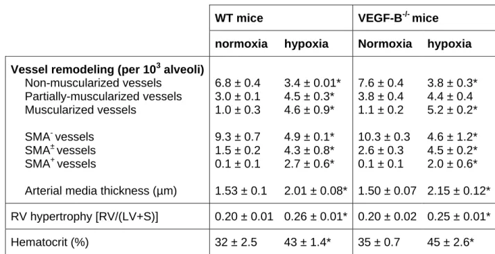
Table I: Negligible role of VEGF-B in pulmonary vessel remodeling after hypoxia
WT mice VEGF-B-/- mice
normoxia hypoxia Normoxia hypoxia Vessel remodeling (per 103 alveoli)
Non-muscularized vessels Partially-muscularized vessels Muscularized vessels SMA- vessels SMA± vessels SMA+ vessels
Arterial media thickness (µm)
6.8 ± 0.4 3.0 ± 0.1 1.0 ± 0.3 9.3 ± 0.7 1.5 ± 0.2 0.1 ± 0.1 1.53 ± 0.1 3.4 ± 0.01* 4.5 ± 0.3* 4.6 ± 0.9* 4.9 ± 0.1* 4.3 ± 0.8* 2.7 ± 0.6* 2.01 ± 0.08* 7.6 ± 0.4 3.8 ± 0.4 1.1 ± 0.2 10.3 ± 0.3 2.6 ± 0.3 0.1 ± 0.1 1.50 ± 0.07 3.8 ± 0.3* 4.4 ± 0.4 5.2 ± 0.2* 4.6 ± 1.2* 4.5 ± 0.2* 2.0 ± 0.6* 2.15 ± 0.12* RV hypertrophy [RV/(LV+S)] 0.20 ± 0.01 0.26 ± 0.01* 0.20 ± 0.02 0.25 ± 0.01* Hematocrit (%) 32 ± 2.5 43 ± 1.4* 35 ± 0.7 45 ± 2.6*
Pulmonary vessel remodeling, induced by chronic exposure to hypoxia and resulting in pulmonary hypertension, was examined in WT and VEGF-B-/- mice. The density of pulmonary vessels was determined, while discriminating between non-muscularized vessels (with only an internal elastic membrane (IEL)), partially-muscularized vessels (IEL and partial external elastic membrane (EEL)) and muscularized vessels (complete IEL and EEL). To evaluate right ventricle (RV) hypertrophy, the dry weight of the RV was measured and divided by the total dry weight of the left ventricle plus septum (LV+S). Values are means ± SEM. * P<0.05 versus normoxia; P=NS for WT mice versus VEGF-B
-
mice.
at SWETS SUBSCRIPTION SERVICE on February 11, 2010 atvb.ahajournals.org
4
SUPPLEMENTAL FIGURE LEGENDS
Figure I: As an alternative method to deliver VEGF-B to cardiomyocytes, we implanted in the myocardial wall of the infarct border, mouse myoblasts, transduced ex vivo with a retrovirus, to constitutively produce mouse VEGF-B167, since a similar strategy using
VEGF-A-expressing myoblasts was previously shown to stimulate vessel growth in muscle
6-8
. A,B, Immunofluorescent staining for B (green) confirmed the absence of VEGF-B in control myoblasts expressing only LacZ (A) and production of mVEGF-VEGF-B167 in
myoblasts expressing both VEGF-B and LacZ (B). Nuclei are counterstained with Dapi (blue). The production of mVEGF-B167 by engineered myoblasts in vivo was further
confirmed by determining mVEGF-B expression levels in cell lysates and muscle extracts. Indeed, myoblasts, transduced with a retrovirus encoding mVEGF-B167, secreted 26 ± 2
ng/106 cells/24h of VEGF-B, while control LacZ-expressing myoblasts failed to express detectable amounts of VEGF-B. At 3 days after implantation of myoblasts into the gastrocnemius muscle, increased amounts of VEGF-B protein were found in the muscles implanted with the VEGF-B167-expressing myoblasts (299 ± 67 pg/mg protein versus 22 ±
2 pg/mg protein in control muscles; N=6; P<0.05). Further in vitro experiments revealed
that the secreted VEGF-B protein was bio-active (not shown). C,D, Double
immunofluorescent staining for ß-galactosidase (site of myoblast engraftment) and CD31 (blood vessels) in the ischemic myocardium revealed no signs of vessel growth around the implantation site of control myoblasts (C), but a robust angiogenic induction around the site of VEGF-B167-expressing myoblast implantation (D). Thus, intramyocardial
implantation of VEGF-B167-expressing myoblasts enhanced vessel growth in the ischemic
myocardium at 28 days post-MI, while control LacZ+ myoblasts failed to promote angiogenesis. Scale bars: 50 µm.
Figure II: We showed previously that adenoviral gene transfer of PlGF into the skin of ears enlarged pre-existing vessels with subsequent stabilization by acquisition of a pericyte coat 9. We therefore injected adenoviruses, expressing hVEGF-B167
(Ad.hVEGF-B167), hVEGF-B186 (Ad.hVEGF-B186) or mPlGF-2 (Ad.mPlGF) intradermally into the ear
skin. A-I, Ears were whole-mount immunostained for CD31 (endothelial cells; brown) in panels A,D,G, for CD31 (green) and SMA (smooth muscle cells; red) in panels B,E,H, and for F4/80 (macrophages, green) and SMA (red) in panels C,F,I. As expected, gene transfer of mPlGF-2 increased the density, tortuosity and size of pre-existing vessels, and
at SWETS SUBSCRIPTION SERVICE on February 11, 2010 atvb.ahajournals.org
5
stimulated their coverage by mural cells (A,B). In contrast, Ad.hVEGF-B167 or
Ad.hVEGF-B186 gene transfer minimally enlarged the pre-existing vessels without increase in number,
tortuosity or mural cell coverage (D,E,G,H). Consistent herewith, we found that many F4/80+ macrophages had infiltrated after Ad.mPlGF gene transfer, while only few macrophages were present after Ad.hVEGF-B167 or Ad.hVEGF-B186 gene transfer (C,F,I).
A control virus (Ad.CMV) failed to affect any of these parameters (not shown). Scale bars: 100 µm in panels A,B,D,E,G,H and 50 µm in C,F,I.
Figure III: A-D, To further analyze the effects of VEGF-B on vessel remodeling and recruitment of mural cells, we examined in VEGF-B-/- mice the remodeling of pulmonary vessels in response to chronic hypoxia 10. Hart’s elastin staining revealed no genotypic differences between WT and VEGF-B-/- mice in pulmonary vessel structure in normoxia (A,B; see also Table S1). In WT mice, continuous hypoxic conditions for 4 weeks increased the number of thick-walled muscularized vessels by ~4.6-fold (C; Table S1). A comparable 4.7-fold increase in the number of thick-walled muscularized vessels was observed in VEGF-B-/- mice (D; Table S1), suggesting that loss of VEGF-B did not impair mural cell recruitment. Similar results were obtained when hypoxic vessel remodeling was analyzed by immunostaining for SMA (Table S1). Consistent herewith, no genotypic differences were observed in the development of right ventricle (RV) hypertrophy, which normally is caused by pulmonary hypertension (Table S1). Compared to WT mice, VEGF-B-/- mice were as sensitive to chronic hypoxia, as evidenced by the similar increase in hematocrit levels (Table S1). The lack of an effect of VEGF-B on pulmonary hypertension is consistent with earlier findings by Louzier et al. 11. E,F, To further analyze the effects of VEGF-B on angiogenesis, we studied in VEGF-B-/- mice retinal neovascularization in response to ischemia, using an established model 10. Neovascularization of the ischemic retina, analyzed by H&E staining on cross-sections, was comparable in WT (E) and VEGF-B-/- mice (F; arrows indicate neovessels). Indeed, compared to WT mice, loss of VEGF-B failed to reduce the number of endothelial cells, forming new intravitreal vessel sprouts (per retinal cross-section: 102 ± 13 in WT mice versus 118 ± 10 in VEGF-B-/- mice; N=5; P=NS) or the number of neovascular tufts (per retinal cross-section: 42 ± 4 in WT mice versus 50 ± 8 in VEGF-B-/- mice; N=5; P=NS; Figure S3E,F). These findings are consistent with findings by Reichelt et al. 12. Scale bars: 50 µm in panels E,F and 25 µm in panels A-D.
at SWETS SUBSCRIPTION SERVICE on February 11, 2010 atvb.ahajournals.org
6
Figure IV: A-D, At 7 days after ligation, laser Doppler perfusion analysis on ischemic limbs also failed to show any genotypic differences (A,B; see also Table 1). Ischemic limb perfusion was also measured in mice lacking both VEGF-B and PlGF – the rationale for this experiment being that limb perfusion was reduced in PlGF-/- mice 10 and that, possibly, the consequences of VEGF-B deficiency might be more apparent when limb revascularization was already impaired by prior loss of PlGF. However, at 7 days after ischemia, laser Doppler imaging revealed that, compared to WT or VEGF-B-/- mice (Table 1; P<0.05), limb perfusion was comparably reduced in mice lacking PlGF alone or in mice lacking both VEGF-B and PlGF (C,D; % perfusion of non-ligated limb: 47 ± 5% in PlGF -/-mice versus 55 ± 9% in VEGF-B-/-:PlGF-/- mice; N=5; P=NS). Please note the color scale: from blue (low perfusion) to red (high perfusion). E,F, H&E staining of transverse sections through the adductor muscles revealed a comparable size of the main collateral vessel in WT (E) and VEGF-B-/- mice (F). G-I, Morphometric analysis revealed a similar amount of F4/80+ macrophages around the collaterals in WT and VEGF-B-/- adductor muscles (G), as also illustrated by microscopic pictures (H,I). In addition, VEGF-B failed to stimulate the production by cultured macrophages of TNF-alpha, previously implicated in collateral growth 9 (data not shown). Please note that the black staining inside the vessels results from bismuth-gelatin filling of the vessels, which enables a better macroscopic visualization of the collaterals. Scale bars: 50 µm.
at SWETS SUBSCRIPTION SERVICE on February 11, 2010 atvb.ahajournals.org
7
SUPPLEMENTAL METHODS Animal models
Myocardial ischemia model: Myocardial ischemia was induced by ligation of the left anterior descending (LAD) coronary artery in female mice as described 3, 9. Seven days after LAD ligation, hearts were harvested, sectioned and analysed for vessel densities and/or macrophage infiltration in the infarct area and border zone.
Mouse skin wound model: A standardized 15 mm full-thickness skin incision was made on the back of mice, taking care not to damage the underlying muscle, as described
10
. Wound healing was quantified by daily measuring the width and the length of the wound. Analysis of vessel densities and inflammation was performed in skin sections, harvested 5 days after wounding.
Mouse ear skin assay: Mouse ears, injected with the PlGF or VEGF-B adenoviral vectors, were dissected, fixed in 1% phosphate buffered paraformaldehyde and whole-mount immunostained for endothelial cells, smooth muscle cells and macrophages using fluorescently conjugated secondary antibodies (Alexa 488 or 546, Molecular Probes).
Mouse ischemic retinopathy model: Mice at postnatal day 7 were exposed to hyperbaric (80%) oxygen for 5 days, as described 10. After returning to normoxia for another 5 days, the eyes were harvested, fixed in 1% paraformaldehyde, paraffin embedded and sectioned. After H&E staining, the number of endothelial cells and vascular tufts in the vitreous cavity were counted, as described 10, 13.
Pulmonary vascular remodeling: Eight-weeks old male mice were placed in a chamber under normobaric hypoxia (10% O2) or in normal air (21% O2; control) as
described 14. After 4 weeks, the right ventricular (RV) wall was dissected from the left ventricle (LV) and septum (S), and dried at 55°C before weighing. Alternatively, lungs were perfusion-fixed, dissected and sectioned as described 14. Pulmonary vascular remodeling was assessed by counting the number of non-muscularized (only IEL), partially muscularized (IEL plus incomplete EEL) and fully muscularized (IEL and complete EEL) peripheral vessels (located distal to the bronchi) per 103 alveoli, as described 14.
Ischemic hindlimb model: In male mice, the right femoral artery and vein (proximal to the popliteal artery) and the cutaneous vessels branching from the caudal femoral artery side branch were ligated, avoiding damage of the femorale nerve 9. Seven days after ligation, functional perfusion measurements of the total limb were performed using a Lisca PIM II camera (Gambro). Perfusion, averaged over 3 images per mouse in the total hindlimb, was expressed as a ratio of right (ischemic) to left (normal) limb. Limb motor
at SWETS SUBSCRIPTION SERVICE on February 11, 2010 atvb.ahajournals.org
8
function was determined via treadmill running exercise (Simplex II, Columbus Instruments), after a one-day conditioning training session 15. The test protocol included a graded exercise test at constant inclination of 10 degrees with increases in belt speed of 2 m/min every 5 minutes. Exhaustion was defined as failure to abandon the shock grid within 15 seconds. After training, the femoral artery was occluded, and at 7 days later, mice were re-tested on the treadmill. Recovery of function was expressed as a ratio to the baseline exercise time. For histology, the gastrocnemius and adductor muscles were harvested at 7 or 28 days after femoral artery ligation. Vessel densities in the regenerating gastrocnemius muscle were determined morphometrically by analyzing the capillary-to-myocyte ratio. Remodeling of collateral vessels and macrophage recruitment in the upper hindlimb were analyzed as described 9. In brief, collateral side branches were categorized as smaller or larger than 300 µm2. Total perfusion area was calculated using the total sum of the side branch luminal areas.
Transplantation of VEGF-B167-expressing myoblasts: A MFG retrovirus encoding mouse VEGF-B167 cDNA, generated as described 6, was used to transduce
LacZ-expressing myoblasts. LacZ-LacZ-expressing myoblasts were used for transplantation to visualize the engrafted cells. Specific expression of the VEGF-B gene was verified by immunofluorescent staining of myoblasts using antibody, recognizing mouse VEGF-B167
(R&D Systems). The amount of gene product, secreted by the VEGF-B producing myoblasts in vitro, was quantified by ELISA. The biological activity of medium conditioned by VEGF-B producing myoblasts was compared to medium of VEGF-A expressing cells or control myoblasts, using a HUVEC proliferation assay (Cell Titer Aqueous One Solution Assay, Promega). Myoblasts, co-expressing VEGF-B167 and LacZ or control
LacZ-myoblasts, were injected into the anterior tibial muscle (5x10⁵ cells/injection) in normal conditions or immediately after femoral artery ligation and transection in adult male SCID mice, as described 8, 16. At 28 days following surgery, tibialis and myocardial muscles were harvested and double labeled for endothelial cells and muscle fibers expressing the LacZ reporter gene, using a polyclonal rabbit antibody to LacZ (Eppendorf 5-Prime). Capillaries and muscle fibers were counted and expressed as capillary-to-muscle fiber ratio. Myoblasts were also injected in the infarct border zone (5x10⁵ cells/injection) of female SCID mice, immediately after LAD ligation, and hearts were harvested for histology at 28 days post-MI.
Histology, immunohistochemistry and morphometric analyses
at SWETS SUBSCRIPTION SERVICE on February 11, 2010 atvb.ahajournals.org
9
All tissues were fixed in 1% phosphate buffered paraformaldehyde and embedded in paraffin. Serial parasagittal (hearts) or transverse (skin, retina, lung, limb muscle) sections were cut at 8 µm thickness and stained for H&E or Hart’s elastin. Immunostainings were performed using primary antibodies for endothelial cells (rabbit anti-thrombomodulin (TM), gift from Dr. R.W. Jackman, Boston, MA; rat anti-CD31, Becton Dickinson), smooth muscle cells (mouse anti-smooth muscle alpha-actin (SMA), Sigma), macrophages (rat anti-Mac3 and rat anti-F4/80, both Becton Dickinson), and ß-Gal (rabbit anti- ß-Gal, Cappel). Following primary antibody incubation, sections were incubated with peroxidase-labeled IgGs (Dako), followed by amplification with the proper tyramide signal amplification systems (Perkin Elmer, Life Sciences). Morphometricanalyses of the vessel densities and macrophage positive areas in the ischemic, granulation or wound border tissues were performed using a Zeiss Axioplan microscope with KS300 image analysis software.
Production and administration of VEGF-B protein, plasmid and adenovirus
The recombinant human VEGF-B167 (rhVEGF- B167) protein was obtained from Amrad Corporation (gift from Dr. A. Nash), and its activity was tested using the Ba/F3 pre-B cell viability assay as described 17 (data not shown). Continuous delivery of 1.5
µg/mouse/day rhVEGF-B167 protein was achieved by subcutaneously implanting osmotic minipumps (Alzet, type 2001), as described 3.
Adenoviruses were constructed by cloning the murine PlGF-2, or the human VEGF-B167 or VEGF-B186 cDNA into the pACCMVpLpA plasmid, using previously published
methods 3, 9. RNA analysis and immunoblotting of extracts and conditioned medium of cells, transduced with the respective adenovirus, revealed that the transduced cells produced murine PlGF-2, hVEGF-B167 or hVEGF-B186 protein (not shown). For the ear
assays, 1x10⁹ pfu adenovirus (Ad.mPlGF, Ad.hVEGF-B167, Ad.hVEGF-B186 or control
Ad.CMV) was injected intradermally in the ears of female NMRInu/nu mice. In the ischemic hind limb model, 3x10⁹ pfu adenovirus (Ad.hVEGF-B167 or control Ad.RR5) was
intravenously injected in the tail vein of male C57BL/6 mice, immediately after ligation of the femoral artery.
A plasmid expressing murine VEGF-B167 (pcDNA3.mVEGF-B167) or an empty
pcDNA3 plasmid was administered via muscle electroporation as described 4, 5. Briefly, one day before ligation of the femoral artery, 15 µg of expression plasmid, encoding
at SWETS SUBSCRIPTION SERVICE on February 11, 2010 atvb.ahajournals.org
10
mVEGF-B167, or 15 µg empty plasmid (both at 1mg/ml in 0.9% NaCl) was injected into the
adductor muscle (total volume of 15 µl over 3 injection sites) using a Hamilton syringe, and electrotransfer (five electric pulses of 100V with a fixed pulse duration of 20 ms and an interval of 200 ms; Electro Square Porator ECM 830, BTX, Harvard Bioscience), with Tweezertrode 520 electrodes (BTX, Harvard Bioscience) was performed.
RT-PCR for Flt-1 and ELISA for human and mouse VEGF-B
Quantitative real-time RT-PCR and protein extraction were performed as described 10
. For determining mouse VEGF-B167 levels a modified version of a previously established
ELISA 1 was used. Plates were coated with a monoclonal antibody to murine VEGF-B
(#MAB751, R&D Systems) and mrVEGF-B167 (gift from Dr. A. Nash) was used as
standard. Human VEGF-B167 plasma levels were determined with a home-made ELISA
using antibodies (#MAB3372 as capture antibody and #AF751 as detection antibody) and rhVEGF-B167 (#751-VE-025) from R&D Systems.
at SWETS SUBSCRIPTION SERVICE on February 11, 2010 atvb.ahajournals.org
11
SUPPLEMENTAL REFERENCES
1. Leppanen P, Koota S, Kholova I, Koponen J, Fieber C, Eriksson U, Alitalo K, Yla-Herttuala S. Gene transfers of vascular endothelial growth factor-A, vascular endothelial growth factor-B, vascular endothelial growth factor-C, and vascular endothelial growth factor-D have no effects on atherosclerosis in hypercholesterolemic low-density lipoprotein-receptor/apolipoprotein B48-deficient mice. Circulation. 2005;112:1347-1352.
2. Li X, Aase K, Li H, von Euler G, Eriksson U. Isoform-specific expression of VEGF-B in normal tissues and tumors. Growth Factors. 2001;19:49-59.
3. Heymans S, Luttun A, Nuyens D, Theilmeier G, Creemers E, Moons L, Dyspersin GD, Cleutjens JP, Shipley M, Angellilo A, Levi M, Nube O, Baker A, Keshet E, Lupu F, Herbert JM, Smits JF, Shapiro SD, Baes M, Borgers M, Collen D, Daemen MJ, Carmeliet P. Inhibition of plasminogen activators or matrix metalloproteinases prevents cardiac rupture but impairs therapeutic angiogenesis and causes cardiac failure [see comments]. Nat Med. 1999;5:1135-1142.
4. Jang HS, Kim HJ, Kim JM, Lee YS, Kim KL, Kim JA, Lee JY, Suh W, Choi JH, Jeon ES, Byun J, Kim DK. A novel ex vivo angiogenesis assay based on electroporation-mediated delivery of naked plasmid DNA to skeletal muscle. Mol Ther. 2004;9:464-474.
5. Qian HS, Liu P, Huw LY, Orme A, Halks-Miller M, Hill SM, Jin F, Kretschmer P, Blasko E, Cashion L, Szymanski P, Vergona R, Harkins R, Yu J, Sessa WC, Dole WP, Rubanyi GM, Kauser K. Effective treatment of vascular endothelial growth factor refractory hindlimb ischemia by a mutant endothelial nitric oxide synthase gene. Gene Ther. 2006;13:1342-1350.
6. Springer ML, Chen AS, Kraft PE, Bednarski M, Blau HM. VEGF gene delivery to muscle: potential role for vasculogenesis in adults. Mol Cell. 1998;2:549-558.
7. Ozawa CR, Banfi A, Glazer NL, Thurston G, Springer ML, Kraft PE, McDonald DM, Blau HM. Microenvironmental VEGF concentration, not total dose, determines a threshold between normal and aberrant angiogenesis. J Clin Invest. 2004;113:516-527.
8. von Degenfeld G, Banfi A, Springer ML, Wagner RA, Jacobi J, Ozawa CR, Merchant MJ, Cooke JP, Blau HM. Microenvironmental VEGF distribution is critical for stable and functional vessel growth in ischemia. Faseb J. 2006;20:2657-2659.
9. Luttun A, Tjwa M, Moons L, Wu Y, Angelillo-Scherrer A, Liao F, Nagy JA, Hooper A, Priller J, De Klerck B, Compernolle V, Daci E, Bohlen P, Dewerchin M, Herbert JM, Fava R, Matthys P, Carmeliet G, Collen D, Dvorak HF, Hicklin DJ, Carmeliet P. Revascularization of ischemic tissues by PlGF treatment, and inhibition of tumor angiogenesis, arthritis and atherosclerosis by anti-Flt1. Nat Med. 2002;8:831-840. 10. Carmeliet P, Moons L, Luttun A, Vincenti V, Compernolle V, De Mol M, Wu Y, Bono F,
Devy L, Beck H, Scholz D, Acker T, DiPalma T, Dewerchin M, Noel A, Stalmans I, Barra A, Blacher S, Vandendriessche T, Ponten A, Eriksson U, Plate KH, Foidart JM, Schaper W, Charnock-Jones DS, Hicklin DJ, Herbert JM, Collen D, Persico MG. Synergism between vascular endothelial growth factor and placental growth factor contributes to angiogenesis and plasma extravasation in pathological conditions. Nat Med. 2001;7:575-583.
11. Louzier V, Raffestin B, Leroux A, Branellec D, Caillaud JM, Levame M, Eddahibi S, Adnot S. Role of VEGF-B in the lung during development of chronic hypoxic pulmonary hypertension. Am J Physiol Lung Cell Mol Physiol. 2003;284:L926-L937.
at SWETS SUBSCRIPTION SERVICE on February 11, 2010 atvb.ahajournals.org
12
12. Reichelt M, Shi S, Hayes M, Kay G, Batch J, Gole GA, Browning J. Vascular endothelial growth factor-B and retinal vascular development in the mouse. Clin Experiment Ophthalmol. 2003;31:61-65.
13. Smith LE, Wesolowski E, McLellan A, Kostyk SK, D'Amato R, Sullivan R, D'Amore PA. Oxygen-induced retinopathy in the mouse. Invest Ophthalmol Vis Sci. 1994;35:101-111.
14. Brusselmans K, Compernolle V, Tjwa M, Wiesener MS, Maxwell PH, Collen D, Carmeliet P. Heterozygous deficiency of hypoxia-inducible factor-2alpha protects mice against pulmonary hypertension and right ventricular dysfunction during prolonged hypoxia. J Clin Invest. 2003;111:1519-1527.
15. Umans L, Cox L, Tjwa M, Bito V, Vermeire L, Laperre K, Sipido K, Moons L, Huylebroeck D, Zwijsen A. Inactivation of Smad5 in endothelial cells and smooth muscle cells demonstrates that Smad5 is required for cardiac homeostasis. Am J Pathol. 2007;170:1460-1472.
16. Banfi A, Springer ML, Blau HM. Myoblast-mediated gene transfer for therapeutic angiogenesis. Methods Enzymol. 2002;346:145-157.
17. Makinen T, Olofsson B, Karpanen T, Hellman U, Soker S, Klagsbrun M, Eriksson U, Alitalo K. Differential binding of vascular endothelial growth factor B splice and proteolytic isoforms to neuropilin-1. Journal of Biological Chemistry. 1999;274:21217-21222.
at SWETS SUBSCRIPTION SERVICE on February 11, 2010 atvb.ahajournals.org
at SWETS SUBSCRIPTION SERVICE on February 11, 2010 atvb.ahajournals.org
at SWETS SUBSCRIPTION SERVICE on February 11, 2010
atvb.ahajournals.org
at SWETS SUBSCRIPTION SERVICE on February 11, 2010
atvb.ahajournals.org
at SWETS SUBSCRIPTION SERVICE on February 11, 2010 atvb.ahajournals.org