The Ribonucleoside Diphosphate Reductase Inhibitor (E)-2
*-Deoxy-(fluoromethylene)cytidine as a Cytotoxic Radiosensitizer in Vitro
1Philippe A. Coucke,
2Laurent A. Decosterd, Ye-Xiong Li, Eliane Cottin, Xiaoguang Chen, Lin-Quan Sun,
Sabine Stern, Nicolas Paschoud, and Juliana Denekamp
Laboratory of Radiation Biology, Department of Radiation Oncology [P. A. C., Y-X. L., E. C., L-Q. S., S. S., N. P.] and Division of Clinical Pharmacology Laboratory [L. A. D., E. C., X. C.], Centre Hospitalier Universitaire Vaudois, Laboratory of Flow Cytometry, Clinique La Source, 1011 Lausanne, Switzerland [N. P.], and Translational Research Group, Oncology Department, Umea` University, 90185 Umea`, Sweden [J. D.]
ABSTRACT
(E)-2*-Deoxy-(fluoromethylene)cytidine (FMdC) is known as an inhib-itor of ribonucleoside diphosphate reductase, a key enzyme in the de novo pathway of DNA synthesis. FMdC was tested as a modifier of radiation response in vitro on a human colon carcinoma cell line (WiDr), and the observed radiosensitization was confirmed on two human cervix cancer cell lines (C33-A and SiHa). Using the clonogenic assay, the effect ratio (ER) at a clinically relevant dose level of 2 Gy was 2.10 (50 nMFMdC), 1.70 (30 nMFMdC), and 1.71 (40 nMFMdC) for the three cell lines WiDr, C33-A, and SiHa, respectively. A more detailed analysis of the importance of timing and concentration of FMdC was done on the WiDr cell line alone, yielding an increased ER(2Gy)with increasing concentration and
duration of exposure to the drug, ranging from 1.0 (6 h) to 1.8 (72 h) at 30 nMFMdC and from 1.2 (6 h) to 3.5 (24 h) at 300 nM. We investigated the effect of FMdC on the cellular deoxynucleotide triphosphate pool in WiDr cells and demonstrated a marked depletion of dATP and a significant rise of TTP levels. Cell cycle analysis showed early S-phase accumulation induced by FMdC alone, G2-M block induced by irradiation alone, and an
increased accumulation of cells in G2-M if both modalities are used. Our
data suggest that FMdC is a radiation response modifier in vitro on different cancer cell lines. The observed radiosensitization may in part be explained by alteration of the deoxynucleotide triphosphate pool, which is consistent with the effect of FMdC on ribonucleoside diphosphate reduc-tase.
INTRODUCTION
The radiation response of human tumor cells has been shown to be dependent on the pool of the purines and pyrimidines A, T, C, and G expressed as the dNTP3
(1). This pool is supplied through the “de novo” biosynthesis of NTP and/or by the “salvage pathway.” In the de novo pathway, enzymes like RR and thymidylate synthetase are playing a pivotal role, whereas in the salvage pathway, thymidine kinase is the key enzyme. RR converts the ribonucleoside diphosphate to a deoxynucleotide diphosphate. Higher levels of RR are found in rapidly proliferating tumors than in normal tissues; therefore, inhibi-tion of RR may offer a way for a relatively selective antitumor activity (2–7).
Various compounds have been investigated as potential inhibitors of RR. These compounds are potentially useful as cytotoxic drugs and possibly as radiation response modifiers (2, 6 –15). One of these compounds, hydroxyurea, is widely used in myeloproliferative
disor-ders. Clinical trials have demonstrated its activity as a tumor response modifier if combined with ionizing radiation in cervix and head and neck cancer (12, 15).
New inhibitors of RR have been developed recently (3, 4, 16, 17). Gemcitabine (dFdC) is one of these new drugs and has been shown to be both a cytotoxic drug and a radiation sensitizer in human colon (HT-29), breast (EMT-6), and human pancreatic cell lines (Panc-1 and BxPC-3; Refs. 8 –10). Among this new class of inhibitors of RR, FMdC (MDL 101,731) is of special interest. FMdC has been devel-oped recently as a chemotherapeutic agent (2, 18, 19). It is effective in vitro and in vivo, both on estrogen-dependent and -independent cell lines of human breast cancer origin, on human colon and prostatic cancer cell lines, and on human glioblastoma and neuroblastoma (20 –22). In nude mouse xenografts of MDA-MB-435, it inhibits spontaneous pulmonary metastases at low concentrations below those required for inhibition of primary tumor growth, probably through an apoptosis-mediated mechanism (20). Short exposure of HeLa cells to high concentrations of FMdC induces radiation sensitization and photosensitization in vitro (11). We have demonstrated previously that FMdC has radiosensitizing and antimetastatic effects on human colon, human cervix, and human brain cancer xenografts grown in nude mice (13, 14). In comparison with gemcitabine, FMdC is relatively resistant to inactivation by cytidine deaminase, resulting in a much slower recovery of dNTP; therefore, FMdC may be considered to be a more potent mechanism-based inhibitor of ribonucleotide reductase (23).
We decided, therefore, to investigate whether this drug is able to modify the radiation response of three different human cancer cell lines in vitro. The influence of the duration of exposure and the effect of concentration of FMdC were determined in details for the colon cancer cell line WiDr. In addition, to better understand the cytotoxic and radiosensitizing effects of FMdC in vitro, we ascertained the modification of cell cycle distribution using flow cytometry and the alteration of the cellular dNTP pool by HPLC.
MATERIALS AND METHODS
Chemicals and Cell Cultures. Cell culture media and supplements were
purchased from Life Technologies, Inc. (Basel, Switzerland), and FCS was obtained from Fakola AG (Basel, Switzerland). The cell lines WiDr, C-33 A, and SiHa were purchased from American Type Culture Collection (Rockville, MD). FMdC (MDL 101,731) was kindly provided by Hoechst Marion Roussel, Inc. (Cincinnati, OH).
Cells were passaged twice weekly. A test for Mycoplasma was routinely performed every 6 months and found negative for contamination. Each cell line was maintained in culture medium that had been shown to provide optimum growth conditions. The WiDr cell line was maintained in MEM with 0.85 g/l NaHCO3, supplemented with 10% FCS, 1% nonessential amino acids
(NE-AA), 2 mM L-glutamine, and 1% penicillin-streptomycin solution. The C-33 A and SiHa cell lines were maintained in Eagle’s MEM, supplemented with 10% FCS, 1% nonessential amino acids, 1% sodium pyruvate, and 1% Earle’s salt.
Effect of FMdC on Growth Rates of Cultures. Cells taken from
subcon-fluent cultures by trypsinization were plated at 106cells/culture dish (Falcon
Primaria, 60 3 15-mm) and were allowed to grow exponentially for 72 h. Medium was replaced every 24 and 48 h; in the test group, the medium was
Received 12/9/98; accepted 8/17/99.
The costs of publication of this article were defrayed in part by the payment of page charges. This article must therefore be hereby marked advertisement in accordance with 18 U.S.C. Section 1734 solely to indicate this fact.
1Supported by grants from Fondation Radiobiologie 2000 and from Stiftung zur
Krebsbeka¨mpfung.
2To whom requests for reprints should be addressed, at Laboratory of Radiation
Biology, Department of Radiation Oncology, Centre Hospitalier Universitaire Vaudois, 1011 Lausanne, Switzerland. Phone: 41-21-31-44-603; Fax: 41-21-31-44-601; E-mail: Philippe.Coucke@chuv.hospvd.ch.
3The abbreviations used are: dNTP, deoxynucleotide triphosphate; RR, ribonucleotide
reductase; dFdC, 29,29-difluoro-29-deoxycytidine (gemcitabine); FMdC, (E)-29-deoxy-29-(fluoromethylene)cytidine; HPLC, high-performance liquid chromatography; BrdUrd, bromodeoxyuridine; TCA, trichloroacetic acid; ER, effect ratio; SER, sensitizer enhance-ment ratio.
supplemented with the same concentration of freshly prepared FMdC as that to be used in the clonogenic assays. The total number of cells in each culture dish was counted using a Bu¨rker-Tu¨rk chamber. The viability was checked by the trypan blue dye exclusion method.
Irradiation Technique and Clonogenic Assay. Exponentially growing
cells were trypsinized and seeded in 603 15-mm Falcon Primaria culture flasks with 5 ml of medium, allowed to attach, and incubated for 24 h before adding FMdC. Medium containing the chosen concentration of freshly pre-pared FMdC was added at 0 h and replaced at 24 and 48 h, resulting in a total of 72 h of cell exposure to FMdC. The cells were trypsinized and resuspended in fresh medium before plating them into 100 3 20-mm Falcon Primaria culture dishes containing 10 ml of medium. After a 3-h incubation to allow cell attachment, the dishes were irradiated. The cells were irradiated at room temperature with an Oris IBL 137 Cesium source at a dose rate of 80.2 cGy/min. A range of single doses ranging from 0 to 10 Gy was used. For each radiation dose, four dishes were irradiated, both for control and drug-exposed cells. The dishes were incubated at 37°C in air and 5% CO2for 2 weeks. The
cells were fixed in ethanol, stained with crystal violet, and manually counted. Colonies of.50 cells were considered survivors. All experiments were done in triplicate. For all of the data obtained by clonogenic assays, the surviving fraction of drug-treated cells was adjusted for drug toxicity to yield corrected survivals of 100% for unirradiated but FMdC-treated cells. The effect shown is therefore the sensitizing action, after the subtraction of the direct cytotoxic effect of FMdC.
Cell Cycle Analysis by Flow Cytometry. Total DNA content (red
fluo-rescence) and BrdUrd incorporation (green fluofluo-rescence) were quantified on a FACScan flow cytometer (Becton Dickinson, Sunnyvale, CA). WiDr cells, which had been growing exponentially, from control cultures and those ex-posed to 50 nMFMdC were labeled 30 min prior to harvesting with BrdUrd (final concentration, 10 mM). The cells were processed for the antibody detection of the incorporated BrdUrd with propidium iodide used to determine the total content of DNA. The antibody used was mouse anti-BrdUrd/iodode-oxyuridine monoclonal antibody from Dako, Ltd.
Analysis dNTP and NTP Pools by HPLC. Simultaneous quantitation
of dNTP and NTP in WiDr cells was performed by gradient elution ion-pair reversed phase HPLC with a modification of a method described previously (16) and is reported in detail elsewhere (24). Briefly, exponentially grow-ing WiDr cells were exposed to FMdC at concentrations ranggrow-ing from 0 nM
(untreated control cells) to 300 nM for 24 and 48 h. The cells were trypsinized, washed, centrifuged, and resuspended in ice-cold ultrapure water (dilution according to cells count) and deproteinized with the same volume of TCA 6% (final applied concentration of TCA, 3%). Acid cell extracts were centrifuged, and the resulting supernatants were stored at
280°C before analysis. Before the HPLC assay, samples were thawed, and
aliquots of 100 ml were neutralized with 4.3 ml of saturated Na2CO3
solution. In the present series of experiments, aliquots of 25 ml were injected onto the HPLC column with satisfactory sensitivity. All experi-ments were done in triplicate, with the triplication process starting at the cell culture step to detect variability associated with the culture growth conditions. Results were expressed as the concentration of the four dNTPs (expressed in pmol/106cells) and as the absolute levels of the four NTPs
(as measured by NTP peak areas). The optimization and full validation of the analytical method is described in detail elsewhere (24).
Statistical Analysis. The data within each experiment were averaged
ar-ithmetically. From these averages, the data are presented as the mean6 SE of at least three independent experiments. Surviving fractions were compared using a two-sided paired t test, and the difference was considered significant if
P5 0.05 was reached. The ER was also calculated at the clinically relevant
2-Gy level by comparing the mean SF value for control and drug-treated cells at that dose. Dose-response curves were fitted using a second-degree polyno-mial regression analysis, yielding a linear quadratic equation. This allowed the calculation of a SER at 2, 20, and 50% survival level (SER2, SER20, and SER50). The curve fitting was obtained using Statview 5.0 software on a Power Macintosh G3.
RESULTS
Effect of FMdC on Growth and Clonogenicity. Fig. 1 shows the growth-inhibitory effects of FMdC alone. These three cell lines showed a 7–9-fold increase in cell number over 72 h in the control groups but virtually no growth in the presence of FMdC at concen-trations ranging from 30 to 50 nMin the different cell lines. Fig. 2 shows the changes in plating efficiency after 48 h of exposure with increasing doses of the drug. A wide range of concentrations were tested for WiDr cells but only two dose levels for the cervix cancer cell lines. On the basis of these data, the drug levels were selected for the other experiments to yield a PE of at least 50% after 48 h of exposure.
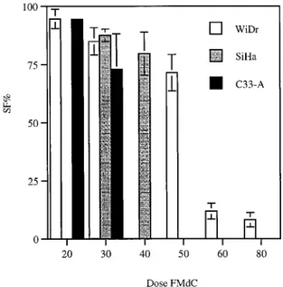
Effect of FMdC on Radiation Response. Fig. 3 shows the cell survival curves as a function of X-ray dose for the three cell lines. FMdC was tested at two dose levels (30 and 50 nM) in WiDr cells and at 30 and 40 nMin the two cervix cancer cell lines C33-A and SiHa, respectively. In each panel, it can be seen that the response to radiation after 48 h of exposure to the drug was considerably steeper than that to radiation alone. An increased effect is seen at every level of radiation dose.
Fig. 1. Growth-inhibitory effect of FMdC on WiDr, C33 A, and SiHa cells, exposed at concentrations of FMdC yielding.50% PE in a clonogenic assay, as compared with untreated controls. The FMdC concentrations were 50, 30, and 40 nMfor WiDr, C33-A, and SiHa cells, respectively. SEs (bars) are given, but most of them are within the size of the symbols used.
Fig. 2. Effect of increasing FMdC concentration (in nM) on the plating efficiency of exponentially growing cells, expressed in percentages compared with controls. Cells were exposed for 48 h. Bars, SE.
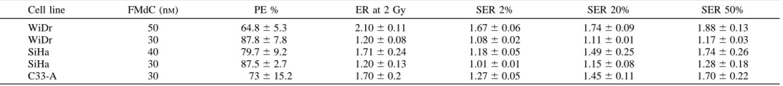
Two quantitative approaches to determine the magnitude of the additional effect due to the addition of the drug can be used: the ratio of the effect levels at the clinically relevant X-ray dose of 2 Gy (ER2Gy); and the ratio of doses to produce a standard level of damage
(SER). Because full dose-response curves are available, this latter SER can be assessed at several levels of damage to indicate whether it is independent of the radiation dose. Table 1 summarizes these data. In the three cell lines, the ER2Gys ranged from 1.2 to 2.1 for 30 – 40
nMexposure over a 48-h period before the irradiation. The SERs show that the sensitizing effect was highest at lower doses (SER50 . SER20 . SER2). There is a concentration-dependent increase of the radiosensitizing effect both in WiDr and in SiHa (Table 1).
Fig. 4 shows the experiments in which a fixed drug dose was used for different periods of exposure. Thirty nMwas used with a range of exposures from 6 –72 h, and a 10-fold higher dose (300 nM) was used with the shorter intervals of 6 –24 h. The dose-effect curves were restricted to the more clinically relevant dose range of 2– 6 Gy. At the lower drug dose, very little sensitization was observed until the exposure time exceeded 24 h. There was a progressive increase in the steepness of the dose-response curve with increasing intervals. The SERs derived for each dose level are shown in Table 2. At the higher drug dose, a very significant effect was already detectable at a 6-h exposure. Table 2 shows the time-dependent SERs showing that, at a survival level of 20%, the SER was similar, i.e., for 24 h at 30 nMand for 6 h at 300 nM.
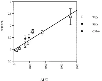
Fig. 5 shows the linear correlation between the area under the curve of FMdC (FMdC concentration3 time of exposure) and the measured
SER20 for the three considered cell lines WiDr, SiHa, and C33-A (R5 0.938 and P 5 0.002).
Cell Cycle Analysis on WiDr. The flow cytometer was used to analyze the fraction of cells in the different stages of the cell cycle, both in untreated controls and in those preexposed to the drug. The results are shown in Fig. 6A. In control cultures,;70% of the cells were in the G0-G1phase,;25% in S phase, and 6% in G2. WiDr cells,
exposed to 50 nMFMdC, were characterized by an emptying of the G1
phase and accumulation in the early S-phase, as judged from pro-pidium iodide staining. However, minimal or no incorporation of BrdUrd was observed if this latter was applied to FMdC-treated cells 30 min before cell cycle analysis (data not shown). Either the cells have been totally arrested at an earlier time or the rate of uptake, as a measure of progress through S, is too slow to be detected.
Irradiated WiDr cells (2 and 6 Gy) did not show a G1block. Rather,
after the higher doses and longer times after irradiation (30 min compared with 24 h), there was a progressive accumulation in G2. The
postirradiation G2block increased with the radiation dose (Fig. 6B).
The WiDr cells exposed to the combined effect of FMdC and irradiation are significantly accumulating in the G2-M phase of the
cell cycle as compared with corresponding controls (Fig. 6C). This G2-M accumulation because of the combination is increased
com-pared with either modality alone. One should be aware that the data in Fig. 6C cannot directly be compared with Fig. 6A, because the experimental conditions are not identical. The cells are partially synchronized by subcultivation at lower density prior to irradiation in the experimental set-up, yielding the data of Fig. 6C.
Fig. 3. A, surviving fraction in % (Y axis) versus dose in Gy (X axis); dose-response curve of WiDr cells after exposure to FMdC (30 and 50 nMfor 48 h; f) as compared with controls (E). B and C, dose-response curve of cervix cancer cell lines (C33 and SiHa) after exposure to FMdC for 48 h (f), as compared with controls (E). SEs (bars) are often within the size of the symbol chosen.
Table 1 Radiosensitizing effect of FMaC on different cell lines, expressed as ER and SER values
Mean and SE of plating efficiency (PE; expressed as a percentage compared with corresponding controls), ER at 2 Gy, and SER at 2, 20, and 50% survival level were calculated from the linear quadratic equation obtained by a second-degree polynomial fit on the dose-response curve.
Cell line FMdC (nM) PE % ER at 2 Gy SER 2% SER 20% SER 50%
WiDr 50 64.86 5.3 2.106 0.11 1.676 0.06 1.746 0.09 1.886 0.13 WiDr 30 87.86 7.8 1.206 0.08 1.086 0.02 1.116 0.01 1.176 0.03 SiHa 40 79.76 9.2 1.716 0.24 1.186 0.05 1.496 0.25 1.746 0.26 SiHa 30 87.56 2.7 1.206 0.13 1.016 0.01 1.156 0.08 1.286 0.18 C33-A 30 736 15.2 1.706 0.2 1.276 0.05 1.456 0.11 1.706 0.22 5221
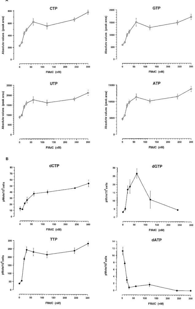
Analysis of NTP and dNTP by HPLC. Our optimized HPLC method provides a simple procedure to measure simultaneously the levels of nucleotides (CTP, GTP, UTP, and ATP) and corresponding amount of deoxynucleotides (dCTP, dGTP, TTP, and dATP) in cell extracts in a single run. It involves minimal chemical manipulation of cells (except protein precipitation with 3% TCA and neutralization). The nucleotides showed approximately a 2–3-fold increase at the 30 nMFMdC concentration, which hardly changed over the range of concentrations up to 300 nM. A similar pattern of activity was ob-served at the two time intervals, i.e., 24 and 48 h of exposure to FMdC (Fig. 7, A and C). This effect could be explained by the increase of total DNA content of those cells treated by FMdC and blocked in S-phase as compared with untreated controls remaining essentially in G0-G1 (Fig. 6A). The dNTP pool showed much larger changes that
were dependent on dose in a much more complex way (Fig. 7, B and D). There was a marked decrease in dATP level in WiDr cells with an essential complete disappearance of dATP at FMdC concentrations above 120 nM. There was a significant increase in TTP and a less pronounced increase in dGTP and dCTP. A plateau was observed in dCTP and TTP.30 nMFMdC and a return to normal of dGTP at the higher doses. These changes in dNTP and NTP were observed both after 24 and 48 h of FMdC exposure. The reduction of dATP is highly significant and reached a nearly zero level. Interestingly, the signifi-cant reduction of the dATP level starts at a concentration of FMdC of
;30–60 nM, concentrations resulting in radiosensitization of WiDr cells.
DISCUSSION
We decided to investigate the effect of a new RR inhibitor FMdC as a potential radiation modifier on a human colon cancer cell line (WiDr) and to check the data obtained in this colon cancer cell line on two human cervix cancer cell lines (C33 A, and SiHa). Preirradiation exposure to FMdC alone resulted in a significant decrease in surviving fraction assessed by colony-forming assay. The radiosensitizing effect at low concentrations of FMdC, i.e., not resulting in.50% cell death by direct cytotoxic effect, was in agreement with observations made by other investigators who described on irradiated HeLa (human cervical cancer cell line) a similar effect (11). However, the radiosen-sitizing effect on HeLa was observed at much higher drug concentra-tions (micromolar) and at very short exposure time (1 h before irradiation). These authors conclude that the increase of sensitivity to UV and X-ray in HeLa cells is most likely explained by repair impairment. The ER2Gyand the SER values, especially at lower doses
we observed in the three different cell lines, are consistent with this hypothesis of repair impairment.
How can this repair impairment be explained? The repair inhibition by FMdC can in theory be the result of three different mechanisms: (a) depletion of dNTP pools with less precursors available for repair; (b) direct inhibition of DNA polymerases (competition between FMdC triphosphate and deoxycytidine triphosphate); or (c) masked chain termination. This masked chain termination has also been ob-served with dFdC (gemcitabine; Ref. 4). The nucleoside dFdC is
Fig. 4. Left, effect of drug exposure duration at a concentration of 300 nMFMdC on WiDr. Right, effect of drug exposure duration at a concentration of 30 nM FMdC on WiDr.
Table 2 Impact of exposure duration on radiosensitizing effect of FMaC on WiDr cells
FMdC Time (h) ER 2 Gy SER 2% SER 20% SER 50%
30 nM 6 0.996 0.04 0.996 0.01 0.986 0.01 0.976 0.02 12 1.016 0.02 0.936 0.02 0.946 0.02 0.976 0.03 24 1.046 0.03 1.046 0.01 1.036 0.01 1.026 0.00 48 1.336 0.04 1.166 0.03 1.256 0.03 1.396 0.05 72 1.796 0.06 1.526 0.03 1.736 0.02 2.066 0.08 300 nM 6 1.196 0.05 1.156 0.04 1.236 0.07 1.346 0.11 12 1.736 0.11 1.296 0.05 1.696 0.09 2.276 0.10 24 3.496 0.84 1.716 0.16 2.376 0.34 3.426 0.62
incorporated into DNA and blocks further DNA synthesis because in its penultimate position it distorts the growing DNA (5). As a result, the growing DNA is no longer an efficient substrate for the DNA polymerase, and the subsequent cell death is characterized by features specific of apoptosis (25). Recent work supports the idea of chain termination as the most likely mechanism in the radiosensitization process by FMdC (5).
On the other hand, FMdC acts on RR; therefore, one would expect an impact on cell cycle progression and alteration of dNTP (15). We decided to assess the former with flow cytometry and the latter with HPLC.
The cell cycle effects of FMdC investigated by flow cytometry lead to the following observations: (a) the S-phase accumulation may be an indirect illustration of the masked chain termination due to the incor-poration of FMdC; (b) subcultivation of unirradiated but FMdC-treated cells to drug-free medium results in a significant accumulation of cells in G2-M, persisting up to 24 h. This particular phase of the cell
cycle is known as one of the most radiation-sensitive cell cycle
Fig. 5. SER 20% as a function of area under the curve of FMdC (drug concentra-tion3 exposure time).
Fig. 6. A, cell cycle redistribution induced by FMdC 50 nMfor 72 h on exponentially growing WiDr. B, cell cycle redistribution induced by irradiation of WiDr cells at doses of 2 and 6 Gy compared with controls (0Gy, no irradiation). Flow cytometric analysis was performed 30 min or 24 h after irradiation. C, cell cycle redistribution induced by 50 nMFMdC for 48 h on WiDr, followed by subcultivation and irradiation at a dose of 6 Gy (F1 RT), compared with subcultivated untreated cells (RT 0Gy), subcultivated and irradiated cells (RT 6Gy), and cells pretreated with FMdC, subcultivated but not irradiated (F).
Fig. 7. Variations of dNTP and NTP levels in exponentially growing human colon carcinoma cells incubated with FMdC. A, NTP, 24 h. B, dNTP, 24 h. C, NTP, 48 h; dNTP, 48 h. D, NTP, 48 h. The data are the mean values of three separate experiments; bars, SE.
Fig. 7 Continued.
phases, and hence, cell synchronization may in part explain the effect on the dose-response curve in vitro; (c) a clear G2block was detected
in irradiated cells, pretreated or not with the nucleoside analogue. For some cell lines, DNA fragmentation and apoptosis are preceded by a period of radiation-induced G2-M block (26). We do not have
exper-imental data to prove that this G2-M block results in apoptosis, but we
are presently investigating whether there is an increase in apoptotic cell death after preirradiation exposure to the drug. In summary, the radiosensitizing effect of FMdC can at least in part be explained by redistribution of cells in a more sensitive cell cycle phase. Even if the synchronization in G2-M is artifactual, i.e., attributable to the
subcul-tivation in drug-free medium, the accumulation of an exponentially growing population of cells in early S-phase may be exploited in vivo. Data in our laboratory on WiDr xenografts grown in nude mice confirm the significant increase of cells in S-phase and the tumor response to irradiation if animals are treated with FMdC (13, 14).
The modifications of the levels of NTP are consistent with the changes we observed in cell cycle distribution. One would expect to detect more NTP in S-phase cells than in G0-G1cells. On the other
hand, the reduction to a nearly zero level of dATP indicates the effect of FMdC on the de novo pathway. For BrdUrd, a radiosensitizer that interacts through thymidine kinase (i.e., the salvage pathway), Law-rence et al. (8) demonstrated a similar significant reduction of dATP. Interestingly, with FMdC we observed a major decrease of dATP level and a simultaneous rise especially of the TTP level. This same observation was made by Takahashi and coworkers (23) on HeLa S3
cells exposed to FMdC. Changes in dNTP as a result of alteration of the de novo pathway should result in a reactive increase of thymidine incorporation, resulting from an increased activity of thymidine kinase (feedback loop on the salvage pathway). This marked reduction of dATP might result in reduction of the precursor of repair, yielding a modification of the dose-response curve.
Experimental data on tumor xenografts of colonic origin (HT-29), prostatic origin (PC-3), and brain (glioblastoma and neuroblastoma) show that FMdC is cytotoxic with only mild effects on bone marrow (13, 14, 20 –22). Furthermore, at nontumoricidal doses, FMdC is able to inhibit the metastatic potential of breast tumor xenografts (20). The lack of long-lasting changes in hematological parameters (21) and the potential to act as a radiation sensitizer in vitro on a variety of human cancer cell lines and in vivo on human colon, cervix, and brain cancer xenografts (13, 14), even at low concentrations, makes FMdC an attractive candidate to be tested in a clinical context. Moreover, in contrast to gemcitabine, there is no metabolization of FMdC to FMdU by human cytidine deaminase and, therefore, no loss of antiprolifera-tive activity by organs containing high levels of cytidine deaminase such as liver, kidney, and solid tumors (23).
Investigations of molecular mechanisms involved in repair of radi-ation damage, regulradi-ation of S-phase transition, and G2-M block and
apoptosis are presently under way. Better knowledge of the mecha-nisms involved in radiosensitization by FMdC may result in a sound rationale for testing this drug as a radiosensitizer in the clinics. REFERENCES
1. Shewach, D. S., Ellero, J., Mancini, W. R., and Ensminger, W. D. Decrease in TTP mediated by 59-bromo-29-deoxyuridine exposure in a human glioblastoma cell line. Biochem. Pharmacol., 43: 1579 –1585, 1992.
2. Baker, C. H., Banzon, J., Bollinger, J. M., and Stubbe, J. 29Deoxy-29methylencytidine and 29-deoxy-29,29-difluorocytidine 59-diphosphate: potent mechanism-based inhibi-tors of ribonucleotide reductase. J. Med. Chem., 34: 1879 –1885, 1991.
3. Grindey, G. B., Hertel, L. W., and Plunkett, W. Cytotoxicity and antitumor activity of 29,29-difluorodeoxycytidine (gemcitabine). Cancer Investig., 8: 313, 1990.
4. Hertel, L. W., Boder, G., Kroin, J. S., Rinzel, S. M., Poore, G. A., Todd, G. C., and Grindey, G. B. Evaluation of the antitumor activity of gemcitabine (2 9,29-difluoro-29-deoxycytidine). Cancer Res., 50: 4417–4422, 1990.
5. Huang, P., Chubb, S., Hertel, L. W., Grindey, G. B., and Plunkett, W. Action of 29,29-difluorodeoxycytidine on DNA synthesis. Cancer Res., 51: 6110–6117, 1991. 6. Kinsella, T. J., Kunugi, K. A., and Yen, Y. Hydroxyurea-mediated radiosensitization: an in vitro study in a human squamous cell cancer cell line (KB). In: Abstract II-49 Ninth International Conference on Chemical Modifiers of Cancer Treatment, pp. 181–182. Oxford, United Kingdom: Christ Church, 1995.
7. Kuo, M. L., and Kinsella, T. J. Radiosensitization by hydroxyurea in a cervical carcinoma cell line, Caski. In: Abstract II-50 Ninth International Conference on Chemical Modifiers of Cancer Treatment, pp. 183–184. Oxford, United Kingdom: Christ Church, 1995.
8. Lawrence, T. S., Chang, E., Y., Hahn, T. M., Hertel, L. W., and Shewach, D. S. Radiosensitization of pancreatic cells by 29,29-difluoro-29-deoxycytidine. Int. J. Ra-diat. Oncol. Biol. Phys., 34: 867– 872, 1996.
9. Rockwell, S., and Grindey, G. B. Effect of 29,2-difluorodeoxycytidine on the viability and radiosensitivity of EMT6 cells in vitro. Oncol. Res., 4: 5151–5155, 1992. 10. Shewach, D. S., Hahn, T. M., Chang, E., Hertel, L. W., and Lawrence, T. S.
Metabolism of 29,29-difluoro-29-deoxycytidine and radiation sensitization of human colon carcinoma cells. Cancer Res., 54: 3218 –3223, 1994.
11. Snyder, R. D. Effect of 29-deoxy-29(fluoromethylene) cytidine on the ultraviolet and x-ray sensitivity of HeLa cells. Oncol. Res., 6: 177–182, 1994.
12. Stehman, F. B., Bundy, B. N., Thomas, G., Keys, H. M., Fowler, W. C., Mortel, R., and Creasman, W. T. Hydroxyurea versus misonidazole with radiation in cervical carcinoma: long-term follow-up of a Gynecologic Oncology Group Trial. J. Clin. Oncol., 11: 1523–1528, 1993.
13. Sun, L-Q., Li, Y-X., Guillou, L., Mirimanoff, R-O., and Coucke, P. A. Antitumor and radiosensitizing effects of (E)-29-deoxy-29(fluoromethylene)cytidine, a novel inhibi-tor of ribonucleoside diphosphate reductase on human colon carcinoma xenografts in nude mice. Cancer Res., 57: 4023– 4028, 1997.
14. Sun, L-Q., Li, Y-X., Guillou, L., and Coucke, P. A. (E)-2 9-Deoxy-29(fluoromethyl-ene)cytidine potentiates radioresponse of two human solid tumor xenografts. Cancer Res., 58: 5411–5417, 1998.
15. Vokes, E. E., Stupp, R., Haraf, D. J., Moran, W., Malone, D., Wenig, B., Sweeney, P., and Weichselbaum, R. R. Hydroxyurea with continuous infusion paclitaxel, 5-fluorouracil, and concomitant radiotherapy for poor-prognosis head and neck can-cers. Semin. Oncol., 22 (Suppl. 6): 47–52, 1995.
16. Di Pierro, D., Tavazzi, B., Perno, C. F., Bartolini, E., Calio, R., Giardina, B., and Lazzarino, G. An ion-pairing high-performance liquid chromatography method for the direct simultaneous determination of nucleotides, desoxynucleotides, nicotinic coenzymes, oxypurines, nucleosides and bases in perchloric acid cell extracts. Anal. Biochem., 231: 407– 412, 1995.
17. Heinemann, V., Xu, Y-Z., Chubb, S., Sen, A., Hertel, L. W., Grindey, G. B., and Plunkett, W. Inhibition of ribonucleotide reduction in CCRF-CEM cells by 2 9,29-difluorodeoxycytidine. Mol. Pharmacol., 38: 567–572, 1990.
18. Matthews, D. P., Persichetti, R. A., Sabol, J. S., Stewart, K. T., and McCarthy, J. R. Improved synthesis of (E)-29-deoxy-29-(fluoromethylene)cytidine–a potent inhibitor of ribonucleotide diphosphate reductase. Nucleosides Nucleotides, 12: 115–123, 1993.
19. McCarthy, J. R, Matthews, D. P., Stemerick, D. M., Huber, E. W., Bey, P., Lippert, B. J., Snyder, R. D., and Sunkara, P. S. Stereospecific method to E and Z terminal fluoro olefins and application to the synthesis of 29-deoxy-29-fluoromethylene nucleo-sides as potential inhibitors of ribonucleoside diphosphate reductase. J. Am. Chem. Soc., 113: 7439 –7440, 1991.
20. Bitonti, A. J., Dumont, J. A., Bush, T. L., Cashman, E. A., Cross-Doersen, D. E., Wright, P. S., Matthews, D. P., McCarthy, J. R., and Kaplan, D. A. Regression of human breast tumor xenografts in response to (E)-2 9-deoxy-29-(fluoromethylene)cy-tidine, an inhibitor of ribonucleotide diphosphate reductase. Cancer Res., 54: 1485– 1490, 1994.
21. Bitonti, A. J., Bush, T. L, Lewis, M. T., and Sunkara, P. S. Response of human colon and prostate tumor xenografts to (E)-29-deoxy-29-(fluoromethylene)cytidine, an in-hibitor of ribonucleotide reductase. Anticancer Res., 15: 1179 –1182, 1995. 22. Piepmeyer, J., Rabidou, N., Schold, C., Bitonti, A. J., Prakash, N. J., and Bush, T. L.
In vitro and in vivo inhibition of glioblastoma and neuroblastoma with MDL101,731, a novel ribonucleotide diphosphate reductase inhibitor. Cancer Res., 56: 539 –361, 1996.
23. Takahashi, T., Nakashima, A., Kanazawa, J., Yamaguchi, K., Akinaga, S., Tamaoki, T., and Okabe, M. Metabolism and ribonucleotide reductase inhibition of (E)-2 9-deoxy-29-(fluoromethylene)cytidine, MDL 101,731, in human cervical carcinoma HeLa S3cells. Cancer Chemother. Pharmacol., 41: 268 –274, 1998.
24. Decosterd, L. A., Cottin, E., Chen, X., Lejeune, F., Mirimanoff, R. O., Biollaz, J., and Coucke, P. A. Simultaneous determination of deoxyribonucleosides in the presence of ribonucleoside triphosphates in human carcinoma cells by high performance liquid chromatography. Anal. Biochem., 270: 59 – 68, 1999.
25. Huang, P., and Plunkett, W. Induction of apoptosis by gemcitabine. Semin. Oncol., 22: 19 –25, 1995.
26. Radford, I. R., and Murphy, T. K. Radiation response of mouse lymphoid and myeloid cell lines: different signals can lead to apoptosis and may influence sensi-tivity to killing by DNA double strand breakage. Int. J. Radiat. Biol., 65: 229 –239, 1994.