University of Montreal Cloning and characterisation of the human gene gremlin promoter By Alexander Watson Department of Biomedical Sciences, University de Montreal Faculty of Medicine Thesis presented to the Faculty of Graduate Studies To obtain the distinction of Masters of Science (M.Sc.) In Biomedical Sciences August 2008 © Alexander Watson, 2008
IDENTIFICATION OF THE JURY University of Montreal Faculty of Graduate Studies This Thesis intitled : Cloning and characterisation of the human gene gremlin promoter presented by : Alexander Watson Evaluated by a jury of the following : Dr. Hassan Fahmi President‐reporter Dr. Johanne Martel‐Pelletier Research Director Dr. Mohamed Benderdour Member of the Jury
RÉSUMÉ
L’ostéoarthrose (OA) est une maladie articulaire invalidante caractérisée par la perte de l’intégrité du cartilage articulaire. Les recherches tentent de comprendre les mécanismes moléculaires de la maladie afin de trouver des inhibiteurs efficaces pouvant prévenir la dégradation du cartilage articulaire. Les BMPs (bone morphogenic proteins) jouent un rôle dans le processus pathophysiologique de cette maladie. Cette étude cible le rôle d’un antagoniste des BMPs, le gremlin.
Nous avons étudié la régulation de l’expression de gremlin par le clonage et la caractérisation de son promoteur et en déterminant si gremlin pouvait jouer un rôle autre qu’antagoniste des BMP, en affectant l’expression d’autres gènes par l’activation d’une cascade de signalisation dans la cellule.
Les résultats ont identifié une région importante dans le promoteur de gremlin qui affecte son activité basale et induite, et ont montré que le gremlin ne pouvait pas affecter l’expression génique et l’activation de signalisation intracellulaire indépendamment des BMPs. Cette étude démontre que le rôle de gremlin dans l’OA en est un essentiellement d’antagoniste des BMPs. Mots clés: Osteoarthrose Cartilage Chondrocyte Gremlin BMP BMP antagoniste
SUMMARY
Osteoarthritis (OA) is a disease that affects the integrity of the articular cartilage which leads to serious health issues for many individuals. Research is focused on understanding the molecular mechanisms which lead to this loss in integrity in the hopes of finding a way to turn the tide. The bone morphogenetic proteins (BMPs) have been shown to play a role in the progression of this disease and this study focuses on one of their antagonists, gremlin.
We therefore decided to study what affects the expression of this protein through the cloning and characterization of its promoter region. We also studied the role of this protein in the disease, can it influence gene expression and can it initiate a signalling cascade within the cell on its own.
The results identified a region important for basal and induced activity of its promoter .The results also demonstrated that the main role of this protein in the progression of OA is through BMP antagonism. Gremlin does not initiate a signalling cascade and affect gene expression on its own. Key words: Osteoarthritis Cartilage Chondrocyte Gremlin BMP BMP antagonist
IDENTIFICATION OF THE JURY...ii RESUME...iii SUMMARY……….iv LIST OF FIGURES….……….………vii LIST OF TABLES……….………viii LIST OF ABBREVIATIONS……….……….ix REMERCIEMENTS………..X INTRODUCTION……….……….1 I. Osteoarthritis……….………1 II. Normal Joint Cartilage……….………..2 A. Chondrocytes……….……….2 B. Extracellular Matrix……….3 1.Cartilage……….……...5 2. Aggrecan………...…….6 3. Small Non‐Aggrecating Proteoglycans………..………...7 4. Other Structural Proteins of the ECM………...8 III. OA Cartilage………10 A. Catabolic Factors………..10 1. Matrix Metalloprotease (MMPs).………10 i. Collagenases………11 ii. Stromelysins………..….12 iii. MT‐MMPs, Gelatinases, ADAMTS and ADAMs...12 2. MMP regulation……….14 3. Serine Proteases………...16 4. Thiol Proteases………...17 B. Pro‐Inflammatory Cytokines………17 1. Interleukin‐1...18 2. Tumor Necrosis Factor‐α (TNF‐α)...19 3. Other Pro‐Inflammatory Cytokines in OA……...20 4. Nitric Oxide (NO)...20
C. Anabolic Factors in OA………....21 1. Anti‐Inflammatory Cytokines………...21 2. Growth Factors involved in Cartilage Homeostasis...22 i. Insulin‐like Growth Factor (IGF)...22 ii. Platelet derived growth factor (PDGF) and fibroblast growth factor (FGF)...23 iii. Transforming growth factor‐β (TGF‐β)...23 D. Bone morphogenetic proteins (BMPs)...26 1. BMP receptor signalling...28 III. BMP Antaagonists………...…..31 A. Gremlin………...33 1. Roles of Gremlin in different tissues and stages of development...34 IV. Purpose of the study...37 MATERIAL AND METHODS...38 RESULTS...48 DISCUSSION...77 REFERENCES...83 DECLARATIONS DE L’ETUDIANT CONCERNANT L’ARTICLE...101
FIGURES LIST 1. Figure 1: Zones of the articular cartilage………....3 2. Figure 2: Compartments of the ECM...5 3. Figure 3: Protein interaction in the ECM...9 4. Figure 4. Mechanisms of BMP signaling...29
LIST OF TABLES Table 1 : Primer List………40
LIST OF ABREVIATIONS BMP: BONE MORPHOGENETIC PROTEINS CSA: CYCLOSPORIN A ECM: EXTRACELLULAR MATRIX FGF: FIBROBLAST GROWTH FACTOR GRM: Gremlin IGF: Insulin‐like growth factor IFN‐γ: INTERFERON GAMMA IL‐1β: INTERLEUKIN‐1‐BETA iNOS: Inducible nitric oxide synthase MMP: MATRIX METALLOPROTEASE NO: Nitric Oxide OA: OSTEOARTHRITIS PDGF: Platelet derived growth factor TNF‐α: Tumor necrosis factor‐α TIMP: Tissue inhibitor metalloproteases
REMERCIEMENTS
J’aimerais remercier les docteurs Johanne Martel-Pelletier et Jean-Pierre Pelletier pour leur aide et soutien dans le déroulement de ce projet. Je voudrais également remercier tout le personnel de l’unité de recherche en arthrose pour leur aide: Dre Ginette Tardif, David Hum, François Mineau, Dr. Daniel Lajeunesse, Changshan Geng, Dr Pascal Reboul, Dr. Hassan Fahmi, Dre Christelle Boileau, Saranette Cheng, Katherine Farrajota et Santa Fiori pour leur aide et pour m’avoir enseigné tous ces années.
INTRODUCTION
I. OSTEOARTHRITIS (OA)
Osteoarthritis (OA) is a disease affecting over 500 000 individuals every year in the United States alone and current estimates show that over 20 million people in the United States are dealing with this affliction (1). This disease affects approximately 15% to 20 % of the population and of this number 65 % are 60 years of age or older. Moreover, one of the risk factors is obesity and thus with the advent of an aging baby boomer generation, the large amount of overweight children and adults, the number of cases of OA will only increase in the future, placing an even greater strain on the already stressed medical infrastructure. The cost of treating these patients will be a huge burden to governments and insurance companies around the world.
The causes of OA differ between individuals and are related to various influences such as genetics, obesity, injury, muscle weakness, use over time of the joint which in addition to the previous influences can lead to early onset of OA (2). These changes can lead to disability which greatly reduces the quality of life of those affected. The changes in the weight bearing joints are manifested through pain, stiffness and an overall loss of motion. OA is a condition resulting from the gradual degradation of the articular cartilage but also involving the other tissues of the joint such as the synovial membrane and the subchondral bone (3). In cartilage, there is a breakdown in the matrix resulting in fibrillation, fissure appearance, gross ulceration and full thickness loss of the joint surface (3). These changes in the cartilage are usually coupled with alterations within the subchondral bone in the form
of bone remodeling and the formation of osteophytes (4). The changes also affect the synovial membrane leading to an inflammation of this tissue causing the release of inflammatory mediators into the synovial fluid which diffuses to the cartilage (4).
The progression of the condition hinges upon the homeostasis of the two general processes involved in the joint makeup, the anabolic and the catabolic processes. The anabolic process involves the proteins and molecules which are necessary for the maintainence and management of the cartilage integrity. Examples are the collagens, the proteoglycans and other matrix proteins. The catabolic process involves proteases, cytokines and molecules involved in the breakdown and degradation of the articular cartilage matrix which undermines the structural integrity of the joint. At an early stage of the disease, there is an upregulation in the anabolic process followed by an increasing production of the catabolic factors (5). Eventually the catabolic process becomes the dominant one leading to matrix breakdown, then inflammation and pain (5).
II. NORMAL JOINT CARTILAGE
A-CHONDROCYTES
The structure of normal human adult cartilage is formed of layers that can be divided into different zones; the superficial or tangential zone, the transitional or middle zone, the radial or deep zone and the calcified zone (Figure 1). The transitional areas between these zones is blurry while the one between uncalcified and calcified is sharp and clear and is named the “tidemark”(Figure 1). There is only one cell type in the articular cartilage and it is named chondrocyte (6). Depending on the zone in the cartilage the density of the chondrocytes varies: the highest densities are found in the superficial zone and the least dense in the deep zone (6). The shape of the chondrocytes also changes from zone to zone in relation to the
density, they are most dense in the superficial zone (Figure 1) where due to higher density of cells they exhibit flattened disc like shapes. The chondrocytes begin to take a more circular shape in the radial zone (figure 1) and seem to be randomly dispersed, while as we approach the end plate (Figure 1) they are arranged perpendicularly to the articular surface and grouped in 2-6 cells (6).
Figure 1: Zones of the articular cartilage
(www.kneejointsurgery.com/.../anatomy.html)
B- EXTRACELLULAR MATRIX (ECM)
The human articular cartilage is formed of an ECM which is heavily hydrated and composed of 2-3% cells (7). These chondrocyte cells lack cell to cell contact (7). Communication occurs through the ECM. The lack of blood vessels and nerve signals means that the delivery of nutrients occurs through diffusion (7). The chondrocytes embedded within the matrix assures the maintanance of the area surrounding them; they are
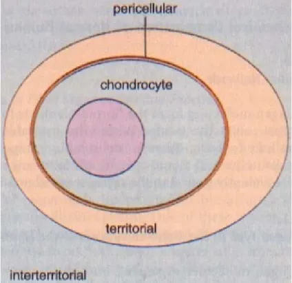
the keepers of the ECM and are responsible for the metabolic environment (7). The ECM is made up of different compartments and the area next to the chondrocytes is called the pericellular or lacunar matrix (8). This area is characterized by proteoglycan aggregates which bind the cell through the interaction of hyaluronic acid with the chondrocyte cells by CD-44-like receptors (8). The pericellular environment is also characterized by a relative lack of fibrillar collagens (8). Next to the pericellular area is the territorial or capsular matrix which is characterized by a large amount of fibrillar collagen that encases the chondrocytes forming a supporting framework for these cells (6). As we move further away from the chondrocyte, the final compartment is called the interterritorial area and it is at this location that the largest amounts of fibrillar collagen and communication between the chondrocytes are found (6). This area occurs through microfilaments and specific matrix molecules like anchorin and CD-44 like receptors on the cellular surface (9). The ECM environment is held together by chondrocytes which rarely divide after adolescence; therefore each loss of a chondrocyte causes a loss in the integrity of the underlying structure of the ECM. The different compartments of the ECM can be seen in Figure 2. It is important to remember that the further one goes from the chondrocyte, the more collagen is present.
Figure 2: Compartments of the ECM (1)
1-COLLAGEN
The collagen framework found in the articular cartilage varies between the different zones and it accounts for most of the dry weight of this region (10). Once this framework is established during development, it cannot be replaced or created anew, it is up to the chondrocytes to maintain this fibrillar network which gives strength and adaptability to the cartilage (11). The concentration of the collagen in the articular cartilage is directly dependent upon the density of the chondrocytes found within. In the superficial zone, analysis under transmission electron microscopy shows that the collagen framework resembles the shape and direction of the chondrocytes in that they are parallel to the articular cartilage and thin in diameter (11). As we move deeper into the cartilage, the diameter of the fibrils increases in size as do the size and shape of the chondrocyte cells,
reflecting an inherent larger space dimension in these areas (11). The fibrous elements found in this region are made up predominantly of collagen II but also of collagen type IX, XI and small amounts of type V, other small non collagenous proteins including proteoglycan and glycoproteins (12). During maturation the content of the fibrils change from a predominantly collagen type IX and makeup to a 90% of the collagen type II constitution reflecting an increase in strength of the fibrils needed with age (14). The type of collagen found closest to the calcified zone in the articular cartilage is the collagen type X (14). As we move further away from the chondrocyte into the interterritorial matrix the collagen fibrils become coarser in their constitution when observed under transmission electron microscopy and more type II collagen in this region is found (15). The fibrils are made up of a mixture of the three types of collagen, type IX, II, XI with this template being the basic one during early development (15). The collagen framework provides the structural support the chondrocytes require and enables the articular joint a manner of flexibility in response to joint load bearing (16). The ability of the chondrocytes to reproduce the dense fibril network decreases with age and it has been observed that only some newly synthesized collagen type II is produced after maturity in response to injuries (12). Besides this fibrillar collagen network there are a number of other proteins that play an important role in the integrity of the articular cartilage, these include proteoglycans, glycoproteins and non-glycosylated proteins (17). The most abundant proteoglycan found in the articular cartilage joint is called aggrecan (18).
2-AGGRECAN
Proteoglycans are proteins with glycosaminoglycan domains; they are found predominantly in the ECM and consist of most of the space in the ECM. These proteins along with water
allow the articular cartilage to compress under load stress thereby giving the joint flexibility (19). Aggrecan makes up 90% of the proteoglycan found in the articular cartilage and has a mass of about 230 kilodaltons (19). Most of aggrecan weight comes from the glycosaminoglycan domains that are made up mostly of keratan sulfate and chondroitin sulfate (20). Each glycosaminoglycan domain can be made up of over 100 different chondroitin sulfate and keratan sulfate chains (20). It is this high content of chondroitin sulfate and keratan sulfate which gives the aggrecan the ability to interact with hyaluronic acid in the ECM (20). Once the aggrecan molecule is synthesized by the chondrocytes and is secreted into the ECM, it aggregates with other aggrecan molecules as it name implies. This aggrecation of over 200 aggrecan molecules and link proteins is essential in forming the link between the aggrecan and hyaluronic acid (8). This proteoglycan aggregation maintains cellular contact through the hyaluronan interaction with CD-44 like receptors on the cell surface. The formation of these large aggregate complexes allows the articular cartilage to resist compression when under stress (8). There are two types of aggregate populations, one that is lighter, weighing approximately 60 kilodaltons, while another termed “superaggregates”, weighing 120 kilodaltons, is found in the middle region of the articular cartilage (21). Interestingly, during the onset of OA there is a distinct loss in superaggregates found in the articular cartilage which demonstrates how the integrity of the ECM is compromised (21).
3-SMALL NON-AGGREGATING PROTEOGLYCANS
The aggrecans are not the only proteoglycans to populate the ECM, there are other small non-aggregating proteoglycans like biglycan and decorin (22). These two proteins are part of a group of proteins called Small Leucine-Rich Repeats Proteoglycans (SLRP) that can be
divided into two subfamilies depending upon which type of side chain is attached to it, either keratan sulfate or dermatan sulfate side chain (23). Both decorin and biglycan are populated with dermatan sulfate side chains and have been found in articular cartilage (24). Examples of proteoglycans with keratan sulfate side chains found in articular cartilage are fibromodulin and lumican (25). Depending upon the stage of development these two proteins are found in the articular cartilage with or without their keratan side chain, fibromodulin has a keratan side chain only in the fetus or in a juvenile but loses it later on in the life cycle (25).
The importance of these small leucine rich repeat proteoglycans in the ECM is demonstrated in SLRP-null mice which develop abnormalities in the articular cartilage (17). The SLRP’s interact with many components of the ECM such as collagens, fibronectin and heparin to help form the support structure network for the chondrocytes (17).
4-OTHER STRUCTURAL PROTEINS OF THE ECM
There are other types of proteins that play an important role in the structural support of the ECM that are neither collagenous nor proteoglycan in type (17). These proteins are also necessary to help maintain the integrity of the ECM by their interactions with the other proteins present within the matrix like collagens and proteoglycans (17). An example of this type of protein is COMP, cartilage oligomeric matrix protein, which is a member of the thrombospondin superfamily and has a mass of 85 kilodaltons (26). This pentameric protein has been shown to interact with collagen through its carboxy-terminal regions (27). When a mutation occurs within this gene, two conditions have been known to occur, pseudoachondroplasia or epiphyseal dysplasia (27). During the early stages of OA, it has been shown that COMP is degraded and fragments of this protein have been found in
synovial fluid (28). These fragments are used as biomarkers for early signs of OA in patients (28).
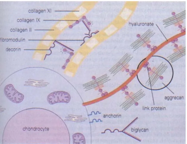
Anchorin is also a structural protein found in the ECM, it has been shown to mediate chondrocyte interactions with the matrix from its position on the cell membrane surface (29). Finally, fibronectin is another of these structural proteins with an essential role in matrix support (30). It seems to mediate interactions between the cellular membrane and other matrix proteins like collagen type II and thrombospondin (30). During the late stages of OA, fragments of degraded fibronectin are found in the joint fluid and have been shown to increase the catabolic breakdown of the matrix and decrease the synthesis of aggrecans (31). Figure 3 depicts the different matrix proteins and how they interact with each other and with the chondrocyte (1).
III. OA CARTILAGE
OA is the result of the action of a number of different factors, which lead to cartilage degeneration over time. The catabolic factors involved in the pathophysiology of OA include the proteases, the pro-inflammatory cytokines and some growth factors. The major family of proteases that have been shown to be involved in OA are the matrix metalloproteases (32). Proteases from this family are able to degrade the major cartilage matrix macromolecules (32). From this family of proteases three groups have been found to be elevated in OA, the collagenases, the stromelysins and the gelatinases (32). Collagenases are responsible for the degradation of collagen, the stromelysins have been linked to proteoglycan degradation and the gelatinases are involved in the degradation of denatured collagen (32).
A. CATABOLIC FACTORS
1-MATRIX METALLOPROTEASES (MMPs)
The MMP family consists of the classical MMPs, the membrane bound MMPs called MT-MMP, the ADAMs (a disintegrin and metalloprotease) also called the adamlysins and the ADAMTS which are a disintegrin and metalloprotease with a thrombospondin motif (33). In cartilage, studies have shown that MMP, MT-MMP and ADAMTS are implicated during the disease process (33). There are over 20 members in the MMP family that include the collagenases, the stromelysins, the gelatinases, some elastases and the aggrecanases ADAMTS-4 and ADAMTS-5 (33). MMPs depend upon the presence of a zinc ion in their active site for their catalytic activity (33). Usually, these proteases are active at neutral pH and are either secreted into the extracellular matrix or as for the MT-MMP found on the
cellular membrane surface (34). Their ability to maintain activity at a neutral pH enables them to play an important role in the overall protein turnover taking place in the ECM (33). Unlike most proteases, MMP are examples of multidomain proteases with additional protein modules that coordinate the localization and character of the protease itself (35). For example the C-terminal domain of collagenases called hemopexin is responsible for its ability to bind to triple-helical collagen (35). The cooperation between this domain and its catalytic domain is still unclear but it is evidently necessary to allow for the triple helical collagen strands to be separated and cleaved (35). Another example of multidomain functionality is found in the gelatinases which contain additional fibronectin type II repeats that are important in the binding of this proteases with its substrate denature collagen (35). These unique characteristics allow the MMPs the ability to be specific and diverse in their role as mediators of macromolecule turnover in the ECM.
i. Collagenases
Three main collagenases have been shown to be elevated in OA, collagenase-1 (MMP-1), collagenase-2 (MMP-2) and collagenase-3 (MMP-13) (32). This family of MMP can be separated into groups based on substrate specificity and their location in the cell (36). The three types of collagenases all cleave type II collagen to varying degrees, with collagenase-3 being five times more likely to cleave collagen II than collagenase-1 (37). Collagenase-1 has higher substrate specificity for collagen type III while collagenase-2 has a higher specificity for collagen type I (37). The presence of these three collagenases varies during the progression of OA. Collagenase-1 has been shown to be involved mainly in the catabolic or inflammatory phase of OA while collagenase-3 has been shown to be involved in the remodelling phase (37). The role of collagenase-2 in OA is still being investigated and like many proteases, this role cannot be narrowed down to one specific activity but to a
general activity important in maintaining the balance between the anabolic/catabolic mechanisms in the ECM. It has been shown in immunohistochemical studies in animal models that with the increase of lesions in OA, the levels of collagenase-1 increased in the superficial zones of the cartilage while the presence of collagenase-3 increased in the deeper zones (38). Interestingly, it has been shown that in OA, collagenase-1 is found also in the synovial fluid further indicating its role in the inflammation process (38).
ii. Stromelysins
Like the collagenases three stomelysins have been described in human articular tissue, stromelysin-1 (MMP-3), stromelysin-2 (MMP-10) and stromelysin-3 (MMP-11) (37). Only stromelysin-1 has been found to be elevated in OA tissues; this is shown in histological studies which correlated an increase in OA lesions with an increase in MMP-3 (37). Stromelysin-2 could not be found in OA synovium but studies have shown it to be present at minimal levels in synovial fibroblasts of rheumatoid arthritis patients, while stromelysin-3 has also been shown to be found in synovial fibroblasts of rheumatoid arthritis patients (39). Stromelysin-1 has also been found in synovial fluid and further studies have linked elevated levels of this stromelysin and proteoglycan degredation (39). Stomelysin-1 has also been shown to be involved in the degradation of collagen type IX which plays an important role in the stability of the ECM (40). Collagen type IX is necessary for the linking of proteoglycans and type II collagen in the ECM (40).
iii. MT-MMPs, Gelatinases, ADAMTS and ADAMs
The membrane bound MMPs are bound to the cellular membrane through their C-terminal domains and this allows for restricted protease activity (41). The MT-MMPs were found to be expressed in articular cartilage and their specific role in OA is yet to be determined (41).
So far there have been four MT-MMP expressed in the cartilage and these have been called MT1-MMP through MT4-MMP and all possess collagenase activity (41). One of the roles attributed to these membrane bound MMPs is the activation of other metalloproteases such as 72 kD gelatinase (MMP-2) and collagenase 3 (MMP-13) (42). Other members of the MMP family that are membrane located are the ADAMs which are also multidomain proteases (43). Theses proteases contain a disintegrin unit and a catalytic unit (43). The disintegrin unit allows it to bind to integrins on the cellular membrane surface and in doing so enables it function as a disrupter of the interactions between the matrix and the cell (43). Among the ADAMs studied, the most notable is the ADAM-17, which is also called TACE for Tumor Necrosis Factor alpha Converting enzyme and has been shown to be involved in OA pathology (43).
The ADAMTS are closely related to the ADAMs family but differ in that they do not bind to the cell surface and contain a thrombospondin motif (35). This motif allows for the protease to bind to glycosaminoglycans (35). The most notable of this family of proteases are ADAMTS-4 and ADAMTS-5 also known as aggrecanases. They are responsible for the degradation of the aggrecan structure in the ECM and cleave the aggrecan molecules at glutamic residues, specifically at the residues GLu373 –Ala374 (44). In OA there is evidence of an increasing number of aggrecan fragments found in the synovial fluid which plays a role in the inflammation of the synovium (44). The increasing amount of aggrecan fragments has been linked to the activity of the aggrecanase proteases and the levels of aggrecanase have been shown to be increased by the inflammatory cytokine interleukin-1β (45).
The last group, the gelatinases, has been found in human articular tissue, specifically gelatinase 72kD and gelatinase 92kD; only the gelatinase 92kd has been shown to be increased in OA (37). Although the MMP family is the major player in the progression of OA in terms of cartilage degradation, there are other proteases that have been found to play a role in cartilage turnover which will be discussed later. The regulation and activation of MMPs is essential in OA. It is often a problem in their regulation that plays an important part in the progression of OA and an overexpression in MMP would lead to a complete destruction of the articular cartilage (37).
2-MMP regulation
The regulation of MMPs occurs at both the transcriptional and posttranslational levels (46). The expression of MMPs in normal articular joint tissue is low (46). The expression of these proteases can be induced by proinflammatory cytokines such as interleukin-1(IL-1), tumor neucrosis factor alpha (TNF-α) and certain growth factors like epidermal growth factor (EGF), platelet derived growth factor (PDGF), basic fibroblast growth factor (bFGF) and transforming growth factor beta (TGF-β) (37). At the transcriptional level, these cytokines and growth factors affect MMP levels of expression by interacting with the Activator protein site (AP-1) in the promoter region of the MMP genes (47). This activator protein site is present in all of the MMP genes except for MMP-2 (47). Transcriptional factors that bind to this site are members of the Fos and Jun family which bind as heterodimers (Jun/Jun or Fos/Jun) to activate transcription of the MMP genes (47). The cytokine TNF-α has been shown to increase MMP expression by inducing a prolonged increase in Jun and Fos while TGF-β and glucocorticoids suppress MMP expression through the AP-1 site (47). Another transcription site that has been shown to play an important role in the induction of MMP
expression is the polyomavirus enhancer A (PEA-3) site and this site has been shown to act in concert with the AP-1 site in some MMP transcription (37).
At the posttranslational level most of the MMPs are synthesized as proenzymes which are inactive until they are activated usually through cleavage by a protease (46). The activation process of MMPs occurs in two different stages. An intermediate is first generated by an activator and then the partially activated MMP is then fully activated by other intermediates or partially activated MMPs (46). Besides being activated by MMPs themselves, they can also be activated by other factors (46). An example of this is the prohormone convertase, furin, which is a serine protease found in the Golgi apparatus that cleaves 11, MMP-14 and aggrecanases to activate them (47). In the case of the other MMPs, they are activated either through the action of serine proteases or other MMPs (47). These posttranslational modifications are necessary as a regulatory mechanism to control the levels of active MMPs found the ECM (47). The activity of MMPs is also regulated by inhibitors called tissue inhibitors of MMPs or TIMPs (48). These proteins contain two domains, one which binds to the active site of the MMPs and in doing so inhibit their catalytic ability (48). There are currently four TIMPs identified and they are called TIMP 1-4 (48). Only three of these molecules have been found in human articular tissue (48). It has been shown that chondrocytes produce TIMPs -1, -2, -3 and TIMP-3 has been found exclusively in the ECM (48). Most of the TIMPs show little binding selectivity as to which MMP they bind but TIMP-3 has been shown to inhibit specifically ADAM-17 (TACE) and the aggrecanases ADAMTS-4 and ADAMTS-5 (48). Studies have demonstrated that in OA there are elevated levels of TIMP-1 and TIMP-3 and that there is a distinct imbalance between the levels of the TIMPs and MMPs (37). The inhibitory function of TIMPs is not the only role that these molecules may play in OA (49). The activation of MMP-2 requires the presence of TIMP-2
to interact with MT-MMP-1 to cleave the pro-MMP-2 (50). Once again, one can see the importance of maintaining a balance between the factors present in the human articular joint; any disruption in this balance leads to a breakdown in the support matrix structure.
3-SERINE PROTEASES
The serine proteases are also active at a neutral pH as the metalloproteases and contain a catalytic group which is a hydroxyl moiety (51). These proteases are also activators of the MMPs (8). An example of this family of proteases is urokinase-type (uPA) and tissue-type (tPA) plasminogen activators (52). These enzymes are responsible for activating the plasmin protease which has been shown to play a role in the activation of MMPs (52). Elevated levels of plasmin have been seen in OA (53). The PA/plasmin system can be stimulated by the proinflammatory cytokine IL-1β (53). Plasmin has the ability to digest glycoproteins which form part of the supporting structure of the ECM (53). As in the case for MMPs, the serine proteases also have inhibitors which help in maintaining a balance between the catabolic and anabolic mechanisms of the articular joint tissue. The serine protease inhibitor is called the PA inhibitor, and PAI-1 has been shown to decrease in OA cartilage (54). As in the case of the TIMPs, the serine protease inhibitor experiences a decrease in level during the progression of OA, thereby demonstrating again the importance of a balance within the articular joint tissue environment.
4-THIOL PROTEASES
The thiol proteases are actively involved in the degradation of the ECM molecules and studies indicate that they are capable of doing so due to their ability to maintain activity at acidic pH levels (55). An example of this is cathepsin B which is active both in the degradation of collagen and proteoglycan but also has been shown in vivo to be an activator of MMPs (55). As in the case of the other proteases, cathepsin B has inhibitors which are important in its regulation. In OA their levels are decreased, while the levels of cathepsin B are increased (56). Therefore an imbalance is created again which favors the catabolic over the anabolic process.
B. PRO-INFLAMMATORY CYTOKINES
The role of the various proteases in the progression of OA is essential but their expression and subsequent involvement in OA is driven by the effect of cytokines released from an inflamed synovial membrane and the chondrocyte of the articular cartilage (57). Cytokines are small proteins that act as a messenger for cell to cell sharing of information. The term cytokine includes some growth factors, interleukins and interferons (58). In the progression of OA these molecules transmit information from cell to cell depending upon the environment of the articular cartilage matrix at the time. In the catabolic state in which the degradation of the cartilage is taking place, the presence of pro-inflammatory cytokines is elevated which causes the release of degradative enzymes in the tissue matrix (59). In patients with OA, it has been shown that the chondrocyte cells produce IL-1β, IL-6, IL-8 , IL-17, IL-18, TNF-α, the inflammatory mediators prostaglandins and nitric oxide (NO) (59). These factors help increase the levels of the degradative proteases like MMPs thereby destabilizing the balance between the anabolic and catabolic states. Studies have also shown that the pro-inflammatory cytokines IL-1β and TNF-α are the major cytokines involved in
the progression of OA; they increase their own expression as well as the expression of other inflammatory cytokines such as IL-6, IL-8 and the leukocyte inhibitory factor from chondrocyte and synovial cells (60). On the other side of the spectrum there are anti-inflammatory cytokines which are essential in maintaining the balance in the articular tissue. The anti-cytokines help restrict the expression of the degradative enzymes into the ECM. Examples of anti-inflammatory cytokines are IL-4, IL-10 and IL-13 which are essential in moderating inflammatory cytokine and protease synthesis (61, 62). They also play a role in regulating and increasing the expression of proteins which in turn help regulate MMP activity such as TIMPs and IL1-Ra which is an antagonist for the IL-1 receptor (62).
1-Interleukin-1β (IL-1β)
The presence of the pro-inflammatory cytokine IL-1β in OA chondrocytes has been shown and is found predominantly in the superficial layer of the articular cartilage joint (63). The cytokine IL-1β has been linked to causing many different changes in the environment of the articular cartilage leading to the overall destruction of the cartilage in OA (64). IL-1β is synthesized as an inactive precursor and activated by the IL-1β converting enzyme ICE (caspase 1), which is responsible for the mature active form of the cytokine (37). Two IL-1β receptors exist, type I and II, and they have different affinities for the cytokine (63). Type I IL-1R receptor has a higher affinity for IL-1β than the type II IL-1R receptor and is responsible for signal transduction (63). An increased number of this receptor type has been seen in chondrocytes during OA (63). Both of these receptors can exist in a soluble form which acts as an antagonist for the membrane bound form in that it binds IL-1β without signal transduction occurring (65).
This cytokine causes an imbalance in the type of collagen produced by the chondrocytes (37). It increases the synthesis of type I and type III collagen without increasing the amount of type II collagen (37). This affects the integrity of the structure of the ECM in that the concentrations of the different types of collagen are not at the right proportions (37). This has been shown to cause damage to articular cartilage repair (37). Other roles for the cytokine in the progression of OA involved decreasing the synthesis of TIMP-1 and increasing the synthesis of MMPs (66). Similarly, IL-1β has been shown to create an imbalance within the plasminogen system by increasing plasmin synthesis while decreasing the synthesis of the plasmin inhibitor PAI-1 (66). These roles share a common thread in that they all create an imbalance within the articular joint by increasing the catabolic process of the joint over the anabolic system of maintenance. IL-1β causes these changes by activating various signal transduction cascade to influence gene expression. Some of the signal transduction cascades affected by the cytokine are PKA, ERK 1 / 2, p38, SAPK/JNK, PKC and other protein kinases (37).
2-Tumor Necrosis Factor-α (TNF-α)
The pro-inflammatory cytokine TNF-α has been shown to play an important role in the progression of OA and also in the inflammation of the synovial membrane (67). The cytokine is produced in an inactive pro-form and cleaved by a TNF-α converting enzyme called TACE which is part of the adamalysin protease family (67). The TACE levels are increased in OA which is indicative of the overall catabolic state of the joint during the disease (67). Once the cytokine is active, it forms a trimer with other TNF-α molecules to bind to one of two types of specific receptors (68). TNFR55 is the dominant receptor in OA and TNF75 can be found on the cell membrane (69). Both of these receptors can exist in a soluble form (69). The soluble forms of the receptors help the binding of TNF-α to the TNF
receptor on the cell membrane (70). However at high concentrations the soluble forms of the receptor can act as an antagonist by competing with free TNF-α and preventing it from binding to its receptor and initiating a signal transduction (71). The signal transduction occurs by the activation of different signaling pathways such as NFκB, the SAPK/JNK, ERK 1/2 kinase and the PKC (72). The concomitant signaling cascade causes an increase in AP-1 synthesis and proteins involved in AP-1 site binding such as Jun/Fos which have all been demonstrated to be involved in MMP expression (72).
3-Other Pro-Inflammatory Cytokines in OA
The cytokines IL-6, IL-17 and leukemia inhibitory factor (LIF) have all been shown to be elevated in OA although exact roles for each of these cytokines still remain to be determined (37). The role of IL-6 has been linked to progression of OA in that it is induced by the pro-inflammatory cytokines IL-1β and TNF-α (73). LIF has also been shown to be induced by IL-1β and TNF-α and plays a role in the inflammation process by causing the degreadation of proteoglycans and MMP and induction of nitric oxide (NO) (74). IL-17 has also been shown to be elevated in OA and has been linked to the induction of NO (75).
4- NITRIC OXIDE (NO)
The inorganic molecule NO has been implicated in the catabolic process of OA and studies have shown that during the progression of the disease elevated levels of NO can be found in the synovial fluid and articular cartilage (76). The elevated level of NO has been tied to both cytokine induction and constitutive induction by the chondrocytes themselves (77). The role of NO plays in the catabolic mechanism occurs through the inhibition of aggrecan and proteoglycan production (79). Studies have also shown that NO increases MMP production while inhibiting the synthesis of IL-1Ra which contributes to catabolism (76,
79). During OA, an increased level of NO has been observed in the articular cartilage when compared to normal cartilage and an increase in the amount of nitrate/nitrite has been found in the synovial fluid (80). The increase in nitrogen products in the synovial fluid is a result of an increase in the amount of inducible NO synthase (iNOS) in the cartilage which is responsible for NO production (81). Finally, elevated levels of NO have been linked to cell death; the presence of this factor in the articular cartilage was also reported to lead to apoptosis (82). Apoptosis is an important part of OA in that one of the characteristics of OA is a lack of chondrocyte cells in the articular cartilage and this phenomenon has been observed during the progression of the disease (83).
C. ANABOLIC FACTORS IN OA
1-ANTI-INFLAMMATORY CYTOKINES
Three main anti-inflammatory cytokines have been studied in OA: IL-4, IL-13 and IL-10 (10). Each of these has been shown to counter the inflammatory process of the pro-inflammatory cytokines by decreasing the production of pro-inflammatory cytokines and increasing the production of TIMPs (58). The effect that these anti-inflammatory cytokines have on the cell seems to be much more specific in that they require specific conformations of certain types of receptors on the cell membrane to initiate a signal transduction. IL-13 and IL-10 receptors have different conformations depending upon the cell type and this affects the ability of the cytokines to initiate signaling (85). This level of complexity in regards to the receptors points to another difficulty in maintaining its potential.
Another inhibitor of the pro-inflammatory cytokines is IL-1Ra which competitively binds to the IL-1 receptor and inhibits the actions of IL-1β (86). IL-1Ra needs to be at a high
level to inhibit IL-1β activities in the articular tissues, once again pointing to the cartilage’s inability to respond or stop the progression of OA (86).
2- GROWTH FACTORS INVOLVED IN CARTILAGE HOMEOSTASIS
The literature suggests that anabolic effects occur through growth factors that synthesize matrix molecules and that are necessary for continued balance in the joint, thus homeostasis.
i. Insulin-like Growth Factor (IGF)
IGF-1 and IGF-2 are important for proteoglycan synthesis in the articular joint and are essential players in cartilage homeostasis in all stages of life (87). IGF-2 has been shown to be predominantly active in early stages of life while IGF-1 has been shown to be active in the adult life stages explaining its studies during OA (87). The presence of these growth factors in the articular joint occurs through their synthesis by the articular tissue cells (37). As it is the case for all factors involved in OA, these growth factors also have specific inhibitory molecules that limit their actions and they are called IGF-binding proteins (IGF-BPs) (88). Studies have shown that the presence of IGF-BPs is elevated in OA through treatment with TNF-α and IL-1β and explains, in part, the reason why the anabolic repair mechanisms fail (89). Another interesting observation is that the levels of IGF-1 are elevated in OA due to to an increase in cytokine presence in chondrocytes during progression of the disease (89). The increase in IGF-1 synthesis does not lead to an increase in cartilage anabolic factor synthesis not because of the limited amount of IGF-1 receptors on the cell surface but because of a high level of IGF-BPs released by the cells (88).
ii. Platelet derived growth factor (PDGF) and fibroblast growth factor (FGF)
PDGF has been shown to also play a role in the balance between the anabolic and catabolic mechanisms of the articular cartilage joint (90). Studies have demonstrated its role in proteoglycan degradation and synthesis (90). The presence of both PDGF/FGF has been found in both synoviocytes and chondrocytes during the progression of OA (90). Although FGF has been shown to increase mitotic activity in chondrocytes by increasing cellular division and proliferation, its precise role in OA is still under investigation (91). One should note that FGF constitutes a family in which different effects could be mediated by different FGF. Studies demonstrate that it is not one growth factor or cytokine alone that helps promote the anabolic mechanism of the articular joint but the presence of all of these factors working in synergy creating an overall anabolic environment (91).
iii. Transforming growth factor-β (TGF-β)
The TGF-β family of proteins consists of 30 members, three of which are TGF-βs, five activins and over 20 bone morphogenetic proteins (92). TGF-β plays an important role in inhibiting cell proliferation in many types of cells such as epithelial and hematopoietic cells and has been shown to limit tumor cell proliferation (92). In OA it plays a role in inhibiting enzyme release from different cells types and promoting cell synthesis of TIMPs (93). TGF-β has been found in both chondrocytes and synoviocytes and is activated during inflammation periods of the joint (94). Another role associated with this growth factor in OA, is the increased synthesis of proteoglycan and specifically the smaller proteoglycans found in the ECM (95).
The two 12.5 polypeptides that make up TGF-β are joined by a disulfide bond (96). The synthesized TGF-β is present in an inactive form called LTBP with a prosegment which designates the protein for the ECM (96). The effects of the TGF-β are often affected by other factors in the ECM which can bind the growth factor, thereby inhibiting it from binding to its receptor to mediate a cellular response (96). The two main receptors used by TGF-β are receptors type I and type II, which are structurally similar to serine/threonine kinases (97). There are far more ligands which are specific for these receptors than there are TGF-β on the cellular surface thereby creating a competition for the ligands to bind the appropriate receptor (97). These receptors are able to cooperate together in complexes with the effector Smad molecules within the cell to affect a greater versatility of signaling (92). Smad molecules also interact with various transcription factors to initiate or inhibit gene transcription (92). Indeed, from a relatively simple pathway of signaling, TGF-β ligand to receptor type I or II to the Smad signaling cascade, the TGF-β superfamily is able to mediate many different cellular functions (98).
The Smads are the principal signaling cascade that is used by TGF-β in the cell and they can be divided into three subgroups 1) R-Smads which are receptor activated Smads and are the first in the cascade, 2) Common Smads (Smad 4) which are the subsequent Smads activated in the cascade, and 3) Inhibitory Smads which regulate and control the signaling cascade from within the cell (92). The R-smads are characterized by a C-terminal SXS motif in which both serines are phosphorylated by the receptor type I (92). TGF-β binding to the receptor type I can lead to the receptor recruitment of the R-smads, such as Smad 2 and Smad 3, and Smad 1, 5 and 8 with BMP signaling (92). So depending upon the type of ligand, a different type of signaling Smad is activated and thus a different type of cellular response is initiated (97). Other than the interaction of the R-Smads with the receptor type
1, R-Smads can also be activated by the presence of various cytoplasmic kinases (99). Many kinases are able to interact with the different Smads, the Protein Kinase C, Calcium/ Calmodulin dependent protein kinase and Akt (99).
The inhibitory Smads, Smad 6 and Smad 7 are able to inhibit the Smad signaling cascade through their ability to interact with the R-Smads (100). They do so by binding to the active site on the receptor type I complex thereby blocking the binding of the R-Smads and the continuous Smad signaling cascade (100). The level of complexity of the signaling in terms of different effects it can have on the cell is increased by the fact as mentioned above that the Smads are able to intereact with many different transcription activators (100). They activate transcription of genes through their cooperation with these other transcription factors (97). Indeed, R-Smads alone, excluding Smad 2, have a relatively low affinity for DNA but this is increased through interactions with a number of different transcription factors (101). They interact with these transcription factors through their MHI or MH2 domains depending upon the type of cofactor it binds to (92). The first cofactor found was the FAST/FoxH1 which is a forkhead transcription factor and was shown to interact with with Smad 2/4 complexes in response to activin binding to the TGF-β receptor (102). An example of this cooperation is seen in endothelial cells in which TGF-β activates the expression of p21, the FoxO cofactor binds to the Smad 3 /4 complexe to help induce transcription of p21 gene (103). In the p21 promoter, Smads are also able to bind to the Sp1 transcription factor site to regulate the expression of the gene indicating a possibility of many Smad complexes binding to one particular promoter in an effort to influence its expression (104).
Finally TGF-β is able to activate signaling cascades other than the Smad; it can activate the MAPK kinases, ERK 1/2, JNK and p38 which can in turn and as discussed above, influence
the Smad signaling cascade (105). This once again demonstrates the level of complexity of the reactions that these ligands can have on the cell and how it is very difficult to anticipate the response of the cell to these ligands (105).
D. Bone Morphogenetic proteins (BMPs)
BMPs are part of the TGF-β superfamily of proteins and have been shown to play a role in embryonic development and other various cellular functions in both adults and infants (99). Over 20 different members of this subgroup have been identified based on their sequence homology (106). BMPs are secreted from the cell with a hydrophobic stretch of 50-100 amino acids (106). This precursor is cleaved upon activation and is characterized by seven cysteines that are used to dimerize with another monomer (106). The BMP precursor is cleaved by convertases such as the serine endoprotease furin (107). Most BMPs have a molecular mass in the range of 20 000 to 30 000 daltons (107). Interestingly, BMP-1 is unrelated to other BMPs in that it is not involved in cell differentiation and growth (107). This BMP acts as a protease and can cleave procollagen fibers, BMP-2, BMP-7 and the antagonist chordin (107).
BMPs have been linked to many different processes within the cell including cell proliferation, differentiation, migration, apoptosis and in angiogenesis (108). Although their name is indicative of their primary role in bone morphogenesis, some of these proteins, in particular BMP-2, -7 and have been shown to stimulate proteoglycan synthesis in chondrocytes (108). This anabolic effect on the articular cartilage is followed by another possible effect in that stimulation by BMPs can lead to dedifferentiation of chondrocytes, calcification and bone formation (108). BMPs have also been shown to stimulate chondrocyte maturation and chondrocyte function which stimulates the production of
collagens type II and type X (107). When BMP-2 and -4 are overexpressed in developing limbs an increase in the number of chondrocytes has been observed and subsequent increase in matrix cartilage (107). BMPs play an important role in chondrogenesis which is the differentiation and maturation of chondrocyte cells by stimulating the expression of the Sox-9 gene which is involved in the evolution of the chondrocyte (107). Studies have shown that BMP-7 is a more potent stimulator of synthesis of cartilage molecules than TGF-β (109). Indeed, in vivo studies have shown that stimulation of the knee joint with BMP-2 and BMP-9 causes an increase in proteoglycan synthesis that is higher than the effect of TGF-β stimulation (110).
Interestingly while the stimulation by BMP is higher, it is shorter in duration than the effect that TGF-β has on the proteoglycan synthesis in the knee joint (110). Another interesting finding is that both TGF-β and BMP signaling converge upon the same effector which is Smad-4 leading to a possible competition for the Smad-4 to initiate changes within the cell (107). This convergence of pathways can either inhibit or increase the effects of the TGF-β and BMP ligands in the cell depending upon the cellular environment at the time.
1-BMP receptor and signaling
The receptors responsible for BMP signaling are two distinct serine/threonine kinase receptors that can pass along the signal to effector Smad proteins, the same type of receptors used by TGF-β ligands; receptor type I and receptor type II (107). The type 1 receptor can be subdivided into four different type; type IA or activin receptor-like kinase ALK-3, type IB or ALK-6, ALK-2 and ALK-1 (108). When the BMP ligand binds to the type I receptor, the receptor type II forms a heterodimer with it and the kinase activity of the type II receptor activates the type I receptor leading to the initiation of the signaling cascade (111).
The different types of receptor type I allows for diversity in signaling which is essential in determining the impact of the ligand binding on the cell. The type of signaling cascade depends upon wether the receptor heterodimer formation occurred prior to or after BMP ligand binding to the type I receptor (107). If the heterodimer formation was already in place before BMP ligand binding, then the Smad signaling cascade is activated (107). When the BMP ligand itself induces the formation of a receptor heterodimer, it is the MAPK pathway that is activated (107). When the BMP ligand binds first to the receptor type II, there is activation of the receptor type I which intitiates the signaling cascade (108). As for the type 1 receptor, the type II can also be subdivided into three different types; BMPR2, ActR-II and ActR-IIB (108). The main reason why BMPs bind to the receptor type II is because the ligands have a low affinity for the type I receptor and that the type II receptor is specific for the BMP ligand (107).
A number of signaling pathways other than Smads have been associated with the BMP ligand, these include the JAK/STAT, the calcium/calmodulin and p38 (106). The latter leads to cell apoptosis but the main signaling cascade used by the BMP ligand is the same as the TGF-β ligand; the SMAD signaling cascade (108). The Smads activated by the receptor type I from BMPs are Smad -1, -5 and -8 which then bind to Smad 4 which aids in the translocation to the nucleus for cellular gene transcription initiation or inhibition (108). As mentioned above the difference between the BMP mediated signaling and the TGF-β mediated Smad signaling is that TGF-β uses Smads 2 and 3 while BMPs use Smad-1 and -5 (107). Both pathways converge upon Smad-4 which is necessary for translocation to the nucleus and DNA binding (107). BMPs activated signaling can be potentiated or inhibited by a number of cofactors within the cell which increases the diversity of the effect BMPs can have on the cell.
Figure 4. Mechanisms of BMP signaling (107)
Studies have shown that the transcription factor Runx-2/Cbfa-1 interact with Smad-1 and Smad-5, which play an important role in translocation to the nucleus of the BMP signal (112). It has also been show that in chondrocytes, BMP-2 induces Runx-2/Cbfa-1 expression as this cofactor is necessary in BMP-2 signaling (113) Figure 4 illustrates BMP initiated signaling within the cell, the different inhibitors and factors involved and the two
major pathways activated by the BMP ligand; Smad and MAPK kinases (107). As is the case in many pathways in the cell, there are often inhibitors both within the cell and outside that play an important role in the regulation of signaling.
Within the cell there are two main mechanisms of BMP signaling repression: inhibitory proteins that block the activities of the Smad molecules and pseudoreceptors incapable of initiating a cellular response (114). The pseudoreceptor that binds both BMP and TGF-β ligands is called BAMBI and it contains extracellular domains similar to the type I receptor for both ligands (114). The BAMBI receptor is able to associate with the BMP type I receptors thereby inhibiting their actions without interfering with the BMP ligands themselves (114). Studies have shown that BAMBI is co-expressed with BMP-4 indicating a possible negative feedback loop in which the BMP pathway is regulating itself (107). The second pathway is through proteins within the cell blocking the signaling cascade, the major proteins are the inhibitory Smads; Smad 6 and Smad 7 (115). They both contain MH2 domains which are necessary for protein interactions, but lack the MH1 domain which enables them to bind to DNA and affect gene transcription (107). These inhibitory Smads replace Smad 1 and 5, are phosphorylated by the BMP type I receptor, and effectively blocking further signaling (115). They can also bind transcription factors preventing association with other Smads (107). For example Smad 1 binds the repression transcription factor Hoxc-8 to remove it from DNA (116). The inhibitory Smad 6 will bind Hoxc-8 to block the action of Smad 1 allowing the Hoxc-8 transcription factor to continue inhibiting gene expression (116).
Besides the inhibitory Smads there are other proteins that inhibit Smad signaling such as the Ski and Tob (117). The Tob protein is a member of a family of anti-proliferative proteins and inhibits BMP activity in osteoblasts by blocking the activity of Smad-1 and -5 (117).
The Tob gene is expressed in osteoblasts when BMP-2 is increased indicating a negative feedback loop in osteoblasts (117). The other is the Ski protein which is an oncogene and blocks BMP signaling by binding to the MH2 protein binding domain of Smad -1, -4 and -5 before the formation of heterodimeric protein complex forms (118). Studies have shown that the lack of this protein in embryonic development leads to death of mice indicating the importance of this protein in regulating the actions of BMPs (119).
The main intracellular mechanism of BMP inhibition occurs through the blocking of the Smad pathway directly through the inhibition of Smad-protein interactions or the inhibition of Smad-DNA interactions. The following section will discuss the extracellular regulation of BMP actions; the BMP antagonists and their role in OA.
IV. THE BMP ANTAGONISTS
BMP antagonists are molecules that inhibit the activities of BMPs by binding to them and blocking them from binding to the BMP receptors. There are many different types of BMP antagonists; noggin, chordin, follistatin, follistatin-related gene (FLRP), ventroptin, twisted gastrulation (Tsg) and the Dan/Cerberus family of genes which includes the head inducer Cerberus, the tumor suppressor Dan, the protein related to Dan/cerberus (PRDC), caronte, Dante (Dte) and gremlin (107). The number of antagonists that exist to inhibit the functions of the BMPs indicates the level of importance these factors play in development and overall cellular functions. Differing roles have been found for the BMP antagonists depending upon the cell type and the stage in the life cycle at which the BMPs are expressed.The BMP antagonist noggin is a polypeptide of about 22 kDa but is secreted as a homodimer of 64 kDa and has been shown to block with varying degrees of specificity BMP-2, -4, -5, -6 and -7 (107). In osteoblasts the levels of noggin expression are increased in response to elevated
BMPs and in chondrocytes elevated noggin expression has been linked to induction by Ihh (Indian hedgehog) (120). Noggin is able to block the effect of BMPs on collagen and non-collagenous protein synthesis in cells of osteoblastic lineage and also has been show to inhibit chondrogenesis and limb development (107).
The BMP antagonist chordin has a molecular weight of around 105 kDa but is secreted as a 120 kDa molecule (121). This is probably due to posttranslational modifications and it is characterized by four cysteine rich domains (107). In osteoblasts chordin expression is low and has not been shown to play a significant role in these cells differentiation (122). However, in chondrocytes it has been shown to play a role in the maturation of chondrocytes (107). Follistatin was initially identified as an activin inhibitor but it has also been shown to inhibit BMP-4 and is involved skeletal development (123). Recently it has shown that the follistatin gene expression was upregulated in human OA chondrocytes and synoviocytes indicating a possible role for the antagonist in the progression of the disease (123). This study also found that some factors were responsible for its up-regulation (123). The ones with the strongest effects were IFN-γ and TNF-α (123). The study also suggested that follistatin appears at a later stage of the OA process as it is induced by inflammatory cytokines (123). Another indication of a linkage between TNF-α and follistatin is that both have been found in higher levels in the superficial zones of OA cartilage, thus nearest to the inflamed synovial membrane (123). Other factors found that affect the expression of the follistatin are TGF-β which induces expression while BMP-2 down regulates its expression (123). Another antagonist is the Twisted Gastrulation (Tsg) which is able to bind chordin/Sog and BMP-4, creating a tertiary complex that has a higher specificity for BMP-4 than either antagonist alone (107).
The Dan family is a group of related glycoproteins that all share a common function of blocking the action of BMPs (107). This family includes seven members: Dan, Cerberus, PRDC, dante, caronte, gremlin and sclerostin (107). Specifically these proteins share a region of homology that unites them: the presence of a cysteine knot in the carboxyl terminal domain which is important in their functionality and can be seen in the tertiary structure (107). Dan has tumor suppressor activity and the protein is about 19 kDa; when it is secreted it usually dimerizes into a homodimer (124). This protein is expressed in embryonic tissues and is not induced by BMP (107). Cerberus as it names indicates is involved in head formation acting as a head organizer and plays a critical role in neural tissue formation (107). The protein is about 31 kDa in size and like the other members of this family undergoes post-translational modifications which enable it to bind primarily BMP-4 (125). Another role for this antagonist is its ability to bind and prevent the actions of the Wnt 8 protein thereby affecting the Wnt signaling pathway (125). Of note, other members of the Dan family have been shown to be involved in this pathway and also play important roles in development as well as in the regulation of the bone morphogenetic proteins (107). As my project focuses on one member in particular, gremlin, it will be discussed in greater detail.
A. GREMLIN
The BMP antagonist gremlin is a protein that has been studied primarily for its involvement in development during the early stages of the life cycle. New evidence suggests that it may also play a role during the progression of OA and respond to the elevated BMP levels found in the articular cartilage during the disease.
The gremlin gene was first cloned from a Xenopus ovarian library (107). It has a role in limb development and axial patterning (126). The rat homolog is Drm (down-regulated by mos), mos is a viral oncogene, interestingly other viral oncogenes including raf and v-ras were found to effect the expression of gremlin/Drm in rat fibroblasts (127). The Drm gene encodes a 20.7 kDa protein that is glycosylated, increasing it weight to 28 kDa at secretion from the cell (127). Gremlin and its rat homolog Drm share 80% amino acid homology, and all forms of this gene across species share this high level of homology (107). It is interesting to note that Drm can be secreted in a glycosylated form but also in a non-glycosylated form, and that both forms undergo phosphorylation (127). The protein can also be found in a soluble and cell associated form, and both have BMP inhibiting capabilities (107). Gremlin expression has been seen in various types of tissue; such as in the brain, the kidneys, the testis and recently in the articular cartilage and synovial membrane (107). The main activity of this protein is to inhibit BMP activities in these different tissue types (107).
1-Roles of Gremlin in different tissues and stages of development
Gremlin plays a role in fibrosis which is characterized by an increase in the production of the ECM components collagens I and II, proteoglycans, fibronectins and hyaluronic acid (128). This cellular event occurs in all types of tissue: the liver, pancreas, kidney, lung, skin as well as in the synovial membrane (128). Boers et al. used hepatic stellate cells (HSC) which are the liver cells in which fibrosis occurs and determined which genes were upregulated when the cells were undergoing fibrosis (128). Their study demonstrated an increase in the expression of the two BMP antagonists follistatin and gremlin. They suggest that a possible role for gremlin, as a pro-fibrotic factor, is its ability to block BMPs (128). This was based upon results indicating that stimulation with BMP-7 reduces the fibrotic
change of the cells (128). This is interesting considering that gremlin binds preferentially to BMP-2,-4 and -7 (128).
Gremlin overexpression in transgenic mice has been shown to cause osteopenia and to reduce bone density by 20-30% resulting in impaired bone formation (129). Gazerro et al. showed that transgenic mice overexpressing the gremlin gene had disorganized collagen bundles and a 70% decrease in the number of osteoblasts (129). These changes in phenotype were related to the gremlin’s inhibition of BMPs (129). However, they postulated that gremlin might have the ability to bind and inhibit other signaling pathways since their results demonstrated a reduction in Wnt signaling (129). Wnt proteins and signaling are involved in many processes that occur in development including cell proliferation and osteoblast differentiation (129). They tested this hypothesis by using another BMP antagonist, noggin, which binds BMP-2,-4 and -7 but with a higher specificity than gremlin in the same type of cells (129). Interestingly, they found that noggin had the same ability to reduce Wnt signaling thereby indicating that gremlin principal role was the antagonism of BMPs (129).
On the other hand, Chen et al. showed a new role for the gremlin protein by demonstrating that overexpression of gremlin in tumor derived cell lines increased the expression of the cell cycle inhibitor p21 (130). They demonstrated that most tumor derived cell lines fail to express the gremlin gene, they induced its expression to observe the effects that this change would have on the cells (130). Data indicated that gremlin has tumor suppressor ability in that its overexpression was able to inhibit the neoplastic phenotype of the tumor cell lines (130). This ability coincided with an increase in the expression of the transcription factor p21 indicating a possible role of gremlin (130). They tested various pathways to determine
how gremlin exerts its effects (130). Data showed that it was not through the signaling cascades p53 or through the MAPK kinases including ERK 1/ 2 and p38 (130). Although, it is well know that gremlin’s principal action is to antagonize the activities of BMPs, they stimulated the cells with BMP -2 and -4 and found that they did not increase tumor cell proliferation or growth (130). Another interesting finding was that they failed to detect the presence of these BMPs in the tumor cell lines (130). They concluded that gremlin was exerting its effects through a novel pathway which is able to increase the expression of the p21 cell cycle inhibitor (130). However questions that remain to be answered are: is gremlin causing this change, if so, is it through its own receptor, is it gremlin itself within the cell causing these changes and can gremlin act as a transcription factor itself?
Gremlin’s role other than only being an antagonist of BMP was suggested by Stabile et al by showing that gremlin could act as a pro-angiogenic factor (131). Gremlin is a well known BMP antagonist and BMPs have been shown to be involved in angiogenesis, specifically BMP-4 (131). However, the authors demonstrated that the presence of BMP-4 has no influence on gremlin’s ability to interact with the endothelial cell receptors, and their results indicate that gremlin exerts its effects through a BMP-independent pathway (131). The authors also suggest that gremlin may be acting through a specific receptor and initiating changes within the cell (131). Lastly gremlin has been shown to be involved in the pathogenesis of OA (123). In this disease, gremlin was found to be upregulated in chondrocytes (123). This upregulation may be linked to the elevated levels of BMPs in the OA pathogenesis.