SYNERGISTIC GELATION PROPERTIES OF GELATIN AND POLYSACCHARIDE AQUEOUS MIXTURES
CHANGSHENG WANG
DÉPARTEMENT DE GÉNIE CHIMIQUE ÉCOLE POLYTECHNIQUE DE MONTRÉAL
THÈSE PRÉSENTÉE EN VUE DE L’OBTENTION DU DIPLÔME DE PHILOSOPHIAE DOCTOR
(GÉNIE CHIMIQUE) AOÛT 2017
UNIVERSITÉ DE MONTRÉAL
ÉCOLE POLYTECHNIQUE DE MONTRÉAL
Cette thèse intitulée :
SYNERGISTIC GELATION PROPERTIES OF GELATIN AND POLYSACCHARIDE AQUEOUS MIXTURES
présentée par : WANG Changsheng
en vue de l’obtention du diplôme de : Philosophiae Doctor a été dûment acceptée par le jury d’examen constitué de : M. FAVIS Basil, Ph. D., président
Mme HEUZEY Marie-Claude, Ph. D., membre et directrice de recherche M. VIRGILIO Nick, Ph. D., membre et codirecteur de recherche
Mme WOOD-ADAMS Paula, Ph. D., membre et codirectrice de recherche M. CARREAU Pierre, Ph. D., membre
ACKNOWLEDGEMENTS
I would like to take this opportunity to thank all those who have supported and helped me during my study in École Polytechnique de Montréal.
First and foremost, I would like to express my deep acknowledgements to my supervisors: Prof. Marie-Claude Heuzey, Prof. Nick Virgilio and Prof. Paula Wood-Adams for their fundamental advices, constructive comments and unrelenting support throughout this project. Their meaningful instructions and recommendations were invaluable to my time as a PhD student and will be remembered and looked upon in years to come. I appreciate all of their contributions, time, and ideas. It was a great honor to work with them.
I would like to take this opportunity to thank Xiaoyan and Jun for their continuous support in my project and my life. Special thanks are extended to Dr. Giovanniantonio Natale for his contributions to the first part of this project.
I am very grateful for all the technical assistance of our previous and current research associates, Ms Melina Hamdine and Mr. Matthieu Gauthier. A sincere acknowledgement to Dr. Nicolas Tran-Khanh for his help in performing CLSM observations. Many thanks go to Prof. Françoise M. Winnik for allowing me to use their micro-DSC instrument, Dr. Evgeniya Korchagina for her help using micro-DSC and useful discussions, Ms Helia Sojoudiasli for useful discussions, Dr. Ross Clark from CP Kelco U.S., Inc for providing the xanthan gum samples, and Mr Bertrand Floure (Malvern Instruments Ltd) for kindly allowing the use of the Malvern Zetasizer Nano ZSP instrument and for useful discussions.
I would also like to express my gratitude to friends and colleagues for their support and help during my PhD study and my life, especially Shiming, Xiang, Qinghua, Yinghao, Nury, Mounia, Faezeh, Gilles, Quentin, Benoît, Fatemeh, Davood, Hanan, Chao and Lu.
I gratefully thank the China Scholarship Council (CSC), the Natural Sciences and Engineering Research Council of Canada (NSERC) and the Center for Applied Research on Polymers and Composites (CREPEC) for financial support.
Finally, I would like to thank my family for all their love, encouragement and support. In particular to my parents who raised me with love and supported me in all my pursuits; to my wife Lan who always encouraged me and took care of our kid to give me the time to work and write; to my brother
RÉSUMÉ
Les protéines et les polysaccharides sont deux composants essentiels de la nourriture qui contribuent à leurs microstructures et textures. En conséquence, des gels composés de mélanges de protéines et de polysaccharides sont actuellement développés et utilisés, à des fins de recherche, en tant que modèles pour comprendre la structure et les propriétés des aliments réels. Récemment, ces gels multicomposants ont également attiré beaucoup d'attention comme systèmes d'encapsulation et de distribution de molécules bioactives, car la gélification peut se produire sans l'utilisation d'agents de réticulation, d’enzyme, ou de traitement thermique. Les gels composés de mélanges de protéines et de polysaccharides, ou gels mixtes, peuvent souvent être formés à des concentrations beaucoup plus faibles et affichent des propriétés mécaniques améliorées par rapport aux gels composés d'un seul composant. Ces gels mixtes sont connus pour être sensibles à des facteurs environnementaux tels que le pH, la force ionique, le rapport massique protéine/polysaccharide et les caractéristiques intrinsèques des biopolymères (type de polysaccharide, poids moléculaire, etc.). Par conséquent, une compréhension fondamentale des interactions entre protéines et polysaccharides en solutions est nécessaire pour concevoir de nouveaux épaississants et/ou gélifiants, systèmes d'encapsulation et de livraison. Dans ce projet, nous avons systématiquement étudié le comportement gélifiant de mélanges binaires composés de quatre types de gélatine (la protéine gélifiante la plus connue et l’une des plus utilisées) et de deux polysaccharides aux charges opposées: le chitosane, à charge positive (le deuxième biopolymère naturel le plus abondant) et la gomme de xanthane, chargée négativement (XG) (en raison de ses applications importantes dans les industries alimentaire, cosmétique et pharmaceutique). L'objectif général de cette thèse est de comprendre et de proposer un mécanisme général de gélification pour des mélanges de gélatine et de polysaccharides.
La première partie de cette thèse démontre qu'un équilibre délicat des charges est nécessaire pour former un gel mixte de gélatine et de gomme de xanthane à des concentrations très diluées (à une concentration environ 10 fois moindre par rapport à la concentration de gélification critique de la gélatine B uniquement) - illustrant le rôle important des interactions électrostatiques dans ces systèmes. Les expériences de rhéométrie ont montré que des systèmes composés de gélatine B de faible indice de Bloom (L-GB) et de XG gélifient légèrement au-dessus du point isoélectrique (pI) de L-GB (pI = 5.3) et affichent un module de stockage beaucoup plus élevé (G') par rapport à des solutions composées de XG ou de L-GB uniquement, avec G' atteignant un maximum à pH 5.5.
enfin des réseaux complémentaires à des pH plus élevés (par exemple pH 8.0) - la structure du réseau se brisant avec l’addition de sel. Ces microstructures corrèlent avec les résultats de rhéologie et de potentiel zêta. Enfin, comme pour les gels de gélatine purs, les gels mixtes sont thermoréversibles, ce qui indique que les liaisons hydrogène jouent également un rôle important pendant le processus de gélification.
Dans la deuxième partie de cette thèse, nous avons étudié les effets de l'indice de Bloom pour la GB, du rapport GB à XG, et du poids moléculaire de la XG sur les propriétés de gélification des mélanges GB/XG. Comme pour les mélanges L-GB/XG (faible indice de Bloom), les mélanges composés de GB à indice de Bloom élevé H-GB/XG présentent un G' maximum à un pH proche du pI de H-GB. Les mélanges (L- et H-GB)/XG possèdent des compositions optimales au-delà desquelles G' diminue. L'augmentation de l'indice de Bloom entraîne l’augmentation de G', alors que l'augmentation du poids moléculaire de la XG provoque l'effet inverse (ce qui peut sembler contre-intuitif) en raison des limitations de transfert de masse (diffusion). En fait, au pH optimal, le CLSM révèle que les mélanges GB/XG présentent également des transitions microstructurales reliées à la composition: d’agrégats discontinus (rapport GB/XG ≤ 1) à des réseaux superposés de GB et XG (ratio = 2-6), puis finalement une fragmentation du réseau (ratio = 8-10). Ces transitions microstructurales sont également corrélées aux propriétés rhéologiques mesurées. La micro-calorimétrie (micro-DSC) a finalement révélé que la XG adopte une conformation moléculaire plus stable avec l'addition de GB, ce qui augmente la gélification GB par la formation de triples hélices, comme l'indiquent la position et la surface des pics de transition.
Dans la troisième partie de cette thèse, nous avons étendu notre étude en analysant les effets des types de gélatine (type A et B) et de la charge des polysaccharides (XG chargé négativement, chitosane chargé positivement (CHI)), afin d'évaluer si les caractéristiques observées dans les systèmes GB/XG constituent un comportement général, ou sont spécifiques à cette combinaison protéine/polysaccharide. Les deux types de gélatine ont des compositions différentes en acides aminés, et donc différents points isoélectriques. Les mélanges GB/polysaccharides présentent toujours un G' à un pH sensiblement supérieur au pI, tandis que les mélanges gélatine A
(GA)/polysaccharides se comportent quelque peu différemment. Par exemple, les mélanges GA/XG montrent un G' maximum à un pH bien au-dessous du pI de GA (un résultat qui, nous soupçonnons, pourrait être dû à la distribution des charges dans la molécule de GA, mais cette hypothèse reste à être validée), tandis que les systèmes GA/CHI montrent une augmentation monotone de G' avec le pH, jusqu'à ce que le chitosane ne soit plus soluble en solution (au-delà de pH 6,0-6,5). Dans tous les cas, les microstructures des gels mixtes, dans les conditions optimales, sont caractérisées par des domaines pauvres et riches en biopolymères, et les résultats de micro-DSC révèlent que l’ajout des polysaccharides mène à l’augmentation de la formation de triples hélices de gélatine.
La synthèse des résultats suggère que le mécanisme de gélification des systèmes mixtes de gélatine/polysaccharide peut être divisé en trois étapes principales: 1) la formation de complexes de gélatine/polysaccharide par attraction électrostatique à une température initialement élevée (au-dessus de la température de transition pelote/hélice de la gélatine); 2) la réduction de la distance entre les molécules de polysaccharide avec une modification de la conformation du polysaccharide, à des températures intermédiaires; 3) l'augmentation de la concentration locale en biopolymères due à un effet de pontage provoqué par la formation de triples hélices de la gélatine, sous la température de transition pelote-hélice de la gélatine, ainsi que la formation d’un réseau.
Enfin, dans la quatrième et dernière partie de ce projet, nous avons commencé à étudier l'impact de la cinétique de gélification et du pH initial en comparant les propriétés des gels de gélatine/polysaccharide préparés selon 3 méthodes différentes: 1) par addition de HCl ou de NaOH en solution - c'est-à-dire par titration, un processus très rapide (~ 1 s) qui a été utilisé dans les trois premières parties de cette thèse; 2) par l’addition de glucono delta-lactone (GDL), ce qui ralentit l'acidification du milieu (~ 4-5 h); 3) par gélification induite par une phase vapeur (acidification très lente sans agitation, ~ 24 h). Nous avons constaté que les procédés d'acidification lente étendent significativement la gamme des compositions et des pH pour la formation de gels. Les gels mixtes obtenus par exposition à une vapeur d’acide présentent les meilleures propriétés mécaniques. Il apparaît que le processus de gélification est affecté par l'agitation, le taux d'acidification, le pH initial et le pH final.
et de polysaccharides. Il ouvre la voie à l’exploration et à la compréhension du comportement de systèmes plus complexes, tels que des émulsions préparées avec des mélanges aqueux de protéines et de polysaccharides. En résumé, ce projet fournit un ensemble de lignes directrices fondamentales pour la conception de nouveaux épaississants et/ou gélifiants à base de protéines et de polysaccharides, pour des applications alimentaires ou pharmaceutiques.
ABSTRACT
Proteins and polysaccharides are two essential components in food, which contribute to structural and textural properties. As a result, gels comprising mixtures of proteins and polysaccharides are currently used, for research purposes, as models of multicomponent structures found in real foods. Recently, these multicomponent gels have attracted much attention for the protection of bioactive molecules when used as encapsulation and delivery systems, since gelation can occur without the use of crosslinking agents, heating or enzymes. Proteins/polysaccharides mixed gels can often be formed at much lower concentrations, and display enhanced mechanical properties compared to gels composed of only one component. These mixed gels are known to be sensitive to environmental factors like pH, ionic strength, protein to polysaccharide ratio, and biopolymer intrinsic characteristics (polysaccharide type, molecular weight, etc.). Therefore, a fundamental understanding of the interactions between proteins and polysaccharides in solutions is required to provide the guidelines to design such novel thickeners and/or gelling agents, encapsulation and delivery systems. In this project, we have systematically investigated the gelation behavior of binary mixtures comprising four types of gelatin (the most well-known and employed gelling protein), and two oppositely charged polysaccharides: the positively charged chitosan (the second most abundant natural biopolymer), and the negatively charged xanthan gum (XG) (due to its extensive applications in food, cosmetics and pharmaceutical industries). The general objective of this thesis is to understand and to propose a general gelation mechanism for gelatin/polysaccharide mixtures.
The first part of this thesis demonstrates that a delicate charge balance is required to form a mixed gel of gelatin and xanthan gum at very dilute concentrations (about 10 times less concentrated compared to the critical gelling concentration of gelatin B (GB) alone) – illustrating the important role of electrostatic interactions in these systems. Rheometry experiments showed that mixed gels comprised of low Bloom index gelatin B (L-GB) and XG form slightly above the isoelectric point (pI) of L-GB (pI = 5.3) and display much higher storage modulus (G’) compared to neat XG and L-GB solutions, with G’ reaching a maximum at pH 5.5. Furthermore, salt addition causes a significant decrease in G’ due to charge screening. Moreover, confocal laser scanning microscopy (CLSM) has revealed that L-GB and XG form dispersed aggregates at pH 5.0 and below, colocalized networks at pH 5.5, and finally complementary networks at higher pHs (e.g. pH 8.0) - the network structure however breaks down when salt is added. These microstructures correlate
In the second part of this thesis, we further investigated the effects of GB Bloom index, GB to XG ratio, and XG molecular weight on the gelation properties of GB/XG mixtures. Similar to L-GB/XG, high Bloom index gelatin B (H-GB)/XG mixtures exhibit a maximum G’ at a pH near the pI of H-GB. Both (L- and H-GB)/XG mixtures possess optimal compositions, beyond which G’ decreases. Increasing the GB Bloom index results in a higher G’, whereas increasing XG molecular weight causes the opposite effect (which might seem counter-intuitive) due to mass transfer (diffusion) limitations. In fact, at the optimum pH, CLSM reveals that GB/XG mixtures also display composition-dependent microstructural transitions: from discontinuous aggregates (GB/XG ratio ≤ 1) to continuous GB and XG colocalized networks (ratio = 2-6), followed by a fragmentation of the network (ratio = 8-10). These microstructural transitions also correlate well with the measured rheological properties. Micro-calorimetry (micro-DSC) finally revealed that XG adopts a more stable molecular conformation with the addition of GB, which in turn enhances GB gelling by triple helix formation, as indicated by the position and area of the transition peaks. In the third part of this thesis, we extended our investigation by analyzing the effects of gelatin types (Type A and B) and polysaccharide charge (negatively charged XG, positively charged chitosan (CHI)), to assess whether or not the observed features observed in GB/XG systems constitute a general behavior, or are particular to this protein/polysaccharide combination. The two types of gelatin have different compositions of amino acids, and thus isoelectric point. Gelatin B (GB)/polysaccharides mixtures always exhibit the highest G’ at a pH near the pI of GB, whereas gelatin A (GA)/polysaccharides mixtures behave somewhat differently. For example, GA/XG displays the highest G’ at a pH far below the pI of GA (a result which we suspect could be due to the charge distribution in GA at a molecular level, but that remains to be validated), while GA/CHI shows a monotonous increase in G’ with pH, until chitosan is no longer soluble over pH (~6.0-6.5). In all cases, the microstructures of the mixed gels under the optimal conditions are characterized by biopolymer-rich and biopolymer-poor domains, and micro-DSC results reveal that both polysaccharides always enhance gelatin gelling by triple helix formation.
Overall, our results indicate that the gelation mechanism of gelatin/polysaccharide mixed systems can be divided into three main steps: 1) the formation of gelatin/polysaccharide complexes via electrostatic attraction at an initially elevated temperature (above the coil-to-helix transition temperature of gelatin); 2) the reduction of the distance between polysaccharide molecules along with a change in polysaccharide conformation at intermediate temperatures; 3) the increase in local biopolymer concentration due to a bridging effect caused by gelatin triple helices formation below the coil-to-helix transition temperature of gelatin, along with a network formation.
Finally, in the fourth and last part of this project, we have started to investigate the impact of the gelling kinetics and initial starting pH by comparing the properties of gelatin/polysaccharide gels prepared following 3 different methods: 1) by the addition of HCl or NaOH solutions – i.e. titration, a very fast process (~ 1 s), which was used in the first three parts of this thesis; 2) by the addition of glucono delta-lactone (GDL), which slows down the acidification of the medium ( 4-5 hrs); 3) by vapor-induced gelification (very slow acidification without stirring, 24 hrs). It was found that slow acidification methods extend the range of compositions and pHs for gel formation. The vapor-induced mixed gels display the best mechanical properties. The gelation process is affected by stirring, acidification rate, initial pH and final pH.
This project has used an extensive array of techniques including rheometry, calorimetry and microscopy, which could be used to explore and understand the gelation mechanism of other linear protein/polysaccharide or globular protein/polysaccharide aqueous mixtures. It paves the way to understand the behavior of more complicated systems, such as emulsions prepared by protein/polysaccharide aqueous mixtures. In summary, this project provides a set of fundamental guidelines to design novel thickeners and/or gelling agents based on proteins and polysaccharides, for food or pharmaceutical applications.
ACKNOWLEDGEMENTS ... IV RÉSUMÉ ... VI ABSTRACT ... X TABLE OF CONTENTS ... XIII LIST OF TABLES ... XVIII LIST OF FIGURES ... XIX LIST OF SYMBOLS AND ABBREVIATIONS... XXIV
INTRODUCTION ... 1
1.1 Background and problem identification ... 1
1.2 Organization of the thesis ... 3
LITERATURE REVIEW ... 4
2.1 Proteins ... 4
2.1.1 Protein structure ... 4
2.1.2 Proteins functional properties ... 6
2.1.2.1 Gelation ... 6
2.1.2.2 Interfacial properties ... 8
2.1.3 Gelatin ... 11
2.2 Polysaccharides ... 13
2.2.1 Polysaccharide functional properties ... 16
2.2.1 Xanthan gum ... 16
2.2.2 Chitosan ... 18
2.4 Protein/polysaccharide gels ... 22
2.4.1 Interpenetrating networks ... 23
2.4.2 Phase-separated networks ... 23
2.4.3 Coupled gel networks ... 24
2.5 Factors influencing protein/polysaccharide gels ... 26
2.6 Rheology of gels ... 27
2.7 Summary ... 32
RESEARCH OBJECTIVES AND COHERENCE OF ARTICLES... 33
3.1 Research objectives ... 33
3.2 Presentation of articles and coherence with research objectives ... 33
ARTICLE 1: SYNERGISTIC GELATION OF GELATIN B WITH XANTHAN GUM ... 35
4.1 Abstract ... 35
4.2 Introduction ... 35
4.3 Materials and methods ... 37
4.3.1 Materials ... 37
4.3.2 Preparation of GB, XG and mixed GB/XG solutions ... 38
4.3.3 Zeta potential measurements ... 38
4.3.4 Rheological measurements ... 38
4.3.4.1 Small amplitude oscillatory shear (SAOS) ... 38
4.3.4.2 Time-resolved small amplitude oscillatory shear ... 39
4.3.5 Confocal laser scanning microscopy (CLSM) ... 39
4.4 Results ... 40
4.4.1 Zeta potential ... 40
4.4.3.2 Time-resolved small amplitude oscillatory shear ... 48 4.4.4 CLSM analysis ... 49 4.5 Discussion ... 56 4.6 Conclusions ... 58 4.7 Acknowledgement ... 58 4.8 Supporting information ... 58 4.9 References ... 60
ARTICLE 2: A MECHANISM FOR THE SYNERGISTIC GELATION PROPERTIES OF GELATIN B AND XANTHAN GUM AQUEOUS MIXTURES ... 64
5.1 Abstract ... 64
5.2 Introduction ... 64
5.3 Materials and methods ... 66
5.3.1 Materials ... 66
5.3.2 Preparation of GB, XG and GB/XG solutions ... 66
5.3.3 Zeta potential measurements ... 66
5.3.4 “Table-top” rheology ... 67
5.3.5 Time-resolved small amplitude oscillatory shear ... 67
5.3.6 Confocal laser scanning microscopy (CLSM) ... 67
5.3.7 Micro-differential scanning calorimetry (Micro-DSC) ... 68
5.4 Results and discussion ... 69
5.4.1 Zeta potential of GB and XG ... 69
5.4.2 “Table-top” rheology ... 71
5.4.4 CLSM ... 77
5.4.5 Micro-DSC ... 82
5.4.6 Proposed synergistic gelation mechanism ... 85
5.5 Conclusions ... 87
5.6 Acknowledgements ... 88
5.7 Supporting Information ... 88
5.8 References ... 99
ARTICLE 3: A GELATION MECHANISM FOR GELATIN/POLYSACCHARIDE AQUEOUS MIXTURES ... 102
6.1 Abstract ... 102
6.2 Introduction ... 102
6.3 Materials and methods ... 104
6.3.1 Materials ... 104
6.3.2 Sample preparation ... 104
6.3.3 Zeta potential measurements ... 104
6.3.4 “Table-top” rheology ... 105
6.3.5 Time-resolved small amplitude oscillatory shear ... 105
6.3.6 Confocal laser scanning microscopy ... 105
6.3.7 Micro-differential scanning calorimetry (Micro-DSC) ... 106
6.4 Results and discussion ... 107
6.4.1 Zeta potential ... 107
6.4.2 “Table-top” rheology ... 109
6.4.3 Time-resolved small amplitude oscillatory shear ... 111
6.4.4 CLSM ... 114
6.6 Acknowledgements ... 122
6.7 Supporting information ... 122
6.8 References ... 123
ARTICLE 4: PROTEIN/POLYSACCHARIDE BASED HYDROGELS PREPARED BY VAPOR-INDUCED PHASE SEPARATION ... 126
7.1 Abstract ... 126
7.2 Results and discussion ... 126
7.3 Experimental section ... 134
7.3.1 Materials ... 134
7.3.2 Preparation of GB, XG and GB/XG solutions ... 135
7.3.3 Preparation of GB/XG mixed gels ... 135
7.3.4 Confocal laser scanning microscope (CLSM) ... 135
7.3.5 Mechanical properties ... 136
7.4 Acknowledgements ... 136
7.5 Supporting information ... 137
7.6 References ... 140
GENERAL DISCUSSION ... 142
CONCLUSIONS AND RECOMMENDATIONS... 146
9.1 Conclusions ... 146
9.2 Recommendations ... 147
LIST OF TABLES
Table 2.1: The 20 amino acids composing proteins ... 4
Table 2.2: Molecular characteristics of some food-grade proteins [28] ... 6
Table 2.3 Published studies on emulsifying properties of some protein/polysaccharide complexes ... 10
Table 2.4: Amino acid composition of collagen and gelatin per 1000 residues [25] ... 12
Table 2.5: Molecular characteristics food-grade polysaccharides [27] ... 13
Table 2.6: Different structural levels of polysaccharides ... 14
Table 2.7: Published studies on protein/polysaccharide mixed gels ... 24
Table 5.1 Specific enthalpies and transition temperatures (peak maximum) of L-GB, R-XG and their mixtures during the second micro-DSC heating segment. ... 84
Table 6.1 Specific enthalpies and transition temperatures (peak maximum) of neat components and their mixtures during the second micro-DSC heating segment. ... 119
Figure 2.2 Schematic of unfolding and aggregation of a protein. ... 7
Figure 2.3 Schematic of stranded and particulate gels formed by globular proteins. The circles with or without thick red borders represent the native or denatured soy proteins, respectively. Modification of sketch by Peng et al. [39]. ... 8
Figure 2.4 Overview of the different steps in the formation of an adsorbed protein layer: A) diffusion and adsorption protein molecules to the interface; B) unfolding and rearrangement of protein molecules at interface; C) film formation at interface. ... 9
Figure 2.5 Illustration of two alternative procedures for stabilization of oil droplets by protein– polysaccharide complexes. A) layer-by-layer procedure, with polysaccharide (ps) added after emulsification with protein (pr); (B) premixing procedure, with both biopolymers present together during emulsification. ... 9
Figure 2.6 The thermoreversible gelation process for gelatin [25]. ... 11
Figure 2.7 Thermally reversible helix-to-coil (order-to-disorder) conformational transition of polysaccharides for a) single- and b) double-helical structures [54]. ... 15
Figure 2.8 Molecular structure of xanthan gum [57]. ... 17
Figure 2.9 Schematic model for the XG order–disorder transition [57]. ... 18
Figure 2.10 Chemical structure of chitin and chitosan [82]. ... 19
Figure 2.11 Possible interactions for proteins/polysaccharides mixtures and consequences in the formation of mixed gels. Based on the work by Ghosh et al. [17], and Stokes [53]. ... 21
Figure 2.12 Different structures formed by the electrostatic complexation of proteins and polysaccharides: (a) coacervates; (b) insoluble complexes; and (c) electrostatic gel. The scale bar is 40 µm [95]. ... 22
Figure 2.13 Network structures: (a) coupled network, (b) interpenetrating network, (c) phase-separated network [96]. ... 23
Figure 2.14 Schematic of the interaction and gel formation process between β-lg (circles) and XG (rod shapes); rectangle presents aggregation zones of β-lg on XG chains, arrow presents electrostatic cross-linking zones of XG chains by β-lg [24]. ... 26 Figure 2.15 Stress versus strain response of a Hookean solid, Newtonian liquid and a viscoelastic
material in dynamic oscillatory tests. ... 28 Figure 2.16 The mechanical spectra of four principle categories: a) dilute solution, b) entangled
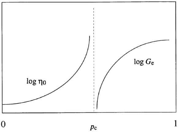
solution, c) strong gel and d) weak gel [125]. ... 29 Figure 2.17 Schematic of the divergence of zero-shear viscosity, η0, and equilibrium modulus, Ge.
The extent of crosslinking is marked by p [126]. ... 31 Figure 4.1 Zeta potential of GB, XG and their mixtures. ■: GB, ●: GB:XG = 5:1, ▲: XG. ... 41 Figure 4.2 a) Effects of pH (3.5-7.0) and NaCl concentration (0-300 mM) on the visual aspect of
GB/XG mixtures (GB:XG = 5:1, total concentration = 1.2% w/v); b) close-up view of mixtures without NaCl at pHs 5.0-7.0. ... 43 Figure 4.3 a) Storage modulus (G’) and b) complex viscosity (|η*|) as functions of frequency at
20oC for the mixtures (GB:XG=5:1, total concentration = 1.2 %) at various pHs (pH=5.0-10.0). XG at a concentration of 0.2 % (w/v) is shown for comparison purposes. No salt added. ... 45 Figure 4.4 Comparisons of storage modulus (G’) and complex viscosity (|η*|) at different pHs for
the mixtures (GB:XG = 5:1, total concentration = 1.2 % w/v) and XG solutions (c = 0.2-0.8 % w/v, pH = 6.0) at = 1 rad/s. No salt added. ... 46 Figure 4.5 Comparison of storage modulus (G’) of the mixtures (GB:XG = 5:1, total concentration
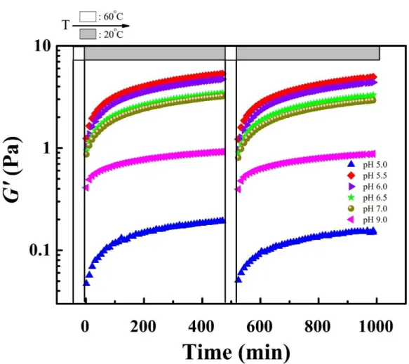
= 1.2 % w/v) at different pHs and salt concentrations (0 - 100 mM) at a = 1 rad/s. ... 47 Figure 4.6 Evolution of G’ of the mixtures (GB:XG=5:1, total concentration = 1.2 % w/v) at
different pHs (5.0-9.0) as a function of time. The samples were first heated up to 60oC, then cooled down to 20oC, and the process was repeated a second time. ... 48
Figure 4.7 CLSM images of mixtures (GB:XG = 5:1, total concentration = 1.2 % w/v) at different pHs, at 0 h and 24 h after solution preparation. GB was stained with Nile Blue A (red). Image size: 210 μm x 210 μm. No salt added. ... 52
Figure 4.9 CLSM images of mixtures (GB:XG = 5:1, total concentration = 1.2 % w/v) at different pHs. XG is labelled with DTAF (green). Image size: 210 μm x 210 μm. No salt added. The pictures were taken 24 h after solution preparation. ... 55 Figure 4.10 Effect of salt addition on the microstructure of GB/XG mixture (GB:XG = 5:1, total
concentration = 1.2 % w/v) at pH = 5.5, salt concentration = 50 mM. Image size: 210 μm x 210 μm. The pictures were taken after 24 h. Left: XG stained with DTAF; right: GB stained with Nile Blue A. ... 55 Figure 5.1 Zeta potential values of the GB (a) and XG (b) grades used in this work. ... 70 Figure 5.2 a) Effect of L-GB concentration (cGB = 0.2-2.0 % w/v) on the visual aspect of
L-GB/R-XG aqueous mixtures, at pH 5.5; b) effects of pH (4.0-7.0) and H-GB concentration (cGB =
0.2–1.6 % w/v) on the visual aspect of H-GB/R-XG mixtures, H-GB/R-XG ratio = 1-8; c) effects of pH (4.0-7.0) and XG molecular weight on the visual aspect of L-GB/XG mixtures (L-GB:XG ratio = 6, cXG = 0.2 % w/v). The photos were taken after overnight storage. ... 73
Figure 5.3 a) Evolution of G’ as a function of time for the L-GB/R-XG mixtures at ratios (1-10), at pH 5.5; b) G’ and G” after 8 hrs, as a function of L-GB/R-XG ratio. cXG = 0.2 % w/v, ω =
1 rad/s. ... 75 Figure 5.4 G’ as a function of time, for the mixtures of L-GB and Low-XG, Med-XG and High-XG respectively, at ratio 6 and pH 5.5. High-XG concentration = 0.2 % w/v, ω = 1 rad/s. ... 76 Figure 5.5 Comparison of G8h’ of H-GB solution with and without R-XG after 8 hrs in the
rheometer at 20oC, ω = 1 rad/s, cXG = 0.2 % w/v. The insert shows the ratio of the G8h’ of
H-GB/R-XG mixtures over the sum of the G8h’ of neat H-GB and R-XG at concentrations in the
corresponding mixtures, as a function of H-GB concentration. ... 77 Figure 5.6 Microstructures of L-GB (red) and R-XG (green) domains in the mixtures at different
ratios (1, 2 6 and 10) and merge of the two imaging, at pH 5.5. The images were taken after storage for 24 hrs. Image size: 210 μm x 210 μm. ... 79
Figure 5.7 Microstructure of L-GB (red) when mixed with Low-XG, Med-XG and High-XG, respectively, at different ratios (2, 6, and 8) and pH 5.5. The images were taken after storage for 24 hrs at room temperature. Image size: 210 μm x 210 μm. ... 80 Figure 5.8 a) Average size of biopolymer-poor (BP-poor) domains in L-GB/R-XG and H-GB/R-XG mixtures, as a function of GB/R-H-GB/R-XG ratio; and b) average size of biopolymer-poor domains in GB/Low-XG, GB/Med-XG and GB/High-XG mixtures, as a function of L-GB/XG ratio. ... 82 Figure 5.9 Micro-DSC heating curves, shifted vertically for clarity. Scanning rate = 1 oC/min. .. 83 Figure 5.10 Proposed gelation mechanism in GB/XG mixtures, based on their interactions and
molecular conformations. ... 85 Figure 5.11 Evolution of G’ during heating of three systems: (◄) L-GB = 4.0 % w/v, (▲) L-GB/R-XG = 6, total concentration = 1.4 % w/v and (■) R-L-GB/R-XG 0.2 % w/v. Heating rate: 0.2 oC/min
... 87 Figure 6.1 Zeta potential values of the gelatins (a), XG (b) and CHI (c) used in this work. ... 108 Figure 6.2 Effects of pH and gelatin concentration on the visual aspect of different mixtures: a) L-GA/XG, b) H-GA/CHI, c) L-GA/CHI and d) H-GB/CHI. The photos were taken after overnight storage. ... 110 Figure 6.3 Evolution of G’ as a function of time for a) the L-GA/ XG mixtures at ratio = 3 and
different pHs, b) the L-GA/ XG mixtures at different ratios and pH 6.0, cXG = 0.2 %, c) the
H-GB/CHI mixtures at ratio = 2.5 and different pHs, d) the H-H-GB/CHI mixtures at different ratios and pH 5.0, cCHI= 0.4 %, e) the L-GA/CHI mixtures at ratio= 3 and different pHs, and
f) the L-GA/XG mixtures at different ratios and pH 6.0, cCHI = 0.4 % ... 112
Figure 6.4 The ratio of G’ of a) H-GB/CHI and b) L-GA/CHI mixtures over the sum of the G’ of neat compositions at concentrations in the corresponding mixtures, as a function of gelatin concentration, 8 h after solution preparation (cCHI = 0.4 % w/v). ... 114
Figure 6.5 Microstructures of L-GA/XG (Panel A, ratio 3, 120 μm x 120 μm), H-GB/CHI (Panel B, ratio 4, 105 μm x 105 μm) and L-GA/CHI (Panel C, ratio 4, 105 μm x 105 μm) mixtures.
Figure 6.7 Gelation mechanism in gelatin/polysaccharide mixtures, based on their interactions and molecular conformations. XG molecules undergo a disorder-to-order transition and CHI molecules become less flexible upon cooling in step b. ... 120 Figure 6.8 Time-resolved rheological and turbidity measurements at different temperatures, a) and
c) H-GB/CHI, ratio = 4, pH 5.0; b) and d) L-GB/XG, ratio = 6, pH 5.5. Turbidity data were obtained at 600 nm. ... 121 Figure 7.1. pH of a gelatin B solution (1.0 % w/v) as a function of time under different acidification
conditions: acetic acid vapor (solid symbols, wt% in water) and GDL addition (open symbols). ... 128 Figure 7.2. State of the mixtures in terms of gelation (upside down vials) and syneresis (tilted vials)
after 24 h: a) initial pH = 5.5 and prepared (i) by exposing to vapor of 80 wt% acetic acid; (ii) with 0.1 wt% GDL; (iii) with 0.5 wt% GDL; (iv) gel destabilization after heating; b) initial pH = 10.0 prepared (i) by exposing to vapor of 80 wt% acetic acid and (ii) with GDL addition; c) effects of (i) XG and (ii) GB concentration. ... 129 Figure 7.3. Microstructures of GB/XG (cXG = 0.2 wt%) mixed gels prepared from an initial pH of
5.5 by VIPS using 80 wt% acetic acid (Panel A), and by GDL addition (Panel B); Panel C: mixed gels prepared from an initial pH of 10.0 by VIPS using 80 wt% acetic acid vapor. GB appears in red and XG in green with colocalized networks appearing yellow. The images were taken after 24 h storage. ... 132 Figure 7.4. Force at break in compression of mixed gels at different GB/XG ratios, prepared by
LIST OF SYMBOLS AND ABBREVIATIONS
GA Gelatin type A
GB Gelatin type B
XG Xanthan gum
CHI Chitosan
CLSM Confocal laser scanning microscope DDA Degree of deacetylation
5-DTAF 5-(4,6-Dichlorotriazinyl) Aminofluorescein pI Isoelectric point
SAOS Small amplitude oscillatory shear WPI Whey protein isolate
LVE Linear viscoelastic regime FITC Fluorescein isothiocyanate VIPS Vapor induced phase separation GDL Glucono delta-lactone
1.1 Background and problem identification
Food formulations are complicated mixtures since they usually contain water, proteins, polysaccharides, fats and other minor components. Proteins and polysaccharides are two essential ingredients in food products, and perform complementary nutritional, structural and textural functions [1]. The interactions between proteins and polysaccharides greatly affect the rheological behavior of the final product [2], and their importance in food formulations has been emphasized by Tolstoguzov [3]. In addition, proteins and polysaccharides are also commonly used in the cosmetic, pharmaceutical and biomedical industries.
The interactions between proteins and polysaccharides were first reported at the turn of the 19th century by the observation of the incompatibility between gelatin and starch, and since then they have been extensively investigated. Two general cases occur when mixing proteins and polysaccharides in aqueous solution, depending on the pH and ionic strength: segregative phase separation (thermodynamic incompatibility) and associative phase separation (thermodynamic compatibility) [4,5]. Further details on protein/polysaccharide interactions are presented in section §2.3. Protein/polysaccharide interactions have gained much attention since they can result in the enhancement of functional properties (stability, interfacial and gelation properties, encapsulation) as compared to those of the individual components.
Polysaccharides can enhance the stability of proteins via complexation, which is of great importance in protein beverages [6]. For example, the ideal pH for whey protein beverages (e.g. sports drinks) is at pH 4-6, where astringency and off-flavors can be avoided [7,8]. However, the stability of whey protein is a concern since the isoelectric point of whey protein is located in the pH range mentioned above, which may result in protein precipitation, microphase separation or gelation after heating [9,10]. The problem can be solved by adding pectin to form whey protein/pectin soluble complexes having a narrower size distribution, reduced hydrodynamic volume and greater magnitude of surface charge [11]. This successfully overcomes the limitation mentioned above and allows the manufacture of high whey protein beverages at pH 4.
Protein/polysaccharide complexes can also be used as emulsion or foam stabilizers. As it is well known, the mechanical strength of the interfacial layer, the electrostatic (repulsion between the
emulsion droplets) and the steric (barrier of thick interfacial layer) effects are the most important factors contributing to the stability of emulsions [12]. There are two ways to prepare emulsions using proteins and polysaccharides: a) premix proteins and polysaccharides under appropriate conditions to form complexes, followed by emulsion processing with the formed complexes [13,14]; b) use a layer-by-layer method by adding proteins and polysaccharides sequentially during emulsion preparation [15,16]. Both techniques can result in a thicker adsorption layer. For example, the interfacial elastic modulus is higher for an interfacial film formed by β-lactoglobulin/acacia gum electrically neutral complexes at pH 4.2 compared to the protein alone. In addition, complexation increases the magnitude of the zeta potential, which enhances the stability of emulsions. Finally, the presence of polysaccharides provides steric stabilization and increases the viscosity of the continuous phase, which reduce emulsion droplets movements and collisions. Protein/polysaccharide mixed gels are receiving more and more interest because they can be formed without heat, enzyme or crosslinking agent and at extremely low concentrations. As a result, they are of high interest for the development of novel thickeners and gelling agents [4,17] for the protection of bioactive molecules, when used as encapsulation and delivery systems [4,5]. Therefore, understanding the underlying principles of how proteins and polysaccharides interact in solution with each other is a prerequisite to design and prepare systems with the desired properties and functionalities.
Protein/polysaccharide mixed gel formation depends on the nature and characteristics of the biopolymers. For both proteins [18] and polysaccharides [19], a higher biopolymer concentration is needed to form a gel when the molecular weight and charge density are lower. Electrostatic forces are the dominant interactions between proteins and polysaccharides in solution, but other interactions such as hydrogen bonding and hydrophobic interactions can also be involved [6,20]. Proteins and polysaccharides can both repel and attract each other even when they carry the same net charge due to the amphiprotic properties of proteins [4,21,22]. Electrostatic forces are affected by the protein/polysaccharide ratio, pH, ionic strength and biopolymer charge density [4,20]. The gelation properties of protein/polysaccharide electrostatic hydrogels are the result of a delicate balance between repulsive and attractive interactions [4,23].
The gelation properties of different protein/polysaccharide mixtures have been investigated extensively, as summarized later in Table 2.7. However, no related studies have been found for the
lactoglubulin/XG mixtures [24]. However, it has several limitations: a) it neglects the conformation of xanthan gum when complexing with β-lactoglubulin, which can also be a driving force for the gelation process; b) it is not ideal to explain the gelation systems at a constant pH due to the used acidifier (glucono delta-lactone, GDL), which changes pH gradually with time; c) it is not suitable to explain the gelation process of gelatin/polysaccharide mixtures due to the differences between globular and linear proteins.
In this project, we aim at understanding the interactions between gelatin and two polysaccharides (xanthan gum, an anionic polysaccharide and chitosan, a cationic polysaccharide), which still remain unknown. We also target to propose a general mechanism on a molecular level to explain the gelation behavior of gelatin/polysaccharide aqueous mixtures, in order to better control the mechanical properties according to needs and to design novel thickeners and/or gelling agents, encapsulation and delivery systems.
1.2 Organization of the thesis
This thesis is based on three articles that have been published by or submitted to scientific journals, and consists of the following chapters:
• Chapter 2 provides a literature review considering the related issues. • Chapter 3 describes the objectives and the coherence of the articles.
• Chapters 4, 5, 6 and 7 present the four articles describing the main achievements obtained in this study.
• Chapter 8 reports a general discussion about the main results.
LITERATURE REVIEW
2.1 Proteins
2.1.1 Protein structure
Proteins constitute one of the most important class of biopolymers, which are encountered in areas such as food, cosmetics, pharmaceutical, medicine, packaging, coatings, etc [25]. Proteins can be composed of up to 20 different amino acids (Table 2.1).
Table 2.1: The 20 amino acids composing proteins Acidic and polar
(positive) pI
Neutral and
non-polar pI
Aspartic acid (Asp) 2.77 Alanine (Ala) 6.00 Glutamic acid (Glu) 3.22 Glycine (Gly) 5.97 Basic and polar (negative) Isoleucine (Ise) 6.02 Arginine (Arg) 10.76 Leucine (Leu) 5.98 Histidine (His) 7.59 Methionine (Met) 5.74 Lysine (Lys) 9.74 Phenylalanine (Phe) 5.48 Neutral and polar Proline (Pro) 6.30 Asparagine (Asn) 5.41 Serine (Ser) 5.68 Cysteine (Cys) 5.07 Tryptophan (Try) 5.89 Glutamine (Gln) 5.65 Valine (Val) 5.96
Threonine (Thr) 5.60 Tyrosine (Tyr) 5.66
Each amino acid contains a primary amine and a carboxylic acid group with the general formula:
The R group (guanidinium of arginine, imidazole of histidine, carboxyl group of aspartic acid, etc.) differs for various amino acids and determines the polarity and charge. Amino acids are linked together through peptide bonds (amide bonds between -NH2 of one amino acid and -COOH of
another). Sequences with fewer than 50 amino acids are referred to as peptides, while longer sequences are termed as polypeptides or proteins. A protein can consist of one or more polypeptides.
its positive, negative or neutral charge, depending on the pH. The isoelectric point (pI) is defined as the pH at which a protein possesses no net charge. The amphiprotic charge properties of proteins are important for understanding protein/protein and protein/polysaccharide interactions.
Figure 2.1 The four different levels of protein structure [26].
Protein structure has four different levels – primary, secondary, tertiary and quaternary (Figure 2.1). The primary structure is defined by the unique sequence, composition and distribution of amino acids in the polypeptide chain. Certain amino acids within the chain give rise to local secondary structures such as α-helix and β-sheets. After complex arrangement and/or folding, protein molecules adopt linear conformations or globular structures, which is referred to as tertiary structure. The tertiary structure is mainly stabilized by hydrogen bonding, disulfide bonding and salt bridges. Globular proteins are formed by folding polypeptide chains into a compact spherical shape with an irregular surface; hydrophobic amino acids tend to reside inside globular proteins with hydrophilic amino acids being outside; they are usually soluble in an aqueous environment and are involved in the transport processes or dynamic functions in cells (e.g. enzymes, bovine serum albumin). Linear proteins are characterized by their long parallel polypeptide chains. They function as structural elements, such as in the connective tissue of animals (e.g. collagen, keratin). The structure of protein can vary in response to changes in environmental conditions, for example, pH, temperature, salts and nature of solvent. Usually, protein molecules exist in the lowest
attainable free energy. The free energy may not be the global minimum, but it will be the lowest that the protein can achieve in a reasonable period of time [27]. Generally, proteins are made of multiple polypeptide chains, which are defined as protein subunits. The associations between multiple protein subunits are referred to as the quaternary structure.
2.1.2 Proteins functional properties
The unique sequence, composition and distribution of amino acids (primary structure) endow a given protein with different physicochemical characteristics, such as solubility, thermal stability, hydrophobicity and hydrophilicity, and further determine its functional properties, such as gelation, foaming and emulsifying properties. Some common proteins are listed in Table 2.2.
Table 2.2: Molecular characteristics of some food-grade proteins [28]
Name Source Structure pI
Bovine serum albumin Bovine blood/milk Globular 4.7 Gelatin Collagen Linear 7-9.4a; 4.8-5.5b
Ovalbumin Egg white Globular
(backbone)
4.5-4.7
Soy glycinin Soybean Globular 5
β-Lactoglobulin Whey protein Globular 4.8-5.1 a Type A gelatin; b Type B gelatin.
2.1.2.1 Gelation
Since a protein is usually stable in solution, denaturation/destabilization is a prerequisite for gelation [29]. The denaturation/destabilization techniques include heating, pressure [30,31], enzymatic crosslinking [32,33] and denaturants [34,35]. Denaturation also occurs at extremes of solution pH and ionic strength. The protein molecules under these conditions are unfolded and then aggregate into a network through non-covalent crosslinks such as electrostatic forces, hydrogen bonding, hydrophobic and covalent bonds (e.g. disulfide bonds) (Figure 2.2). The functional properties of protein hydrogels (gel strength, elasticity, water holding capacity, etc.) depend on the protein intrinsic characteristics, protein concentration, pH, salt concentration and type, as well as denaturation conditions (heating temperature, time, pressure, etc.) [4].
Figure 2.2 Schematic of unfolding and aggregation of a protein.
Globular proteins can form two categories of gels: particulate gels and fine-stranded gels. Particulate gels are obtained when heated at a pH close to the pI and/or at high ionic strength when electrostatic repulsion is low [36,37], whereas the fine-stranded gels are formed at pH far from the pI at low ionic strength when electrostatic repulsion is high [38] (Figure 2.3). The former gels have coarse, opaque and brittle structures; in contrast, the latter ones are usually transparent, more elastic, smoother and less sticky. The gels formed by globular proteins are usually thermally irreversible. In contrast, linear proteins are able to form thermally reversible gels, such as collagen and gelatin. More details are given in §2.1.3.
Figure 2.3 Schematic of stranded and particulate gels formed by globular proteins. The circles with or without thick red borders represent the native or denatured soy proteins,
respectively. Modification of sketch by Peng et al. [39]. 2.1.2.2 Interfacial properties
Proteins are commonly used as emulsifiers due to their surface-activity properties. During emulsification, protein molecules adsorb at the newly-created interfaces by diffusion and undergo a structural rearrangement in order to optimize their conformation to pack inside the adsorbed layer (Figure 2.4). Smaller protein molecules are expected to be more effective since they diffuse to the interface at a faster rate. Solvent conditions like pH or ionic strength also affect the adsorption and emulsion stability due to their effects on the net charges and conformations of protein molecules. It has been shown that increasing the exposed hydrophobic segments on a protein reduces its kinetic barrier [40] for adsorption while increasing the net charge increases the kinetic barrier [41].
Figure 2.4 Overview of the different steps in the formation of an adsorbed protein layer: A) diffusion and adsorption protein molecules to the interface; B) unfolding and
rearrangement of protein molecules at interface; C) film formation at interface.
Figure 2.5 Illustration of two alternative procedures for stabilization of oil droplets by protein–polysaccharide complexes. A) layer-by-layer procedure, with polysaccharide (ps)
added after emulsification with protein (pr); (B) premixing procedure, with both biopolymers present together during emulsification.
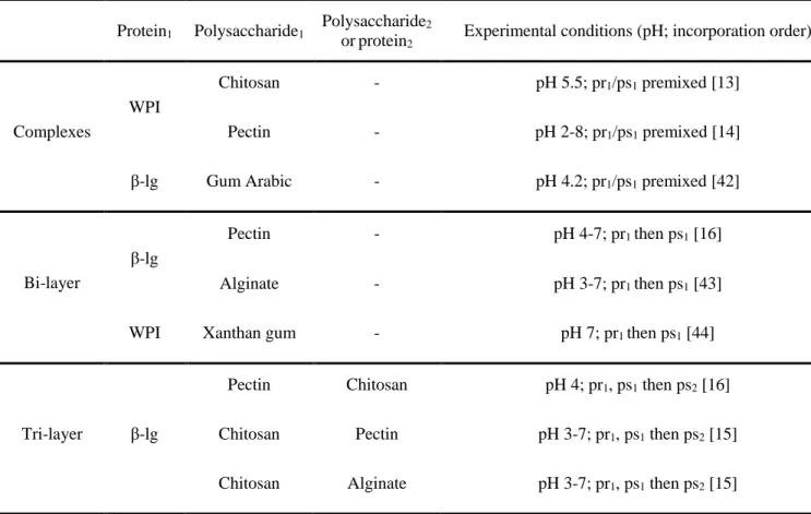
Table 2.3 Published studies on emulsifying properties of some protein/polysaccharide complexes
Protein1 Polysaccharide1 Polysaccharide2
or protein2 Experimental conditions (pH; incorporation order)
Complexes
WPI
Chitosan - pH 5.5; pr1/ps1 premixed [13]
Pectin - pH 2-8; pr1/ps1 premixed [14]
β-lg Gum Arabic - pH 4.2; pr1/ps1 premixed [42]
Bi-layer
β-lg
Pectin - pH 4-7; pr1 then ps1 [16]
Alginate - pH 3-7; pr1 then ps1 [43]
WPI Xanthan gum - pH 7; pr1 then ps1 [44]
Tri-layer β-lg
Pectin Chitosan pH 4; pr1, ps1 then ps2 [16]
Chitosan Pectin pH 3-7; pr1, ps1 then ps2 [15]
Chitosan Alginate pH 3-7; pr1, ps1 then ps2 [15]
Note: pr: protein; ps: polysaccharide; WPI: whey protein isolate; β-lg: β-lactoglobulin; 1 and 2 indicates the different types of proteins or polysaccharides.
Protein molecules associate with neighboring molecules after adsorbing at an interface, resulting in the formation of a viscoelastic interfacial layer. The interfacial layer stabilizes oil droplets against flocculation and coalescence through electrostatic repulsion (at a pH far from the pI) and steric effects (at a pH close to the pI). However, the adsorbed protein layer is too thin to provide steric stabilization in many cases. In order to overcome this, protein/polysaccharide complexes are of great interest since they combine the advantages of proteins (e.g. surface-activity, fast adsorption) and polysaccharide (e.g. steric repulsion). There are two alternative procedures to prepare emulsions using protein/polysaccharide complexes (Figure 2.5) and related work has been reported in Table 2.3. One approach is to prepare emulsions initially with a protein, followed by the addition of a polysaccharide which can interact with the adsorbed proteins, forming a bilayer [15,16] . Larger charge and thickness of the bilayer increase the stability of oil droplets against
2.1.3 Gelatin
Gelatin is one of the most well-known gelling proteins. It has numerous applications in many areas, such as confectionery, pharmaceutical/medical, and cosmetic products. The global gelatin market is expected to reach 4.08 billion USD by 2024, growing at a compound annual growth rate (CAGR) of 5.3% from 2016 to 2024, according to a report by Grand View Rearch, Inc [45]. It is a denatured protein derived from collagen by either acid (type A) or alkaline (type B) treatment [46]. The two types of gelatin differ in amino acid composition. Type A gelatins have similar amino acid composition to collagen, whereas type B gelatins do not contain glutamine and asparagine due to the alkaline processing [25](Table 2.4). Therefore, gelatin A has an identical isoelectric point as collagen in a pH range of 7-9.4, while gelatin B has a lower pI in a range of 4.8-5.4 [46]. Typically, the gelatin primary structure consists of repeating sequences of glycine-X-Y, where proline occurs in the X and Y positions and hydroproline exclusively in the Y position [46]. Glycine, as the smallest amino acid, allows the three peptides units to come closely together, whereas proline and hydroproline enhance rigidity due to their pyrrolidine rings and stabilize gelatin structure by hydrogen bonding [47].
Table 2.4: Amino acid composition of collagen and gelatin per 1000 residues [25]
* Type A gelatin: acid-pretreated pigskin gelatin.
**Type B gelatin: alkali-pretreated bone gelatin
Like collagen, gelatin also has a helix-to-coil transition temperature, which is located at about 15-40 oC depending on gelatin sources (e.g. fish gelatin: ~15 oC and bovine ~40 oC). Above this temperature, gelatin molecules exist in solution as random coils, and once cooling down below this temperature, the coils start to form a triple helices structure driven by hydrogen bonding (Figure 2.6), which can induce chain associations and a thermally reversible three-dimensional network [47,48].
Gelatin gel formation is proposed as a two-step process: the formation of locally ordered regions by partial random return (renaturation) of gelatin to collagen-like helices, followed by a continuous
upon heating. The gel state rarely reaches an equilibrium since the junctions are continuously reorganizing and new junctions are slowly formed with time [25,49]. Rheology and optical rotation measurements tests reveal that the gel strength is determined by the helix concentration [50,51]. Industrially, the Bloom index (also called Bloom value, Bloom number or Bloom strength), rather than the storage modulus, is used to characterize the gel strength. It is defined as the weight in grams that is required for a 12.7 mm diameter flat bottomed cylindrical plunger to depress the surface of a 6.67 % (w/w) gelatin gel (matured at 10 oC for 16-18 h) to a depth of 4 mm. A linear relationship can be found between the Bloom index and storage modulus.
2.2 Polysaccharides
Polysaccharides are another family of very important biopolymers widely used in different areas such as food, cosmetics, pharmaceutical, medicine, packaging, coatings, etc. [52]. Polysaccharides are long chains of carbohydrate polymers and are composed of at least 20 repeating monosaccharide residues linked by O-glycosidic linkages [52,53]. The number of monosaccharide units in a polysaccharide is referred to as the degree of polymerization (DP). Some common polysaccharides are listed in Table 2.5.
Table 2.5: Molecular characteristics food-grade polysaccharides [27]
Name Main structure type Major monomer Charge
Chitosan Linear
2-Amino-2-deoxy-β-D-glucose
Positive
Xanthan gum Branched Glucose, mannose Negative
Carrageenan Linear/helical Sulfated galactan Negative Gum Arabic Branched coil domains on
protein scaffold
Galactose Negative
Pectin Highly branched coil Glucuronate (backbone) Negative
Alginate Linear β-D-Mannuronic Acid,
Guluronic acid
Negative
Starch Linear/branched Glucose Neutral
Unlike proteins and nucleic acids, polysaccharides are both poly-disperse and poly-molecular. A particular polysaccharide possesses a range of degree of polymerizations (DPs) and molecular weights rather than a defined monomeric unit or a defined molecular weight. In addition, most polysaccharides are chemically heterogeneous [54]. They are poly-molecular in the sense that individual molecules within a polysaccharide type may differ from one to another with respect to monosaccharide sequence, composition, linkage type, branching frequency, etc. [54]
Table 2.6: Different structural levels of polysaccharides
Structural level Description
Primary structure Monosaccharide composition, sequence, linkage types Secondary structure Helix, ribbon and random coil
Tertiary structure Double helices
Quaternary structure Aggregates of secondary or tertiary structure
Analogous to proteins, the polysaccharide structure can also be defined on several different organization levels (Table 2.6). The primary structure of a polysaccharide refers to the monosaccharide composition, sequence, linkage types, and it determines the development of secondary, tertiary and quaternary structures. Glycosidic linkage type is believed to exert a greater influence on molecular conformation than the monosaccharide type [55], as illustrated by the comparison of three polysaccharides, cellulose, amylose and dextran, which are all linear chains of polyglucose, differing only in the nature of their glycosidic linkages. The secondary structure refers to the helix, ribbon and random coil conformations, and the arrangements such as double helices and aggregates of helices and ribbons can be regarded as tertiary or higher levels of structure [54,56]. Most polysaccharide secondary and tertiary structures are stabilized by intra- and intermolecular hydrogen bonding. Therefore, temperature influences the adoption of an ordered secondary and tertiary structure. A polysaccharide in an ordered conformation typically undergoes an order-to-disorder (helix-to-coil) transition upon increasing temperature, which disrupts the hydrogen bonds that stabilize the ordered conformation (Figure 2.7). When cooling down below
such as electrostatic forces, hydrogen bonding and hydrophobic interactions. The intermolecular associations in the quaternary structure level leads to the development of gels or crystalline structures.
Figure 2.7 Thermally reversible helix-to-coil (order-to-disorder) conformational transition of polysaccharides for a) single- and b) double-helical structures [54].
Polysaccharides can be neutral (starch, cellulose), negatively charged (xanthan gum, alginate, carrageenan) or positively charged (chitosan) depending on the ionic groups along the chain background and solution conditions. The electrical charge magnitude is related to the pHs. Anionic polysaccharides are neutral at pHs sufficiently below their pKa but negative above, whereas cationic polysaccharides are neutral at pHs sufficiently above their pKa but positive below [27]. The electrical charge of polysaccharides alters their solubilization in water and interactions with other ionic species, such as salt and other charged biopolymers. The alteration of electrical charges induces changes in the secondary and/or tertiary structures of polysaccharides, which in turn results in different functional properties.
2.2.1 Polysaccharide functional properties
The monosaccharide composition, sequence, linkage types, chain shapes and degree of polymerization dictate the molecular properties, such as molecular weight, degree of branching, flexibility and electrical charges, which in turn determine the functional properties, such as solubility, gelation, thickening and interfacial properties, water holding capacity and digestibility [27,52].
The polysaccharide gel formation depends on polysaccharide intrinsic characteristics, such as flexibility, charge density, molecular weight, monosaccharide composition, etc., and extrinsic factors, such as ionic strength, pH and temperature. Variations of extrinsic factors may lead to a disorder-order conformation change, and the intermolecular associations between ordered domains result in the formation of physical links and then subsequently a network [53]. The driving forces for crosslink formation vary between polysaccharides. For example, agar gelation is driven by hydrogen bonding, whereas for alginate and low methoxyl pectin, gelation is induced by ionic interactions [53]. Some polysaccharides cannot form gels due to conformational restriction or repulsive conditions. For example, λ-carrageenan is a non-gelling polysaccharide mainly due to the repulsive conditions caused by its high density of sulfate groups [57]. Mixing two or more polysaccharides is another way to produce gels. For example, thermally reversible gels are formed when mixing xanthan gum with galactomannan, carob gum or tara gum [58].
2.2.1 Xanthan gum
Xanthan gum (XG) (Mw ~ 2-6 x 10⁶ kDa) is a microbial polysaccharide produced by Xanthomonas
campestris by a distinct fermentation process. It has extensive applications, such as thickener,
stabilizer, cleaner, etc., in areas such as food, cosmetics, pharmaceutical industries and some water-based systems [59]. The XG market is expected to reach a value of 452.8 million USD by 2022, at a compound annual growth rate (CAGR) of 3.25% from 2016 [60]. XG has a cellulosic backbone with trisaccharide side chains (Figure 2.8). The trisaccharide side chain contains two mannose units separated by a glucuronic acid unit. Approximately half of the terminal mannose units are linked to a pyruvate group at C4- and C6-positions; whereas the non-terminal residue is usually acetylated at C6. The carboxyl groups on the side chains make XG an anionic polysaccharide.
X-Figure 2.8 Molecular structure of xanthan gum [57].
XG molecules in solution undergo a disorder-to-order transition (coil-to-helix) depending on salinity and temperature (Figure 2.9) [57,59,63,64]. In the absence of salt or at intermediate salt levels, XG molecules are partially ordered due to the electrostatic repulsion between the charged carboxylic groups on the side chains [64-66]. Adding salt causes a disorder-to-order transition due to charge screening effects, in which the backbone takes on a helical conformation and the charged side chains collapse down onto the backbone and stabilize the ordered conformation [64]. The ordered structures allow the molecules to be easily aligned and associate with each other. Heating a XG solution above a certain temperature results in the “melting” of the ordered structures; the structures return to their original state upon cooling [67,68]. This temperature is defined as the helix-to-coil transition temperature (usually at 50-55 oC). Salt addition pushes the helix-to-coil transition temperature to a much higher value by promoting helix formation [64].
Figure 2.9 Schematic model for the XG order–disorder transition [57].
XG is commonly regarded as a non-gelling polysaccharide [61,69]. It can however form a gel in the presence of trivalent ions or when mixed with other polysaccharides [57,70,71], or even proteins [24]. Weakly associated XG microgels (XG aggregates) can be formed by side-by-side associations between the ordered regions, which gives a tenuous network structure and endows XG dispersions with a “gel-like” behavior [57,68,72]. The presence of XG microgels accounts for many unusual features, such as a highly shearing-thinning behavior. Under shear, the weak links between the microgels break, leading to the deformation of microgels, chain disentanglement and alignment, whereas the network re-establishes itself upon removal of the applied shear [66]. This rheological feature enhances sensory qualities (mouth feel, flavor release) in food products, and makes XG easy to mix, pump and pour during processing and/or actual use despite its high molecular weight [59].
2.2.2 Chitosan
Chitin is the second most abundant natural biopolymer derived from exoskeletons of crustaceans and also from cell walls of fungi and insects [73,74]. Alkaline deacetylation of chitin results in chitosan (CHI). Chitin and CHI have excellent properties like biodegradability and biocompatibility, as well as low toxicity. As a result, they have been of great interest due to their various applications, such as chelator, drug release, texture controlling agent, dietary fiber, etc, in food technology, biomedical, and pharmaceutical industries [75-77]. The global CHI market is
use the degree of acetylation (DA) or deacetylation (DDA). Generally, the DDA of chitosan is ≥ 60 % [79]. The DDA and degree of polymerization (DP) are two important parameters dictating the physical, chemical and biological properties of CHI, such as the ability to chelate metal ions, its immunoadjuvant activity, the tensile strength of films, etc. [80] The DDA also influences the CHI solubility and solution properties, such as charge and viscosity [57,81]. The apparent charge density increases as DDA increases (i.e. the number of NH2 groups increases at the expense of
acetylamine groups). The protonated amine groups make CHI to expand in solution at low ionic strength, thus increasing excluded volume by electrostatic repulsions, whereas acetylated units increase the rigidity of chitosan [82]. Molecular weight is another parameter that affects the properties of chitosan, such as solubility and viscosity. The relation between viscosity of chitosan in solution and molecular weight can be described by the Mark-Houwink equation ([η] =KM α; where [η] is the intrinsic viscosity and M is the viscosity average of the molecular weight, K and α are constants. Decreasing the molecular weight of chitosan can thus decrease its viscosity and improve its solubility.
Figure 2.10 Chemical structure of chitin and chitosan [82].
CHI is composed of randomly distributed deacetylated units (β-(1-4)-linked glucosamine, or D-unit) and acetylated units (N-acetyl-D-glucosamine, or A-D-unit), as shown in Figure 2.10. It is a cationic polysaccharide with a pKa around 6.3-6.5, and that explains why it is only soluble in acidic
solutions. The amino groups of D-units allow chemical modification of chitosan by covalent attachment of various chemical groups, according to the targeted applications [81].
There are several ways to make CHI to form a gel. One way is to re-acetylate CHI with acetic anhydride in hydroalcoholic media, which then gives rise to a chitin gel through hydrophobic interactions [83]. Another way is to use β-glycerol phosphate (β-GP) combined with temperature [84-87]. The gel mechanism suggests that heat-stimulated proton transfer from CHI to β-GP reduces electrostatic repulsion, thus leading to CHI aggregation [88]. Other ionic crosslinker and complexing agents can also be used to gelify CHI, such as anionic molecules like phosphates and citrates [89] and metal ions like molybdate [90]. CHI gels can even be obtained without any additives by exposing CHI to well-defined conditions (e.g. using water-alcohol mixed solvent + heating) [91,92]. Generally, soft and easily degraded gels are formed from highly acetylated chitosan while more solid gels are obtained from highly deacetylated chitosan [93].
2.3 Protein-polysaccharide interactions
When mixing two biopolymers together in aqueous environment, two different types of interactions can occur, mainly depending on pH and ionic strength (Figure 2.11): segregative phase separation (thermodynamic incompatibility) and associative phase separation (thermodynamic compatibility) [4,5].
Segregative phase separation occurs when one or both biopolymers are uncharged or both biopolymers have similar charges. A one-phase solution is formed at sufficiently low biopolymer concentrations, whereas a two-phase solution containing protein-rich and polysaccharide-rich phases is formed once the biopolymer concentration exceeds a certain level [24,27]. In contrast, associative phase separation is induced by relatively strong attractions between the two biopolymers. It has been established that such interactions are primarily electrostatic in nature and will be inhibited at higher ionic concentration. The resulting two-phase system consists of solvent-rich and biopolymer-solvent-rich phases.
Figure 2.11 Possible interactions for proteins/polysaccharides mixtures and consequences in the formation of mixed gels. Based on the work by Ghosh et al. [17], and Stokes [53]. The biopolymer-rich phase may either form coacervates or precipitates (insoluble complexes) (Figure 2.12). There is some confusion in the scientific literature where in some instances complexes are referred to as “coacervates”, since the two are related to phase separation. In fact, coacervates are obtained by a liquid-liquid phase separation, whereas precipitates are formed by a solid-liquid phase separation. The two structures follow the same initial path, i.e. the two biopolymers form primary soluble complexes at a critical pH (pHc), followed by the formation of interpolymer complexes when the pH is decreased to a second critical pH (pHφ), and finally bulk phase separation [5]. However, it is still not clearly understood why some protein/polysaccharide systems result in precipitates whereas others lead to coacervates, but it may be related to the flexibility and charge density of the molecules [5]. Proteins or polysaccharides with low charge density and/or very flexible backbone tend to form coacervates, e.g. gelatin, acacia gum, dextran
![Table 2.4: Amino acid composition of collagen and gelatin per 1000 residues [25]](https://thumb-eu.123doks.com/thumbv2/123doknet/2351605.36402/36.918.194.740.150.731/table-amino-acid-composition-collagen-gelatin-residues.webp)


![Figure 2.16 The mechanical spectra of four principle categories: a) dilute solution, b) entangled solution, c) strong gel and d) weak gel [125]](https://thumb-eu.123doks.com/thumbv2/123doknet/2351605.36402/53.918.151.779.233.903/figure-mechanical-spectra-principle-categories-solution-entangled-solution.webp)