O
pen
A
rchive
T
oulouse
A
rchive
O
uverte
(OATAO)
OATAO is an open access repository that collects the work of some Toulouse
researchers and makes it freely available over the web where possible.
This is
an author'sversion published in:
https://oatao.univ-toulouse.fr/23110Official URL :
https://doi.org/10.1016/j.jse.2018.07.023
To cite this version :
Any correspondence concerning this service should be sent to the repository administrator:
Collin, Philippe and Thomazeau, Herve and Walch, Gilles and Gerber, Christian and Mansat,
Pierre and Favard, Luc and Colmar, Michel and Kempf, Jean François and Hervé, Anthony
and Betz, Michael Clinical and structural outcome twenty years after repair of isolated
supraspinatus tendon tears. (2018) Journal of Shoulder and Elbow Surgery, 28 (1). 196-202.
ISSN 1058-2746
OATAO
Clinical and structural outcome twenty years after
repair of isolated supraspinatus tendon tears
Philippe Collin, MD, PhD
*
, Herve Thomazeau, MD, PhD, Gilles Walch, MD,
Christian Gerber, MD, Pierre Mansat, MD, PhD, Luc Favard, MD, Michel Colmar, MD,
Jean François Kempf, MD, PhD, Anthony Hervé, MD, Michael Betz, MD
Institut Locomoteur de l'Ouest Saint Gregoire (Vivalto Sante), Saint Gregoire, France
Background: This study evaluated the clinical and structural outcome 20 years after repair of isolated supraspinatus tendon tears. We hypothesized that the results would deteriorate over time.
Materials and Methods: For this retrospective multicenter study, 137 patients were recalled for a clini
-cal and imaging assessment. Six patients (4.3%) had died from unrelated causes, 52 (38.0%) were lost to follow-up, and 13 (9.5%) had undergone reoperations. This left 66 patients for clinical evaluation. Ra
-diographs and magnetic resonance imaging were additionally performed for 45 patients, allowing assessment of osteoarthritis, tendon healing, fatty infiltration (Fl), and muscle atrophy.
Results: The Constant Score (CS) improved from 51.5 ± 14.1 points preoperatively to 71 points (P < .05) with a mean Subjective Shoulder Value (SSV) of 77 .2% ± 22%. Tendon discontinuity (Sugaya IV-V) was present in 19 of 45 patients (42 %), and there was advanced FI (Goutallier ill-IV) of the supraspinatus in
12 (27%) and of the infraspinatus muscle in 16 (35%). Supraspinatus atrophy was present in 12 patients
(28%), advanced arthritis in 6, and cuff tear arthropathy in 12 (30%). The CS and SSV were significantly
inferior for shoulders with Fl of stages
m
to IV (P < .05). The CS was lower in cuff tear arthropathy and correlated with infraspinatus Fl.Conclusions: At 20 years after surgical repair of isolated supraspinatus tears, the clinical outcome remains significantly above the preoperative state. Fl of the infraspinatus is the most influential factor on long
-term clinical outcome.
Level of evidence: Level IV; Case Series; Treatment Study
Keywords: Rotator cuff; tear; long follow-up; MRI; open repair; supraspinatus
Rotator cuff tears are among the most frequent shoulder pathologies causing pain and functional impairment. It is es-timated that rotator cuff tears are responsible for 4.5 million annual patient visits in the United States and for nearly 250,000 The Medical University of Strasbourg Ethical Committee approved this
clin-ical research (Number IDRCB 2013-AOJ788-37).
*Reprint requests: Philippe Collin, MD, PhD, Institut Locomoteur de
l'Ouest Saint Gregoire (Vivalto Saute), 7 blvd de la Boutière, Saint Gregoire 35740, France.
E-mail address: [email protected] (P. Collin).
operative repairs.6
•17 Numerous authors have reported con-siderable improvement in clinical and radiographie outcomes after repair of isolated supraspinatus tears using various sur-gical techniques, although most studies have limitations due
to small cohort sizes or short follow-up. To date, true long-term clinical and structural results after operative supraspinatus repair have not been reported.
The objective of this study was to report the clinical and structural outcome 20 years after repair of isolated supraspi-natus tears in a sizeable cohort. We hypothesized that 20 years
after surgery, the benefit of surgery would have substantial-ly decreased or disappeared due to a high tendon failure rate and advanced osteoarthritis, with a need for revision with pro-cedures such a reverse shoulder arthroplasty.
Materials and methods
Study design
We retrospectively studied the records of all patients who under-went surgical repair of isolated supraspinatus tears by 6 surgeons in 6 different centers in 1994. To be included in the study, the pa-tients had to be adults with full-thickness, isolated supraspinatus tears that were treated with an open operative repair. Patients were ex-cluded if they had partial-thickness tears, history of previous shoulder surgery, partial tendon repair, or concomitant lesions of other rotator cuff tendons. A total of 137 patients fulfilled these criteria and were recalled in 2014 for evaluation at a follow-up of 20 years. All pa-tients provided informed consent. For clinical assessment, we used the Constant Score (CS), the Subjective Shoulder Value (SSV), and the Simple Shoulder Test (SST).
We analyzed on standard x-ray images the rate of
glenohu-meral arthritis with the Samilson-Prieto classification.21We considered
stage 0, 1, and 2 as nonarthritic and 3 and 4 as arthritic. Cranial head migration was analyzed on true anteroposterior radiographs taken
in neutral rotation using the Hamada and Fukuda classification,9,10
as modified by Walch et al.26
The magnetic resonance imaging (MRI) protocol included T2-weighted fat-suppression sequences (nonproton density T2-weighted) in the oblique coronal, oblique sagittal, and transverse planes, in-cluding the entire scapula, to analyze tendon healing, and T1-weighted sequences in the transverse and sagittal planes to analyze fatty infiltration (FI) and muscle conditions. FI was analyzed
ac-cording to Goutallier/Fuchs.5,8We grouped stages 0/1 and 2 as
functional muscles and stages 3 and 4 as nonfunctional muscles. Su-praspinatus tendon healing was analyzed with the Sugaya
classification.23We defined types I/II and III as healed and types
IV and V as retorn tendons. We analyzed muscle atrophy with the
tangent sign described by Zanetti et al.27
Surgical technique
The repairs were performed with the patient in a beach chair po-sition using an open, anterosuperior approach with the use of
nonabsorbable transosseous sutures.18All repairs were watertight at
the end of the operation. An adjuvant anterior acromioplasty was performed in all shoulders.
Postoperative rehabilitation
After surgery, the arm was supported in a sling at 20° of abduction
for 5.6± 1.0 weeks (median, 6; range, 1-8 weeks). Passive-motion
exercises were initiated on the first postoperative day, and when pos-sible, hydrotherapy was initiated after skin healing. Active shoulder
motion was allowed after 8.2± 6.8 weeks (median, 6; range, 3-50
weeks). Patients were not allowed to perform any strengthening or strenuous work for 6 months after surgery. Low-demand sports and activities were allowed after 6 months.
Statistical analysis
Statistical analyses were performed using R 3.2.2 software (R Foun-dation for Statistical Computing, Vienna, Austria). Descriptive statistics were used to summarize the data. For non-Gaussian quan-titative data, intergroup differences were evaluated using Wilcoxon rank sum tests (Mann-Whitney U test). When 3 or more groups were compared, Kruskal-Wallis tests were used. Categoric data were
ana-lyzed using Pearsonχ2tests or Fisher exact tests. Stepwise descending
multivariate linear and logistic regression was performed. Model as-sumptions were checked before the analyses were performed. P
values of<.05 were considered statistically significant.
Results
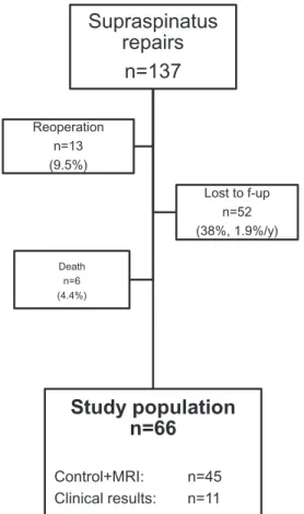
Of the original cohort, 6 patients (4.4%) had died from un-related causes, 52 (38.0%) were lost to follow-up, corresponding to a 1.9% loss per year (Fig. 1), and 13 (9.5%) had undergone reoperations (4 repeat repairs, 4 total shoul-der arthroplasties, 3 infections, 1 tenotomy of long biceps, 1 other) before the 20-year follow-up and were not in-cluded in the final functional analysis. This left a cohort of 66 patients (35 women [53%]), aged 52 years (range, 25-65 years), for the final analysis (Table I). Of this study cohort, 45 patients (41% women), aged 56.1± 7.6 years (range,
Supraspinatus
repairs
n=137
Study population
n=66
Control+MRI: n=45 Clinical results: n=11 Reoperation n=13 (9.5%) Lost to f-up n=52 (38%, 1.9%/y) Death n=6 (4.4%)Figure 1 Flowchart detailing inclusion and exclusion of pa-tients from the original cohort. F-up, follow-up; MRI, magnetic resonance imaging.
32-67 years), consented to standard radiographs and MRI at the 20-year follow-up.
In the 66 shoulders evaluated clinically, the CS im-proved from 52 points (range, 16-83 points) preoperatively to 71 points (range, 16-94 points) at 20 years, from 6 to 13 points for pain (15 points, freedom from pain; 0 , worst imag-inable pain), and from 6.5 to 9.2 points for strength (1 point representing 0.5 kg of strength at 90° of scapular plane ab-duction). The SSV at 20 years was 77% (range, 2%-100%), and the SST was 9.5 (range, 2-12;Table II). A postopera-tive complication had occurred in 7 patients (11%), consisting of 6 stiff shoulders and 1 infection, which were treated nonoperatively.
Analysis of 53 standard radiographs showed no arthritis in 18 patients (34%), arthritis stage 1 in 16 (30.2%), stage 2 in 7 (13.2%), stage 3 in 5 (9.4%), and stage 4 in 1 (1.9%). Cuff tear arthropathy (Hamada-Fukuda stage 4) was diag-nosed in 12 patients (30%).
Of the 45 shoulders evaluated using MRI, repair integri-ty was Sugaya integri-type I in 4 patients (8.8%), integri-type II in 8 (17.8%), type III in 14 (31.1%), type IV in 8 (17.8%), and type V in 11 (24.4%). Thus, repair integrity (Sugaya I, II, and III) was 58%, and repair failure was 42%. Integrity of the supraspi-natus (healed tendon) was associated with an albeit not statistically significant better total CS (75 vs. 68 points, P= .1). FI of the supra supinatus was stage 1/2 or 3 (functional muscles) in 33 shoulders (73.3%) and stage 3 or 4 (nonfunc-tional muscles) in 12 (26.7%). Supraspinatus atrophy, as judged with the tangent sign of Zanetti et al,27was absent in 21
pa-tients (48.8%) mild in 10 (23.2%), moderate in 4 (9.3%), and severe in 8 (18.7%).
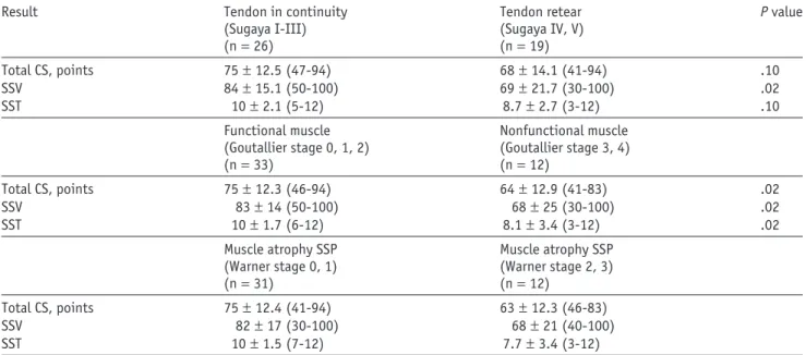
The influence of supraspinatus repair integrity, supraspi-natus muscle functionality, and trophicity on clinical results is summarized inTable III.
The association of rotator cuff FI (supraspinatus, infra-spinatus, and subscapularis) with CS, SSV, and supraspinatus tendon retear rate is reported inTable IV. Multivariate anal-ysis confirmed significant associations between clinical outcome and FI of any of the rotator cuff muscles. The most predictive parameter for postoperative CS and SSV was post-operative infraspinatus FI (Table IV).
Minor supraspinatus discontinuities (Sugaya type IV) were associated with less infraspinatus FI and better clinical results (CS) compared with major supraspinatus discontinuities (Sugaya type V) at 20 years of follow-up (Table V).
Multivariable regression analysis revealed no additional significant associations between other independent vari-ables of preoperative tear size, chronicity of symptoms, occupational injuries, level of activity, and smoking habits. Surgical procedures on the biceps tendon or acromioclavicu-lar joint resection were not associated with the quality of the clinical outcome (CS, SSV) or tendon healing.
Discussion
The principal findings of this study are, first, that less than 10% of the patients required further surgery, and only
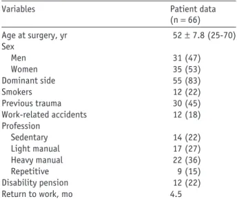
Table I Patient demographics
Variables Patient data
(n= 66) Age at surgery, yr 52± 7.8 (25-70) Sex Men 31 (47) Women 35 (53) Dominant side 55 (83) Smokers 12 (22) Previous trauma 30 (45) Work-related accidents 12 (18) Profession Sedentary 14 (22) Light manual 17 (27) Heavy manual 22 (36) Repetitive 9 (15) Disability pension 12 (22) Return to work, mo 4.5
Data are shown as mean± standard deviation (range), as number (%), or as indicated.
Table II Postoperative assessment
Outcome Preoperative Postoperative
(n= 66) (n= 66) Complications Total 7 (10.6) Infection 1 (1.5) Stiffness 6 (9.8) CS, points Pain 6.4± 3.1 (0-15) 13.1± 2.7 (5-15) Activity 10.5± 3.6 (4-20) 16.4± 4.2 (0-20) Mobility 27.2± 8.9 (0-40) 32.2± 7.4 (0-40) Strength 6.5± 4.9 (0-19) 9.2± 5.3 (0-20.8) Total CS 51.5± 14.1 (16-83) 71.3± 15.1 (16-94) SFA Sedentary 1 (2%) 6 (9.8%) Occasionally active 6 (11.8%) 16 (26.2) Active 15 (29.4%) 19 (31.1) Very active 29 (56.9%) 20 (32.8) SSV 77.2± 22.7 (2-100) SST 9.5± 2.6 (2-12) Sugaya classification I 4 (8.9) II 8 (17.8) III 14 (31.1) IV 8 (17.8) V 11 (24.4)
CS, Constant Score; SFA, Shoulder Function Assessment; SSV, Subjec-tive Shoulder Value; SST, Simple Shoulder Test.
Data are presented as mean± standard deviation (range) or as number (%).
approximately 3% required revision into reverse shoulder re-placement. Second the remaining patients have a CS that is a mean of 19 points higher than the preoperative score, a dif-ference that markedly exceeds the minimal clinically important difference established for this scoring system.12In addition,
two-thirds of the patients subjectively had an excellent or good shoulder (SSV≥ 80) at 20 years.
Structurally, 58% of all repaired tendons were still in con-tinuity, but only 8.9% of the tendons were “normal” (Sugaya type I). Therefore, the hypotheses that 20 years after surgery, the clinical benefit of supraspinatus tendon repair is lost and revision surgery is very frequently necessary must be refuted. Our analysis demonstrated that clinical outcome (CS, SSV) at 20 years was significantly associated with postoperative FI of the rotator cuff. The most predictive parameter for post-operative clinical outcome was postpost-operative infraspinatus FI (Table IV).
Minor supraspinatus discontinuities (Sugaya type IV) were associated with less infraspinatus FI and better clinical results (CS) than major supraspinatus discontinuities (Sugaya type V;Table V). This suggests that complete supraspinatus dis-continuities (Sugaya V) progressively extend toward posteriorly, leading to infraspinatus FI (Table V) and less fa-vorable clinical results (CS, SSV, SST) (Table III).
Outcomes after repair of isolated supraspinatus tears have been reported in 13 studies2,18-25(Table VI). The respective
cohort sizes range from 22 to 67 shoulders, with 1 study re-porting 129 shoulders.11 Follow-up periods are generally
between 1.4 and 8.6 years, with the exception of 1 study that extends up to 20 years.24The CS is the most frequently
re-ported clinical outcome (11 of 13 studies).2-7,14-16,18,20,25It tends
to be higher in studies with short follow-up2,4,7,11,15,16,25than
in studies with longer follow-up.20,24
The mean 20-year CS in the present series was 71 points, which is within the range reported in the literature. Consid-ering that postoperative CSs are often correlated with preoperative CSs, our 20-year results indicate a 50% improvement, which compares favorably with most pub-lished studies reporting improvement between 32% and Table III Influence of supraspinatus repair integrity, supraspinatus muscle functionality, and trophicity on clinical results
Result Tendon in continuity Tendon retear P value
(Sugaya I-III) (Sugaya IV, V)
(n= 26) (n= 19)
Total CS, points 75± 12.5 (47-94) 68± 14.1 (41-94) .10
SSV 84± 15.1 (50-100) 69± 21.7 (30-100) .02
SST 10± 2.1 (5-12) 8.7± 2.7 (3-12) .10
Functional muscle Nonfunctional muscle
(Goutallier stage 0, 1, 2) (Goutallier stage 3, 4)
(n= 33) (n= 12)
Total CS, points 75± 12.3 (46-94) 64± 12.9 (41-83) .02
SSV 83± 14 (50-100) 68± 25 (30-100) .02
SST 10± 1.7 (6-12) 8.1± 3.4 (3-12) .02
Muscle atrophy SSP Muscle atrophy SSP
(Warner stage 0, 1) (Warner stage 2, 3)
(n= 31) (n= 12)
Total CS, points 75± 12.4 (41-94) 63± 12.3 (46-83)
SSV 82± 17 (30-100) 68± 21 (40-100)
SST 10± 1.5 (7-12) 7.7± 3.4 (3-12)
CS, Constant Score; SSV, Subjective Shoulder Value; SST, Simple Shoulder Test. Data are presented as the mean± standard deviation (range).
Table IV Effect of fatty infiltration of supraspinatus, infra-spinatus, and subscapularis on the CS, SSV, and tendon retear
Variable CS↓ SSV↓ Tendon retear
FI postoperatively
Supraspinatus 0.02 0.02 NS
Infraspinatus 0.01 0 0
Subscapularis 0.02 0.03 NS
CS, Constant Score; SSV, Subjective Shoulder Value; FI, fatty infiltra-tion; NS, not significant.
Table V Impact of supraspinatus tendon discontinuity on in-fraspinatus fatty infiltration and Constant Score
Variable Sugaya type IV CS Sugaya type IV Sugaya type V CS Sugaya type V (n= 8) (n= 11) FI ISP 5 82 2 67 Grade 2 Grade 3 2 79 4 60 Grade 4 1 66 4 53
Table VI Literature review
Author Year Journal Approach Indications/technique Cohort Age FU Constant Score SSV SST
Pre-op Post-op Impr Pre-op Post-op Impr Pre-op Post-op Impr
(No.) (yr) (mo) (%) (%) (%)
Djahangiri et al3
2013 J Shoulder Elbow Surg
A/O Full-thickness SSP tear
O: acromioplasty, Mason-Allen, TOE fixation A: anterolateral acromioplasty, SR mattress
suture
58 69 78 49 78 59 42 83
Liem et al16
2007 JBJS Am A Isolated SSP tear, subacromial
decompression, Mason-Allen, single row
53 60.9 26.4 53.5 83.4 56
Boileau et al2
2005 JBJS Am A Chronic full-thickness SSP tear, tension-band 65 Healed tendon 57.8 Partially healed or unhealed 68 29 51.6 83.8 62 Fucentese et al4 2012 JBJS Am N Nonoperative treatment 24 52 42 75 74 Gerhardt et al7 2012 Am J Sports Med
A SSP tear, SR modified Mason-Allen vs. DR 40 SR 61.5 DR 61.2 SR 16.8 DR 23.4 SR 82.2 DR 77 SR 91 DR 92.9 Ikemoto et al11 2012 Rev Bras Ortop
A Small and medium-sized SSP tear, acromioplasty 129 55 39 McCormick et al18 2014 Int J Shoulder Surg
A SR, DR, TOE fixation, subacromial decompression 62 SR 62.5 DR 54.3 TOE 61.8 48 SR 72 DR 78 TOE 76 SR 10.1 DR 9.3 TOE 10.2 Meyer et al19 2012 Am J Sports Med
A/O Complete SSP tear, O: TOE fixation, Mason-Allen stitches 33 24 Nich et al20 2014 Orthop Traumatol Surg Res
O SSP full thickness tear combined with adjacent ISP tear (A), SSP and adjacent SSC tear (B), SSP and adjacent ISP and SSC tear (C), TOE fixation
22 58 75 A 56.5 B 62 C 55 A 72 B 76 C 84 A 27% B 22% C 53% Vastamäki et al24 2013 Clin Orthop Relat Res O SSP (53%), SSP+ ISP (36%), SSP+ ISP + SSC (6%), SSP+ SSC (4%), (n = 12), reconstruction using free tendon grafts (n= 52), acromioplasty or acromial osteotomy 67 52 240 Intact cuff 71 Cuff retear 58 Intact cuff 9.1 Cuff retear 7.6 Voigt et al25 2010 Am J Sports Med
A SSP tear, suture-bridging technique 51 62 24 58 88 51 12
Kukkonen et al14
2015 J Bone Joint Surg Am
A/N SSP tear,
group 1 physiotherapy only
group 2 acromioplasty and physiotherapy, all operations arthroscopically, subacromial débridement
group 3 rotator cuff repair, acromioplasty and physiotherapy, SR technique in smaller tears, DR technique in larger tears, subacromial débridement, acromioplasty 180 Group 1 64 Group 2 65 Group 3 65 24 Group 1 57.8 Group 2 59.6 Group 3 58 Group 1 76.2 Group 2 80.1 Group 3 80.6 Group 1 31% Group 2 34% Group 3 39% Liem et al15
2007 Arthroscopy A/O SSP, A: SR technique, modified Mason-Allen stitches O: Mini-open 38 A 61.9 O 62.1 A 25 O 17.6 A 53.8 O 53.5 A 83.9 O 83.7 A 56% O 56%
62%.2,3,14-16,20,25This means that the primary benefit of surgery
remains over time, even at long follow-up. There is little con-sensus on the optimal strategy to manage isolated supraspinatus tears due to the lack of literature reporting long-term outcomes4,13,22and conflicting results in those studies with the
longest follow-up:
• Vastamäki et al24
reported outcomes of 67 patients at 20 years. The retear rate was 94%. The remaining 4 pa-tients had a partial supraspinatus tendon tear. FI was marked in the supraspinatus and infraspinatus tendons. Cuff integrity correlated with better clinical results. • Nich et al20
reported outcomes of 27 patients at 8.6 years. They analyzed 49 shoulders. At a minimum follow-up of 60 months, the age- and sex-adjusted relative CS im-proved from 67% to 95%. MRI analysis showed a retear rate of 12%. Retear did not negatively influence the func-tional result.
A major strength of this study is the availability of clin-ical and imaging results, which were specifclin-ically collected and evaluated in a very standardized fashion 20 years after surgery for the purpose of this study. Our data therefore allow for analysis of tendon repair integrity, correlation between structural and clinical results, and information about longev-ity of the results.
The study has some limitations. First its retrospective design, without a control group, makes it a level 4 observa-tional study. Nonetheless, it provides previously unavailable information on a defined patient group with a well-defined pathology treated in a homogenous manner and also analyzed in a highly standardized fashion. It may be said that current arthroscopic techniques are different and that ante-rior acromioplasty is no longer a routine. Nonetheless at least the clinical results presented document that previous tech-niques were already valuable and a benchmark for the assessment of alleged improvements is provided. Moreover, Collin and al1
showed that that there were no differences between open and arthroscopic repair at 10 years of follow-up. Second, the loss of patients to follow-up is regrettable but at 20 years inevitable in the environment of the 6 centers. We consider that a yearly loss of follow-up of 1.9% over 20 years is reasonably good.
Third, we would certainly have preferred to obtain imaging studies in all patients but had to accept the refusal of the re-spective patients who declined imaging studies for very various reasons mostly because they felt it would not change any-thing for them.
Conclusion
At 20 years after open repair of an isolated supraspina-tus tear, two-thirds of the patients have an excellent or good subjective result, less than 10% are revised, and only 3%
need a reverse total shoulder arthroplasty. Significantly better results can be obtained if the repair remains intact and specifically if fatty infiltration of the infraspinatus muscle can be prevented.
Acknowledgments
The authors thank the members of the Société Française de Chirurgie Orthopédique et Traumatologique (SoFCOT), especially Arnaud Godeneche, Laurent Lafosse, Laurent Nové-Josserand, Pierre-Henri Flurin, and Pascal Boileau, for their participation in the 2015 SoFCOT symposium about rotator cuff repair at 20 years of follow-up.
Disclaimer
This study was financed by the Société Française de Chirurgie Orthopédique et Traumatologique (SoFCOT). Pierre Mansat is deputy editor for the Journal of
Shoul-der and Elbow Surgery and a paid consultant for
Wright-Tornier and Stryker. The other authors their immediate families, and any research foundation with which they are affiliated have not received any financial payments or other benefits from any commercial entity related to the subject of this article.
References
1. Agout C, Berhouet J, Bouju Y, Godenèche A, Collin P, Kempf JF, et al. Clinical and anatomic results of rotator cuff repair at 10 years depend on tear type. Knee Surg Sports Traumatol Arthrosc 2018;26:2490-7.
https://dx.doi.org/10.1007/s00167-018-4854-1
2. Boileau P, Brassart N, Watkinson DJ, Carles M, Hatzidakis AM, Krishnan SG. Arthroscopic repair of full-thickness tears of the supraspinatus: does the tendon really heal? J Bone Joint Surg Am 2005;87:1229-40.http://dx.doi.org/10.2106/JBJS.D.02035
3. Djahangiri A, Cozzolino A, Zanetti M, Helmy N, Rufibach K, Jost B, et al. Outcome of single-tendon rotator cuff repair in patients aged older than 65 years. J Shoulder Elbow Surg 2013;22:45-51.http://dx.doi.org/ 10.1016/j.jse.2012.03.012
4. Fucentese SF, von Roll AL, Pfirrmann CW, Gerber C, Jost B. Evolution of nonoperatively treated symptomatic isolated full-thickness supraspinatus tears. J Bone Joint Surg Am 2012;94:801-8. http:// dx.doi.org/10.2106/JBJS.I.01286
5. Fuchs B, Weishaupt D, Zanetti M, Hodler J, Gerber C. Fatty degeneration of the muscles of the rotator cuff: assessment by computed tomography versus magnetic resonance imaging. J Shoulder Elbow Surg 1999;8:599-605.
6. Genuario JW, Donegan RP, Hamman D, Bell JE, Boublik M, Schlegel T, et al. The cost-effectiveness of single-row compared with double-row arthroscopic rotator cuff repair. J Bone Joint Surg Am 2012;94:1369-77.
http://dx.doi.org/10.2106/JBJS.J.01876
7. Gerhardt C, Hug K, Pauly S, Marnitz T, Scheibel M. Arthroscopic single-row modified Mason-Allen repair versus double-row suture bridge reconstruction for supraspinatus tendon tears: a matched-pair analysis.
Am J Sports Med 2012;40:2777-85. http://dx.doi.org/10.1177/ 0363546512462123
8. Goutallier D, Postel JM, Bernageau J, Lavau L, Voisin MC. Fatty muscle degeneration in cuff ruptures. Pre- and postoperative evaluation by CT scan. Clin Orthop Relat Res 1994;(304):78-83.
9. Hamada K, Fukuda H, Mikasa M, Kobayashi Y. Roentgenographic findings in massive rotator cuff tears. A long-term observation. Clin Orthop Relat Res 1990;(254):92-6.
10. Hamada K, Yamanaka K, Uchiyama Y, Mikasa T, Mikasa M. A radiographic classification of massive rotator cuff tear arthritis. Clin Orthop Relat Res 2011;469:2452-60. http://dx.doi.org/10.1007/s11999-011-1896-9
11.Ikemoto RY, Murachovsky J, Nascimento LG, Bueno RS, Almeida LH, Strose E, et al. Arthroscopic repair of small and medium tears of the supraspinatus muscle tendon: evaluation of the clinical and functional outcomes after two years of follow-up. Rev Bras Ortop 2012;47:436-40.
http://dx.doi.org/10.1590/S0102-36162012000400005
12. Kukkonen J, Joukainen A, Itälä A, Äärimaa V. Operatively treated traumatic versus non-traumatic rotator cuff ruptures: a registry study. Ups J Med Sci 2013;118:29-34.http://dx.doi.org/10.3109/03009734 .2012.715597
13. Kukkonen J, Joukainen A, Lehtinen J, Mattila KT, Tuominen EK, Kauko T, et al. Treatment of non-traumatic rotator cuff tears: a randomised controlled trial with one-year clinical results. Bone Joint J
2014;96-B:75-81.http://dx.doi.org/10.1302/0301-620X.96B1.32168
14. Kukkonen J, Joukainen A, Lehtinen J, Mattila KT, Tuominen EK, Kauko T, et al. Treatment of nontraumatic rotator cuff tears: a randomized controlled trial with two years of clinical and imaging follow-up. J Bone Joint Surg Am 2015;97:1729-37.http://dx.doi.org/10.2106/JBJS.N .01051
15. Liem D, Bartl C, Lichtenberg S, Magosch P, Habermeyer P. Clinical outcome and tendon integrity of arthroscopic versus mini-open supraspinatus tendon repair: a magnetic resonance imaging-controlled matched-pair analysis. Arthroscopy 2007;23:514-21.http://dx.doi.org/ 10.1016/j.arthro.2006.12.028
16. Liem D, Lichtenberg S, Magosch P, Habermeyer P. Magnetic resonance imaging of arthroscopic supraspinatus tendon repair. J Bone Joint Surg Am 2007;89:1770-6.http://dx.doi.org/10.2106/JBJS.F.00749
17. Mather RC 3rd, Koenig L, Acevedo D, Dall TM, Gallo P, Romeo A, et al. The societal and economic value of rotator cuff repair. J Bone Joint Surg Am 2013;95:1993-2000.http://dx.doi.org/10.2106/JBJS.L.01495
18. McCormick F, Gupta A, Bruce B, Harris J, Abrams G, Wilson H, et al. Single-row, double-row, and transosseous equivalent techniques for isolated supraspinatus tendon tears with minimal atrophy: a retrospective comparative outcome and radiographic analysis at minimum 2-year follow up. Int J Shoulder Surg 2014;8:15-20.http://dx.doi.org/10.4103/ 0973-6042.131850
19. Meyer DC, Wieser K, Farshad M, Gerber C. Retraction of supraspinatus muscle and tendon as predictors of success of rotator cuff repair. Am J Sports Med 2012;40:2242-7. http://dx.doi.org/10.1177/ 0363546512457587
20. Nich C, Dhiaf N, Di Schino M, Augereau B. Does partial tear repair of adjacent tendons improve the outcome of supraspinatus tendon full-thickness tear reinsertion? Orthop Traumatol Surg Res 2014;100:721-6.http://dx.doi.org/10.1016/j.otsr.2014.07.014
21. Samilson RL, Prieto V. Dislocation arthropathy of the shoulder. J Bone Joint Surg Am 1983;65:456-60.
22. Strauss EJ, Salata MJ, Kercher J, Barker JU, McGill K, Bach BR Jr, et al. Multimedia article. The arthroscopic management of partial-thickness rotator cuff tears: a systematic review of the literature. Arthroscopy 2011;27:568-80.http://dx.doi.org/10.1016/j.arthro.2010 .09.019
23. Sugaya H, Maeda K, Matsuki K, Moriishi J. Functional and structural outcome after arthroscopic full-thickness rotator cuff repair: single-row versus dual-row fixation. Arthroscopy 2005;21:1307-16.http:// dx.doi.org/10.1016/j.arthro.2005.08.011
24. Vastamäki M, Lohman M, Borgmästars N. Rotator cuff integrity correlates with clinical and functional results at a minimum 16 years after open repair. Clin Orthop Relat Res 2013;471:554-61.http:// dx.doi.org/10.1007/s11999-012-2494-1
25. Voigt C, Bosse C, Vosshenrich R, Schulz AP, Lill H. Arthroscopic supraspinatus tendon repair with suture-bridging technique: functional outcome and magnetic resonance imaging. Am J Sports Med 2010;38:983-91.http://dx.doi.org/10.1177/0363546509359063
26. Walch G, Edwards TB, Boulahia A, Nové-Josserand L, Neyton L, Szabo I. Arthroscopic tenotomy of the long head of the biceps in the treatment of rotator cuff tears: clinical and radiographic results of 307 cases. J Shoulder Elbow Surg 2005;14:238-46.http://dx.doi.org/10.1016/ j.jse.2004.07.008
27. Zanetti M, Gerber C, Hodler J. Quantitative assessment of the muscles of the rotator cuff with magnetic resonance imaging. Invest Radiol 1998;33:163-70.