Geng Meng,aXinzheng Zhang,aPavel Plevka,a* Qian Yu,bPeter Tijssen,bMichael G. Rossmanna Department of Biological Sciences, Purdue University, West Lafayette, Indiana, USAa
; INRS-Institut Armand-Frappier, Université du Québec, Laval, Québec, Canadab
The 3.5-Å resolution X-ray crystal structure of mature cricket parvovirus (Acheta domesticus densovirus [AdDNV]) has been
determined. Structural comparisons show that vertebrate and invertebrate parvoviruses have evolved independently, although
there are common structural features among all parvovirus capsid proteins. It was shown that raising the temperature of the
AdDNV particles caused a loss of their genomes. The structure of these emptied particles was determined by cryo-electron
mi-croscopy to 5.5-Å resolution, and the capsid structure was found to be the same as that for the full, mature virus except for the
absence of the three ordered nucleotides observed in the crystal structure. The viral protein 1 (VP1) amino termini could be
ex-ternalized without significant damage to the capsid. In vitro, this externalization of the VP1 amino termini is accompanied by
the release of the viral genome.
P
arvoviruses are small (⬃250- to 300-Å-diameter), single-
stranded DNA (ssDNA), icosahedral (T
⫽1), nonenveloped
viruses whose genomes are approximately 5 kb long (
1
). The
Par-voviridae family has been subdivided into viruses that infect
ver-tebrates (Parvovirinae) and those that infect inverver-tebrates
(Denso-virinae) (
2
). Parvoviruses replicate in dividing cells such as in
tissues from insect larvae and fetuses. Densoviruses are highly
pathogenic, and those that use insect hosts usually kill 90% of the
larvae within a few days (
2
). Densoviruses pose a threat to
com-mercial invertebrates such as shrimp (
3
), silkworms (
4
), and
crick-ets (
5
,
6
). Some highly pathogenic densoviruses are potential
se-lective pesticides for vectors that transmit mosquito-borne
diseases (
7
). Parvovirinae generally have three types of proteins
(VP1, VP2, and VP3) in their capsids (
8
), whereas Densovirinae
generally have four types of proteins (VP1 to VP4) in their capsids
(
2
). In densoviruses there are 200 additional amino acids in VP1 at
the N terminus. These different proteins result from different
ini-tiation sites for translation of the capsid gene and from
posttrans-lational modification of their N termini (
8
). Generally, each of the
60 subunits within a capsid has the same amino acid sequence and
is structurally the same, except that the different proteins start at
different amino acids. The VP2s of some densoviruses are unique
among VP2s of parvoviruses since they are not completely
con-tained within corresponding VP1s (
Fig. 1A
).
Parvoviruses enter cells by dynamin-dependent
receptor-me-diated endocytosis and escape the endosome by the phospholipase
(PLA2) activity within the amino-terminal domain of VP1 (
9–13
).
Although there is often less than 5% amino acid identity among
the structural proteins of parvoviruses, the sequence of the PLA2
N-terminal domain of VP1 has more than 30% amino acid
iden-tity (
Fig. 1A
and
B
). The PLA2 domain is not exposed in
assem-bled, full parvoviruses such as minute virus of mice (MVM) (
13
)
and human parvovirus B19 (
14
), and it therefore has to be exposed
during endocytosis (
9
,
11
,
13–15
). However, the mechanism by
which the VP1 amino-terminal PLA2 domain is exposed has not
been elucidated in detail (
16
).
The structures of six autonomous vertebrate parvoviruses
(ca-nine parvovirus [CPV] [
17
], feline parvovirus [FPV]) [
18
],
por-cine parvovirus [PPV] [
19
], MVM [
20
], H-1 parvovirus [H-1PV]
[
21
], and human parvovirus B19 [
22
]) and three invertebrate
par-voviruses (Galleria mellonella densovirus [GmDNV] [
23
],
Bom-byx mori densovirus [BmDNV] [
24
], and Penaeus stylirostris
densovirus [PstDNV] [
25
]) have been determined (
Table 1
).
Fur-thermore, extensive studies have been made of the human
adeno-associated dependoviruses (
26
,
27
). The structures of these
parvo-viruses consist of 60 structurally equivalent capsid proteins
assembled with icosahedral symmetry. Each capsid protein has a
“jelly roll” fold, a motif that is common to many viruses, including
the nonenveloped RNA picornaviruses (
28
) and small RNA plant
viruses (
29
) as well as larger double-stranded DNA (dsDNA)
ad-enoviruses (
30
), the enveloped bacteriophage PRD1 (
31
), the
fun-gal virus Paramecium bursaria chlorella virus 1 (PBCV-1) (
32
),
vaccinia virus (
33
), and probably also mimivirus (
34
). The jelly
roll fold is a
-barrel consisting of two opposed antiparallel
-sheets with adjacent-strand BIDG and CHEF, where the strands
along the polypeptide chain are named A and B to H. The interior
of the barrel is exceedingly hydrophobic.
Parvoviruses have a channel along the 5-fold axes formed by
five symmetry-related DE loops (the “DE” loop is between the
-strands D and E). Residues lining the channel are mostly
hydro-phobic and guide the externalization of a conserved glycine-rich
sequence near the amino ends of the VPs (
35–37
). The loops
con-necting the
-strands of the jelly roll fold are usually exceptionally
large in parvoviruses compared with the loops in picornaviruses
(
28
,
38
) and form the exterior of the virus and intersubunit
con-tacts (
Fig. 1C
). These loops are more variable in sequence than the
core jelly roll structure.
Here, we describe the crystal structure of mature virions of
cricket parvovirus (Acheta domesticus densovirus [AdDNV]) at
3.5-Å resolution and the cryo-electron microscopic (cryoEM)
structure of the emptied virus at 5.5-Å resolution. We also report
on the externalization of the VP1 N-terminal region and
subse-quent genome release by an increase in temperature.
Received 3 July 2013 Accepted 3 September 2013 Published ahead of print 11 September 2013
Address correspondence to Michael G. Rossmann, [email protected].
* Present address: Pavel Plevka, CEITEC, Masaryk University, Brno, Czech Republic. Copyright © 2013, American Society for Microbiology. All Rights Reserved.
MATERIALS AND METHODS
Virus purification and preparation of the emptied virus particles. The
original virus was isolated from infected crickets (5). Further purification was achieved using CsCl equilibrium density gradient centrifugation. Of the two bands with different densities, the lower band contained the full particles and represented 99% of all the particles. The upper band con-tained empty particles assembled mainly from VP4. The two bands were separately transferred into Tris-buffered saline (TBS) (10 mM Tris-Cl, 100 mM NaCl, 1 mM CaCl, and 1 mM MgCl at pH 7.5) for further usage. Aliquots of the full virus particles were incubated at 26, 37, 45, 55, 65, 75, and 100°C for 1 h. The heat-treated emptied particles were frozen on holey carbon (Quantifoil) EM grids and checked by cryoEM. The numbers of full, empty, and broken particles were counted by eye (Fig. 2) and aver-aged over three holes on two different EM grids. Each hole had roughly 100 particles.
Determination of the crystal structure of the full virus particle.
Crystals of the full particles were obtained by hanging-drop vapor
diffu-sion in the presence of 20% polyethylene glycol (PEG) 400 and 100 mM MgCl2at 16°C. Further optimization of the crystallization conditions pro-duced crystals of up to 0.5 mm in length. Crystals were soaked for at least 20 min in the presence of 20% glycerol cryoprotectant prior to freezing.
X-ray diffraction data were collected at 100 K at the Advanced Photon Source (APS) beamline 23ID (Table 2). Diffraction data from about 20 crystals were indexed and merged, using the HKL2000 computer program (39) to generate the final 3.5-Å resolution data set. The space group was P42212 with a⫽ 412.67 Å and c ⫽ 278.80 Å. The Matthews coefficient was 3.64 Å3/Da, assuming half a virus particle per crystallographic asymmetric unit. Thus, the virus was located on a crystallographic 2-fold axis. A self-rotation function, calculated with the GLRF program (40) using 8- to 3.5-Å resolution data, gave the accurate orientation of the particle about the crystallographic 2-fold axis. This showed that one of the icosahedral 2-fold axes of the virus was roughly parallel to the 42crystallographic axis, with a 1.6° rotation away from being exactly parallel. As a consequence, the position of the particle along the crystallographic 2-fold axis could be
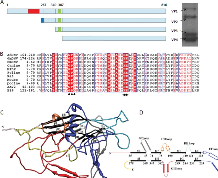
FIG 1 Structure of the AdDNV capsid protein. (A) Left, capsid protein organization. Red area, PLA2 domain; green area, glycine-rich region; blue area, extra
sequence at the N termini of VP2. Right, SDS-polyacrylamide gel of the AdDNV full particles. (B) Alignment of PLA2 sequences in VP1s of various parvoviruses, including AdDNV, GmDNV, BmDNV, CPV, mink parvovirus, FDV, rat parvovirus, mouse parvovirus, PPV, AAV2, and B19. The stars indicate the His-Asp catalytic site, and the triangles locate the Ca2⫹-binding site. (C) Three-dimensional structure of AdDNV capsid protein, showing the core jelly roll in black. The surface loops connecting the strands of the core jelly roll are colored as follows: BC loop, gray; CD loop, orange; DE loop, sky blue; EF loop, dark blue; and GH loop, red. (D) Diagrammatic representation of the capsid protein structure (heavy lines represent-strands) (color coding is the same as in panel C).
determined from the big Patterson peak generated by the large number of parallel equal-length vectors.
The structure was determined using the molecular replacement
method (41) with the structure of GmDNV (Protein Data Bank [PDB]
code1DNV) (23) as the initial phasing model to 15-Å resolution. The phases were then extended to 3.5-Å resolution in steps of one reciprocal lattice interval (1/c) at a time. Three cycles of 30-fold noncrystallographic symmetry (NCS) averaging and solvent flattening were performed for each extension step. The averaging and extension processes were per-formed using the program AVE in the Uppsala Software Factory (42) and
FFT, SFALL in CCP4 programs (43). The final overall correlation coeffi-cient between the observed structure amplitudes and the calculated struc-ture factors corresponding to the final averaged and solvent-flattened map was 0.866. The atomic model was built into the 3.5-Å resolution map
using COOT (44). The model coordinates were refined with the CNS
program (45) while applying NCS constraints and reasonable model re-straints, including group temperature factor refinement. No attempt was made to identify water molecules, as the data extended to only 3.5-Å resolution. The structures of the three ordered nucleotides bound to the inside surface of the capsid (see Results and Discussion) were included in
TABLE 1 Structural studies of autonomous parvoviruses
Virus Description of particle
Structural protein(s) in particles Resolution (Å) Icosahedral ordered genome structure (bp) PDB code (reference) Vertebrate parvoviruses
Canine parvovirus Full virus VP1, VP2, VP3 2.9 11 4DPV (17)
Empty particle VP1, VP2 3.0 None 2CAS (56)
Feline parvovirus Empty particle VP3 3.3 None 1FPV (18)
Porcine parvovirus Virus-like particle VP2 3.5 None 1K3V (19)
Human parvovirus B19
Virus-like particle VP2 3.5 None 1S58 (22)
Minute virus of mice Full virus VP1, VP2, VP3 3.5 11 1MVM (20)
Rat H-1 parvovirus Full virus VP1, VP2, VP3 2.7 10 4G0R (21)
Empty particle Unknown 3.2 None 4GBT
Invertebrate parvoviruses
GmDNV Full virus VP1, VP2, VP3, VP4 3.7 None 1DNV (23)
BmDNV Virus-like particle VP3 3.1 None 3P0S (24)
PstDNV Virus-like particle VP4 2.5 None 3N7X (25)
AdDNV Full virus VP1, VP2, VP3, VP4 3.5 3
Induced emptied particle VP1, VP2, VP3, VP4 5.5 None
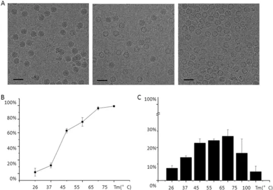
FIG 2 Heat treatment and PLA2 activity of the virus particles. (A) CryoEM micrographs showing the virus after incubation for 1 h at 26°C, 45°C, and 65°C,
resulting in only full particles (left), about an equal number of full and emptied particles (middle), and emptied particles (right), respectively. (B) Percentage of emptied particles after incubating full particles at different temperatures. (C) Phospholipase activity as a function of temperature with respect to the PLA2 activity of honey bee PLA2 at 26°C.
the final stages of refinement. After more than 5 cycles of refinement and model rebuilding, the R factor had dropped from 34% to 28.9%. In the presence of the 30-fold NCS redundancy, there will be no significant dif-ference between Rfreeand Rworking.
Detecting the externalized VP1 N termini and their phospholipase A2 activity. Full particles and the heat-treated emptied particles were
digested by trypsin at room temperature. About 30l of the particle suspension at a concentration of 5g/l was incubated with trypsin for 1 h at 26°C. The trypsin had a final concentration of 1g/l in the mixture. The samples were then checked for VP1 cleavage using a 15% SDS-poly-acrylamide gel (Fig. 3).
The PLA2 activities of the full particles and the heat-treated emptied particles were measured at 26°C by a colorimetric assay (sPLA2 assay kit; Cayman Chemical, Ann Arbor, MI), using the 1,2-dithio analog of dihep-tanoyl phosphatidylcholine (dihepdihep-tanoyl thio-PC) as the substrate for PLA2. The absorbance at 405 nm was determined every minute for 30 min. Measurements of the PLA2 activity were normalized relative to the activity of 1 ng bee venom PLA2. The activity of the PLA2 in 1.5g virus is equivalent to the PLA2 activity in 1 ng bee venom.
CryoEM and three-dimensional structural reconstruction of emp-tied particles. The optimal condition for obtaining the largest percentage
of emptied, unbroken particles was 55C° for 1 h (Fig. 2). Three microliters of the heat-treated emptied particles at a protein concentration of 5g/l
was applied to holey grids (Quantifoil) and blotted for 6 s in an FEI Mark 3 Vitrobot chamber at 90% humidity. The grids were then fast-frozen in liquid ethane. Cryo-electron microscopy (CryoEM) images were acquired on an FEI Titan Krios operated at 300 keV. Images were recorded with a 4k⫻ 4k charge-coupled device (CCD) detector. As a control, grids of untreated particles were prepared and viewed in the same way. The as-sumed magnification of 59,000 was calibrated with respect to a known specimen and was shown to correspond to a pixel separation of 1.51 Å in the image. The electron dose was⬃20 e/Å2, and the image was defocused
by between⬃1.6 and 2.6 m. About 150 cryoEM micrographs, each
showing roughly 100 particles, of the emptied particles were recorded. The defocus and the astigmatism of each micrograph were estimated with the EMAN1 fitctf program (46) and further confirmed with the program ctfit. Image processing and three-dimensional reconstruction were per-formed using the EMAN suite of programs (47). The final reconstruction was computed using⬃15,000 particles out of about 17,000 initial boxed images and was found to have 5.5-Å resolution based on the separate structure determinations of two randomly selected independent sets of images using the Fourier shell correlation threshold of 0.143 (Fig. 4) (48).
Sequence alignment of the PLA2 domain and structural compari-sons. The sequence of the AdDNV VP1 N-terminal PLA2 domain (GI
326392953) was aligned with the corresponding sequences of adeno-as-sociated virus 2 (AAV2) (GI 110645923), human parvovirus B19 (GI 169212578), CPV (GI 116646110), MVM (GI 332290), rat parvovirus (GI 410443463), mink parvovirus (GI 425696394), PPV (GI 46404508), GmDNV (GI 23334609), and BmDNV (GI 18025360) using Clustal X (49).
The crystal structure of AdDNV was compared with those of other invertebrate densoviruses, i.e., GmDNV (23) and BmDNV (24), as well as
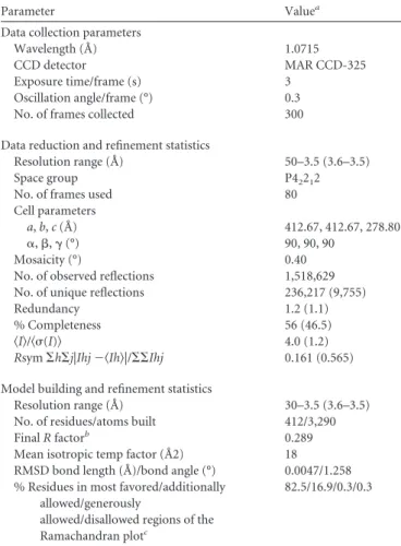
TABLE 2 X-ray data collection and structure refinement
Parameter Valuea
Data collection parameters
Wavelength (Å) 1.0715
CCD detector MAR CCD-325
Exposure time/frame (s) 3
Oscillation angle/frame (°) 0.3
No. of frames collected 300
Data reduction and refinement statistics
Resolution range (Å) 50–3.5 (3.6–3.5)
Space group P42212
No. of frames used 80
Cell parameters
a, b, c (Å) 412.67, 412.67, 278.80
␣, , ␥ (°) 90, 90, 90
Mosaicity (°) 0.40
No. of observed reflections 1,518,629
No. of unique reflections 236,217 (9,755)
Redundancy 1.2 (1.1)
% Completeness 56 (46.5)
具I典/具(I)典 4.0 (1.2)
Rsym⌺h⌺j|Ihj ⫺具Ih典|/⌺⌺Ihj 0.161 (0.565)
Model building and refinement statistics
Resolution range (Å) 30–3.5 (3.6–3.5)
No. of residues/atoms built 412/3,290
Final R factorb 0.289
Mean isotropic temp factor (Å2) 18
RMSD bond length (Å)/bond angle (°) 0.0047/1.258
% Residues in most favored/additionally allowed/generously
allowed/disallowed regions of the Ramachandran plotc
82.5/16.9/0.3/0.3
aValues in parentheses refer to the highest-resolution shell. b
No Rfreevalue was calculated, because the high NCS redundancy interrelates
reflections, causing the free reflections to be dependent on all other reflections. As a result, there would be little difference between Rworkingand Rfree.
cPercentage of a total of 360 nonglycine, nonproline residues as defined in the program PROCHECK.
FIG 3 SDS-PAGE of heat-treated AdDNV particles. Lanes: M, protein
mark-ers; H, heat-treated emptied AdDNV particles; E, non-heat-treated empty par-ticles among the purified mature AdDNV parpar-ticles; H⫹T, further trypsin di-gestion of the heat-treated emptied AdDNV particles.
FIG 4 Fourier shell correlation (FSC) based on the independent structure
determinations of two randomly selected equally sized sets of images, showing the resolution of the emptied AdDNV particle reconstruction to be⬃5.5 Å when the FSC is 0.143.
mammalian autonomous parvoviruses CPV (17), PPV (19), FPV (18),
MVM (20), and B19 (22) using the HOMOlogy program (50). These
structural comparisons do not include the disordered PLA2 domain, whose positions in the virus are random and therefore cannot be observed in the crystal structure.
Accession numbers. The atomic coordinates of the AdDNV crystal
structures have been deposited with the Protein Data Bank (www.pdb
.org) (PDB code 4MGU); the cryo-EM maps of the emptied AdDNV
particle have been deposited with the Electron Microscopy Data Bank (www.emdatabank.org) (EMDB code EMD-2401).
RESULTS AND DISCUSSION
Crystal structure of the full AdDNV particles. The structure of
AdDNV was determined to 3.5-Å resolution. The position of the
core jelly roll relative to the icosahedral symmetry axes was
essen-tially the same in AdDNV as in other known parvovirus structures
(
Table 3
and
4
).
The four structural proteins VP1 (88.1 kDa), VP2 (65.3 kDa),
VP3 (50.8 kDa), and VP4 (46.9 kDa) are in an approximate 1:11:
18:30 proportion in AdDNV full particles based on scanning the
gel with Kodak Image Station 2000R and analyzing with software
Kodak MI (
Fig. 1A
). The glycine-rich sequence is present in VP1,
VP2, and VP3, but is missing in VP4 (
Fig. 1A
). It may be
signifi-cant that, compared with vertebrate parvoviruses, there is
there-fore only one copy of the PLA2 structure per virion. The
polypep-tide chain of the capsid protein could be traced from residue 23 of
VP4 situated at the base of the 5-fold axis channel to residue 418 at
the carboxy terminus (
Fig. 1C
).
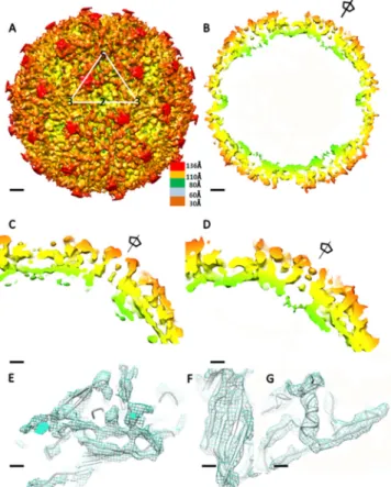
The electron density in this channel ( ⫽ 1.5) of AdDNV is
weak and discontinuous (
Fig. 5C
), which is similar to the density
in the GmDNV 5-fold channel. The glycine-rich motif in AdDNV
consists of about 17 residues, 8 of which are glycines, whereas in
GmDNV the same motif has 7 glycines and about 16 residues (
Fig.
6
). The difference of the sequence length may be partly related to
the structure of the channel in the different parvoviruses.
The low density in the 5-fold channel suggests that only several
of the 12 5-fold channels are occupied, resulting in externalization
of the VP amino termini. A similar lack of amino-terminal
exter-nalization was observed in GmDNV, the only other known
struc-ture of a mastruc-ture DNV. The strucstruc-tures of silkworm and shrimp
densoviruses (
Table 1
) were self-assembled from recombinantly
expressed VP3 and VP4 capsid proteins, respectively. Hence, these
structures are missing the glycine-rich sequence. As there is only
one VP1 per virion, some of the 5-fold channels must be occupied
by VP2 or VP3. However, in the vertebrate parvoviruses CPV (
17
)
TABLE 3 Sequence and structural comparisons of AdDNV capsidprotein with other autonomous parvovirus capsid proteins
Virus Sequence identity (%) RMSD (Å) between C␣ atoms No. of aligned C␣ atoms Total no. of C␣ atoms Canine parvovirus ⬍5 4.8 261 548 Feline parvovirus ⬍5 4.9 263 534 Porcine parvovirus ⬍5 4.9 258 542 Human B19 ⬍5 5.1 260 523 Minute virus of mice ⬍5 5.0 264 549 GmDNV 30.50 2.1 295 415 BmDNV ⬍5 3.7 331 412 PstDNV 8.30 4.1 224 299
TABLE 4 Superposition of the AdDNV jelly roll core (70 residues) on
other invertebrate parvovirus capsid proteins
Virus RMSD (Å) between C␣ atoms
Canine parvovirus 2.0
GmDNV 0.8
BmDNV 1.4
PstDNV 1.6
FIG 5 Structure of AdDNV emptied particles. (A) CryoEM reconstruction of
emptied particles. Surface features with a triangle showing the limits of one icosahedral asymmetric unit are shown. The scale bars represent 2 nm. (B) Center section of the cryoEM reconstruction. The scale bars represent 2 nm. (C) Enlargement of the 5-fold channel density in the X-ray electron density map. The scale bars represent 1 nm. (D) Enlargement of the 5-fold channel density in the cryoEM density map. The scale bars represent 1 nm. (E, F, and G) Fit of the X-ray structure polypeptide backbone into the cryoEM density for the-sheets of the jelly roll (scale bars represent 5 Å) (E), the BIDG -sheet (the scale bars represent 3 Å) (F), and the␣-helix located in the EF loop (the scale bars represent 3 Å) (G).
FIG 6 Sequence comparisons of the glycine-rich regions of AdDNV and other
autonomous parvovirus, including GmDNV (GI 23334609), BmDNV (GI 18025360), canine parvovirus (CPV) (GI 116646110), mink enteritis virus (MEV) (GI 425696394), feline parvovirus (FPV) (GI 333476), porcine parvo-virus (PPV), and human parvoparvo-virus (B19) (GI 169212578).
and MVM (
20
), all the 5-fold channels are always fully occupied
by the VP amino termini. Furthermore, there is a larger
propor-tion of VP1 subunits per virion in the vertebrate parvoviruses.
However, the externalized amino termini are disordered in both
the vertebrate and invertebrate parvovirus crystal structures.
Pairwise comparisons of the parvovirus structures were used to
determine the number of and root mean square deviation
(RMSD) between equivalent C␣ atoms (
Table 3
). The number of
inserted amino acids between
-strands was used to calculate a
phylogenetic tree using the program MEGA (
51
) (
Fig. 7
). This
shows a closer relationship among the insect densoviruses
(AdDNV, GmDNV, PstDNV, and BmDNV) than between these
viruses and vertebrate viruses. Although the capsid proteins of all
parvoviruses have common structural features, the capsid
pro-teins of vertebrate and invertebrate parvoviruses must have
evolved independently (
Fig. 7
).
The A
-strand is folded back to run antiparallel to -strand B
in the BIDG sheet in all known vertebrate parvovirus structures
(
Fig. 1D
). In previously determined densovirus structures,
includ-ing the AdDNV structure reported here,
-strand A is associated
with
-strand B in the neighboring, 2-fold related BIDG sheet.
Such an exchange of domains between 2-fold-related structures is
an example of “domain swapping” (
Fig. 1C
and
D
). However, in
AdDNV the A
-strand contains three proline residues (Pro24,
Pro26, and Pro28) and therefore diminishes the H bonding with
the B
-strand of the neighboring subunit.
The channel along the 5-fold icosahedral axes of parvoviruses
is formed by the DE loop and is between 16 Å and 18 Å in
diam-eter, measured from atom center to atom center. The
conforma-tion of the DE loop is variable among both vertebrate and
inver-tebrate parvoviruses (
8
). In AdDNV, as also in all other known
parvoviruses, there are several hydrophobic amino acids in the DE
loop (from 116 Ala to 133 Gln in AdDNV) that interact with the
glycine-rich region.
Emptied densovirus particles and phospholipase activity.
Most parvoviruses, including densoviruses, assemble in vivo both
as full infectious particles and as empty particles. However, for
AdDNV and presumably also for other densoviruses, the small
fraction of particles that are empty in a virus preparation consist of
only VP4 (
Fig. 3
) and are missing the glycine-rich sequence,
whereas the dominant infectious virus particles contain all four
types of subunits (VP1 to VP4) (
Fig. 1A
). Therefore, after heat
treatment, nearly all the emptied particles that have a full
comple-ment of all four VPs must have been full of genome, whereas
empty particles containing only VP4 must have been assembled as
empty particles. It had been shown that heating parvoviruses to
70°C generated PLA2 activity, suggesting exposure of the VP1 N
termini (
12
,
13
). However, it was not clear whether only the VP1 N
termini were exposed from intact particles or whether the particles
had disassembled. The loss of the genome associated with a
pre-sumably transient change in the capsid has some resemblance to
the infectious process in picornaviruses (
52
).
Here we used cryoEM to show that on heating of AdDNV for a
defined length of time, the number of emptied particles increased
with temperature (
Fig. 2A
and
B
). When the temperature was
increased beyond 65°C there was also an increase of broken
par-ticles. Concomitant with the increase of emptied particles, there
was also an increase of PLA2 activity (
Fig. 2C
). Above about 65°C,
the virions disintegrated and had reduced PLA2 activity. Unlike
the case for full, infectious AdDNV particles, the VP1 N termini
of the heat-treated emptied particles were sensitive to trypsin
di-gestion, whereas the capsids remained intact as determined with
TABLE 5 Results of a six-dimensional search on fitting of the AdDNV
capsid protein structure into the 5.5-Å cryoEM density by using the EMfit program (55)a
Sumfb Clash (%)c ⫺Den (%)d
40.0 1.7 4.8
24.0 14.6 15.6
21.1 19.4 20.9
a
Three possible fits were found, but the top fit is by far the best.
bSumf, mean density height averaged over all atoms, where the maximum density in the electron density map is set to 100.
cClash, percentage of atoms in the model that approach closer than 5 Å to icosahedral-related capsid protein molecules.
d⫺Den, percentage of atoms in density less than zero density.
cryoEM. This showed that the heat treatment causes
externaliza-tion of the N termini while leaving the capsid intact, with the PLA2
domain remaining a part of the particle.
Externalization of the N termini prior to endocytosis abolishes
infectivity in parvovirus (
53
), suggesting that the sequence of
events during infection is critical. This could explain why all
voviruses harbor the N-terminal part of VP1 within the virus
par-ticle until they are ready to breach the endosomal membrane.
PLA2 requires a Ca
2⫹concentration of greater than 1 mM (
9
).
Such Ca
2⫹concentrations are present in the endosome but not in
the cytoplasm (
54
), further narrowing the viral PLA2 activity to
the endosomal membrane.
The 5.5-Å pseudo-atomic-resolution cryoEM structure of
emptied AdDNV particles. An initial effort to crystallize
heat-treated emptied particles failed to produce crystals, probably
be-cause of the externalized VP1 N termini. However, it was possible
to obtain a 5.5-Å resolution cryoEM structure from
approxi-mately 15,000 images of heat-treated emptied particles (
Fig. 5A
and
B
). The structure of the crystallographically determined virus
could readily be superimposed onto the cryoEM map by aligning
the icosahedral symmetry axes (
Table 5
). The cryoEM map had to
be expanded by 5%, amounting to a radius increase of 6 Å, to
obtain the best fit. This change in size of the EM map is well inside
the error of determining the magnification of the electron
micro-scope. The quality of the cryoEM map was excellent, as indicated
by the resolution of the main chain in some places (
Fig. 5E
to
G
).
A 5.5-Å resolution difference map between the crystal
struc-ture electron density and the cryoEM map was calculated using
the EMfit program (
55
). This showed two regions of density
higher than three standard deviations of the background density,
associated with the inside surface of the protein shell. The first
region had an average height of about 6.5
and could be readily
interpreted in terms of three nucleotides (
Fig. 8A
). This structure
was an extended trinucleotide with the bases facing the inside
capsid surface (
Fig. 8B
), close to the icosahedral 3-fold axes with
the interaction with Tyr337 and Gln252. The second region had
an average height of 3.5
but was not easily interpreted in terms of
a standard nucleotide structure. Icosahedrally ordered genome
structure has been previously observed in canine parvovirus (
36
)
and minute virus of mice (
35
) but not in invertebrate densoviruses
(
Table 1
).
The density in the channel along the 5-fold axes in the cryoEM
map of the emptied particles was similar to that in the
crystallo-graphically determined map of the full infectious particle
calcu-lated to 5.5-Å resolution (
Fig. 5C
and
D
). Thus, the emptied
par-ticles still have the glycine-rich region occupying the channel
along the 5-fold axes in at least some of the 12 channels of each
particle. The externalization of the VP1 termini does not seem to
have caused much damage to the particles. In contrast to the case
for AdDNV, there is little density along the 5-fold axes of the
shrimp (PstDNV) and silkworm (BmDNV) densoviruses.
How-ever, these structures are of recombinant particles that contain
only VP3 or VP4, respectively, and are therefore missing the
gly-cine-rich region. These observations pose the intriguing question
of whether the PLA2 domain has to be refolded to be threaded
through the 5-fold pore or whether the pore opens with
restora-tion of the initial capsid structure after extrusion.
ACKNOWLEDGMENTS
We thank Agustin Avila-Sakar and Valorie Bowman for help with the cryo-electron microscopy. We are grateful to Sheryl Kelly for help in pre-paring the manuscript. We also thank the staff of beamline 23ID, GM/CA-CAT, at Advanced Photon Source, Argonne National Laboratory, for help with the data collection.
Use of the Advanced Photon Source was supported by the U.S. De-partment of Energy, Office of Science, Office of Basic Energy Sciences, under contract DE-AC02-06CH11357. The work was supported by an NIH grant award (AI11219) to M.G.R. The work was also supported by Purdue University funds for Structural Biology and the Electron Micro-scope Facility. P.T. was supported by a grant from the Natural Sciences and Engineering Research Council of Canada, and Q.Y. acknowledges tuition waivers at INRS-Institut Armand-Frappier and a scholarship from the People’s Republic of China.
G.M., X.Z., P.P, Q.Y., P.T, and M.G.R. designed research; G.M., X.Z., P.P., and Q.Y. performed research; G.M. analyzed data; Q.Y. and P.T. contributed reagents/analytic tools; and G.M., P.T., and M.G.R. wrote the paper.
We declare no conflict of interest.
REFERENCES
1. Berns K, Parrish CR. 2006. Parvoviridae, p 2437–2478. In Knipe DM, Howley PM (ed), Fields virology. Lippincott Williams & Wilkins, Phila-delphia, PA.
2. Bergoin M, Tijssen P. 2000. Molecular biology of Densovirinae. Contrib. Microbiol. 4:12–32.
3. Harvell CD, Kim K, Burkholder JM, Colwell RR, Epstein PR, Grimes
DJ, Hofmann EE, Lipp EK, Osterhaus AD, Overstreet RM, Porter JW, Smith GW, Vasta GR. 1999. Emerging marine diseases— climate links
and anthropogenic factors. Science 285:1505–1510.
4. Li Y, Zadori Z, Bando H, Dubuc R, Fediere G, Szelei J, Tijssen P. 2001. Genome organization of the densovirus from Bombyx mori (BmDNV-1) and enzyme activity of its capsid. J. Gen. Virol. 82:2821–2825.
5. Liu K, Li Y, Jousset FX, Zadori Z, Szelei J, Yu Q, Pham HT, Lepine F,
Bergoin M, Tijssen P. 2011. The Acheta domesticus densovirus, isolated
from the European house cricket, has evolved an expression strategy unique among parvoviruses. J. Virol. 85:10069 –10078.
6. Szelei J, Woodring J, Goettel MS, Duke G, Jousset FX, Liu KY, Zadori
Z, Li Y, Styer E, Boucias DG, Kleespies RG, Bergoin M, Tijssen P. 2011.
Susceptibility of North-American and European crickets to Acheta domes-ticus densovirus (AdDNV) and associated epizootics. J. Invertebr. Pathol.
106:394 –399.
7. Carlson J, Suchman E, Buchatsky L. 2006. Densoviruses for control and genetic manipulation of mosquitoes. Adv. Virus Res. 68:361–392. 8. Tijssen P. 1999. Molecular and structural basis of the evolution of
parvo-virus tropism. Acta Vet. Hung. 47:379 –394.
9. Canaan S, Zadori Z, Ghomashchi F, Bollinger J, Sadilek M, Moreau
ME, Tijssen P, Gelb MH. 2004. Interfacial enzymology of parvovirus
phospholipases A2. J. Biol. Chem. 279:14502–14508.
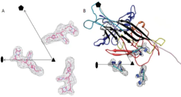
FIG 8 Structure of the three ordered ssDNA bases in AdDNV particles. (A)
Difference map between X-ray electron density and cryoEM density, with black lines showing the limits of one icosahedral asymmetric unit. Three bases of the ssDNA were built into the density. (B) Location of the three bases relative to the AdDNV capsid. The colors of the AdDNV capsid polypeptide are as defined forFig. 1C.
10. Girod A, Wobus CE, Zadori Z, Ried M, Leike K, Tijssen P,
Klein-schmidt JA, Hallek M. 2002. The VP1 capsid protein of adeno-associated
virus type 2 is carrying a phospholipase A2 domain required for virus infectivity. J. Gen. Virol. 83:973–978.
11. Zadori Z, Szelei J, Lacoste MC, Li Y, Gariepy S, Raymond P, Allaire M,
Nabi IR, Tijssen P. 2001. A viral phospholipase A2 is required for
parvo-virus infectivity. Dev. Cell 1:291–302.
12. Tijssen P, Zadori Z. April 2002. Viral phospholipase A2 enzymes, anti-viral agents and methods of use. WO patent 2,002,000,924.
13. Farr GA, Zhang LG, Tattersall P. 2005. Parvoviral virions deploy a capsid-tethered lipolytic enzyme to breach the endosomal membrane dur-ing cell entry. Proc. Natl. Acad. Sci. U. S. A. 102:17148 –17153. 14. Ros C, Gerber M, Kempf C. 2006. Conformational changes in the
VP1-unique region of native human parvovirus B19 lead to exposure of internal sequences that play a role in virus neutralization and infectivity. J. Virol.
80:12017–12024.
15. Mani B, Baltzer C, Valle N, Almendral JM, Kempf C, Ros C. 2006. Low pH-dependent endosomal processing of the incoming parvovirus minute virus of mice virion leads to externalization of the VP1 N-terminal se-quence (N-VP1), N-VP2 cleavage, and uncoating of the full-length ge-nome. J. Virol. 80:1015–1024.
16. Venkatakrishnan B, Yarbrough J, Domsic J, Bennett A, Bothner B,
Kozyreva OG, Samulski RJ, Muzyczka N, McKenna R, Agbandje-McKenna M. 2013. Structure and dynamics of adeno-associated virus
serotype 1 VP1-unique N-terminal domain and its role in capsid traffick-ing. J. Virol. 87:4974 – 4984.
17. Xie Q, Chapman MS. 1996. Canine parvovirus capsid structure, analyzed at 2.9 Å resolution. J. Mol. Biol. 264:497–520.
18. Agbandje M, McKenna R, Rossmann MG, Strassheim ML, Parrish CR. 1993. Structure determination of feline panleukopenia virus empty parti-cles. Proteins 16:155–171.
19. Simpson AA, Hebert B, Sullivan GM, Parrish CR, Zadori Z, Tijssen P,
Rossmann MG. 2002. The structure of porcine parvovirus: comparison
with related viruses. J. Mol. Biol. 315:1189 –1198.
20. Llamas-Saiz AL, Agbandje-McKenna M, Wikoff WR, Bratton J,
Tatter-sall P, Rossmann MG. 1997. Structure determination of minute virus of
mice. Acta Crystallogr. D Biol. Crystallogr. 53:93–102.
21. Halder S, Nam HJ, Govindasamy L, Vogel M, Dinsart C, Salome N,
McKenna R, Agbandje-McKenna M. 2013. Structural characterization of
H-1 parvovirus: comparison of infectious virions to empty capsids. J. Vi-rol. 87:5128 –5140.
22. Kaufmann B, Simpson AA, Rossmann MG. 2004. The structure of human parvovirus B19. Proc. Natl. Acad. Sci. U. S. A. 101:11628 –11633. 23. Simpson AA, Chipman PR, Baker TS, Tijssen P, Rossmann MG. 1998. The structure of an insect parvovirus (Galleria mellonella densovirus) at 3.7 Å resolution. Structure 6:1355–1367.
24. Kaufmann B, El-Far M, Plevka P, Bowman VD, Li Y, Tijssen P,
Rossmann MG. 2011. Structure of Bombyx mori densovirus 1, a silkworm
pathogen. J. Virol. 85:4691– 4697.
25. Kaufmann B, Bowman VD, Li Y, Szelei J, Waddell PJ, Tijssen P,
Rossmann MG. 2010. The structure of Penaeus stylirostris densovirus, a
shrimp pathogen. J. Virol. 84:11289 –11296.
26. Xie Q, Bu W, Bhatia S, Hare J, Somasundaram T, Azzi A, Chapman
MS. 2002. The atomic structure of adeno-associated virus (AAV-2), a
vector for human gene therapy. Proc. Natl. Acad. Sci. U. S. A. 99:10405– 10410.
27. Chapman MS, Agbandje-McKenna M. 2006. Atomic structure of viral particles, p 107–123. In Kerr JR, Cotmore S, Bloom ME, Linden RM, Parrish CR (ed), Parvoviruses. Hodder Arnold, London, United King-dom.
28. Rossmann MG, Arnold E, Erickson JW, Frankenberger EA, Griffith JP,
Hecht HJ, Johnson JE, Kamer G, Luo M, Mosser AG, et al. 1985.
Structure of a human common cold virus and functional relationship to other picornaviruses. Nature 317:145–153.
29. Harrison SC, Olson AJ, Schutt CE, Winkler FK, Bricogne G. 1978. Tomato bushy stunt virus at 2.9 Å resolution. Nature 276:368 –373. 30. Roberts MM, White JL, Grutter MG, Burnett RM. 1986.
Three-dimensional structure of the adenovirus major coat protein hexon. Sci-ence 232:1148 –1151.
31. Benson SD, Bamford JKH, Bamford DH, Burnett RM. 1999. Viral
evolution revealed by bacteriophage PRD1 and human adenovirus coat protein structures. Cell 98:825– 833.
32. Nandhagopal N, Simpson AA, Gurnon JR, Yan X, Baker TS, Graves
MV, Van Etten JL, Rossmann MG. 2002. The structure and evolution of
the major capsid protein of a large, lipid-containing DNA virus. Proc. Natl. Acad. Sci. U. S. A. 99:14758 –14763.
33. Bahar MW, Graham SC, Stuart DI, Grimes JM. 2011. Insights into the evolution of a complex virus from the crystal structure of vaccinia virus D13. Structure 19:1011–1020.
34. Xiao C, Kuznetsov YG, Sun S, Hafenstein SL, Kostyuchenko VA,
Chipman PR, Suzan-Monti M, Raoult D, McPherson A, Rossmann MG. 2009. Structural studies of the giant mimivirus. PLoS Biol. 7:e92.
doi:10.1371/journal.pbio.1000092.
35. Agbandje-McKenna M, Llamas-Saiz AL, Wang F, Tattersall P,
Ross-mann MG. 1998. Functional implications of the structure of the murine
parvovirus, minute virus of mice. Structure 6:1369 –1381.
36. Tsao J, Chapman MS, Agbandje M, Keller W, Smith K, Wu H, Luo M,
Smith TJ, Rossmann MG, Compans RW, Parrish CR. 1991. The
three-dimensional structure of canine parvovirus and its functional implica-tions. Science 251:1456 –1464.
37. Chapman MS, Rossmann MG. 1993. Structure, sequence, and function correlations among parvoviruses. Virology 194:491–508.
38. Hogle JM, Chow M, Filman DJ. 1985. Three-dimensional structure of poliovirus at 2.9 Å resolution. Science 229:1358 –1365.
39. Otwinowski Z, Minor W. 1997. Processing of X-ray diffraction data collected in oscillation mode. Methods Enzymol. 276:307–326. 40. Tong L, Rossmann MG. 1997. Rotation function calculations with GLRF
program. Methods Enzymol. 276:594 – 611.
41. Rossmann MG. 1990. The molecular replacement method. Acta Crystal-logr. A 46:73– 82.
42. Kleywegt GJ, Read RJ. 1997. Not your average density. Structure 5:1557– 1569.
43. Collaborative Computational Project Number 4. 1994. The CCP4 suite: programs for protein crystallography. Acta Crystallogr. D Biol. Crystal-logr. 50:760 –763.
44. Emsley P, Cowtan K. 2004. Coot: model-building tools for molecular graphics. Acta Crystallogr. D Biol. Crystallogr. 60:2126 –2132.
45. Brünger AT, Adams PD, Clore GM, DeLano WL, Gros P,
Grosse-Kunstleve RW, Jiang JS, Kuszewski J, Nilges M, Pannu NS, Read RJ, Rice LM, Simonson T, Warren GL. 1998. Crystallography and NMR
system: a new software suite for macromolecular structure determination. Acta Crystallogr. D Biol. Crystallogr. 54:905–921.
46. Ludtke SJ, Baldwin PR, Chiu W. 1999. EMAN: semiautomated soft-ware for high-resolution single-particle reconstructions. J. Struct. Biol.
128:82–97.
47. Baker ML, Zhang J, Ludtke SJ, Chiu W. 2010. Cryo-EM of macromo-lecular assemblies at near-atomic resolution. Nat. Protoc. 5:1697–1708. 48. Scheres SH, Chen S. 2012. Prevention of overfitting in cryo-EM structure
determination. Nat. Methods 9:853– 854.
49. Higgins DG, Sharp PM. 1988. CLUSTAL: a package for performing multiple sequence alignment on a microcomputer. Gene 73:237–244. 50. Rao ST, Rossmann MG. 1973. Comparison of super-secondary
struc-tures in proteins. J. Mol. Biol. 76:241–256.
51. Tamura K, Dudley J, Nei M, Kumar S. 2007. MEGA4: Molecular Evo-lutionary Genetics Analysis (MEGA) software version 4.0. Mol. Biol. Evol.
24:1596 –1599.
52. Garriga D, Pickl-Herk A, Luque D, Wruss J, Caston JR, Blaas D,
Verdaguer N. 2012. Insights into minor group rhinovirus uncoating: the
X-ray structure of the HRV2 empty capsid. PLoS Pathog. 8:e1002473. doi: 10.1371/journal.ppat.1002473.
53. Boisvert M, Fernandes S, Tijssen P. 2010. Multiple pathways involved in porcine parvovirus cellular entry and trafficking toward the nucleus. J. Virol. 84:7782–7792.
54. Huotari J, Helenius A. 2011. Endosome maturation. EMBO J. 30:3481– 3500.
55. Rossmann MG, Bernal R, Pletnev SV. 2001. Combining electron micro-scopic with X-ray crystallographic structures. J. Struct. Biol. 136:190 –200. 56. Wu H, Rossmann MG. 1993. The canine parvovirus empty capsid