Université de Montréal
14-3-3ζ overexpression improves tolerance to acute and chronic cold exposure in male mice
Présenté par Kadidia Diallo
Faculté de Médecine, Programme de Sciences Biomédicales
Mémoire présenté à la Faculté de Médecine de l’Université de Montréal en vue de l’obtention du grade de Maîtrise (MSc.) en Sciences Biomédicales, option médecine expérimentale
Août 2019
© Kadidia Diallo, 2019 Université de Montréal
Ce mémoire intitulé
14-3-3ζ overexpression improves tolerance to acute and chronic cold exposure in male mice
Présenté par Kadidia Diallo
A été évalué par un jury composé des personnes suivantes :
Dr. Pierre Haddad, Président-rapporteur Dr. Gareth Lim, Directeur de recherche
3
Résumé
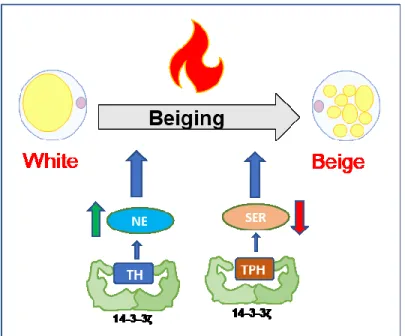
La thermogenèse adaptative est un mécanisme de production de chaleur médié par les adipocytes bruns. En réponse au froid, ou à un stimulus adrénergique, les adipocytes blancs peuvent être convertis en adipocytes beiges lors d’un processus que l’on nomme le « beiging ». Contrairement aux adipocytes blancs, les adipocytes beiges et bruns expriment des taux élevés de la protéine de découplage 1 (UCP1) et dissipent l'énergie sous forme de chaleur grâce à l'oxydation des lipides. Il a été démontré chez les rongeurs que l’activation des adipocytes bruns et beiges entraîne une réduction significative du poids corporel et l’activation de ces adipocytes chez l’humain semble être un traitement prometteur contre l’obésité et le diabète. Nous avons précédemment identifié un rôle essentiel de la protéine d’échafaudage 14-3-3ζ dans l'adipogenèse, mais son rôle dans d'autres processus adipocytaires reste incertain. Une des premières fonctions identifiées de la 14-3-3ζ est sa capacité à réguler l'activité enzymatique de la tyrosine hydroxylase, indispensable à la production de norépinephrine pour la thermogenèse. Notre étude vise donc à déterminer si la 14-3-3ζ influence le développement et la fonction des adipocytes beiges et bruns. Nos données montrent que la délétion d’un allèle du gène de la 14-3-3ζ n’affecte pas la tolérance au froid aiguë. Comparées aux souris de type sauvage (WT), les souris transgéniques mâles surexprimant la 14-3-3ζ (TAP) ont une meilleure tolérance au froid aiguë (3 heures, 4 °C) et chronique (3 jours, 4 °C). On observe chez les TAP une augmentation du beiging due à une élévation significative de l'ARNm et de la protéine UCP1 dans le tissu adipeux blanc inguinal (iWAT). Par ailleurs, les souris TAP présentent également une réduction significative de la conductance thermique lors d’exposition au froid leur permettant de mieux conserver la chaleur. Collectivement, nos résultats soulignent le rôle novateur de la 14-3-3ζ dans le beiging et nous permettent de mieux comprendre comment la thermogenèse adaptative est régulée.
Mots clés : 14-3-3ζ, protéines 14-3-3, beiging, browning, thermogenèse adaptative, adipocytes beiges, adipocytes bruns.
4
Abstract
Adaptive thermogenesis is a mechanism of heat production primarily mediated by brown fat. In some instances, cold exposure or adrenergic stimuli can convert white adipocytes into brown-like or beige adipocytes during a process termed “beiging”. Both beige and brown adipocytes express higher levels of uncoupling protein 1 (UCP1) and can release energy in the form of heat following lipid oxidation. The activation of these thermogenic adipocytes increases energy expenditure to reduce body weight in rodents, and it has been postulated to be a promising therapy for the treatment of obesity and diabetes. We previously identified an essential role of the molecular scaffold, 14-3-3ζ, in adipogenesis, but its roles in other adipocyte processes is uncertain. An early identified function of 14-3-3 was its ability to regulate the enzymatic activity of tyrosine hydroxylase, which is indispensable in the production of norepinephrine for thermogenesis. Thus, our study aims to investigate whether 14-3-3ζ influences the development and function of beige and brown adipocytes.
We report here that one allele deletion of the gene of 14-3-3ζ did not affect acute cold tolerance. On the other hand, transgenic overexpression of 14-3-3ζ in male mice (TAP) improves cold tolerance due to enhanced beiging with a remarkable increase in Ucp1 mRNA and protein in inguinal white adipose tissue (iWAT). Interestingly, beiging is increased in the TAP mice without any changes in sensitivity to beta-adrenergic stimuli, sympathetic innervation, or norepinephrine content being detected between WT and TAP mice. TAP mice also displayed significantly lower thermal conductance decreasing heat loss during the chronic cold challenge. Collectively, our results point to a novel role of 14-3-3ζ in beiging and increases our understanding of how adaptive thermogenesis is regulated.
Key words: 14-3-3ζ, 14-3-3 proteins, beiging, browning, adaptive thermogenesis, beige adipocytes, brown adipocytes
5
Table of contents
Résumé ... 3
Abstract ... 4
Table of contents ... 5
List of Figures and tables ... 7
List of abbreviations and acronyms ... 9
Acknowledgements ... 11
Chapter1: Introduction ... 12
1. White adipose tissue (WAT) ... 14
1.1. Major characteristics of WAT ... 14
1.2. Physiological roles of the WAT ... 16
2. Thermogenic adipose tissues... 18
2.1. Brown adipose tissue (BAT) ... 18
2.1.A. Anatomy and developmental origin ... 18
2.1.B. Regulation of BAT function ... 21
2.2. Beige fat ... 24
2.2.A. Induction and maintenance of beige fat ... 24
2.2.B. Different origins of beige adipocytes ... 25
2.3. Human brown and beige adipocytes... 26
2.4. Physiological role of brown and beige adipocytes ... 28
3. Therapeutic potential of brown and beige fat ... 30
4. Mechanisms of adaptation to cold ... 33
4.1. Non-shivering or adaptive thermogenesis ... 33
4.1.A. UCP1-dependent thermogenesis ... 35
4.1.B. UCP1-independent thermogenesis ... 36
4.1.C. Diet-induced adaptive thermogenesis ... 38
4.2. Shivering Thermogenesis ... 39
4.3. Vasoconstriction ... 42
5. 14-3-3 proteins... 44
5.1. A global characterization of 14-3-3 proteins... 44
5.1.A. Structure and properties of 14-3-3 proteins ... 44
6
5.2. A brief glimpse at 14-3-3 proteins function ... 46
5.3. Metabolic functions of 14-3-3 proteins and their relevance to metabolic diseases ... 48
5.3.A. From glucose metabolism to adipogenesis, metabolic roles of 14-3-3 proteins are diverse ... 48
5.3.B. The emerging importance of 14-3-3 proteins in metabolic diseases. ... 49
6. Hypothesis and objectives ... 51
Chapter 2: Article ... 53
1. Abstract ... 55
2. Introduction ... 56
3. Material and Methods... 59
4. Results ... 65
5. Discussion ... 70
6. Acknowledgements ... 75
7. Conflicts of interests... 75
Chapter 3: General discussion ... 93
Chapter 4: Conclusion ... 100
7
List of Figures and tables
Introduction
Figure 1: Anatomy of Major Fat Depots in Rodents and Humans ... 15
Figure 2: Anatomical locations of thermogenic fat in mice ... 18
Figure 3: Factors in beige and brown adipogenesis and transcriptional regulation ... 20
Figure 4: An overview of the heat-producing pathway in brown adipose tissue ... 22
Figure 5:Anatomical locations of thermogenic fat in humans ... 28
Figure 6:Variant models for the role of fatty acids in H+ transport by UCP1 ... 35
Figure 7: Models of Creatine-Driven Futile Substrate Cycling ... 38
Figure 8: Central circuitry mediating the response to cold ... 40
Figure 9: Regulation of 14-3-3 protein function ... 46
Figure 10: Hypothesis ... 52
Article
Figure 1: Effect of 50% deletion of Ywhaz in response to acute cold exposure ... 76Figure 2: Depletion of 14-3-3ζ in UCP1-Luciferase cells does not affect the induction of UCP1 by isoproterenol ... 77
Figure 3: Effect of 14-3-3ζ overexpression in the response to acute cold exposure ... 78
Figure 4: 14-3-3ζ overexpression increases body temperature but not energy expenditure during chronic cold exposure ... 79
8
Figure 6: 14-3-3ζ overexpression does not change adrenergic content or TH expression in TAP mice during cold adaptation ... 81 Figure 7: Reduced thermal conductance and enhanced beiging in TAP mice following chronic cold stress ... 82 Figure 8: Effect of 14-3-3ζ overexpression on glucose tolerance during cold adaption ... 83
Supplemental Material
Figure S 1: Effect of 50% deletion of Ywhaz in response to acute cold exposure ... 84 Figure S 2: Effect of 14-3-3ζ overexpression in the response to acute cold exposure ... 85 Figure S 3: 14-3-3ζ overexpression does not affect energy intake or body composition following chronic cold exposure ... 86 Figure S 4: Immunofluorescent detection of perilipin in paraffin-embedded gWAT and iWAT sections ... 87 Figure S 5: Immunofluorescent detection of TH in iWAT sections ... 88 Figure S 6: Relative mRNA levels of thermogenic genes (as indicated) in the iWAT following chronic cold exposure ... 89 Figure S 7: Relative mRNA levels of thermogenic genes (as indicated) in the iWAT following chronic cold exposure ... 90
Tables
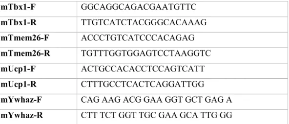
Table 1: List of primers ... 91 Table 2: List of Antibodies ... 92
9
List of abbreviations and acronyms
β-AR: β adrenergic receptor ADRB3: adrenergic receptor beta 3 AS160: AKT substrate 160KD ATP: adenosine triphosphate BAT: brown adipose tissue
cAMP: cyclic adenosine monophosphate CD137: cluster of differentiation 137 CD36: cluster of differentiation 36
CIDEA: cell death-inducing-like effector a
CITED1: Cbp/P300 Interacting Transactivator With Glu/Asp Rich Carboxy-Terminal Domain 1 FFA: free fatty acid
FGF21: fibroblast growth factor 21
FGFR1: fibroblast growth factor receptor 1 GLUT1/4: glucose transporter 1/4
gWAT: gonadal white adipose tissue HSL: hormone sensitive lipase iWAT: inguinal white adipose tissue LPL: lipoprotein lipase
MAPK: mitogen activated protein kinase NE: norepinephrine
PDK4: pyruvate dehydrogenase kinase, isoenzyme 4
10 Pi: phosphate
PPARγ: peroxisome proliferator-activated receptor-gamma PRDM16: PR domain-containing protein 16
scWAT: subcutaneous white adipose tissue
SERCA: sarco/endoplasmic reticulum Ca2+ ATP ase SLN: sarcolipin
SNS : sympathetic nervous system SVF : stromal vascular fraction T3 : triiodothyronine
TBX1: T-box 1 TG: triglyceride
TH: tyrosine hydroxylase
TMEM26: transmembrane protein 26 UCP1: uncoupling protein 1
VEGF: vascular endothelial growth factor vWAT: visceral white adipose tissue
WAT: white adipose tissue
11
Acknowledgements
In completion of this research project, I am extremely grateful to my supervisor Dr. Gareth Lim for his support and his countless encouragements. Thank you for your patience, and your commitment to make us not only students but individuals that stand out in everything we do. You have been an amazing mentor and I have no doubt that I will always benefit from all the lessons and all the advices you gave me. Through these last two years, you taught me a lot more than I thought I would learn, and I am honored and grateful to have you as a supervisor. I would like to thank my lab mates, Yves and Abel who taught me a lot of experiments and many other skills. They were always ready to give me a hand, advice, and feedback on my research project. Thank you to Christina Grech, David Millette the summer interns that did a considerable amount of work for this research project. Thank you to all my colleagues at the CRCHUM, Vanessa, Henry, Ju Jing, Hasna, Pegah who helped me a lot with experiments and feedback on my data. Many thanks to Dr Marc Prentki and Dr Jen Estall for their advice and their feedback on my project. I want to thank my Dad and dedicate this memoir to the amazing man he was and the exceptional father he has been for our family. Thank you, Papa, you were the one who believed in me and saw potential in me before anyone else. Always encouraging and supporting me. You are my pillar, you are my rock, and I always try my best to make you proud. I am grateful to my mother, my siblings, my grandmother and my friends who gave me strength. I don’t think I would have been able to accomplish anything in my life without your love and your constant support. You dried my tears when I cried and made me laughed when I was sad. Thank you for brightening up my days and listened to my complaints. I will finish by thanking God who gave me my family, my friends and who place in my path all the opportunities that allowed me to be here today. Without him none of this would have been possible. I am grateful to him for this wonderful experience and this amazing journey that has been my life!
12
Chapter1:
13
Adipose tissue is a connective tissue mainly formed of adipocytes, which are cells specialized in storage of nutrients synthesized by our body or acquired from our diet [1, 2]. Mammals have several types of adipocytes that are yellow, white, brown and beige; these fat cells are dispersed under the skin and around organs and exert unique roles [3-5]. Over the last decades, the global pandemic of obesity and diabetes has renewed a particular interest in understanding white, brown and beige adipose tissue biology. Bone marrow fat also referred to as yellow fat is a type of adipose tissue located in the medullary canal of the long bones (tibia, femur and humerus) and can account for up to 70% of total bone marrow volume[6-8]. Very little is known concerning the origin of the bone marrow fat cells, but they are thought to be distinct from white, brown and beige adipocytes [9]. They contain a large unilocular lipid droplet, develop postnatally and accumulate with aging. Aberrant bone marrow adipocytes development and function has been associated with diseases such as cancer and osteoporosis [7]. This heterogenous adipose tissue is an active organ that exerts metabolic, endocrine and immune functions. Bone marrow fat has also been implicated in the regulation of hematopoiesis, bone marrow environment, and it serves as energy reservoir that can store up to 5% of total fat mass in adults [8-10]. Pink adipocytes are mammary gland alveolar epithelial cells that are formed during pregnancy and lactation [11, 12]. This fifth type of adipocyte is only found in females, and its primary function is to produce and secrete milk, although they can also secrete leptin that may help to prevent obesity in newborns. During pregnancy subcutaneous white adipocytes transdifferentiate into pink adipocytes, and in the post-lactation phase, they revert back to white adipocytes [11]. These alveolar mammary cells are characterized by abundant cytoplasmic lipids and appear pink in color during pregnancy [11, 12].
14
1. White adipose tissue (WAT)
1.1. Major characteristics of WAT
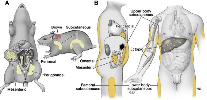
WAT is composed of mature white adipocytes that have triglycerides (TGs) packaged in large unilocular lipid droplets and the stromal vascular fraction (SVF), which contains immune cells, endothelial cells, preadipocytes, and progenitor cells [13]. WAT is localized to several depots that are anatomically, physiologically and patho-physiologically different; and their distribution varies among individuals, sexes and ages. They also differ in size, potential for replication and differentiation, developmental gene expression, and adipokine secretion [14, 15]. Each depot has unique properties and exerts different functions. As depicted in figure 1, mice have 2 major types of WAT: visceral WAT (vWAT) formed by perirenal, perigonadal and mesenteric depots, and the subcutaneous WAT (scWAT) formed by anterior and posterior depots. In humans, the major anatomical fat depots can be generally divided in 3 groups: the intra-abdominal fat that include the omental, mesenteric and pericardial depots, the upper body subcutaneous, and the lower body subcutaneous depots (Figure 1). In both mice and humans, there are also ectopic depots located in the liver, the muscles and the bone [15].
15
Figure 1: Anatomy of Major Fat Depots in Rodents and Humans. Several different names for particular fat depots in rodents (A) and humans (B) are used, as are different groupings of fat depots for physiological and clinical studies. Figure taken from [15].
WAT is formed in the mesoderm during embryogenesis and continues to develop postnatally [16]. Lineages studies show that white adipocytes originate from multipotent mesenchymal stem cells that express α-smooth muscle actin (αSMA) and platelet-derived growth factor receptors (PDGFRα and/or PDGFRβ) and may also be derived from endothelial cells and pericytes [17, 18]. During adipogenesis, progenitor cells and preadipocytes give rise to new fat cells in response to insulin, insulin like growth factor-1 (IGF-1), lipids, glucocorticoids and bone morphogenic proteins (BMPs) [19], while the WNT and Hedgehog signalling pathways are known suppressors of adipogenesis [20, 21]. Adipogenesis can be described as a two-step process: first, the commitment of mesenchymal precursor cells to the adipocyte lineage to form preadipocytes, and secondly, terminal differentiation of preadipocytes into mature adipocytes [22]. Commitment is stimulated by BMPs, notably BMP4 and BMP2, while terminal differentiation involves the master
16
adipogenic regulator, Peroxisome proliferator-activated receptor γ (PPARγ) and CCAAT/enhancer binding protein alpha, beta and delta (C/EBPα, β, δ) [23]. These transcription factors act in concert with specific coactivators and corepressors to activate or repress adipocyte differentiation by regulating the expression of approximately 2500 genes including Krüppel-like transcription factors (KLFs), GATA binding proteins and activators of transcription, early B cell factors (EBFs), and interferon-regulatory factor families [23].
1.2. Physiological roles of the WAT
WAT is the major reservoir of lipids that stores and releases free fatty acids (FFAs) in response to energetic demands [24]. It is a dynamic organ that can expand through processes of hypertrophy (increase in size of adipocytes) and hyperplasia (increase in number of adipocytes) to allow lipid storage during sustained nutrient excess [22, 25]. During nutrient deprivation, hormones such as glucagon and norepinephrine (NE) stimulate lipolysis, which results in the breakdown of TGs and the subsequent release of FFAs and glycerol in circulation [26, 27]. FFAs can serve as fuel for the production of energy (ATP), heat, and they can also serve as signalling molecules that drive the release of cytokines or stimulate insulin secretion [27-29].
WAT is an important regulator of energy homeostasis and excessive fat accumulation, inflammation and fibrosis of WAT have been closely linked to many diseases, including obesity, type 2 diabetes, cardiovascular diseases, and cancer [2, 30, 31]. WAT also exerts immune, endocrine, thermal and mechanical functions. For example, it is an effector of glucose metabolism such that when blood glucose levels rise, insulin is secreted to suppress lipolysis and stimulate glucose uptake in the adipocytes [32]. Furthermore, adipose tissue is a mechanical barrier that increases insulation and thermogenesis to protect against cold [2], and it can protect delicate organs (the eye) and cushion body parts exposed to high levels of mechanical stress (the heels and toes)
17
[2]. WAT also releases hormones termed adipokines that act in an endocrine, paracrine and autocrine fashion to regulate systemic metabolism [28, 33-35]. These adipokines are mostly secreted by adipocytes, preadipocytes, and immune cells [33, 34]. For example, WAT releases pro-inflammatory and anti-inflammatory cytokines, such as Il-6 and TNF-, which help fight bacterial infections and modulate insulin sensitivity [2, 15, 36]. Leptin acts centrally to increase satiety, decrease food intake, regulate body weight, and stimulate lipolysis [22]. Adiponectin sensitizes to insulin, reduces fibrosis and inflammation in peripheral organs, while FGF21 stimulates hepatic gluconeogenesis [22, 37, 38].
18
2. Thermogenic adipose tissues
2.1. Brown adipose tissue (BAT)
2.1.A. Anatomy and developmental origin
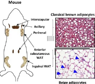
Brown adipocytes are phenotypically and functionally different from white adipocytes. They contain small, multilocular lipid droplets and are rich in mitochondria with abundant expression of uncoupling protein 1 (UCP1) [39]. In mice, they are found in the inter-scapular, cervical, axillary, retroperitoneal and peri-renal depots illustrated in figure 2. Brown adipocytes are formed during embryogenesis prior to WAT development to provide newborns with thermogenic capacities for protection against cold [40].
Figure 2: Anatomical locations of thermogenic fat in mice. Classical brown adipocytes reside in dedicated brown adipose tissue (BAT) depots, including interscapular, axillary, and perirenal BAT depots in mice and infants. Beige adipocytes sporadically reside in subcutaneous white adipose tissue (WAT) depots, such as the inguinal and anterior subcutaneous WAT in mice (arrowheads indicate the multilocular beige adipocytes). Figure taken from [41]
19
In terms of their developmental origin, brown adipocytes develop from the dermomyotomal precursor cells that express transcriptions factors Myogenic Factor 5 (Myf5), Paired-box protein 7 (Pax7), Pax3, and Engrailed 1 (En1); therefore, they are closely related to skeletal muscle, which also originates from Myf5+ progenitor cells [42].
Brown fat development is regulated by the master regulator of adipogenesis PPARγ, which acts in concert with brown fat-specific transcription factors [43]. For example, the developmental switch between brown adipocytes and myocytes is regulated by the transcriptional regulator PR domain zinc finger protein 16 (PRDM16), which is enriched in brown fat compared to white adipocytes and skeletal muscle [42, 44]. PRDM16 is essential for brown fat development, as its overexpression in myoblasts results in the formation of brown adipocytes, whereas reduced expression of this protein in brown adipocyte precursors resulted in a loss of brown adipocyte characteristics and induction of muscle differentiation [42, 44]. The capacity for PRDM16 to repress muscle differentiation is completely dependent on EHMT1, a histone-N- methyltransferase co-regulator that physically interacts with PRDM16. Genetic loss of Ehmt1 in Myf5+ precursor cells impairs brown adipocyte differentiation and
promotes muscle differentiation [45]. PRDM16 forms a transcriptional complex with PPARγ, C/EBP-β and early B cell factor 2 (EBF2) to stimulate expression of genes important for brown phenotype [46]. EBF2 functions as a marker of brown fat precursor cells and establishes brown fat characteristics of BAT by recruiting PPARγ to the brown-selective genes promoters [47]. Accordingly, EBF2-deficient mice lose brown-specific characteristics and thermogenic capacity of their BAT [47, 48]. Prior to differentiation, progenitor cells are committed to the brown adipocyte lineage, and this process is stimulated by BMP7, a member of the transforming
20
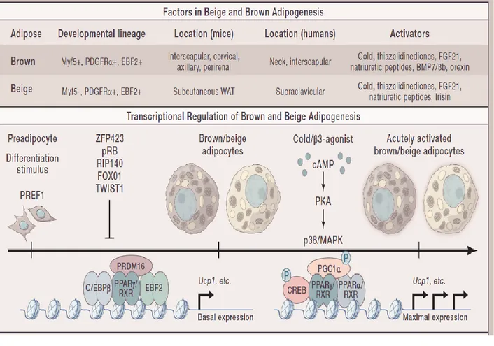
growth factor β family of proteins. BMP7 expression in brown adipocyte precursors is driven by the RNA binding protein EWS, which associates with the transcription factor YBX1 [49]. ZFP516, another PRDM16-interacting partner is also required to suppress the expression of muscle-specific genes during BAT development. Conversely, many transcription factors, including ZFP423, FOXO1, TWIST1, p107, LXRA, pRB, and RIP140, act as repressors of brown adipocyte differentiation [50]. Figure 3 is a brief overview of the transcriptional regulation of brown adipogenesis.
Figure 3: Factors in beige and brown adipogenesis and transcriptional regulation of brown and beige adipogenesis. Figure taken from [39]
21
BAT mass is dynamic and can undergo atrophy or hypertrophy in response to environmental and internal cues. The growth of BAT is principally stimulated by cold exposure and adrenergic stimuli, both of which promote proliferation and de novo differentiation of progenitor cells to increase thermogenic capacity [51]. β-Adrenergic signalling has been directly shown to induce the proliferation of EBF2 and platelet-derived growth factor receptor-α (PDGFRα) positive precursor cells [52]. Brown adipocytes express specific genes markers including PRDM16, PGC1A, CIDEA, ZIC1 AND PDK4 [53].
2.1.B. Regulation of BAT function
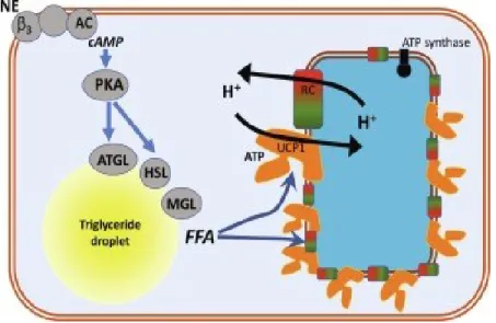
BAT is highly vascularized and innervated by post ganglionic sympathetic nerves [54, 55], and it is activated in response to cold stimuli, beta adrenergic agonists, FGF21, and T3 [51, 56, 57]. Cold is sensed by peripheral tissues such as the skin, spinal cord, abdominal viscera and the brain itself. In general, the preoptic area (POA) is the most thermosensitive site of the brain and is located between the anterior commissure and optic chiasm. When cool temperatures are sensed in this brain region, it activates the sympathetic nervous system (SNS) and causes the release of NE from sympathetic nerve terminals in BAT [58]. NE then binds to β3 adrenergic receptors (β3-AR), triggering the activation of adenylate cyclase, cAMP production, and the activation of protein kinase A (PKA), which phosphorylates perilipin and hormone sensitive lipase (HSL) to increase lipolysis. The released FFAs activate UCP1 and are oxidized in mitochondria to serve as an energy source for thermogenesis (see Figure 4) [54, 59-61]. In parallel, PKA phosphorylates CREB and p38MAPK leading to the activation of PGC1α [59, 62]. This latter coordinates with PPARγ and PPARα to stimulate the transcription of UCP1 [63, 64].
22
Figure 4: An overview of the heat-producing pathway in brown adipose tissue. Norepinephrine (NE) binds to β3-adrenergic receptors (β3) that stimulates adenylyl cyclase (AC) leading to cyclic adenosine monophosphate (cAMP) production. This activates Protein Kinase A (PKA) that will activate adipose tissue triglyceride lipase (ATGL) to break down triglycerides to diglycerides and free fatty acids (FFA). PKA will also activate hormone-sensitive lipase (HSL) that will break down the diglycerides to monoglycerides and more fatty acids; the last fatty acid will be released from the monoglycerides through monoglyceride lipase (MGL) activity. The fatty acids will counteract the inhibitory effect of cytosolic adenosine triphosphate (ATP) on Uncoupling Protein-1 (UCP1). This means that protons (H+) that were pumped out of the
mitochondria by the respiratory chain (RC) can re-enter the mitochondria and respiration can therefore proceed unhampered by the mitochondrial membrane potential. Figure taken from [51]
Thermogenesis rapidly depletes the limited brown fat stores, and a supplemental fuel is required to sustain adaptive thermogenesis. Thus, in addition to using its own lipid stores, brown adipocytes can take up fatty acids and glucose from the circulation to sustain thermogenesis [60, 65, 66]. NE released during cold also increases vascular endothelial growth factor (VEGF) [67] and lipoprotein lipase (LPL) expression [68, 69]. These two proteins, along with CD36, work
23
together to increase FFAs uptake in BAT. VEGF increases capillary permeability for plasma TGs, and LPL degrades TGs into FFAs transported through the plasma membrane by CD36, increasing FFAs availability for combustion by BAT. Glucose uptake in BAT after cold exposure, sympathetic nerve stimulation, and β-AR agonism has also been reported in several in vivo studies [60, 65, 66]. For example, NE stimulates the translocation of glucose transporters (GLUTs) from intracellular stores to the plasma membrane in mouse brown adipocytes, independent of insulin [70]. Both GLUT1 and GLUT4 are located on the plasma membrane of brown adipocytes, and GLUT4 expression is increased in BAT of rats fasted before chronic cold exposure to minimise any effect of insulin [71]. β3-adrenoceptors can also stimulate glucose uptake in BAT via cAMP-mediated increases in GLUT1 transcription and de novo synthesis and via mTORC2-stimulated translocation of GLUT1 [72]. Although glucose is an important contributor to BAT activity, FFA is the major substrate for thermogenesis [73], and it is required to maintain UCP1 function [74]. Succinate, another metabolite in the tricarboxylic cycle, also activates energy expenditure in the BAT, and in comparison to other fat cells, brown adipocytes largely uptake circulating succinate to serve as fuel for thermogenesis [75]. In addition to Glucose and FFAs, BAT can also use other nutrients such as branched-chain amino acids (BCAAs) to drive thermogenesis. In response to cold, BCAAs (leucine, isoleucine, valine) are transported and oxidized in the mitochondria to produce heat [76]. Increased circulating levels of BCAAs have been linked to obesity and diabetes and BCAA clearance by BAT may have implications in these disease states [76, 77]. In fact, impaired BCAA catabolism in BAT was shown to induce obesity and glucose intolerance [76] .
24 2.2. Beige fat
2.2.A. Induction and maintenance of beige fat
In mice kept at thermoneutrality (30 oC) or room temperature (22 oC), WAT is formed
mostly of white adipocytes. However, when exposed to chronic cold or long-term treatment with β3-AR agonists, beige adipocytes appear in subcutaneous depots during a process termed browning or beiging [41, 78-80] illustrated in Figures 2 and 3. Beige adipocytes can be also formed in response to several other external or internal stimuli including caloric restriction or manipulation, exercise, cancer cachexia, bariatric surgery, tissue injury, and FGF21 [80]. Beige adipocytes phenotypically resemble brown adipocytes, but they are inducible thermogenic fat cells. They are rich in mitochondria and express high levels of UCP1, albeit at lower levels when compared to brown adipocytes [39]. Beige adipocytes express specific markers including UCP1,
PGC1A, PRDM16, CITED1, CD137, TMEM26, AND TBX1 [53]. Recent studies, however,
suggest that there are at least 2 subtypes of beige adipocytes that are positive or negative for UCP1 [81].
Similar to BAT, the SNS is strongly involved in the development and function of beige fat. Beige adipocytes are recruited by NE [80]; however, other pathways independent of the SNS have been shown to stimulate beiging. For example, alternatively activated macrophages residing in adipose tissue synthesize and release catecholamines. These M2 anti-inflammatory macrophages are recruited to the scWAT and secrete NE to activate BAT and induce beige adipocyte development [82]; notwithstanding, data against this hypothesis has also been demonstrated [83].
Beige adipocytes are transient, and when mice are returned to thermoneutrality or room temperature, beige adipocytes gradually disappear from the WAT. Some studies have shown that
25
they can revert to unilocular white adipocytes within approximately 2 weeks following re-warming or withdrawal of the β3-AR agonist [78, 84, 85]. This process is thought to be mediated by an increase in mitochondrial degradation or mitophagy [41, 78, 86]. UCP1+-adipocyte-specific
deletion of Atg5 or Atg12 is essential for autophagosome formation and prevents beige adipocytes from reverting back to a white phenotype even after withdrawal of external stimuli [78]. In addition, Parkin is a mitochondrial E3 ubiquitin ligase regulated by the β3-AR and PKA signaling pathway, and its recruitment to the mitochondria is essential for the activation of mitophagy in beige fat and the maintenance of beige adipocyte thermogenesis in vivo [86].
2.2.B. Different origins of beige adipocytes
The cellular origins of beige adipocytes and the mechanism underlying their formation remain unclear and have been source of much debate. To date, some studies have reported de novo differentiation from resident precursor cells [87, 88], and several observations demonstrated a trans-differentiation model during which mature white adipocytes directly convert into functional beige adipocytes [89-91]. Altogether, the available data suggest that both de novo differentiation and trans-differentiation contribute to beige fat biogenesis [80]. Beige fat is heterogeneous, as it is formed by multiple subtypes of thermogenic adipocytes with distinct developmental origins and biological roles [41]. It has been proposed that beige adipocytes are related to vascular smooth muscle cells and mural cells [92]. Some lineage-tracing studies have demonstrated that some beige adipocytes are derived from progenitors expressing Sma, myosin heavy chain 11 (Myh11), Pdgfra, or Pdgfrb (see Figure 3), while some beige adipocytes residing in subcutaneous or retroperitoneal WAT depots are derived from progenitors expressing Pax3 and/or Myf5 [39]. In response to long term cold exposure, Myh11+ muscle cells and mural cells, which express Pdgfrb, can differentiate
26
into beige adipocytes [92]. Similar to brown adipocytes, PRDM16 is also a key transcriptional regulator of beige adipocyte biogenesis and its expression in vascular smooth muscle cells promotes their differentiation into beige adipocytes [52]. Another transcription factor involved in beige adipogenesis is EBF2 whose expression in WAT or primary adipocyte cultures promotes the recruitment of beige adipocytes [47]. A recent study of Chen et al., shows beige adipocytes in murine iWAT originate from a unique population of myoblast determination protein 1 (Myod1+)
progenitors, referred to as Myod1+-derived beige fat. These Myod1+-derived beige adipocytes
accounted for about 15% of UCP1+ beige adipocytes in the iWAT, which supports the hypothesis
of several progenitor cells responsible for beige adipocytes development. The Myod+ derived beige
fat is composed of glycolytic beige adipocytes that display enhanced glucose metabolism when β-AR signalling is blocked or during severe and prolonged cold. They are therefore distinct from classic beige adipocytes activated by β-AR signalling [93].
2.3. Human brown and beige adipocytes
The presence of brown adipocytes in mammals has been described since 1550, and their importance in heat production has been known for over 40 years [73]. In humans, inter-scapular BAT (iBAT) was known to be active in infants (see figure 5) and to regress with aging [94]. Research on BAT has re-initiated in recent years (2007-2009) when positron emission tomography-computerized tomography (PET-CT) scans in adult humans revealed the presence of several BAT depots displaying high uptake rates of the glucose analog 18F-fluoro-2-deoxy-glucose (18F-FDG) [95-98]. BAT activity in humans is stimulated by cold, and no glucose uptake
can be detected in study participants kept under warm conditions [98]. It has also been suggested that outdoor workers exposed to a cold environment have a higher amount of BAT than the general
27
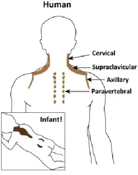
population [99]. In humans, BAT and beige fat consist of small discrete depots principally located in the upper body including the neck area, the supraclavicular depots, suprarenal, paravertebral, and paraaortic depots depicted in Figure 5 [100]. PET‐CT imaging studies show women have greater quantities of BAT and higher expression of thermogenic genes than men, while young persons, in particular newborns, possess more BAT than adults [101, 102].
Human BAT depots are heterogeneous and are thought to resemble both murine classical BAT and inguinal beige adipocytes [53, 80]. For example, Wu et al., have demonstrated that some brown fat in adult humans, notably the supraclavicular depot, has similar molecular characteristics to murine beige adipocytes, while infant iBAT and the deep neck regions in adult humans contain thermogenic fat that resembles classical brown fat in mice [53, 80]. Surprisingly, human and mice have opposing patterns of brown markers expression in vWAT versus scWAT [103]. In rodents, exercise increases beiging of scWAT and improves whole body metabolic homeostasis through the effects of the myokine irisin. However, exercise does not seem to affect human beige fat and is associated with a decreased concentration of circulating irisin [104]. Most knowledge of BAT and beige fat is from rodent studies; consequently, mechanisms underlying human thermogenic fat cells development and their significance to metabolism still remain to be elucidated.
28
Figure 5: Anatomical locations of thermogenic fat in humans. In adult humans, BAT is present in multiple locations, including cervical, supraclavicular, axillary, paravertebral, and abdominal subcutaneous regions. UCP1-positive adipocytes from the supraclavicular region show a molecular signature resembling that of mouse beige adipocytes, whereas the deep neck regions contain thermogenic fat that resembles classical brown adipocytes in mice. Figure taken from [41]
2.4. Physiological role of brown and beige adipocytes
The principal role of brown adipocytes is to dissipate chemical energy in the form of heat. BAT is the principal site of non-shivering thermogenesis (NST), a mechanism that defends the body against hypothermia by maintaining core body temperature in the physiological range [51, 73, 105, 106]. Although BAT only represents a small portion of the total body mass in large mammals and adult humans, it is responsible for at least 60% of NST [39]. BAT also produces several “batokines” that exert autocrine, paracrine and endocrine functions and, thus, play important roles in regulating glucose homeostasis, bone and muscle function [138]. For example, BAT produces neuregulin 4 (NRG4) which reduces fat storage in the liver to improve peripheral
29
insulin sensitivity [107]. Brown and beige fat also secrete WNT10b and insulin-like growth factor-binding protein 2 (IGFBP2) to promote osteoblast activity and bone health [108]. Differentiated brown adipocytes or BAT from newborn and adult rodents exposed to cold for 1–2 days were found to express high levels of nerve growth factor (NGF). This growth factor can increase proliferative capacity of BAT by increasing mitosis of precursor cells in the SVF [109].
In mice, beige adipocytes represent a small percentage of iWAT and are considered to have a negligible role in whole-body energy expenditure [110, 111]. However, recent studies have shown that beige fat may play key roles in the regulation of whole-body energy homeostasis, as well as inflammation and fibrosis of WAT [126, 139]. Beige adipocytes support adaptive thermogenesis by generating heat in response to cold, and they can regulate glucose metabolism even in the absence of adrenergic stimuli [106] and improve adipose tissue function by decreasing fibrosis and inflammation [139]. Beige adipocytes secrete a PRDM16-regulated factor named Slit2, a member of the Slit extracellular protein family. Increased levels of Slit2 promote PKA activation and consequently augment the thermogenic activity of beige adipocytes and improve glucose homeostasis [140]. In humans, plasma SLIT2 is negatively correlated with serum glucose and HbA1c in diabetic individuals [141]. The study of beige fat is quite recent, and many characteristics of these adipocytes remain to be elucidated. One of the grey areas in beige fat biology is the determination of its mass, which is limited by current technology. In addition, beige adipocytes are transient fat cells that are recruited in response to external or internal stimuli. Unlike WAT and BAT which are homogenous and distinct adipose depots, beige fat is heterogenous and dispersed within scWAT [41, 43, 80]. In humans, some depots of beige fat are hardly distinguishable from classical BAT [53], and this has made it challenging to accurately determine beige fat mass. In a study by Blondin et al., BAT mass is estimated to represent ∼1% of total body
30
weight in adult humans [112]. However, this study could not discriminate between human brown fat that is similar to murine beige adipocytes or classical brown adipocytes. This suggests that in mice and humans, beige fat might represent less than 1% of total body weight [112].
3. Therapeutic potential of brown and beige fat
Obesity and diabetes are global pandemics that affect many social classes and demographics. The prevalence of obesity has remarkably increased over the last 50 years [113]. Currently, obesity affects over a third of the world’s population, and it is expected by 2030 that an estimated 38% of the world’s adult population will be overweight of which 20% will be obese [114, 115] . Changes in diet and physical activity are generally the first lines of treatment adopted to combat obesity; however, there are other therapeutic options available including weight loss medications and bariatric surgery [116, 117]. Bariatric surgery is effective, but it is expensive and cannot be afforded by most patients. In addition, it is a selective process that is generally performed on patients with morbid obesity, as defined by a body mass index (BMI) greater than 40 [117]. As of 2017, only 6 medications had been approved for the treatment of obesity [118]. These drugs include Orlistat, which reduces fat absorption by inhibiting gastrointestinal lipases, and Liraglutide, a GLP-1 agonist that decreases appetite and food consumption by slowing gastric emptying and increasing post prandial satiety. Phentermine and Lorcaserin, a serotonin receptor agonist, are also available for weight loss management [118-121].
To combat obesity, it is important to develop new pharmacological treatments accessible to more subjects with obesity. More reports of the potential power of brown and beige fat to combat metabolic disorders are culminating. Obesity results from an imbalance in energy homeostasis with energy expenditure being inferior to energy intake [101]. With their ability to regulate fat mass by increasing energy expenditure through lipid oxidation, brown and beige adipocytes
31
represent promising targets for reducing the excessive fat accumulation that defines obesity [122]. BAT has been also shown to positively impact whole-body metabolism. In addition to clearing stored triglycerides, these thermogenic adipocytes have a high capacity for glucose disposal allowing them to be potential targets in the treatment of diabetes [68]. It has also been demonstrated that the ability of BAT to efficiently dispose of stored energy allows a mouse exposed to cold to eat 3–4 times that of a mouse at thermoneutrality without becoming obese [56]. Although UCP1-deficient mice do not develop obesity under normal chow diet, they gain significantly more weight during a high fat diet (HFD) compared to WT mice [123]. Furthermore, mice with surgical removal or transgenic ablation of BAT become obese supporting a role of brown fat in the development of obesity [124, 125]. Additionally, inducing BAT angiogenesis in obese mice improves glucose metabolism [126, 127].
Recent studies in rodents have reported beneficial effects of beiging by conferring protection against obesity and diabetes. An elegant study by Seale et al. demonstrated improved glucose tolerance and insulin sensitivity in transgenic Ap2-Prdm16 mice due to enhanced beige fat formation compared to littermate controls. The Ap2-Prm16 mice also gain significantly less body weight and adiposity under HFD conditions. In contrast, beige fat-deficient mice, caused by the adipocyte-specific deletion of Prdm16 or Ehmt1, develop obesity and systemic insulin resistance even under ambient temperatures [45, 128]. In adult humans, increased brown fat is correlated with low BMI and low adipose tissue content [129], and in contrast, subjects who are overweight or with obesity have decreased BAT activity compared to lean individuals [130, 131].
By lowering stored lipids and improving glucose metabolism, it is valid to think that increasing BAT activity and stimulating beiging in adult humans can represent potential treatments for obesity, diabetes and the metabolic syndrome [56]. To date selective β3-AR agonists that
32
activate adaptive thermogenesis have been trialed as a treatment for obesity but were unsuccessful. In fact, these agonists are potent activators of the cardiovascular system and increase the risk of developing higher blood pressure and cardiovascular diseases [132]. Consequently, identifying β-AR independent signalling pathways with minimal cardiovascular risks that activate BAT and promote beiging could lead to more effective therapeutic treatments of metabolic disorders.
33
4. Mechanisms of adaptation to cold
Adaptation to cold is a complex process that involves several physiological, behavioural and genetic responses [133]. As described previously, brown and beige adipocytes are fat cells specialized in heat production or thermogenesis and thus play a major role in adaptation to cold environments. Thermogenesis regulates body temperature through two involuntary, mostly autonomic, physiological processes that dissipate heat and include adaptive thermogenesis primarily mediated by brown and beige fat and skeletal muscle shivering [58, 101, 134, 135].
4.1.Non-shivering or adaptive thermogenesis
Adaptive thermogenesis, also known as non-shivering thermogenesis (NST) or facultative thermogenesis, is cold or diet-induced heat generation to defend against cold and to regulate energy balance after changes in diet.
The brain regulates adaptive thermogenesis by activating the SNS, which heavily innervates BAT and WAT [54]. Inhibiting the SNS with adrenergic blockers during cold exposure causes a drastic fall in body temperature; while mimicking its effects with administration of NE, L-DOPA, or isoproterenol increases energy expenditure and thermogenic protein expression in the iWAT and the BAT [136]. Furthermore, Dopamine β-hydroxylase knockout mice lacking noradrenaline and adrenaline are unable to induce thermogenesis in BAT when exposed to cold [136, 137]. The brain can also regulate adaptive thermogenesis by activating the hypothalamus-pituitary-thyroid axis [105, 138]. The mechanisms through which thyroid hormone controls thermogenesis and energy balance are unclear, but is likely via substrate and ion cycling and mitochondrial proton leaks [139]. During adaptive thermogenesis, adrenergic stimuli increase the expression of DIO2 that converts T4 into T3, the ligand for thyroid-hormone receptor TRβ. This
34
is consistent with the hypothesis of a thyroid-sympathetic axis synergism that potentiates adaptive thermogenesis [140, 141].
Several other molecules have been shown to activate adaptive thermogenesis in murine models [51, 142]. For example, PPARγ ligands such as rosiglitazone and FFAs can control adaptive thermogenesis. Some studies demonstrated that rosiglitazone can activate Ucp1 expression in BAT and brown adipocyte cultures [143, 144]. Atrial natriuretic peptide (ANP) is released from the heart and can activate brown fat cells via its signaling receptor NPR-A, that activates guanyl cyclase and elevates cyclic guanosine monophosphate (cGMP). NPR-A has been shown to increase lipolysis via cGMP in human adipose tissue and isolated human adipocytes [145]. Furthermore, cardiac natriuretic peptides also act via p38MAPK to induce the brown fat thermogenic program in mouse and human adipocytes [146]. Another activator of NST is a peptide named fibroblast growth factor 21 (FGF21). The predominant source of FGF21 is the liver although both WAT and BAT release this growth factor in response to chronic cold exposure [147]. FGF21 is markedly increased in BAT of UCP1-ablated mice and is thought to increase glucose uptake and thermogenesis in brown and beige adipocytes via its receptor complex, consisting of FGFR1 and its obligatory co-receptor β-klotho [148]. Several BMPs have also been shown to play a role in brown fat development and activity. For example, BMP7 induces the differentiation of brown preadipocytes, while BMP8b seems to work together with NE to enhance lipolysis and thermogenic activity [149]. Finally, retinoic acid has also been shown to acutely activate UCP1 both in intact brown fat mitochondria and in isolated brown fat cells [51].
35 4.1.A. UCP1-dependent thermogenesis
UCP1, also known as thermogenin, is the primary protein that mediates heat generation during adaptive thermogenesis [51, 73]. In BAT, UCP1 is present at very high levels even during the non-stimulated state and resides in the inner mitochondrial membrane to uncouple the mitochondrial proton gradient from ATP synthesis (Figure 4) [73]. UCP1 functions as a H+
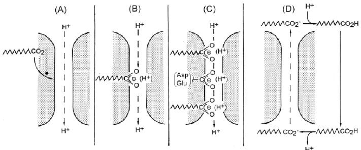
transporter that requires FFAs. In this sense, FFAs are regarded as cofactors of UCP1 that allows H+ transport by two distinct mechanisms, as illustrated in figure 6. In the first, FFAs function as
H+ donors to the UCP1 translocation channel (model A,B,C of figure 6) [74] and in the second,
UCP1 does not transport H+, but rather FFA anions outside the mitochondrial matrix. Once outside,
FFA anions are re-protonated and flip flop back in the inner mitochondrial membrane transferring protons inside the mitochondrial matrix (model D of figure 6) [150].
Figure 6: Variant models for the role of fatty acids (FAs) in H+ transport by UCP1. (A)
Conformational activation of the H+ transport pathway by FA. (B) Cofactor role of FA by
providing H+ translocating groups. (C) Cofactor role of FA by providing H+ translocating groups
in addition to resident groups (Asp/Glu), where FAs fill gaps in the H+ translocating path. (D) FA
anion transport shuttle. FAs are translocated as anions by UCP1 and re-shuttle though the membrane as undissociated acids. Figure taken from [74]
36
Several studies have shown the prominent role of UCP1 in adaptive thermogenesis. UCP1-deficient mice have pronounced cold sensitivity and can only survive severe, long-term cold at
4 oC through gradual reductions in environmental temperature [123]. Acute cold exposure of four
hours or less is enough to increase UCP1 activity and mRNA expression, while chronic cold exposure of several hours or days will cause a significant elevation in the transcriptional coactivator PGC1α, which coordinates with PRDM16 to stimulate UCP1 protein and mitochondrial biogenesis [79, 106]. UCP2 and UCP3 are homologues of UCP1 expressed in BAT and other tissue types, such as the skeletal muscle [151, 152]. Although they have proton transport activity, their expression remains inconsistent in cold exposed rodents; therefore, their importance in adaptive thermogenesis remains unclear [153-155].
4.1.B. UCP1-independent thermogenesis
Recent data show that UCP1 is dispensable for adaptive thermogenesis as thermal homeostasis can be maintained without UCP1 [41]. In support of this claim, UCP1-deficient mice can be acclimated to cold by slowly decreasing the temperature [123]. In addition, discordance in the metabolic phenotypes between brown/beige fat-deficient mice and UCP1-deficient mice has suggested the existence of UCP1-independent mechanisms of adaptive thermogenesis [41]. For example, BAT-deficient mice, caused by the transgenic expression of diphtheria toxin (DTA), or beige fat deficient mice due to adipocyte-specific deletion of PRDM16 or EHMT1, exhibit obese and diabetic phenotypes even under ambient temperature [45, 128]. By contrast, UCP1-deficient mice are not diabetic and develop obesity only when they are kept at thermoneutrality [123, 156]. PRDM16 Tg-UCP1-/- mice (PRDM16 transgenic mice crossed with UCP1-/- mice) are resistant
37
to HFD induced weight gain and have improved glucose tolerance and insulin sensitivity when compared to UCP1 -/- control mice [41, 157]. This implies that the anti-obesity and anti-diabetic effects of beige fat are UCP1-independent.
To date, two thermogenic mechanisms have been described through which beige adipocytes can contribute to the regulation of thermal and energy homeostasis, independent of UCP1. These mechanisms are ATP-dependent and prominent in beige fat, however, they are not relevant to BAT function [81, 157-160]. Ikeda et al., identified a thermogenic mechanism in beige adipocytes that involves ATP-dependent Ca2+ cycling between sarco/endoplasmic reticulum Ca
2+-ATPase 2b (SERCA2b) and ryanodine receptor 2 (RyR2). Ca2+ cycling is a conserved mechanism
present in both adult humans and mice. The mechanisms underlying Ca2+ cycling in beige
adipocytes are not fully understood but is triggered by the binding of NE to α and β-AR, which in turn activates SERCA2b and RyR2 and leads to increased intracellular Ca2+ flux. Ca2+ is
transported in the ER by SERCA2b and this non-canonical form of thermogenesis occurs when Ca2+ transport is uncoupled from ATP hydrolysis by SERCA2b.
A futile creatine cycle has also been identified as an alternative beige fat mechanism that results in increased heat production during cold exposure [81, 158, 159]. Creatine metabolism is an important part of adaptive thermogenesis that regulates energy expenditure in both brown and beige adipocytes. The creatine driven substrate cycle promotes adaptation to cold by stimulating mitochondrial respiration when ADP is limiting [81, 158, 159]. Indeed, creatine reduction diminishes oxidative properties of mouse and human brown adipocytes and, decreases core body temperature in UCP1-/- mice [158]. Furthermore, adipose tissue specific KO of glycine amidinotransferase (GATM), the first and rate-limiting enzyme of creatine biosynthesis increases body weight and fat mass during diet-induced obesity [158]. Another line of evidence supporting
38
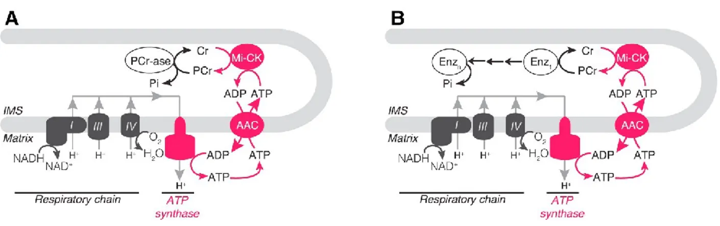
this mechanism is that ablation of UCP1-dependent thermogenesis is compensated by an increase in genes involved in creatine metabolism [158]. In the model presented by Kazak et al., creatine facilitates the regeneration of adenosine diphosphate (ADP) through the futile hydrolysis of phosphocreatine either by a phosphatase or via several phosphate-transfer reactions catalyzed by multiple enzymes depicted in Figure 6. Creatine-driven thermogenesis is present in beige adipocytes regardless of their UCP1 expression but is more prominent in epididymal beige adipocytes that do not express UCP1 [158]. This mechanism might co-exist in the beige adipocytes with UCP1-dependent thermogenesis, and Ca2+ cycling [81].
Figure 7: Models of Creatine-Driven Futile. Substrate Cycling. (A) Model of creatine-driven futile substrate cycling based on direct hydrolysis of phospho-creatine (PCr). (B) Model of creatine-driven futile substrate cycling based on multiple phospho-transfer events catalyzed by multiple enzymes (Enz). Creatine (CR), mitochondrial creatine kinase (miCK), ATP/ADP carrier (AAC), intermembrane space (IMS). Figure taken from [158]
4.1.C. Diet-induced adaptive thermogenesis
Diet-induced adaptive thermogenesis is activated in response to changes in diet, and it has been hypothesized to protect against intake of surplus energy [106]. In humans or rodents, a
39
positive-energy balance induced by excessive caloric intake will cause BAT activation to increase energy expenditure in order to limit weight gain [161]. Many mouse models of obesity and leptin-deficient ob/ob mice display decreased BAT mass and activity [162, 163]. In addition, mice with ablated or a reduced amount of BAT become obese, as well as diabetic and hyperlipidemic [124, 125]. Diet-induced adaptive thermogenesis is thought to be mediated by leptin which increases SNS activity to brown fat [164]. Central and peripheral administration of leptin have been shown to increase UCP1 mRNA and protein levels, and this effect is decreased when leptin levels fall due to starvation [165, 166].
4.2. Shivering Thermogenesis
Skeletal muscle (SKM) plays an important role as a thermogenic organ. SKM represents 40% of total body weight and plays an essential function in regulating the response to acute cold [102]. Within minutes of exposure to cold, heat is produced from the involuntary contractile activity of muscles, which is also known as shivering [5].
As pictured in figure 8, shivering is controlled by many brain regions including the lateral parabrachial nucleus (LPB), the preoptic area (POA), the dorsomedial hypothalamus (DMH), and the rostral raphe pallidus (rRPA) [6-9]. However, the circuitry that connects these brain structures and the precise pathway that leads to motor neuron activation are unclear. It has been proposed that thermal information is received and integrated into the POA and then transmitted to effectors through a descending pathway that exits the brain via the rostral medulla. These medullary output neurons then activate peripheral sympathetic neurons in the case of adaptive thermogenesis and somatic motor neurons in the case of shivering to mediate cold tolerance [167].
40
Figure 8: Central circuitry mediating the response to cold. POA, preoptic area of the hypothalamus; MnPO, median preoptic nucleus; MPO, medial preoptic area; DMH, dorsomedial hypothalamus; rRPa, rostral raphae pallidus nucleus; LPB, lateral parabrachial nucleus; BAT, brown adipose tissue. Figure taken from [133]
41
Shivering consists of fast, repetitive contraction-relaxation cycles of SKM that release heat from the exothermic reaction of ATP hydrolysis (ATP + H2O= ADP + Pi + Heat) [10]. Myocytes
have a great capacity for thermogenesis, and several pathways of heat production are present in these cells. Heat is primarily produced by enzymes hydrolysing ATP, including Na+/K+ ATPase,
myosin ATPase, and SERCA [11]. When Ca2+ is released from the sarcoplasmic reticulum (SR)
into the cytosol via the ryanodine receptor 1 (RYR1), it binds to myofilaments initiating contraction. Myosin ATPase hydrolyses ATP and uses the energy released to bind actin, resulting in contraction. When Ca2+ builds up in the cytosol, it triggers SERCA, which pumps Ca2+ back
into the SR. In this process, SERCA utilises energy from ATP hydrolysis thereby causing relaxation, whereas Na+/K+ ATPase uses energy from ATP hydrolysis to reset resting ion gradients
and membrane potential [10, 12, 13].
With long-term adaptation to cold, shivering decreases and is replaced by a more prominent increase in adaptive thermogenesis [20]. This is because NST protects the shivering muscles from severe damage due to defective Ca2+ handling [22]. In addition, shivering relies largely on muscle
glycogen that can become limiting after few hours . However, recent evidence show SKM also contributes to thermogenesis via a mechanism of non-shivering thermogenesis (NST) mediated by a futile sarcoplasmic reticulum Ca2+ ATPase (SERCA) activity [11, 14-17]. Muscle-based NST
has been known for a long time in animals lacking BAT or functional UCP1 such as birds and fish, but its importance in mammals was described only recently through studies in rodents. NST in the SKM has been reported to be the earliest mechanism of NST in vertebrates preceding BAT-driven thermogenesis. This alternative mechanism of heat production is mediated by a transmembrane Ca2+-ATPase (SERCA) located in the sarcoplasmic reticulum membrane and a
42
slippage occurs decreasing the Ca2+ transported from the cytosol back into the SR. Consequently,
more ATP needs to be hydrolyzed by SERCA to transport the released Ca2+ which leads to heat
production [18]. Studies have shown that SLN protein levels are upregulated during diet overload and cold adaptation. In addition, overexpression of SLN in muscle protects against diet-induced obesity . Of note, muscles can produce myokines, such as FGF21, that stimulates BAT thermogenesis and beiging in iWAT, and as a result enhance thermogenesis in other tissues [19].
4.3.Vasoconstriction
Another physiologic response to cold exposure in mammals is the constriction of blood vessels or vasoconstriction [58, 168]. For the maintenance of body temperature to be efficient, it is important that heat generated through thermogenesis is retained, and this can be done by decreasing blood flow via vasoconstriction. Unlike thermogenesis, vasoconstriction does not generate heat, but prevents heat loss [58, 168]. Cold exposure activates vasoconstriction prior to shivering and BAT thermogenesis, and vasoconstriction is similarly regulated by NE through sympathetic neural stimulation [169, 170]. The importance of NE in controlling vasoconstriction, and therefore preventing heat loss, has been demonstrated in mice deficient in dopamine β-hydroxylase. These mice lack NE and epinephrine and consequently are unable to maintain their body temperature during cold exposure partly due to the failure of peripheral vasoconstriction [137]. In UCP1-deficient mice, reductions in heat loss from the tail through vasoconstriction was shown to be a part of the compensatory mechanism for maintaining homeothermy [168].
There is evidence that another hormone mediating heat conservation during cold exposure is leptin. In fact, leptin-deficient ob/ob mice are also characterized by mild hypothermia when housed
43
at ambient temperature and display profound hypothermia when exposed to cold [171, 172]. This indicates that a physiological role of leptin in thermoregulation consists of reducing thermal conductance, or heat loss, to maintain core body temperature under cold conditions. In addition, leptin treatment in ob/ob mice defends body temperature without increasing energy expenditure or BAT recruitment but by inducing vasoconstriction in the tail to reduce heat loss [173].
44
5. 14-3-3 proteins
5.1. A global characterization of 14-3-3 proteins
5.1.A. Structure and properties of 14-3-3 proteins
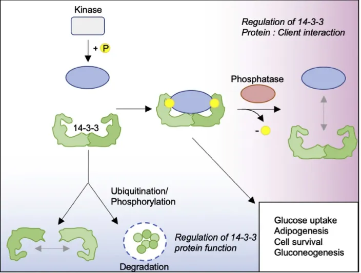
From plants to animals, 14-3-3 proteins are ubiquitously expressed in all eukaryotes [174]. 14-3-3 proteins which were named according to their electrophoretic mobility are found in all subcellular compartments and bind to a wide range of biosynthetic enzymes, signalling proteins, and transcriptions factors [175-177]. In mammals, the 14-3-3 protein family consists of seven isoforms, each encoded by a specific gene, and include β, ζ, ε, γ, η, σ, τ, with α, and δ being the phosphorylated forms of β and γ, respectively [175, 178]. As portrayed in figure 9, the 14-3-3 isoforms are 28-33 KDa acidic proteins that form hetero- or homo-dimers and function as adaptor proteins to stabilize their targets and facilitate interaction of their targets with other proteins [178-180]. Each 14-3-3 monomer comprises nine anti-parallel alpha helices, and the assembly of these monomers creates a negatively-charged amphipathic groove necessary for protein-protein interactions. Conserved and identical regions of each isoform are located in the inner core of this groove, while the variable residues are mostly located on the outer surface of the protein [181, 182]. Three phosphorylation dependent binding motifs of 14-3-3 proteins have been identified including RSXpS/TXP (mode 1) and RXXXpS/TXP (mode 2), where pS/T represents phosphorylated serine/threonine and X a generic amino acid [175, 183]. A third motif (mode 3) was later defined as when binding partners have a phosphorylated serine or threonine as the penultimate residue in the C-terminus [183]. Although 14-3-3 proteins were originally identified as phospho-serine and phospho-threonine binding protein, it is well established now that they bind
45
to unphosphorylated targets and proteins harboring the O-GlcNac post-translational modification [184, 185].
5.1.B. Mechanisms regulating 14-3-3 protein activity
14-3-3 protein activity is regulated by phosphorylation at specific residues, which have inhibitory effects on function. Phosphorylation induces dimer dissociation, and as a consequence, prevents binding to target proteins [179, 186, 187]. For example, phosphorylation at Ser58 at the dimerization interface of 14-3-3ζ by MAPKAPK-2 or SDK1 promotes dimer disassociation, thereby reducing client protein binding [188, 189]. JNK phosphorylates 14-3-3σ and ζ at Ser185 which causes the dissociation of BAX from 14-3-3 proteins and its translocation to mitochondria [190]. Two other important regulators of 14-3-3 proteins interaction with client proteins are protein phosphatases PP1, and PP2A. As shown in figure 9, these enzymes dephosphorylate serine and threonine residues on 14-3-3 docking sites of client proteins and reduces their interactions with 14-3-3 proteins [191]. Mechanisms of transcriptional and post translational regulation of 14-3-3 proteins through proteasomal degradation have also been identified, in particular for the 14-3-3σ isoform [192]. For example, in breast epithelial cells, the oestrogen-induced E3 ligase, EFP, initiates ubiquitination of 14-3-3σ, which results in its rapid degradation [193].
46
Figure 9: Regulation of 14-3-3 protein function. In most cases, 14-3-3 proteins interact with target client proteins at high-affinity phosphorylation binding motifs created by serine and/or threonine residues. Removal of phosphate groups on target proteins by phosphatases results in the loss of 14-3-3 binding sites and subsequent 14-3-3 protein dissociation from target proteins. Regulation of 14-3-3 protein activity is facilitated by their phosphorylation or ubiquitination. These post-translational modifications either promote the dissociation of 14-3-3 protein dimers or target them for degradation. Figure taken from [194]
5.2. A brief glimpse at 14-3-3 proteins function
Since their discovery by Moore and Perez in 1967 [175, 179], the number of identified binding partners of 14-3-3 proteins have been escalating, reaching more than 1000 identified binding partners [175-177]. This has increased our knowledge of the processes regulated by these
47
molecular scaffolds including cell signalling, apoptosis, metabolism, cell cycle, transcription, and DNA repair [177, 180, 195-197]. One of the first assigned roles to 14-3-3 proteins was their regulation of tyrosine and tryptophan hydroxylase, rate-limiting enzymes involved in the synthesis of catecholamines and serotonin, respectively. These functions gave rise to their gene name tyrosine- and tryptophan hydroxylase activators (YWHAs) [176]. Another defining feature of 14-3-3 proteins is their ability to mediate protein-protein interactions. 14-14-3-3 proteins function as adaptor proteins that can induce conformational changes in their target proteins revealing an active site, a ligand–binding region, or a region that interacts with another protein [198]. For example, 14-3-3β binding to the serine/threonine rich B box in the kinase domain in BCR promotes the formation of a RAF-1/BCR complex [199]. 14-3-3 proteins β and θ can facilitate the coupling of PKCζ to RAF-1 [200]. 14-3-3 proteins can also promote steric hindrance or stabilize enzyme substrate complexes. For example, binding of 14-3-3ζ to Serotonin–N-acetyltransferase (AANAT) stabilizes the enzyme active conformation which increases its catalytic rate and its interactions with substrates [201].
14-3-3 proteins also regulate protein trafficking into several cellular compartments, often resulting in the inhibition of their target protein [179, 202]. 14-3-3 isoforms can also sequester client proteins or mask import and export sites on their interacting partners [203, 204]. For example, 14-3-3ε binding to the pro-apoptotic transcription factor FOXO1 causes structural changes that unmask two nuclear export sequences (NESs), leading to its cytoplasmic translocation [205]. Several reports demonstrated an important role for 14-3-3 proteins in the regulation of the cell cycle. These molecular scaffolds can mediate the progression from G1, S, or G2 phases and ensure proper timing of mitosis by regulating the activity and/or localization of several cyclins [198, 203, 206]. For example, 14-3-3 proteins bind to and activate WEE1, a tyrosine kinase, during