HAL Id: tel-01127195
https://tel.archives-ouvertes.fr/tel-01127195
Submitted on 7 Mar 2015
HAL is a multi-disciplinary open access archive for the deposit and dissemination of sci-entific research documents, whether they are pub-lished or not. The documents may come from teaching and research institutions in France or abroad, or from public or private research centers.
L’archive ouverte pluridisciplinaire HAL, est destinée au dépôt et à la diffusion de documents scientifiques de niveau recherche, publiés ou non, émanant des établissements d’enseignement et de recherche français ou étrangers, des laboratoires publics ou privés.
specific of a dietary antigen in a mouse model of
celiac-like enteropathy
Natalia Korneychuk
To cite this version:
Natalia Korneychuk. Analysis of the roles of Interleukin 15 and CD4+ T cells specific of a dietary antigen in a mouse model of celiac-like enteropathy. Immunology. Université René Descartes - Paris V, 2014. English. �NNT : 2014PA05T037�. �tel-01127195�
Université Paris Descartes
Doctorate school Gc2iD
INSERM U1163 (ex-U989) Laboratory of intestinal immunity
Analysis of the roles of Interleukin 15 and CD4
+
T
cells specific of a dietary antigen in a mouse
model of celiac-like enteropathy
Natalia Korneychuk
PhD dissertation in Immunology
Directed by Nadine Cerf-Bensussan
Presented and publicly defended on July 9, 2014
Before a jury panel composed of:
Prof. SIX Adrien President of Jury
Dr. VERHASSELT Valérie Rapporteur
Prof. PABST Oliver Rapporteur
Dr. LANTZ Olivier Examiner
Dr. EBERL Gérard Examiner
3
Abstract
In physiological conditions, robust immunological mechanisms avoid adverse responses to food antigens. In contrast, in celiac disease that affects about 1% of Western populations, exposure to dietary gluten of genetically predisposed HLA-DQ2.5/ DQ8 individuals triggers a chronic small intestinal enteropathy. Previous studies in humans have established the crucial role of HLA-DQ2/DQ8 restricted gluten-specific intestinal CD4 T cell response. This CD4 T cell response is necessary but is however not sufficient to induce tissue damage. Other studies have pointed to the role of interleukin 15 (IL-15). Thus, IL-15 over-expressed in the mucosa of celiac patients can interfere with immunoregulatory mechanisms and stimulate the activation of cytotoxic CD8 T intraepithelial lymphocytes, thought to induce epithelial lesions. Whether and how gluten-specific CD4 T cells and IL-15 interact to activate CD8 T intraepithelial lymphocytes and to drive intestinal tissue damage has not been however established.
To address this question, we have set up a mouse model based on the breeding of OTII mice possessing CD4 T cells specific of a model antigen, ovalbumin, with heterozygous transgenic mice overexpressing a secreted form of human IL-15 in intestinal epithelium (hIL-15Tge mice). Resulting OTII+/- B6 and OTII+/- hIL-15Tge+/- mice were exposed to dietary ovalbumin from the prenatal period until 3 months of age. Upon chronic exposure to ovalbumin, OTII+/- hIL-15Tge+ mice, contrary to their OTII+/- B6 littermates, developed growth retardation, and villous atrophy associated with expansion of intestinal cytotoxic CD8 T cells, as in celiac disease. Moreover, we showed that IL-15 impaired immunoregulation by FoxP3+ T cells and cooperated with IL-2 produced by OVA-activated CD4 T cells to stimulate the expansion of non-cognate cytotoxic CD8 T cells. We suggest that a comparable scenario can operate in celiac disease.
During this study, I observed that chronic overexpression of IL-15 was associated with an expansion of CD103+CD11c+CD11b- mononuclear cells. In the Supplementary results, I have shown that this effect depends on the production of GM-CSF secreted by IL-15-activated NK cells and that CD11c+ DCs differentiated in mice overexpressing IL-15 were enriched in CD103+ cells and displayed enhanced cross-presentation abilities in vitro. The latter results illustrate how IL-15, by orchestrating a crosstalk between NK cells and mononuclear phagocytes, can modulate adaptive immune responses.
Keywords: Celiac disease, Interleukin 15, mouse model, cytotoxic CD8 T cells, CD4 T cell help, regulatory T cells, GM-CSF, CD103+ dendritic cells, cross-presentation
I dedicate this thesis to my family that accompanied me throughout this long journey.
To my parents, thank you for your great affection, your help to realize my dreams, and for having taught me working hardly.
To my mom, thank you for your tenderness, devotion, support and all your love.
To my dad, thank you for sharing with me your passion for science. To my lovely sister Olga, thank you for your confidence in me and all your help and encouragement.
To my dear husband Jonathan, thank you for your limitless optimism and love, for your patience and support.
To my little monster Eneko, you are my little sunshine and my daily source of joy!
This thesis has been a hard work during three years and a half, and it would have never been realized without the support of a great number of persons. I would like to express my sincere thanks to many people who were instrumental in the creation and realization of this project and who supported me in one way or another during my PhD study.
First of all, I would like to express my special thanks to my PhD advisor Dr Nadine Cerf-Bensussan who took me to her lab despite my 6-months pregnancy. I would like to thank you for your presence and availability in spite of your tight schedule, for all regular meetings and discussions that we had. Thank you for having given me a large autonomy and your precious advice at moments when I particularly needed it. Thanks for the time you devoted for improving of our manuscripts and correction of this thesis during long (and sometimes very long..) evenings in the office ;). To work by your side during this time allowed me to particularly appreciate your skilled guidance, commitment, perfectionism, and hard working.
Many thanks to Dr Bertrand Meresse, who directly participated in the designing and initiation of this study and co-supervised this PhD work, for your precious suggestions and comments throughout of three years. I will not forget your jokes about Russians...
I would like to thank Dr Valérie Verhasselt and Prof. Oliver Pabst for having accepted to judge this work and Dr Olivier Lantz and Dr Gérard Eberl for having accepted to be part of the jury. I also thank Prof. Adrien Six for having accepted to preside this jury, you probably do not know but without your agreement to take me on Master 1 in the Université Pierre Marie Curie in 2008 I would have been never able to obtain Eiffel Fellowship that allowed me to come from a faraway Vladivostok to France for Master studies. I would also like to thank Doctoral School ED157 (Gc2iD) of Université Paris Descartes for having attributed me a doctoral fellowship for three years, as well as Fondation pour la Recherche Médicale for their financial support during the last six months.
My special thanks to all present and past members of Nadine’s lab. Particular thanks to Emma who spent three years optimizing all conditions in order to set up the OTII/IL-15Tge mouse model of celiac-like enteropathy. I am deeply grateful to you for initiating me to isolation of those famous intestinal lymphocytes and of all other state-of-the-art techniques that I needed to continue the project. Thanks to Julie for teaching me doing intravenous injections, gavage, and other manipulations. Huge thanks to Julien for assuming a heavy burden of doing breeding of OTII and IL-15Tge mice, since I was occupied with obtaining of TCR/HLA-DQ2/MHCIIKO mice. I appreciated so much your help during so many early and very early mornings for sacrifices of tones of mice. Many thanks to Grand Nico who so often joined us for
for some rare times when you succeeded in waking up sufficiently early in the morning (I know it was too much early for you). Guys, you made it so nice to come to the lab, thank you for all your endless jokes, crazy birthday presents and so on! Overall, I doubt I will find another team like you.
Huge thanks to all the members who shared with me the everyday lab life and work. I can’t not mention you, my dear girls, Juliette and Swellen, with whom we had so many mad laughs, discussions, and simply nice meals. Thanks for your so helpful presence in hard times especially in the beginning of this thesis! Ma chère Bernadette (!! non non pas vieille, ne les écoute pas ;))), je te remercie beaucoup pour ton soutien, pour avoir été ma voisine préférée pendant tout ce temps, malgré tes problèmes informatiques sans arrêt et tes demandes de massages, ça a été un vrai plaisir de t’aider et de masser ton (vieux ;) dos pendant tout ce temps ! Particular thanks to Martin, for sharing your optimism and for all our endless scientific and non-scientific discussions and laughs! Thank you, guys, for all chocolate bars that you shared with me and that I have finished!!
Special thanks to the “SFB team”: Valérie (for your kindness), Sabine (for your notable baking skills), Emelyne (for your diplomatic skills and perseverance), Hélène, Marion (for your courage and straightness) and Marine (for your determination and excellence), thank you girls for sharing your passion for microbiota with “celiac disease” part of the lab. I would also like to thank the “CD71-IgA” team: Corinne (“la pro de la microscopie confocale”), Georgia (The Gastroenterologist), Benoît and Simon (the best duo of the lab), as well as the “exome sequencing” team: Frank, Bernadette, Fabienne (for you determination), Anaïs (for your always good mood). Mirette (formidable pour son caractère), merci d’avoir assuré sans faille pendant tout ce temps le fonctionnement du labo, sans toi on n’aurait pas pu travailler! Manoëlla, thank you for assuring the purchase of all lab goods and for your availability. Overall, I particularly appreciated the solidarity and the cohesion in the lab, events, and cakes that we shared. Finally, I would like to wish best of luck to younger and new PhD students, interns, and members in pursuit of your studies: Marion, Fabienne, Marine, Benoît, Anaïs, Pierre, and Nouara.
I would also like to thank all those who provided mice and technical advice for this project including Hiroshi Kiyono, Lars Fugger, Olivier Lantz, Jim Di Santo, and Sebastian Amigorena.
I would like to acknowledge my outside-lab friends and all talented people with whom I had a luck to work on different associative projects (Commission de la Recherche de Paris V, Forum BioTechno, BioDocs, UNESCO, etc..): Natalia, Aygul, Cécile, Marianna, Zhenia, Alexandre, Nour, Shimeen, Tarek and many others, thank you for your support and for sharing your enthusiasm.
encouragements and I believe that you will be so proud of me. My dear sister (with your bad temper), thank you for all the help and your availability. I am sorry that I was not able to tell you more about my PhD studies, as I needed to change the subject of conversation out of the lab.
My dearest Jonathan, thank you for your immeasurable patience, for your easy-going character, for all nights and week-ends that you took care of Eneko alone when I was doing my experiments and writing this manuscript, for your tolerance of my bad moods and for your efforts to join me (and Eneko) for jogging on Saturday and Sunday mornings. I know I was not very easy the latter time, but I appreciate so much that you were with me all this time. I love you, my boys.
4
Table of contents
LIST OF FIGURES AND TABLES ... 6
LIST OF PUBLICATIONS ... 8
LIST OF ABBREVIATIONS ... 9
GENERAL INTRODUCTION ... 11
I. CHAPTER 1: ORAL TOLERANCE ... 12
1.1INTRODUCTION ... 12
1.2GUT IMMUNE SYSTEM ... 13
1.3MECHANISMS OF UPTAKE OF INTRALUMINAL SOLUBLE ANTIGENS ... 20
1.3.1.ROLE OF M CELLS ... 20
1.3.2.ROLE OF ENTEROCYTES... 22
1.3.3GOBLET CELLS ... 26
1.3.4GUT MONONUCLEAR PHAGOCYTES ... 27
1.4ROLE OF MONONONUCLEAR PHAGOCYTES IN THE PRESENTATION OF SOLUBLE DIETARY ANTIGENS ... 29
1.4.1COMPARISON OF THE ROLES OF CX3CR1+ AND CD103+MNP IN ANTIGEN PRIMING ... 29
1.4.2INFLUENCE OF DISTINCT SUBSETS OF INTESTINAL MNP ON PRIMING AND OUTCOME OF INTESTINAL IMMUNE RESPONSES TO DIETARY ANTIGENS ... 32
1.5SYSTEMIC DISSEMINATION OF INTESTINAL ANTIGENS... 34
1.6MECHANISMS OF ORAL TOLERANCE ... 35
1.6.1CLONAL ANERGY AND DELETION ... 35
1.6.2REGULATORY CD4T CELLS ... 36
1.6.3CD8T CELLS ... 40
1.7OPEN ISSUES:FACTORS INFLUENCING ORAL TOLERANCE INDUCTION ... 40
II. CHAPTER 2: ABNORMAL RESPONSE TO FOOD GLUTEN IN CELIAC DISEASE ... 43
2.1INTRODUCTION ... 43
2.2ADAPTIVE RESPONSE ... 45
2.2.1GLUTEN-SPECIFIC CD4T CELL RESPONSE ... 45
2.2.2B CELL RESPONSE ... 50
2.3ACTIVATION OF INTRAEPITHELIAL LYMPHOCYTES ... 53
2.3.1ROLE OF IL-15 ... 54
2.3.2ROLE OF TCR AND NK RECEPTORS IN TRIGGERING OF IEL CYTOTOXICITY AGAINST EPITHELIUM... 58
2.4OPEN ISSUES: FACTORS TRIGGERING THE BREAK OF TOLERANCE TO GLUTEN ... 59
2.4.1SUPPLEMENTAL GENETIC FACTORS (IL2/IL21) ... 59
2.4.2FROM GLUTEN-SPECIFIC CD4T CELL RESPONSE TO IEL ACTIVATION ... 62
2.4.3ROLE OF NEONATAL INFECTIONS IN ACTIVATION OF INNATE IMMUNE RESPONSE ... 63
2.4.4ACTIVATION OF TG2 ... 64
2.4.5INDUCTION OF IL-15 ... 65
5
2.5CONCLUSION AND PERSPECTIVES ... 66
III. CHAPTER 3: ANIMAL MODELS OF CELIAC DISEASE ... 67
3.1MODELS USING WILD TYPE ANIMALS ... 67
3.1.1GLIADIN INDUCES ORAL TOLERANCE IN WILD TYPE MICE ... 67
3.1.2GLUTEN-INDUCED ENTEROPATHY IN NEWBORN RATS ... 68
3.1.3ENTEROPATHY MODEL USING IMMUNODEFICIENT MICE ... 69
3.2MODELS USING HLA-DQ TRANSGENIC MICE ... 71
3.2.1GENERATION OF HUMANIZED TRANSGENIC MICE ... 71
3.2.2USE OF HLA-DQ2.5/8 TRANSGENIC MICE TO STUDY ADAPTIVE IMMUNE RESPONSE TO GLUTEN ... 73
3.3LESSONS LEARNED FROM MOUSE MODELS AND PERSPECTIVES ... 85
3.3.1IMPORTANCE OF AGE OF GLUTEN INTRODUCTION AND DURATION OF EXPOSURE ... 86
3.3.2CD4T GLIADIN-SPECIFIC RESPONSE IS NECESSARY BUT NOT SUFFICIENT ... 86
3.3.3NEED FOR SUPPLEMENTAL TRIGGERS FOR DISEASE ONSET ... 87
EXPERIMENTAL WORK ... 89
A. THESIS AIMS AND EXPERIMENTAL APPROACH ... 90
B. MAIN RESULTS: COOPERATION BETWEEN ANTIGEN-SPECIFIC CD4 T CELLS AND IL-15 IN THE MOUSE MODEL OF CELIAC-LIKE SMALL INTESTINAL ENTEROPATHY INDUCED BY DIETARY ANTIGEN 94 B.1RESULTS ... 94
B.2ARTICLE ... 94
B.3DISCUSSION ... 95
C. SUPPLEMENTARY RESULTS: IL-15-ACTIVATED NK CELLS TRIGGER CD103 EXPRESSION ON MONONUCLEAR PHAGOCYTES AND ENHANCE THEIR CROSS-PRIMING ACTIVITY ... 105
C.1INTRODUCTION ... 105
C.2MATERIALS AND METHODS ... 106
C.3RESULTS ... 108
C.4DISCUSSION ... 119
CONCLUSION ... 125
6
List of Figures and Tables
Figure I.1: Anatomy of gastrointestinal tract ... 14
Figure I.2: Organisation of small intestinal mucosa ... 14
Figure I.3: Types of intestinal epithelial cells ... 15
Figure I.4: Structural organisation of intestinal mucosa ... 15
Figure I.5: Schematic representation of lymphoid elements of the intestinal immune system ... 17
Figure I.6: Antigen uptake and priming of effector T and B cells ... 19
Figure I.7: Mechanisms of antigen uptake in gut-associated lymphoid tissue (GALT) and lamina propria ... 21
Figure I.8: Paracellular and transcellular transport pathways ... 22
Figure I.9: Immunoglobulin-mediated retrotransport of luminal antigens ... 25
Figure I.10: The emerging intestinal lamina propria dendritic cell compartment ... 28
Figure I.11: Transfer of antigen from lamina propria macrophages to DCs through gap junctions ... 30
Figure I.12: Conditioning of CD103+ DCs by the intestinal microenvironment ... 33
Figure I.13: Routes of systemic dissemination of antigen ... 35
Figure I.14: A multistep model of oral tolerance to soluble antigens ... 39
Figure II.1: Normal and celiac-disease small intestinal mucosae ... 43
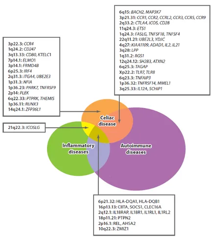
Figure II.2: Overlap of genetic risk factors for celiac disease with autoimmune and inflammatory diseases ... 44
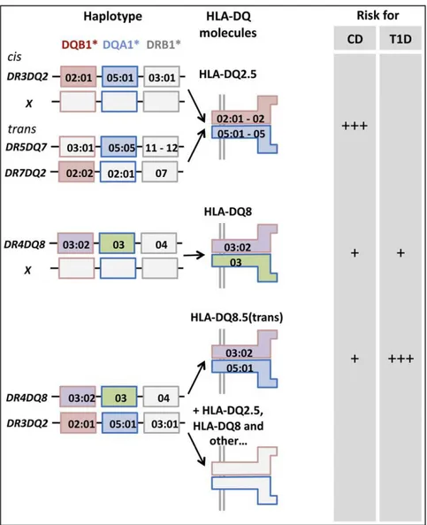
Figure II.3: Human leukocyte antigen (HLA) associations in celiac disease (CD) ... 46
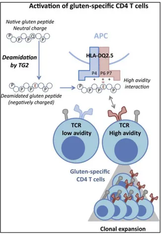
Figure II.4: Posttranslational modification of gluten ... 48
Figure II.5: Activation of gluten-specific CD4 T cells responses by HLA-DQ2.5 molecule ... 50
Figure II.6: B cell response in celiac disease ... 52
Figure II.7: T cell response in celiac disease ... 57
Figure II.8: History of celiac disease genetics ... 60
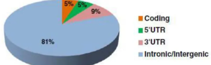
Figure II.9: Location and effect of CD risk single nucleotide polymorphisms (SNPs) ... 61
Figure II.10: Hypothetical role of non-HLA genes in celiac immune responses ... 62
Figure III.1: Effect of feeding mothers gluten-containing diet (ND) during pregnancy and/or breast-feeding on the immune response of their offspring in (Troncone and Ferguson, 1988) ... 68
Figure III.2: Experimental protocol of gluten-sensitization used in (Freitag et al., 2009) ... 70
Figure III.3: Schematic illustration of the generation of HLA-DQ8+. H-2Ab0 mice ... 75
Figure III.4: Schematic illustration of HLA-DQ8.hCD4.Aβ0.mCD40 mice generation and gluten-sensitization protocol used in (Black et al., 2002) ... 76
Figure III.5: Schematic illustration of gluten-sensitization protocol used in (Marietta et al., 2004) ... 77
Figure III.6: Regulated expression of the HLA-DR3-DQ2 haplotype encoded by yeast artificial chromosome. ... 78
Figure III.7: Schematic illustration of gluten-sensitization protocol used in (de Kauwe et al., 2009) ... 79
Figure III.8: Structure of the Dd-IL-15 transgene. ... 82
Figure III.9: Schematic illustration of experimental protocol used in (Abed et al., 2014) ... 85
Figure A.1: Experimental hypothesis ... 91
Figure A.2: Experimental mouse model (1) ... 92
Figure A.3: Experimental mouse model (2) ... 93
Figure B.1: Tetracycline on/off system ... 97
Figure B.2: Scheme depicting cooperation between IL-15 and diet-activated CD4 T cells in the activation of intestinal cytotoxic CD8 T cells ... 101
Figure C.1: Gating strategy for identification of small intestinal lamina propria mononuclear phagocytes (SI LP MNP) ... 109
7 Figure C.2: Comparative analysis of frequency and numbers of CD11c+CD11b- and CD11c+CD11b+ MNP
in WT B6, hIL-15Tge and IL-15KO mice ... 110
Figure C.3: Significant increase in frequency of CD103+ cells among CD11c+CD11b- but not CD11c+CD11b+ MNP in hIL-15Tge mice ... 111
Figure C.4: Strong increase of intensity of CD103 expression by CD11c+CD11b- cells but not CD11c+CD11b+ of ... 111
Figure C.5: Strong expansion of CD103+CD11c+CD11b- and CD103+ CD11c+CD11b+ cells in hIL-15Tge mice ... 112
Figure C.6: Increased CD86 expression by lamina propria CD11c+CD11b- and CD11c+CD11b+ mononuclear phagocytes in hIL-15Tge mice. ... 112
Figure C.7: No changes in CD40 and MHC class II expression by MNP in hIL-15Tge mice ... 113
Figure C.8: Increased CD103 expression on bone marrow differentiated dendritic cells in the presence of Flt3L and IL-15 accompanied by emergeance of NK cells... 114
Figure C.9: CD103 expression on DCs is not directly triggered by IL-15 but is mediated by IL-15-activated NK cells ... 115
Figure C.10: CD103 induction on DCs does not require cell-cell contact between DCs and NK cells ... 116
Figure C.11: GM-CSF can recapitulate the effect of IL-15-activated NK cells on the CD103 induction on DCs ... 117
Figure C.12: Csf-2 transcripts in duodenum of CD patients and controls ... 118
Figure C.13: Cross-presentation of OVA by DCs subsets from B6 and hIL-15Tge mice to OTI CD8 T cells ... 119
Table I-1: Routes of uptake of soluble and particulate antigens in the small intestine (SI) of mice. ... 27
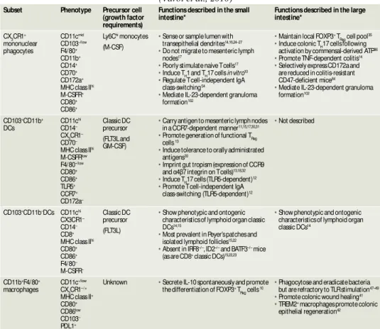
Table I-2: Intestinal lamina propria mononuclear phagocyte subsets in the mouse ... 31
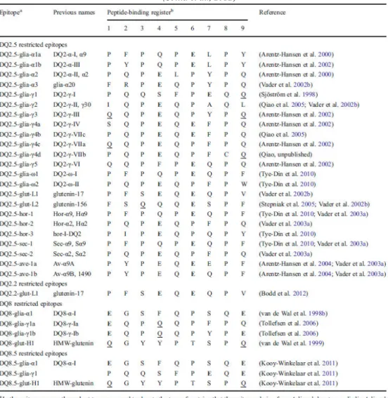
Table II-1: List of celiac disease relevant T cell epitopes recognized by CD4 T cells ... 49
8
List of Publications
Korneychuk, N., Ramiro-Puig, E., Ettersperger, J., Schulthess, J., Montcuquet, N., Kiyono, H., Meresse, B., and Cerf-Bensussan, N. (2014). Interleukin 15 and CD4(+) T cells cooperate to promote small intestinal enteropathy in response to dietary antigen. Gastroenterology 146, 1017-1027.
Korneychuk, N. (2014). [Innate lymphoid cells control the intestinal commensal bacteria adaptive response]. Medecine sciences : M/S 30, 253-257.
9
List of Abbreviations
ARA Anti-reticulin antibody
APC Antigen-presenting cell
BM Bone marrow
CD Celiac disease
CFA CFSE
Complete Freund’s adjuvant
Carboxyfluorescein succinimidyl ester
CX3CR1 CX3C chemokine receptor 1
DC Dendritic cell
DH Dermatitis herpetiformis
DT Diphtheria toxin
DTH Delayed type hypersensitivity
EAE Experimental autoimmune encephatilis
ELISA Enzyme-linked immunosorbent assay
EmA Anti-endomysium antibody
FACS Fluorescence activated cell sorting
FAE Follicle-associated epithelium
Flt3L FMS-like tyrosine kinase 3 ligand
Foxp3 Forkhead box P3
GALT Gut-associated lymphoid tissue
GAP Goblet-cell-associated antigen passage
GFD Gluten-free diet
GFP Green fluorescent protein
GM-CSF Granulocyte-macrophage colony-stimulating factor
GvHR Graft versus host reaction
GWAS Genome-wide association study
HEV High endothelial venule
HLA Human leukocyte antigen
HRP Horseradish peroxidase
IEC Intestinal epithelial cell
IEL Intraepithelial lymphocyte
IFN Interferon
Ig Immunoglobulin
ILF Isolated lymphoid follicle
iTreg Induced regulatory T cell
LP Lamina propria
LPL Lamina propria lymphocyte
M cell Microfold cell
MBP Myelin basic protein
MHC Major histocompatibility complex
10
MNP Mononuclear phagocyte
MIIC MHC class II-enriched compartment
nTreg Natural regulatory T cell
OVA Ovalbumin
PBL Peripheral blood lymphocytes
PCR Polymerase chain reaction
pDC Plasmacytoid dendritic cell
PI3K Phosphoinositide 3 kinase
Poly I:C Polyinosinic:polycytidylic acid
PP Peyer’s patches
RA Retinoic acid
SED Subepithelial dome
SI Small intestine
SIgA Secretory IgA
SNP Single nucleotide polymorphism
TED Transepithelial dendrite
TGF Transforming growth factor
TG2 Transglutaminase 2
Th T helper cell
Treg Regulatory T cell
TSLP Thymic stromal lymphopoietin
T1D Type 1 diabetes
11
General Introduction
In physiological conditions, robust immunological mechanisms avoid adverse responses to food antigens. Loss of tolerance to dietary antigens can cause different inflammatory diseases. In my thesis, I have focused on the pathogenesis of celiac disease, an autoimmune-like disease, in which loss of tolerance to dietary gluten leads to severe epithelial damage. CD pathogenesis has been studied in depth in humans. A first set of studies demonstrated that gluten-specific CD4 T cells were necessary but insufficient to trigger tissue damage. A second set of studies suggested that IL-15 over-produced in the intestine of CD patients was necessary to drive the expansion of cytotoxic CD8 T cells. It remains however unclear how these two mechanisms cooperate and whether they are sufficient to induce tissue damage. The largest part of the experimental work performed during my thesis addresses this question with the use of a mouse model. In the introduction of this manuscript, I review the data currently available in literature that have helped us to design experimental approaches and to interpret our results. In Chapter 1: Oral Tolerance, I discuss the mechanisms that control the uptake of soluble antigens in the gut and the mechanisms that allow the establishment and the maintainance of tolerance to dietary proteins in and beyond the intestine. In Chapter 2: Abnormal response to food gluten in Celiac disease, I summarize the advancements in the study of celiac disease pathogenesis and address the issues that remain unknown or uncertain to date. Finally, in Chapter 3: Animal models of Celiac disease, I analyze the previous attempts to set up a relevant mouse model of celiac disease, notably using HLA-DQ2.5 or HLA-DQ8 humanized mice, and discuss why they have remained largely unsuccessful. I notably address the question of the missing elements required to trigger tissue damage.
12
I. Chapter 1: Oral Tolerance
1.1 Introduction
Oral tolerance is defined as the specific suppression of humoral and/or cellular systemic immune responses to non-replicative antigens by administration of the same antigen through the oral route (Faria and Weiner, 2005). The hyporesponsive state observed after challenges with the same antigen can be demonstrated by the down-modulation of cellular responses, including delayed-type hypersensitivity (DTH) in vivo and lymphocyte proliferation in vitro, as well as by the inhibition of humoral IgE and IgG responses (Faria and Weiner, 1999). The fact that systemic tolerance to soluble dietary antigens is initiated in gut-associated lymphoid organs and avoids inadverant T cell responses in the intestinal mucosa allows extending the concept of oral tolerance to the local immune response. Importantly, the local regulatory T cell response does not inhibit the production of intestinal IgA against dietary proteins, an important mechanism preventing absorption of non-digested immunogenic proteins. Of note, the immune response to dietary soluble antigens differs from the response to microbiota by the outcome of peripheral responses after systemic injection of the antigen. In contrast to the systemic tolerance observed upon challenge with a soluble antigen initially given orally, systemic administration of a commensal bacterium triggers activation of spleen CD4 T cells and production of serum IgG (Slack et al., 2009). Oral tolerance therefore represents a major continuous immune process guaranteeing local and systemic tolerance to exogenous soluble (or non-replicative) antigens, by analogy with central tolerance assuring the unresponsiveness to self-antigens.
Since the pioneer experiments demonstrating active tolerance to orally ingested proteins performed over a century ago by Wells and then Besredka in guinea pigs, which became protected from anaphylaxis to hen’s egg or milk proteins by prior feeding of these proteins (Faria and Weiner, 1999), immunological mechanisms of oral tolerance have been progressively unknotted. Depending on dose of ingested antigen, nature of the antigen, duration of exposure and timing of administration, multiple mechanisms were shown to contribute to the establishment of oral tolerance, including anergy and deletion of antigen-specific T cells and induction of various types of regulatory T cells (Weiner et al., 2011). Regulatory T cells are now viewed as absolutely crucial in preventing systemic and local intestinal immune responses to dietary antigens (Pabst and Mowat, 2012), a view substantiated by the onset of severe food
13 allergy in patients lacking FoxP3 or in mice deficient for CNS1 unable to develop peripherally induced Tregs (Josefowicz et al., 2012; Torgerson et al., 2007) (see 1.6.2 Regulatory CD4 T cells). Loss of tolerance to dietary antigens can cause different diseases. The best characterized are IgE-dependent food allergies and celiac disease (CD). CD was defined in the past as type IV allergy and today is generally considered as an autoimmune-like disease induced by dietary gluten (see Chapter 2: Abnormal response to food gluten in Celiac disease).
In addition to an important protective role in physiology, oral tolerance may have a therapeutic potential. Indeed, simplicity and non-toxicity of this method based on oral administration of a specific antigen are particularly attractive (Wang et al., 2013). This approach has been successfully tested in multiple mouse models of autoimmune diseases (EAE, diabetes, arthritis, colitis, etc) but mainly as a preventive approach. It has however demonstrated only limited success in clinical trials for diabetes, multiple sclerosis, and rheumatoid arthritis, likely because it is more difficult to induce tolerance once a proinflammatory immune response has been initiated (Faria and Weiner, 2006). Better defining immunological processes implicated in oral tolerance might help for developing of therapeutic applications. Indeed, oral administration of antigen is currently tested for desensitization of IgE-dependent food allergies
Herein, I will present mechanisms that drive oral tolerance to dietary proteins. Since digestion and absorption of food proteins takes place in small intestine (SI), I will more particularly describe this part of the gastrointestinal tract, which contains the largest number of immune cells.
1.2 Gut immune system
The gut is anatomically divided into small intestine and colon (Figure I.1). The human intestine (Figure I.1) with a surface of more than 300 m2 (Moog, 1981) is the main physical barrier between internal and external environments. Food digestion largely takes place in the duodenum (Figure I.1), the short proximal part of the small intestine. Partially digested in the stomach, the food is further broken down by enzymes delivered with pancreatic juice into the duodenum. Bile and pancreatic juice secreted into the duodenum also neutralize the acidic pH of gastric content. The enzymes located in the brush border of epithelial layer throughout the small intestine participate in further protein degradation, which is completed into the lysosomal compartment of epithelial cells. The formation of villi considerably increases the absorptive and digestive surface of the SI (Figure I.2). This surface is further increased through the formation of folds, called Kerckring’s valves, and the development of microvilli at the apical
14 surface of enterocytes that form the brush border and harbour digestive enzymes. It is estimated that the intestinal surface can be thus multiplied by 600 fold (Figure I.2).
Figure I.1: Anatomy of gastrointestinal tract
Small intestine is located after the stomach and the colon. Small intestine consists of three segments: duodenum, jejunum and
ileum. http://missinglink.ucsf.edu/lm/ids_106_lowergi/lower%20gi/mainpages/smallintestine.htm
Figure I.2: Organisation of small intestinal mucosa
http://physiologie.envt.fr/spip/IMG/pdf/Phys_digest_14.pdf
The SI epithelium is made of a monolayer of intestinal epithelial cells (IECs) comprising four cell types: absorptive enterocytes representing approximately 80% of SI
Microvilli *600
Villi *30
15 epithelial cells, mucus-secreting goblet cells, hormone-secreting enteroendocrine cells and Paneth cells secreting antimicrobial molecules (van der Flier and Clevers, 2009) (Figure I.3, Figure I.4). All epithelial cells arise from stem cells in the crypts but, while Paneth cells remain at the bottom of the crypts, the three other cell types migrate up and form villi that are supported by an axis made of connective tissue, lamina propria (LP). LP contains blood vessels and lymphatics and remains separated from the epithelial layer by the basal membrane (Figure I.4).
Figure I.3: Types of intestinal epithelial cells
a. H&E staining showing the morphology of the mouse intestine. Signle layer of epithelial cells is organized into crypts and villi. Immunohistochemical analysis for main four differentiated cell types present in the intestinal epithelium: b, periodic acid-Schiff (PAS) to stain goblet cells; c, anti-synaptophysin to stain enteroendocrine cells; d, lysozyme to stain Paneth cells; e, alkaline phosphatase to stain enterocytes (van der Flier and Clevers, 2009).
Figure I.4: Structural organisation of intestinal mucosa
Schematic representation of the small intestinal barrier consisting of a mucosal layer (1) and gradients of IgA and microbial factors, epithelial cells (2) made up by enterocytes, Paneth cells, goblet cells and enterocrine cells, intraepithelial lymphocytes (γ), and the lamina propria (4). Secondary lymphoid structures, such as Peyers’ patches are present in the lamina propria
16 Digested peptides and nutrients are mainly absorbed in duodenum and jejunum (Figure I.1). The distal part of SI, is however indispensable for the absorption of bile salts and the concomitant absorption of liposoluble vitamins. Of note, due to the higher pH and to the lesser concentration in bile salts, the number of bacteria increases considerably in the ileum (about 108 organisms per gram of luminal content), compared to stomach and proximal portion of SI (103-105 per ml of luminal content respectively) but remains less than in colon (1010-1012 per ml) (Macpherson and Harris, 2004). In contrast to the large intestine, which is protected by two mucus layers, notably by a thick inner layer almost deprived of bacteria, the small intestine is protected only by a single partially discontinuous layer of mucus permeable to bacteria (Hansson, 2012). Interactions between bacteria and epithelial cells can thus become intimate in the ileum, notably with segmented filamentous bacteria. These interactions are thought to play an important role in the development of the lymphoid structures that are more particularly abundant in this part of the intestine. However, up to now it remains poorly understood to which extent the responses to microbiota overlap with the responses to dietary antigen, even if recent data suggest that some bacterial species can induce regulatory responses promoting tolerance to food proteins (Atarashi et al., 2013).
The intestine has evolved an elaborated defence system in order to cope with the vast spectrum of intraluminal antigens and to ensure its integrity and homeostasis. It is estimated that the gut contains 1012 immune cells, mostly lymphocytes (Mestecky and McGhee, 1987) that represents about 70% of all immune cells in mammals. These immune cells are organized within a complex gut-associated immune system comprising inductive and effector sites (Brandtzaeg et al., 2008) (Figure I.5). Inductive sites comprise gut-associated lymphoid tissue (GALT) composed of Peyer’s patches (PP) scattered along the mesenteric side of the SI and isolated lymphoid follicles (ILF) distributed throughout the mucosa of the small and large intestine (Mowat, 2003), but also gut-draining mesenteric lymph nodes (MLN) (Figure I.5). PP, ILF and MLN are sites where gut adaptive immune response can be initiated through the priming of naïve T and B cells by mucosal antigens processed by antigen-presenting cells (APC). Lymphocytes primed in gut-associated inductive sites follow a hemolymphatic cycle that allows them to colonize the whole intestinal length of the intestine, where they become mature effector cells, and to distribute in lamina propria and epithelium, effector sites of the gut immune system.
17
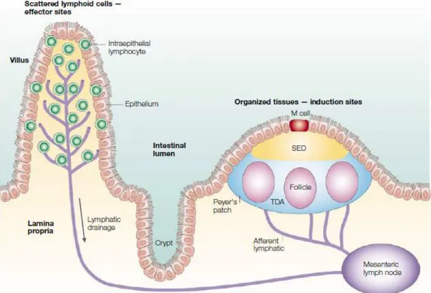
Figure I.5: Schematic representation of lymphoid elements of the intestinal immune system
The organized tissues of the Peyer’s patches and mesenteric lymph nodes (εδN) are involved in the induction of immunity and tolerance, whereas the effector sites are scattered throughout the lamina propria and the epithelium of the mucosa. Both Peyer’s patches and lamina propria are drained by afferent lymphatics that go to the MLN. SED, subepithelial dome; TDA-thymus-dependent area (Mowat, 2003).
Besides GALT, intraepithelial and lamina propria lymphocytes are distributed all over the intestine. The intestinal epithelium contains several subsets of intraepithelial lymphocytes (IELs) (Figure I.4), the origin of which remains still debated. IEL are heterogenous, and adults comprise mainly effector CD8 T cells displaying TCRα or TCR receptors. Due to their proximity to the gut lumen, IELs form a first line of defence. Thus, they are enriched in cytotoxic T cells that can be activated both via their TCR and/or via innate receptors. IELs can also secrete cytokines and may even exert regulatory functions (Cheroutre et al., 2011). Lamina propria contains much more diverse immune cell populations including resident macrophages, dendritic cells (DCs), and effector T cells, mainly CD4+ TCRα +, plasma cells, innate lymphoid cells, eosinophils and mast cells.
Peyer’s patches and isolated lymphoid follicles. Peyer’s patches are macroscopic
lymphoid aggregates located in LP and submucosa of small intestine. They are regularly scattered across the SI but are more particularly abundant in the ileum. Mature PP consist of B-cell follicles separated by T-B-cell zones (Figure I.6) (Mowat, 2003). B follicles are overlaid by the dome covered by the follicle-associated epithelium (FAE) that contains a specialized cell type, microfold cells (M cells), which lack microvillous brush border and basement membrane
18 (Figure I.6). M cells that differentiate under the influence of adjacent B cells are characterized by their capacity to capture and rapidly transport soluble and particular antigens under intact form from gut lumen into the subepithelial dome (SED) of PP where they can be taken up by APC (Figure I.6). The mechanism of the transepithelial antigen transport across M cells remains not fully delineated (Mowat, 2003). Dendritic cells loaded with antigens in the SED were shown to migrate to the T zone where they can present antigen to naïve B and T cells (Iwasaki and Kelsall, 2000). They are also likely to migrate to MLN through the efferent lymphatics. ILF resemble PP and notably possess an SED, but consist of only one or two B cell follicles, with no distinct T cell zone (Eberl and Lochner, 2009). As PP, ILF develop as a consequence of a genetically determined programme. In contrast to PP that develop in utero, ILF appear only during the post-natal period as cryptopatches. The full development of PP and ILF is triggered by intestinal bacterial colonization (Eberl and Lochner, 2009). Of note, some bacteria and notably segmented filamentous bacteria, but also inflammation, can stimulate de
novo development of tertiary lymphoid follicles well beyond the neonatal period (Lecuyer et al., 2014; Lochner et al., 2011).
Mesenteric lymph nodes. Mesenteric lymph nodes are the largest lymph nodes of the body. They acquire antigen-loaded dendritic cells migrating in a CCR7-dependent manner from the gut through the afferent lymphatics draining the intestinal mucosa and PP (Jang et al., 2006; Worbs et al., 2006). They can also acquire soluble proteins from the bloodstream captured by DCs numerous in the paracortex (Sixt et al., 2005). These DCs can prime naïve T and B cells entering MLN via high endothelial venules (HEV). Similarly to PP DCs, they can stimulate expression of the gut homing receptors α4 7 and CCR9 and thereby imprint lymphocytes for homing back into the gut mucosa (Agace, 2008) (Figure I.6).
T and B cells that are primed in gut associated lymphoid tissues (PP and ILF) and in MLN can indeed leave the gut wall and MLN via efferent lymphatics, circulate in the thoracic duct and home into mucosa due to their expression of gut-homing receptors. Primed lymphocytes may also localize in the spleen, which possesses a terminal circulation and can thereby collect all circulating lymphocytes (Figure I.6).
19
Figure I.6: Antigen uptake and priming of effector T and B cells
Antigen might enter through the microfold (M) cells in the follicle-associated epithelium (FAE) (a), and after transfer to local dendritic cells (DCs), might then be presented directly to T cells in the Peyer’s patch (PP) (b). Alternatively, antigen or antigen-loaded DCs from PP might gain access to draining lymph (c), with subsequent T-cell recognition in the mesenteric lymph noes (MLN) (d). A similar process of antigen or antigen-presenting cell (APC) dissemination to MLN might occur if antigen enters
through the epithelium covering the villus lamina propria (e), but in this case, there is the further possibility that MHC class II+
enterocytes might act as local APC (f). In all cases, the antigen-responsive CD4 T cells acquire expression of the α4 7 integrin
and the chemokine receptor CCR9, leave the MLN in the efferent lymph (g) and after entering the bloodstream through the thoracic duct, exit into the mucosa through vessels in the lamina propria. T cells, which have recognized antigen first in the MLN, might also disseminate from the bloodstream throughout the peripheral immune system. Antigen might also gain direct access to the bloodstream from the gut (h) and interact with T cells in the peripheral lymphoid tissues (i) (Mowat, 2003).
What are the respective roles of MLN, PP and ILF in oral tolerance? As highlighted by recent work from our laboratory, PP partially relayed by ILF play a crucial role for development of IgA and specific Th17 gut immune responses against the microbiota (Lecuyer et al., 2014). In contrast, several studies indicate that MLN, but not PP, are indispensable for inducing tolerance to the soluble antigens present in the intestinal lumen. Thus, H.δ. Weiner’s group has demonstrated that δTα-/- mice lacking both PP and MLN could not develop oral tolerance, while δT -/- or δTα/δT +/-
mice lacking only PP were orally tolerized (Spahn et al., 2001). Along the same line it was shown that mice prenatally treated with δT R-IgG that blocked PP development readily developed oral tolerance contrary to mice treated with TNFR I-IgG, which lacked both PP and MLN (Spahn et al., 2002). Similar conclusions were done in another study that introduced a soluble antigen in the lumen of ligated small bowel loops containing or not PP (Kraus et al., 2005). The impossibility to induce systemic tolerance (as
20 reflected by DTH) after surgical removal of MLN further emphasized the key role of MLN in oral tolerance (Worbs et al., 2006). Discrepant results were however obtained by another group that observed impaired systemic tolerance in mice lacking PP after prenatal treatment with δT R-IgG (Fujihashi et al., 2001). Moreover, they observed that M cell-dependent delivery of antigen favoured the increase in regulatory cells in both systemic and mucosal lymphoid tissues (Suzuki et al., 2008). Overall, it seems clear that MLN play a crucial role in oral tolerance. A complementary role of PP and perhaps of ILF is possible but may not be sufficient for the induction of oral tolerance.
1.3 Mechanisms of uptake of intraluminal soluble antigens
1.3.1. Role of M cells
M cells represent particular absorptive columnar enterocytes lacking surface microvilli that makes easier the uptake of large antigens. M cells are mainly found in follicle-associated epithelium of PP and ILF, but some cells similar to M cells are dispersed throughout of the epithelium, although their numbers remain low (Jang et al., 2004). Unlike enterocytes, M cells deliver luminal antigens to the SED via a transepithelial vesicular pathway largely protected from lysosomal degradation, thus allowing the transport of antigens under an intact form (Neutra et al., 2001). The lack of microvilli, but also the expression of specific receptors may favour adherence of bacteria and viruses to M cells (Schulz and Pabst, 2013). DCs located in SED may then capture luminal antigens transported by M cells and then participate in priming of naïve B and T cells locally in PP or after migrating to MLN (Iwasaki and Kelsall, 2000; Kelsall and Strober, 1996; Ruedl et al., 1996).
In contrast to well studied role of M cells in transport and initiation of immune responses to some bacteria and viruses (Schulz and Pabst, 2013), the exact role of M cells in the transport of protein macromolecules is less well delineated. A very ancient study showed that M cells may quickly take up horseradish peroxidase injected into normal mouse intestine (Owen, 1977). Furthermore, it has been shown that M cells may mediate the transport of immune complexes of secretory IgA (SIgA) bound to bacteria through a non-identified IgA receptor (Kadaoui and Corthesy, 2007). It is thus not excluded that this mechanism can participate to the transport of large proteins, for example gluten presented bound to SIgA.
As discussed above, PP are not likely to be obligatory for oral tolerance induction. Thus, one hour after intragastric administration, OVA was detected not only in PP but also in lamina propria dendritic cells, indicating that PP cannot be the exclusive site of entry of soluble proteins (Chirdo et al., 2005). Yet, villous M cells present at distance from PP in the villous
21 epithelium may perhaps allow the entry of proteins. Several other routes of entry and sampling have been suggested for soluble antigens, including paracellular diffusion between epithelial cells or transcellular transport across entrocytes, goblet-cell-associated antigen passages (GAPs) and uptake by intestinal mononuclear phagocytes (MNP) (Figure I.7). Their respective contribution, either in physiological or pathological situations, remains poorly delineated. We will examine below the different routes of entry of soluble antigens and how they may influence the onset or, on the contrary, the loss of tolerance to dietary antigens.
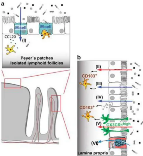
Figure I.7: Mechanisms of antigen uptake in gut-associated lymphoid tissue (GALT) and lamina propria
(a) In organized tissues of the GAδT such as Peyer’s patches and isolated lymphoid follicles, specialized microfold cells (ε cells) in the epithelium overlying the lymphoid follicles mediate transcellular transport of particulate material notably intestinal microbiota (I). This antigen is then passed onto dendritic cells that lie either below the epithelium or in the basolateral pocket of
M cell. Production of CCL20 by surrounding epithelial cells can attract further DCs via their expression of CCR6. (b) CD103+
DCs in the lamina propria underlying normal villus epithelium also play a critical role in presenting antigen for the induction of tolerance. They may acquire soluble antigens that have diffused through epithelial tight junctions (II), or that have been
transferred across epithelial cells by transcellular routes (III). Exosomes containing antigen derived from MHCII+ enterocytes
may be taken up by DCs (IV). CX3CR1high macrophages have also been reported to capture luminal antigens by extending
processes through the epithelial layer and they may pass this onto neighbouring CD103+ DCs, probably via gap junction
transfer (V). Soluble antigen may also be taken up by Goblet cells via goblet-cells-associated antigen passages (GAPs).
22
1.3.2. Role of enterocytes
Enterocytes play a major role in the absorption of nutrients. The absorption of various molecules of different sizes by enterocytes is termed intestinal permeability (Menard et al., 2010). Several mechanisms have been established, globally separated in two groups:
paracellular diffusion of small molecules (<600 Da, (Watson et al., 2001) through tight junctions between adjacent intestinal epithelial cells (IECs) (Figure I.8A) and transcellular
transport of macromolecules from apical to basolateral surface of IECs known as transcytosis (Figure I.8B) (Menard et al., 2010). Dietary proteins are largely degraded within intestinal lumen into small non-immunogenic peptides. Yet, some proteins or peptides can reach the epithelial surface in intact form, notably allergenic proteins and gluten-derived peptides that are characterized by their resistance to enzymatic digestion (Faria and Weiner, 1999).
Figure I.8: Paracellular and transcellular transport pathways
A. The paracellular pathway relates to structures joining adjacent intestinal epithelial cells and is delineated by tight junction, adherent junctions and desmosomes. Paracellular diffusion of molecules is mainly restricted by tight junctions, network of transmembrane proteins. Under steady-state condition, these highly regulated structures allow the diffusion of ions and inert molecules of small size <600 Da and can be increased in pathological situation. B. Transcellular transport pathways. Under steady-state condition, molecules of MW>600 Da, such as food antigens, peptides, are sampled by the epithelial cells by endocytosis at the apical membrane and transcytosis toward the lamina propria. During transcytosis, full-lenght peptides or proteins are partly degraded in acidic and lysosomal compartments and released in the form of amino acids (total degradation) or breakdown products (partial degradation) at the basolateral pole of enterocytes. Early endosomes containing partially degraded food antigens meet the major histocompatibility complex class II-enriched compartment (MIIC) where exogenous peptides are loaded on MHC class II molecules. Inward invagination of MIIC compartment leads to the formation of exosomes, small membrane vesicles (40-90nm) bearing MHC classII/peptide complexes at their surface. Exosomes can diffuse in the basement membrane and interact with local immune cells (Menard et al., 2010).
Paracellular pathway. The role of the paracellular pathway in the intestinal entry of immunogenic peptides has been suggested notably in inflamed gut. This pathway that depends on the very narrow pores formed by tight junction proteins allows the passage of ions and very small molecules across epithelium. It has been advocated notably by A. Fasano that in celiac disease immunogenic gluten peptides can however pass by paracellular diffusion through tight junctions (Fasano et al., 2000; Lammers et al., 2008; Tripathi et al., 2009). While there is ample
23 evidence that paracellular permeability is increased and that tight junctions are modified during intestinal inflammation, notably due to the action of inflammatory cytokines, there is as yet no formal demonstration that immunogenic peptides can pass through this pathway (Heyman et al., 2012; Menard et al., 2012). I will therefore focus on the role of transcellular pathways in the intestinal entry of dietary proteins.
Transcellular pathway. Internalisation of luminal macromolecules within enterocytes can be divided into two main mechanisms depending or not on the implication of a receptor at the apical cell surface.
Non-receptor mediated transcellular pathway. Large particles with size of 200nm up to 2µm can be endocytosed by IECs, and detected in LP within 5-30 min and in MLN within 30min-1h after oral administration (Hodges et al., 1995; Snoeck et al., 2005). Internalization of macromolecules by enterocytes involves clathrin- or caveolae-mediated or independent endocytosis, but is unlikely to be mediated by macropinocytosis or phagocytosis (Conner and Schmid, 2003). Most proteins or peptides internalized by passive endocytosis undergo strong degradation within IECs. In vitro analysis of transcellular transport of HRP molecules by HT29 epithelial cells using Ussing chambers has shown that only 0.1% of native proteins resisted to the degradation while the largest part was degraded into small peptides with average molecular weight of about 1,500 Da (Terpend et al., 1998). Interestingly, this molecular size is close to that of immunogenic peptides that can be uploaded to MHC class II molecules (Terpend et al., 1998). Further analysis by high-performance liquid radiochromatography (HPLC) demonstrated that about 40% of proteins applied at the mucosal surface of biopsies mounted in Ussing chambers were transformed by IECs into peptides of size compatible with MHC pockets, while over 50% were completely degraded into amino acids (Figure I.8B) (Menard et al., 2010). The fact that peptides processed by enterocytes had a size close to the size of immunogenic peptides led to suggest that the peptides may be protected from degradation through the binding to MHC class II molecules expressed by enterocytes. In keeping with this hypothesis, it was shown that enterocyte cell lines could present soluble proteins to CD4 T cell hybridoma (Brandeis et al., 1994; Hershberg et al., 1997; Kaiserlian et al., 1989). The in vivo relevance of direct presentation is however uncertain, knowing that IECs are separated from CD4 T cells by the basement membrane, but may perhaps occur in recall responses of the small subset of CD4+ IELs. Alternatively, experimental work performed in our laboratory has shown that IECs can release endocytosed antigens within exosomes, small membrane vesicles of 30-90nm size formed through the fusion of early endosomes and MHC class II-enriched compartments (MIICs) (van Niel et al., 2001) (Figure I.8B). Following this fusion, exosomes
24 may be formed by the inward invagination of MIIC membrane, thus leading to the formation of highly immunogenic peptide/MHC class II complexes on their surface. Exosomes released at the basal side of enterocytes (Figure I.8B) may then be captured by LP mononuclear phagocytes and thereby promote the antigen presentation. This mechanism can be probably enhanced in inflamed gut due to the increased expression of MCH class II molecules on epithelial cells, notably under the influence of IFN released by activated T cells.
Receptor-mediated transcellular transport of immune complexes. Small intestine harbors great numbers of antibody secreting cells. 80% of the plasmocytes are IgA-producing cells, 15-20% - polymeric IgM-secreting cells and 3-4% - IgG-secreting. IgE-secreting cells can be found only in food allergy or helminth infection (Brandtzaeg and Johansen, 2005). Polymeric IgA and IgM are transported from the basal to apical side of enterocytes by the polymeric IgA receptor and released in the lumen as secretory IgA (SIgA) or IgM bound to the extracellular part of the receptor (secretory component). The primary function of these antibodies is to neutralize bacterial and food antigens and to limit their entry to the mucosa. However, it has been suggested that these immune complexes may play a role in protected transport of dietary antigens via receptor-mediated route. The impact of this protected transport on the induction of consequent tolerogenic or proinflammatory response remains poorly understood. Three receptors have been respectively implicated in the transport of IgA, IgG or IgE immune complexes.
Studies performed in our laboratory have shown that in the pathological situation of coeliac disease SIgA may play a role in the retrotransport of large gluten peptides. In fact, the cross-linking of secretory IgA by poorly digested gluten peptides may lead to the formation of large complexes able to bind CD71. CD71 was first described as a high avidity receptor for transferrin, but can also serve as a weak-affinity receptor for IgA (Moura et al., 2001). CD71 expression is restricted to the basolateral surface of IECs in healthy individuals, but in celiac disease it can be induced also at the apical surface. It has been therefore shown that SIgA complexes could bind to apically expressed CD71 and allow the retrotransport of intact gluten peptides from the lumen into LP (Lebreton et al., 2012; Matysiak-Budnik et al., 2008) (Figure I.9A). In vivo study using mice that had upregulated CD71 apical expression and bore hybridomas secreting ovalbumin (OVA)-specific IgA suggested that OVA/IgA complexes could be transported via CD71 across the epithelium and favour the appearance of IFN -producing T cells in MLN in response to OVA (Abed et al., 2014). Moreover, the group of I. Moura has shown that binding of IgA1 to CD71 can induce the activation of proinflammatory MAPK/ERK-mediated signalling in mesanglial cells associated with glomerular damage in IgA
25 nephropathy (Tamouza et al., 2012). It should be therefore verified if the binding of gluten peptide/IgA complexes to CD71 in enterocytes could also induce proinflammatory pathways.
The neonatal Fc receptor (FcRn) was initially reported to play a role in protective immunity in suckling rats by allowing the absorption of IgG from maternal milk (Brambell, 1966; Jones and Waldmann, 1972). In humans, this receptor was also found at the apical and basolateral poles of adult intestinal enterocytes where it was shown to favour the bidirectional transport of IgG (Israel et al., 1997; Shah et al., 2003). As determined by in vitro and in vivo studies, FcRn-mediated transport of IgG bound to ovalbumin may protect the antigen from degradation (Figure I.9B) and favour the presentation of the antigen by CD11c+ DCs to OVA-specific CD4 T cells (Yoshida et al., 2004). Other data in newborn mice have suggested that the apico-to-basal transport of IgG/OVA immune complexes from maternal milk may participate in the establishment of oral tolerance (see 1.7 Open issues: Factors influencing oral tolerance induction) (Matson et al., 2010; Mosconi et al., 2010). The relevance of IgG-mediated transport in humans is unclear, given the very low concentration of IgG in human maternal milk and in intestinal secretion, but it is not excluded that it can have a pathogenic function during inflammatory bowel disease associated with a marked increase in intestinal IgG plasma cells.
Figure I.9: Immunoglobulin-mediated retrotransport of luminal antigens
A. In healthy individuals, undigested gliadin peptides are taken up by non-specific endocytosis in enterocytes and entirely degraded during transepithelial transport. In some pathological situations, notably in celiac disease, secretory IgA allow the protected transcytosis of gliadin peptides. The ectopic expression of CD71 at the apical membrane of epithelial cells favors the retrotransport of IgA immune complexes and inapproriate immune responses. B. IgG have been shown to bind the neonatal Fc receptor expressed on intestinal epithelial cells (FcRn). This receptor-mediated transcytosis allows a protected transport of IgG and their release on the basal side of enterocytes. IgG immune complexes can also be shuttled from basal to apical pole of the enterocytes. In food allergy, an overexpression of CD23 at the apical side of enterocytes can drive the transport of intact IgE/allergen immune complexes from the intestinal lumen to the lamina propria, a phenomen triggering mast cell degranulation and allergic inflammatory cascade (Menard et al., 2010).
Finally, IgE induced in food allergy may also play a role in the protected transport of dietary antigens and thus promote gut allergic responses. It has been shown that CD23 (Fc RII), the low-affinity receptor for IgE (Bonnefoy et al., 1987), can be up-regulated by IL-4
26 in rats (Yang et al., 2000; Yu et al., 2001) and overexpressed on IEC apical and basolateral surfaces in human cow’s milk allergy (Kaiserlian et al., 1993). Moreover, this receptor was shown to promote the translocation of IgE bound to HRP in sensitized rats and thereby to stimulate the degranulation of intestinal mast cells (Bevilacqua et al., 2004) (Figure I.9).
Contrary to pathways discussed below, the protected transport of dietary antigens mediated by IgA/peptide and IgE/peptide immune complexes is likely to be specifically induced in pathological situations, although the role of IgG/peptide complexes in induction of tolerance or inflammation remains unclear.
1.3.3 Goblet cells
One group has recently shown using a minimally disruptive in vivo imaging that small intestinal goblet cells (Figure I.3, Figure I.4) may deliver intraluminal fluorescent proteins (Mw<70kDa), like dextran or OVA, into lamina propria, a process that they termed goblet-cell associated antigen passages (GAPs) (McDole et al., 2012). Presence of goblet cells throughout the intestine makes this mechanism of potential interest. In vivo imaging suggested that the uptake of orally administered or intraluminally injected large molecules such as fluorescent OVA, dextran or LPS, occured via GAPs throughout the epithelium and via PP within 30min-1h after antigen administration, while IECs internalized only nanoparticles (<40nm) during this time lapse (Table I-1) (Howe et al., 2014). Imaging also suggested that antigens delivered through GAPs were captured by CD103+ LP DCs found in close contact with goblet cells, while CX3CR1+ DCs were much less efficient in this process (McDole et al., 2012). The factors that determine the preferential capture of soluble antigen by CD103+ DCs remain to be identified. Providing further support to the role of this pathway in loading LP DCs, it was observed that LP DCs isolated from Math1-/- mice specifically lacking goblet cells were not able to present OVA to OVA-specific CD8 (OTI) T cells. Yet, the molecular basis of this pathway needs to be further investigated. Although GAPs seem to be implicated in transport of antigen in physiological conditions, it remains unknown whether this pathway may amplify the antigen entry in pathological situations.
27
Table I-1: Routes of uptake of soluble and particulate antigens in the small intestine (SI) of mice
Analysis performed in living mice by in vivo imaging one hour after intraluminal injection of fluorescent molecules
(Howe et al., 2014)
1.3.4 Gut mononuclear phagocytes
Different studies have suggested that lamina propria CD11c+ mononuclear phagocytes (MNP) can directly take up bacterial antigen by extending dendrites inbetween epithelial cell tight junctions into the lumen (transepithelial dendrites, TEDs) (Chieppa et al., 2006; Niess et al., 2005; Rescigno et al., 2001) (Figure I.10). The dissemination of Salmonella enterica has thus been associated with CX3CR1+ DCs able to send dendrites into the lumen, while CX3CR1GFP/GFP mice, which were unable to send such dendrites, were protected from
Salmonella invasion (Hapfelmeier et al., 2008; Niess et al., 2005). One recent study (Farache et al., 2013) suggested that CD103+ DCs could also extend TEDs for sampling luminal bacteria, but this mechanism did not allow an efficient uptake of soluble antigens. It is therefore likely that TED-dependent mechanism is irrelevant to the uptake of proteins, probably because of the lack of proinflammatory signals required for the recruitment of DCs to the epithelium.
28
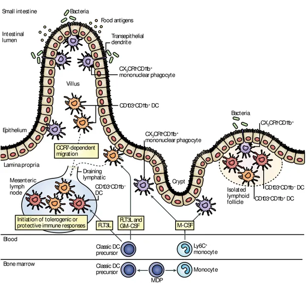
Figure I.10: The emerging intestinal lamina propria dendritic cell compartment
A common precursor, the macrophage-dendritic cell precursor (MDP), gives rise to classical DC precursor and monocytes in
the bone marrow, which enter the circulation and home to the gut. Locally, classical DC precursor give rise to CD103+ lamina
propria DCs that can be subdivided into a CD11b- and a CD11b+ population, which reside in the gut-associated lymphoid tissue
(GALT) and lamina propria. Differentiation of CD11b+ lamina propria DCs requires FMS-related tyrosine kinase 3 ligand
(Flt3L) and granulocyte-macrophage colony-stimulating factor (GM-CSF). Following antigenic challenge, CD11b+CD103+
lamina propria DCs migrate in a CC-chemokine receptor 7 (CCR7)-dependent manner to the mesenteric lymph nodes and
promote antigen-specific tolerance or protective immunity. Ly6C+ monocytes differentiate locally into CX3CR1+ lamina
propria mononuclear phagocytes that can penetrate epithelium by extending transepithelial dendrites. It remains controversial
whether CX3CR1+ lamina propria mononuclear phagocytes are able to migrate to lymph nodes but seem to have pro and
anti-inflammatory functions locally (Varol et al., 2010).
However, CX3CR1+ MNP may play an indirect role in the uptake of soluble antigens. One hour after injection of fluorescent ovalbumin into the gut lumen of anaesthetized CX3CR1+/GFP mice, a small part (about 10%) of lamina propria CX3CR1+ MNP, but not CD103+ MNP, took up the fluorescent dye (Farache et al., 2013; Schulz et al., 2009). Of note, a recent study has suggested that CX3CR1+ lamina propria MNP may not only participate in the capture of luminal antigens but may also in the uptake of blood-derived soluble antigens circulating in fenestrated capillaries located just beneath intestinal epithelium (Chang et al., 2013). Epithelium Crypt Isolated lymphoid follicle Intestinal lumen Villus Bacteria Bacteria Food antigens Transepithelial dendrit e CD103+CD11b+ DC CD103+CD11b– DC CD103+CD11b– DC CD103+CD11b+ DC CX3CR1+CD11b+ mononuclear phagocyte CX3CR1+CD11b+ mononuclear phagocyte CX3CR1+CD11b+ Small intestine Lamina propria Mesenteric lymph node Draining lymphatic CCR7-dependent migration Blood Bone marrow Ly6C+ monocyte Monocyte MDP Initiation of tolerogenic or
protective immune responses FLT3L FLT3L and GM-CSF
Classic DC precursor Classic DC precursor – M-CSF β ‘ ’ –/ β
Figure 2 | The emerging intestinal lamina propria dendritic cell compartment. A common precursor, the macrophage–
– —
–
–
29
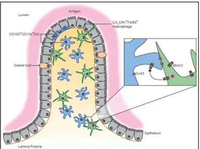
1.4 Role of monononuclear phagocytes in the presentation of
soluble dietary antigens
1.4.1 Comparison of the roles of CX3CR1
+and CD103
+MNP in
antigen priming
Despite the ability to take up fluorescent ovalbumin, CX3CR1+ MNP were suggested to have only poor in vitro and in vivo priming capacities and failed to induce gut-homing receptors on T cells (Schulz et al., 2009). In contrast, CD103+ MNP demonstrated much superior ability to prime and to induce CCR9 and α4 7 gut-homing receptors on T cells (Annacker et al., 2005; Jaensson et al., 2008; Johansson-Lindbom et al., 2005; Schulz et al., 2009). Moreover, due to their intrinsic capacity to produce retinoic acid (RA) and transforming growth factor (TGF ), CD103+ DCs were shown to be very efficient in inducing Foxp3+ CD4regulatory T cell from naïve T cells (Coombes et al., 2007; Scott et al., 2011; Sun et al., 2007a). On the basis of these results, it has been suggested that CX3CR1+ cells are resident antigen-capturing macrophages, while CD103+ MNP represent DCs able to migrate to MLN and to prime antigen-specific cells (Persson et al., 2010; Persson et al., 2013a; Varol et al., 2010). This concept was reinforced by the data suggesting that two subsets may have distinct ontogenic origins, CX3CR1+ cells may derive from circulating Ly6Chi monocytes and depend on M-CSF for their differentiation, while CD103+ cells may derive from bona fide DC precursors and depend on FMS-like tyrosine kinase 3 ligand (Flt3L) and granulocyte-macrophage colony-stimulating factor (GM-CSF) (Bogunovic et al., 2009; Varol et al., 2009). Although this concept seems to be generally accepted, several questions persist. First, it is unclear by which mechanisms CX3CR1+ MNP specifically take up fluorescent antigens. As TED-dependent mechanism is unlikely to contribute for the uptake of soluble proteins, other mechanisms should exist. It has been speculated that some CX3CR1+ cells are found close to IECs may capture antigens delivered to lamina propria, but this hypothesis has not been formally demonstrated. Second, since CD103+ DCs do not directly capture luminal antigens but are critical for the antigen transfer to MLN, it is unclear how and from which cells they receive the antigen. A recent study suggests that CX3CR1+ macrophages may deliver ovalbumin to CD103+ DCs through connexin 43-dependent gap junctions (Mazzini et al., 2014) (Figure I.11), but the in vivo relevance of this mechanism remains unknown. Third, two groups have shown that CX3CR1+ phagocytes may efficiently deliver at least bacterial antigens to MLN and promote tolerogenic or inflammatory immune responses (Cerovic et al., 2013; Diehl et al., 2013). CX3CR1+ CD11chigh LP MNP were also shown to efficiently cross-present OVA to CD8 T cells, which produced IL-10 and in