�����
���� ������������������
������������������������������������
��������� ���!������"������ �� "��#�$�% ��� � ����
%��"���������"� ��� ��� �
� � &������
������������ �������
����'������"�� � (����� �"�� ���"�� �������"�&)�����"����*����
(�+ "" ��"��� ��������"�����!���
������������������
,�,�$�,����-��.�� ���.�,����+&����-��"
������������
�,����/��������������
��������������������
�,�0�%�$�� ��� ���������,����-������� � �������0���+ � ����� ������'�������
%����1�� �����
�����������������
/��������/�,��
���
���������� ������������������� ������������������������������ �� ����������������� ����������%��"���������"� ��� ��� �
� � &������
������������ �������
����'������"�� � (����� �"�� ���"�� �������"�&)�����"����*����
(�+ "" ��"��� ��������"�����!���
,�,�$�,����-��.�� ���.�,����+&����-��"
��������������������
�,�0�%�$�� ��� ���������,����-������� � �������0���+ � ����� ������'�������
%����1�� �����
�����������������
/��������/�,��
���
���������� ������������������� ������������������������������ �� ����������������� ����������Actively transcribed genes can be the source of genome instability through numerous mechanisms. Those genes are characterized by the formation of secondary structures such as RNA-DNA hybrids. They are formed when nascent RNA exiting RNA polymerase II hybridizes single stranded DNA. Numerous studies have shown that RNA-DNA hybrids accumulation can lead to DNA damages. Among those damages, DNA double strand breaks (DSB) are the most deleterious for cells since they can generate mutations and chromosomal rearrangements. Two major repair mechanisms exist in the cell: Non-Homologous End-Joining (NHEJ) and Homologous recombination (HR). My lab showed recently that DSB occurring in transcribed genes are preferentially repaired by HR. Moreover, multiple studies have shown a cross talk between transcription and DSB repair. Those results led us to propose that actively transcribed genes could be repaired by a specific mechanism implicating proteins associated with transcription: “Transcription-coupled DSB repair”. During my PhD, using the DIvA (DSB Induction via AsiSI) cell line allowing the induction of annotated DSB through the genome, I worked on 2 projects focusing on DSB repair in transcribed genes.
First, we showed that DSB repair in transcribed loci requires a known RNA: DNA helicase: senataxin (SETX). After DSB induction in an active gene, SETX is recruited which allows RNA-DNA hybrid resolution (mapped by DRIP-seq). We also showed that SETX activity allows RAD51 loading and limits DSB illegitimate rejoining and consequently promotes cell survival after DSB induction. This study shows that DSB in transcribed loci require specific RNA-DNA hybrids removal by SETX for accurate repair.
Second, we showed an interplay between SETX and Bloom (BLM) a G4 DNA helicase in DSB repair induced in transcribed loci. We showed that BLM is also recruited at DSB in transcribed loci where it promotes resection and repair fidelity. Strikingly, we showed that BLM depletion rescued the survival defects observed in SETX depleted cells following DSB induction. Knock down of other G4-helicases (RTEL1, FANCJ) also promoted cell survival in SETX depleted cells upon damage. Those data suggest an interplay between G4 helicases and RNA: DNA resolution for DSB repair in active genes.
Altogether, these studies promote a better understanding of the specificity of DSB repair in transcriptionally active genes, and notably identification of proteins involved in “Transcription-coupled DSB repair”.
Les gènes transcriptionellement actifs peuvent être la source de l'instabilité du génome via de nombreux mécanismes. Ces gènes sont caractérisés par la formation de structures secondaires telles que les hybrides ADN : ARN. Ils se forment lorsque l'ARN sortant l'ARN polymérase II s'hybride au simple brin d'ADN. De nombreuses études ont montrées que l’accumulation de ces hybrides peut mener à la création de dommages à l'ADN.
Parmi ces dommages, les Cassures Double Brins (CDB) sont les plus dangereuses pour la cellule puisqu'elles peuvent produire des mutations et des réarrangements chromosomiques. Il existe deux mécanismes de réparation majeurs dans la cellule : la Jonction Non-Homologue des Extrémités (NHEJ) et la Recombinaison Homologue (HR). Mon équipe a récemment montré que les CDB localisées dans les gènes transcrits sont préférentiellement réparés par HR. De plus, de nombreuses études ont montrées une interaction entre transcription et réparation des CDB. Au vue de ces résultats, nous avons donc émis l'hypothèse que les gènes transcriptionellement actifs pourraient être réparés par un mécanisme spécifique nécessitant l'activité de protéines associées à la transcription : "Réparation couplée à la transcription". Durant ma thèse, je me suis intéressée au rôle de deux protéines dans la réparation des régions transcrites en utilisant la lignée cellulaire DIvA (DSB Induction via AsiSI) qui permet l'induction de cassures annotées sur tout le génome.
Premièrement, nous avons montré que la réparation des CDB dans des loci transcrits nécessitent une hélicase ADN : ARN connue : sénataxine (SETX). Après induction d'une cassure dans un gène, SETX est recrutée ce qui permet la résolution d'hybride ADN : ARN (cartographié par DRIP-seq). Nous avons aussi montré que SETX permet le recrutement de RAD51 et limite les jonctions illégitimes des CDB et par conséquent promeut la survie des cellules après induction des cassures. Cette étude montre que les CDB dans les loci transcrits requièrent la résolution spécifique des hybrides ADN : ARN par SETX pour permettre une réparation précise et est absolument indispensable pour la survie cellulaire.
Deuxièmement, nous avons montré une interaction entre SETX et Bloom (BLM) une G4 DNA hélicase dans la réparation des CDB dans les régions transcrites. Nous avons montré que BLM est aussi recrutée au CDB dans les loci transcrits où elle est nécessaire à la résection et à la fidélité de réparation. De façon importante, nous avons montré que la déplétion de BLM restaure le défaut de survie cellulaire observé dans les cellules déplétées pour SETX après induction des CDB. La déplétion d'autres hélicases G4 (RTEL1, FANCJ) promeut aussi la survie des cellules déplétées pour SETX après dommages. Ces résultats suggèrent une interaction entre les hélicases G4 et la résolution des hybrides ADN : ARN dans la réparation des gènes actifs.
En conclusion, ces études permettent une meilleure compréhension de la spécificité de la réparation des régions transcrites du génome, et notamment l'identification de protéines impliquées dans la "Réparation couplée à la Transcription".
Table of contents
Summaries ... 2 Table of contents... 5 Introduction ... 9 I. Transcription ...10 A. Steps of transcription...10 Initiation ...10 Elongation/Splicing ...11 Termination...12B. Non coding RNAs processing...12
Small RNA ...12
Long non-coding RNA...13
C. Secondary structure: R-loops ...13
Formation...14
Processing ...15
Detection techniques...16
D. Transcription induces genome instability ...17
Common fragile site and replication collapse ...17
Topoisomerase II activity ...18
R-loops ...19
R-loops in human diseases ...22
II. DNA double strand breaks repair...25
A. DSB detection and signaling ...25
B. Mechanisms...26
Non-Homologous End Joining...26
Homologous Recombination ...27
Alternative End-joining/Microhomology Mediated End-joining ...27
C. Factors influencing the choice between the 2 major repair pathways ...28
Chromatin state around DSB ...29
Cell cycle dependent...30
Type of breaks ...31
III. Transcription role in DSB repair ...33
A. RNA: active player for DSB repair? ...33
Transcription initiation following DSB induction ...33
Non-coding RNAs target DDR proteins at the break ...34
RNA template for repair during Homologous Recombination ...35
B. RNA: roadblock for repair ...36
Transcription inhibition: stopping RNA production at the break ...36
Removing RNA: DNA hybrids from DSB ...38
Results ...41
I. RNA: DNA hybrid resolution is crucial for DSB repair in active genes ...42
II. RNA: DNA and G4 helicases interplay for DSB repair in active genes ...43
A. BLM is recruited at DSB produced in G4-rich loci...43
B. BLM is mainly recruited at DSBs induced in active genes. ...44
C. BLM is recruited at HR-prone DSB and required for RAD51 loading ...45
D. SETX and BLM interplay for DSB repair...46
BLM has no impact on cell survival but is harmful in SETX depleted cells46 BLM promotes RNA-DNA hybrids accumulation ...47
Discussion ...49
I. RNA: DNA hybrid resolution is crucial for DSB repair in active genes ...50
A. RNA: DNA hybrids mapping ...50
RNA: DNA hybrids loss on the gene body...50
RNA: DNA hybrids accumulation around the DSB ...51
RNA: DNA hybrids function at DSB...52
B. RNA: DNA hybrids resolution required for repair ...53
C. In human diseases ...54
II. RNA: DNA and G4 helicases interplay for DSB repair in active genes ...55
A. BLM is recruited at DSB in active genes and required for HR...56
Bibliography ...59 Annexes ...74
I.
Transcription
DNA (DesoxyriboNucleic Acid) molecule carries the genetic information for all living organisms. This genetic information is held in DNA sequences called genes. In human, protein-coding genes produce around 20000 proteins by a mechanism called transcription. This mechanism is taken care of by the 12 sub-unit enzyme RNA Polymerase II leading to the production of a messenger RNA (mRNA) from the transcription of the DNA template. Transcription is a complex, dynamic and timely regulated process in the nucleus. This regulation occurs at every steps notably by the DNA structure and sequence itself but also by different factors such as chromatin and non-histone proteins (Shandilya and Roberts, 2012).
A.
Steps of transcription
Initiation
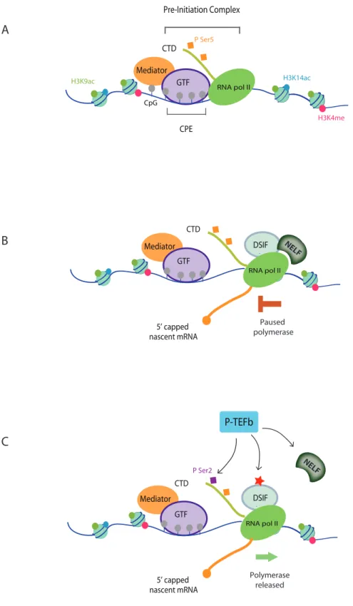
In mammalians, transcription is commonly initiated at the core promoter elements (Figure 1A). It is defined as the minimal contiguous DNA elements required for accurate transcription. This core promoter element can be located upstream or downstream of their targeted gene transcription sites. The first core promoter element discovered in 1979 is the TATA box (Goldberg, 1979). It can function independently or synergistically and is located about -31 to -30 relative to the transcription site. A second class of core promoter elements are CpG islands which are DNA stretches of CG dinucleotides (Deaton and Bird, 2011) (Figure 1A). They are preferentially located around transcription start sites and CpG methylation correlates with gene expression levels (Cross and Bird, 1995). Promoter regions with unmethylated CpG are usually active promoters whereas inactive/silenced promoters are characterized by important methylated 5mCpG (Boyes and Bird, 1992). TATA-box dependent
promoters represent a minority of mammalian promoters (10-16%) whereas a majority of human promoters are associated with CpG islands (around 70%) (Baumann et al., 2010). The promoter-proximal region is also characterized by histone marks specific of active transcription such as H3K9/K14 acetylation and H3K4 methylation (Figure 1A). Those epigenetics marks allow a more open and permissive chromatin (Li et al., 2007a).
The core promoter elements serve as a platform for the binding of the Pre-Initiation Complex (PIC) (Baumann et al., 2010; Roeder, 1996; Sainsbury et al., 2015). The PIC is composed of RNA polymerase II associated with the general transcription factors (GTF): TFIIA, TFIIB, TFIID, TFIIE TFIIF and TFIIH (Buratowski et al., 1989; Sainsbury et al., 2015) (Figure 1A). Those GTFs and the mediator complex are assembled with RNA polymerase II in a timely manner for a productive initiation.
Mediator Mediator Mediator GTF RNA pol II CTD H3K9ac H3K14ac H3K4me CpG P Ser5 Pre-Initiation Complex P Ser2 GTF CTD RNA pol II NELF DSIF Paused polymerase 5’ capped nascent mRNA RNA pol II NELF DSIF Polymerase released P-TEFb GTF 5’ capped nascent mRNA CTD
A
B
C
CPEFigure 1: Transcription initiation
A. Pre-initiation complex (PIC) assembly: Binding of the PIC is required for transcription inhibition.
The PIC is composed of the General Transcription Factor (GTF: TFIIA, TFIIB, TFIID, TFIIE TFIIF and TFIIH) together with mediator omplex and RNA polymerase II. They are recruited at the Core Promoter Element (CPE) which in a majority of human promoters are associated with CpG islands. Ser5 phosphorylation of RNA polymerase II CTD repeats is also necessary for initiation. Epigenetic modifications involved in initiation are H3K9/K14 acetylation and H3K4 methylation.
B. RNA Polymerase II promoter proximal pausing: 25 nascent RNA nucleotides are produced before Ser5P allows 5’end capping. DSIF and NELF binding lead to RNA polymerase II pausing.
C. RNA Polymerase II release: Positive-Transcription Elongation Factor b (P-TEFb) complex made of cyclin T1
and Cyclin Dependent Kinase 9 phosphorylates Ser2, NELF and DSIF (red star). NELF is then evicted from RNA polymerase II and DSIF becomes a positive factor. RNA polymerase II is then released from pausing.
Finally, post-translational modification of RNA polymerase II C-Terminal Domain (CTD) is also necessary for transcription initiation. In human, CTD is composed of 52 tandem hepta-peptide repeats of Y-S-P-T-S-P-S. One of the most important modification is Serines phosphorylation. Serines at positions 2, 5 and 7 are known to be phosphorylated at different steps of transcription. Notably Ser 5 needs to be phosphorylated to initiate transcription (Figure 1A) (Shandilya and Roberts, 2012).
After the transcription of around 25 nucleotides of nascent RNA, Ser5P recruits the capping enzyme allowing the addition of a cap structure to the 5' end and therefore productive transcription initiation (Figure 1B). DSIF and NELF proteins can bind and inhibit RNA polymerase II leading to promoter proximal pausing (Jonkers and Lis, 2015). RNA polymerase II is released from pausing by the Positive-Transcription Elongation Factor b (P-TEFb) complex Transcription elongation complex made of cyclin T1 and Cyclin Dependent Kinase 9 (Figure 1C). P-TEFb phosphorylates Ser2, NELF and DSIF. NELF is then evicted from RNA polymerase II and DSIF becomes a positive factor. Following release, RNA polymerase II proceeds across the coding gene to engage in elongation (Shandilya and Roberts, 2012).
Elongation/Splicing
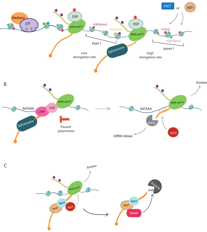
Gradual increase of Ser2 and the loss of Ser5 could explain RNA polymerase II increase elongation rates from 0.5kb per minutes within the first few kilobases to 2-5kb per minutes after 15 kb. Several other factors influence the elongation rate such as certain histone marks. Indeed, histone marks such as H2B ubiquitylation and H3K79 dimethylation are enriched in the first intron and associated with higher elongation rate whereas H3K36 trimethylation and H4K20 methylation are enriched in exons and associated with reduced elongation rate (Li et al., 2007a) (Figure 2A). Histone chaperones like FACT and ASF1 can also affect elongation rate. Finally, the factor with the strongest negative effect on elongation rate are exons. Transcription is delayed by 20-30 seconds per exon and this delay could be caused by their high GC content. This delay through the gene might be necessary for co-transcriptional splicing (Saldi et al., 2016). Indeed, most eukaryote genes are expressed as precursors mRNA (pre-mRNA) requiring a step of splicing to remove introns (non-coding sequences) and ligate exons together (Herzel et al., 2017). This mechanism is catalyzed in the nucleus by a multi-megadalton ribonucleoprotein complex called the spliceosome (Figure 2A). The main spliceosome building blocks are 5 U-rich nuclear small ribonucleoproteins (snRNPs) which are named after their small nuclear RNA component U1, U2, U4, U5 and U6. They are also associated with Sm proteins and are implicated in splicing in a sequential manner (Herzel et al., 2017; Saldi et al., 2016; Wahl et al., 2009).
Mediator GTF RNA pol II DSIF RNA pol II DSIF Spliceosome Exon 1 Intron 1 FACT ASF1 or H3K79me2 H2Bub H3K36me3 H4K20me Low elongation rate High elongation rate
A
AATAAA RNA pol II CPSF CstF Paused polymerase AATAAA XRN2 RNA pol II SETX EvictionB
Nrd1 Nab3 AAA TRAMP Exosome RNA pol II Nrd1 Nab3 Sen1 Eviction mRNA releaseC
SpliceosomeFigure 2: Transcription elongation/splicing and termination
A. Transcription elongation and splicing: RNA polymerase II is released while the rest of the PIC remains at the promoter as a transcription reinitiating scaffold. Elongation rate is influenced by histone marks in introns and exons. In exons, elongation rate is slow and associated with H3K36me3 and H4K20me whereas in introns, elongation is high and associated with H2Bub and H3K79me. Histone chaperones FACT and ASF1 promote histone marks specific to introns. Elongation delay contributes to co-transcriptional splicing by the spliceosome.
B. Transcription termination in mammalian cells: after transcription of the polyadenylation site, Cleavage and Polyadenylation Specificity Factor (CPSF) and the Cleavage Stimulatory Factor (CstF) activity leads to transcription pausing and to RNA
polymerase II eviction. In the torpedo model, after cleavage the RNA unprotected 5’ end still tethered to RNA polymerase II can be degraded by XRN2 5’-3’ exoribonuclease. RNA: DNA helicase SETX can promote XRN2 access to RNA by unwinding R-loop formed co-transcriptionally.
C. Transcription termination in S.cerevisiae: NNS pathway promote non-coding RNA termination by binding Nrd1 and Nab3 to neo-transcript RNA. This binding promotes Sen1 recruitment allowing RNA unwinding and RNA polymerase II. 3’ end non-coding RNA is then generated by endoribonucleotilyc cleavage or/and by the nuclear exosome/TRAMP complex and are lacking a polyA tail.
Termination
After elongation, transcription termination occurs when RNA polymerase II and the nascent mRNA dissociate from the DNA. There are 2 main pathways in eukaryotes. In human, mRNA termination is coupled with a maturation event in which the nascent RNA 3’ end is cleaved and polyadenylated (Figure 2B). The major effectors of termination are the Cleavage and Polyadenylation Specificity Factor (CPSF) and the Cleavage Stimulatory Factor (CstF) (Kuehner et al., 2011; Porrua et al., 2016; Porrua and Libri, 2015). After transcription of the polyadenylation site, their activity leads first to transcription pausing and then to RNA polymerase II release. Release of RNA polymerase II might also require the activity of XRN2, 5’-3’ exoribonuclease. In the torpedo model, creation of an unprotected 5’ end after cleavage event will allow XRN to degrade RNA still tethered to RNA polymerase II (Figure 2B). Their collision would promote termination. The RNA:DNA helicase SETX has also be involved in RNA polymerase II release by unwinding R-loops and access to XRN2 after nascent RNA cleavage (Wagschal et al., 2012).
In S.cerevisiae, the other major pathway dedicated for non-coding RNAs termination is the NNS (Nrd1-Nab3-Sen1) pathway (Figure 2C). In this case, 3’-ends of yeast non-coding RNAs are generated by endoribonucleotilyc cleavage or/and by the nuclear exosome/TRAMP complex and are lacking a polyA tail. In this model, RNA binding proteins Nrd1 and Nab3 serve as adaptors to position the SETX homolog Sen1. Sen1 can therefore unwind RNA and release the polymerase (Arndt and Reines, 2015).
B.
Non coding RNAs processing
In eukaryotes, RNA polymerase II is responsible for the transcription of most non-coding RNAs except for tRNA and rRNA. They are generated from the gene body but also from extragenic loci such as enhancers and upstream and antisense of mRNA genes. Those non-coding RNA are usually less than 1kb. They are highly unstable and restricted to the nucleus.
Small RNA
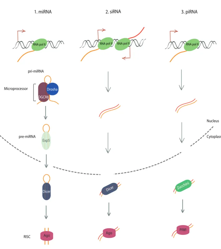
Small RNAs were first discovered in 1993 by genetic screens in C.elegans. They are highly conserved and have various functions such as heterochromatin formation, RNA silencing and translational control. Small RNAs involved in RNA silencing are by definition short, around 20-30 nucleotides and associated with Argonautes (Ago) family proteins. They are classified in 3 families in animals: micro RNA (miRNA), small interfering RNA (siRNA) and PIWI-interacting RNA (piRNA) (Castel and Martienssen, 2013).
RNA pol II RNA pol II RNA pol II RNA pol II Nucleus Cytoplasm Exp5 pre-miRNA pri-miRNA DGCR8 Drosha Microprocessor Dicer Ago RISC Dicer Ago Zucchini PIWI
1. miRNA 2. siRNA 3. piRNA
Figure 3: Small non-coding RNA processing
A. micro-RNA (miRNA) processing: RNA polymerase II generate short hairpin RNAs called pri-miRNA which are then cleaved by the microprocessor: Drosha and DGCR8 in the nucleus. Pre-miRNA forms a complex with exportin 5 which is exported in the cytoplasm. Finally, pre-miRNA is cleaved by Dicer and forms the RNA-Induced Silencing Complex (RISC) with Ago protein. B. Small Interfering RNA (siRNA) processing: dsRNA can be generated for example by convergent RNA polymerase II
transcription. They are exported to the cytoplasm and processed by Dicer and form a complex with Ago proteins. C. PIWI-Interacting RNA (piRNA) processing: piRNA precursors are produced as dsRNA and exported to the cytoplasm. They are processed by Zucchini and associated with PIWI proteins.
miRNA are generated from short hairpin RNAs that are successively processed by 2 RNAse III type proteins (Ha and Kim, 2014; Kim et al., 2009) (Figure 3A). In the nucleus, pri-miRNA maturation in pre-pri-miRNA is first initiated by Drosha in association with DGCR8, also called microprocessor complex. The pre-miRNA is then exported in the cytoplasm by forming a complex with the exportin 5 (EXP5). This complex translocates through the nuclear pore complex. The pre-miRNA is then finally processed by Dicer and then loading to an Ago protein to form the effector RNA-Induced Silencing Complex (RISC). siRNA are produced as dsRNA that are then processed in the cytoplasm by Dicer where they are also associated in a complex with Ago proteins (Ghildiyal and Zamore, 2009) (Figures 3B).
piRNA have a different processing and does not require the action of RNAse III type proteins or the loading on Ago proteins. On the contrary, they are processed in the cytoplasm by an endonuclease called Zucchini and then associated with PIWI proteins. (Figure 3C)
Long non-coding RNA
Long Non Coding RNA (lnc RNA) are very similar to mRNAs (reviewed in(Quinn and Chang, 2016). They are 5' capped, spliced and polyadenylated. The main differences between those 2 categories are their length, lnc RNA are usually no longer than 200 nucleotides and they do not have an Open Reading Frame (ORF). Their expression usually follows mRNA expression patterns therefore they are expressed in a cell type-, tissue-, developmental stage-or disease state-specific manner.They also are estimated to outnumber mRNAs. The function of a majority of lnc RNAs has not been identified however a few of them have been well described. For example, lnc RNA XIST (X inactive specific transcript) is necessary for chromosome X inactivation and TERC (telomerase RNA component) is required for telomere elongation. Their degradation is mediated by the exosome or by cytosolic nonsense-mediated decay. Moreover, lncRNA called TERRA (Telomere repeat-containing RNA) are also produced at telomeres where they control telomerase activity and heterochromatin formation at telomeres (Dianatpour and Ghafouri-Fard, 2017). Finally, two lncRNA are processed by RNA P: MALAT1 (metastasis associated-lung adenocarcinoma transcript 1) and NEAT1 (nuclear enriched abundant transcript 1) in mammals (Quinn and Chang, 2016). It has been proposed that they both participate in expression regulation: MALAT1 by regulating alternative splicing and NEAT1 by keeping mRNA in the nucleus for editing (Yoshimoto et al., 2016; Yu et al., 2017). Their overexpression has been associated with different types of cancer
C.
Secondary structure: R-loops
Transcription can produce secondary structure such as R-loops. R-loops are triple helix composed by an RNA-DNA hybrid highly stable and a single strand DNA. R-loops
are formed co-transcriptionally when RNA exiting RNA polymerase II hybridizes to DNA leaving the second DNA strand free. They differ from RNA: DNA hybrids forming transiently during transcription elongation by their span: from 100 to 2000 base pairs. They participate to various biological phenomenon such as transcription regulation and can generate DNA damages (Crossley et al., 2019; Santos-Pereira and Aguilera, 2015).
Numerous studies showed that this structure is not a rare event from transcription. Instead they form across the whole genome throughout the cell cycle and R-loops are highly conserved in bacteria, yeast and higher eukaryotes. Recent studies showed they are abundant structures: estimated occupation on the genome is up to 5% of the mammalian genome (Sanz et al., 2016) and 8% of the budding yeast genome (Wahba et al., 2016).
Formation
DNA sequence and conformation
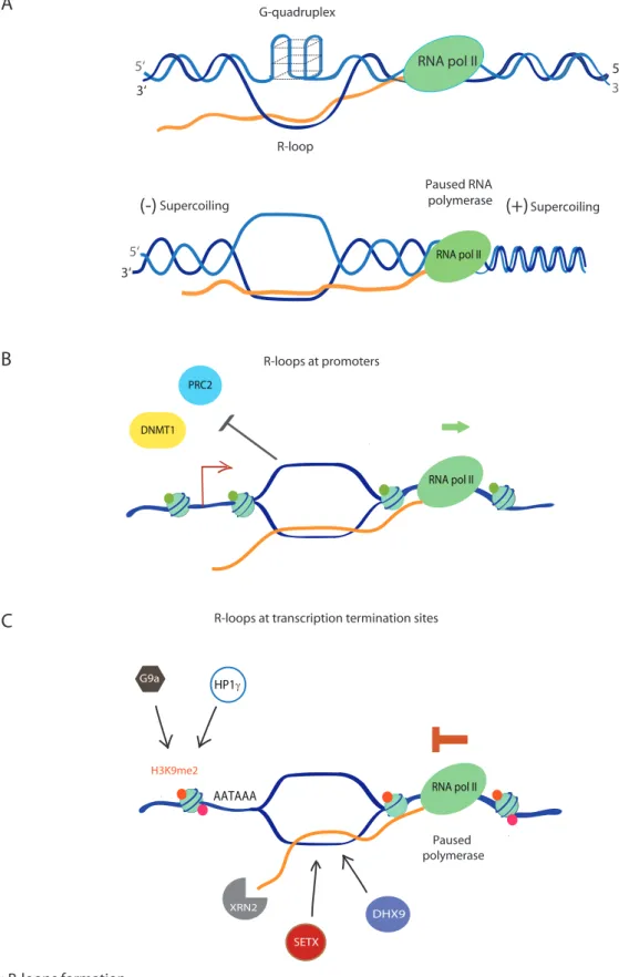
R-loops formation was first studied in vitro by the Davis lab in 1976 (Thomas et al., 1976). This formation is ensured by RNA: DNA hybridization being more stable than DNA: DNA hybridization. Sequence specificity takes part in this phenomenon. Indeed, it has been shown in vitro and in vivo that G-rich RNA are thermodynamically more stable with C-rich DNA than G-rich DNA. Moreover, studies showed that G-rich sequence can promote the formation of secondary DNA structure called G-quadruplex or G4 (Duquette et al., 2004). Only 4 consecutive guanines are necessary for the formation of G4. Interestingly, G4 can be formed co-transcriptionally and are associated with R-loops formation (Figure 4A) (Duquette et al., 2004). They could promote dsDNA “opening” by maintaining the non-templated ssDNA stabilized. (Hamperl and Cimprich, 2014; Quinn and Chang, 2016)
DNA conformation can also play a role in R-loops formation. It has been showed that RNA polymerase II passing through DNA duplex can create negative and positive DNA supercoiling. Positive supercoiling in front of RNA polymerase II can lead to transcription pausing/block whereas negative supercoiling behind RNA polymerase II can lead to an opening of the DNA duplex and facilitate RNA annealing (Figure 4A) (Corless and Gilbert, 2016).
Pausing
Several studied showed that RNA polymerase II pausing is associated with R-loops formation. For example, G4 genome-wide mapping in human cells has shown that promoters and some termination pause sites are highly enriched in G4 (Hänsel-Hertsch et al., 2018; Marsico et al., 2019). However, it is not clear if pausing is responsible for R-loops formation or
RNA pol II
R-loops at promoters
R-loops at transcription termination sites
DHX9 Paused polymerase XRN2 SETX G9a RNA pol II AATAAA
(+)
(-)
Supercoiling Supercoiling Paused RNA polymerase RNA pol II 5‘ 3‘ R-loop 5‘ 3‘ RNA pol II G-quadruplex 5‘ 3‘A
B
C
DNMT1 PRC2 H3K9me2 HP1γFigure 4: R-loops formation
A. Influence of the DNA sequence and conformation: top panel, R-loops formation can be promoted by G-rich sequences and notably by G-quadruplex (G4) formation on the ssDNA. G4 could promote dsDNA “opening” by maintaining the non-templated ssDNA stabilized. Bottom panel: RNA polymerase II elongation can produce supercoiling: negative behind RNA polymerase II leading to DNA “opening” and therefore RNA annealing, positive in front of RNA polymerase II leading to its pausing.
B. Influence of pausing at promoters: Genome-wide mapping of R-loops showed an accumulation at promoter proximal pausing sites. R-loops can act as an epigenetic mark allowing transcription regulation. Indeed, R-loops inhibits transcription silencing for example, by blocking DNMT1 DNA methyltransferase which as binds poorly to R-loops and therefore blocking methylation. R-loops can also inhibit chromatin remodelers such as polycomb repressive complex 2 (PRC2) and therefore inhibits transcriptional repressive marks.
C. Influence of pausing at transcription termination sites (TTS): Genome-wide mapping of R-loops also showed an accumulation at TTS. R-loops promote pausing by inducing repressing chromatin marks via G9a histone lysine methyltransferase recruitment (G9a). H3K9me2 is formed and allows HP1γ recruitment. R-loops resolution at TTS is dependent on RNA: DNA helicases SETX and DHX9. Following RNA degradation is achieved by XRN2.
on the contrary if R-loops formation leads to RNA polymerase II pausing. Nevertheless, a number of studies reveal a function of R-loops on those pausing sites (Mayer et al., 2017).
(1) At promoters
R-loops mapping showed that they accumulate at promoters (Chen et al., 2017; Sanz et al., 2016) and they are formed immediately after the transcription start site (Figure 4B) (Dumelie and Jaffrey, 2017). Their formation could be facilitated by chromatin opening at promoters (Boque-Sastre et al., 2015). They could act as an epigenetic mark allowing transcription regulation. Indeed, they can block transcription silencing for example, by inhibition of methylation as DNA methyltransferase binds poorly to R-loops (Grunseich et al., 2018). Studies have shown that R-loops can also inhibit chromatin remodelers and therefore inhibits transcriptional repressive marks (Boque-Sastre et al., 2015; Chen et al., 2015). By promoting chromatin opening, R-loops could facilitate transcription (Cloutier et al., 2016).
(2) At termination sites
They are evidences that R-loops might participate to efficient transcription termination (Figure 4C). R-loops mapping showed that they form at termination site (Sanz et al., 2016; Skourti-Stathaki et al., 2011). It has been proposed that they allow RNA polymerase stalling downstream of the polyadenylation sequence (Sanz et al., 2016). R-loops could promote RNA polymerase II pausing by inducing repressive chromatin marks via G9a histone lysine methyltransferase recruitment. Consequently, H3K9me2 is formed and heterochromatin protein 1γ is then recruited (Skourti-Stathaki et al., 2014). R-loops are then resolved by RNA-DNA helicases such as SETX (Skourti-Stathaki et al., 2011) and DHX9 (Cristini et al., 2018). Finally, the free RNA strand can be degraded by exonucleases such as XRN2 (Morales et al., 2016).
Processing
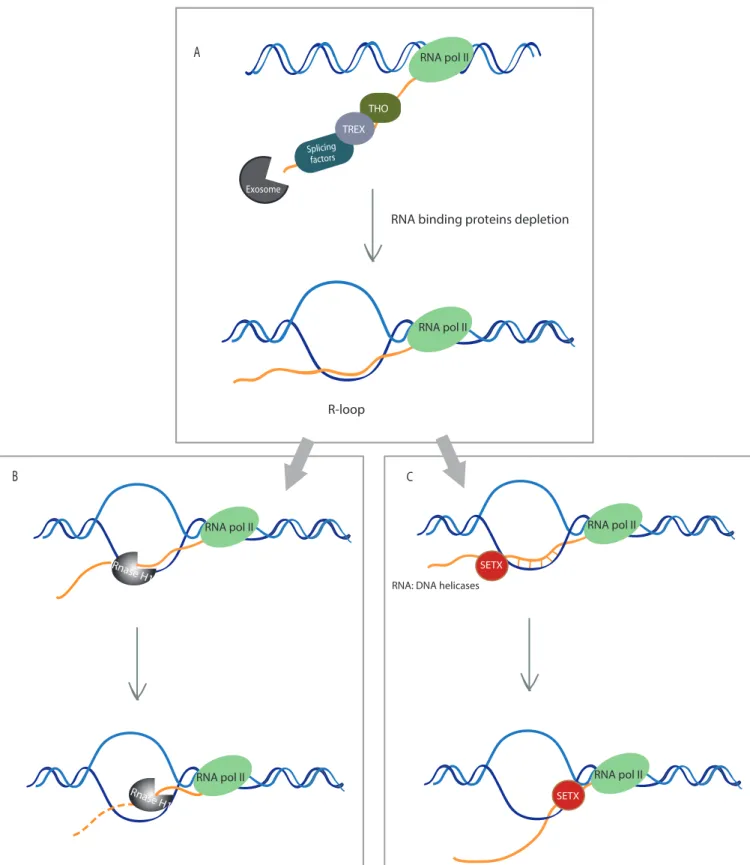
Numerous factors exist to prevent R-loops formation but also to promote R-loops resolution or degradation.
(1) RNA processing factors: Tho/TREX and mRNP
Prevention of R-loops formation requires to "coat" the nascent RNA, making it unavailable for hybridization with the template DNA (Figure 5A). Binding of RNA processing on the nascent RNA factors has been shown to prevent R-loops formation and mutations of those factors lead to R-loops accumulation. In yeast, mutants for RNA binding protein Pbp1 and protein involved in the Tho complex (mRNP biogenesis and elongation) and TREX complex (TRansport/Export) promote R-loops accumulation (Huertas and Aguilera, 2003;
Jimeno et al., 2006; Salvi et al., 2014). RNA surveillance protein such as trf4 has also been involved in R-loops resolution (Gavaldá et al., 2013). Depletion of splicing factors such as ASF1/SF2,BuGZ and Bub3 promotes R-loops accumulation as well (Li and Manley, 2005; Li et al., 2007b; Wan et al., 2015). Finally, the exosome complex through notably Rrp6 exoribonuclease activity is thought to resolve R-loops (Pefanis et al., 2015).
(2) Rnase H
Rnase H enzymes can specifically degrade RNA strand involved in R-loops (Figure 5B) (Cerritelli and Crouch, 2009). Both prokaryotes and eukaryotes encode for 2 different Rnase H: Rnase HI/RnaseH1 and RnaseHII/Rnase H2. Eukaryotic Rnase H2 is composed of 3 sub-units. The catalytic sub-unit is similar to the monomeric prokaryotic RNase HII whereas the 2 other sub-units are specific to eukaryotes with unidentified functions. Rnase HII/Rnase H2 also takes a role in removing ribonucleotides incorporated during replication.
(3) Helicases
Once R-loops are formed they can be resolved by helicases that unwind specifically RNA: DNA hybrids (Figure 5C). For example, during transcription termination R-loops can be resolved by Rho in bacteria (Hong et al., 1995) and by SETX/Sen1 (Becherel et al., 2013; Kim et al., 1999; Mischo et al., 2011) and DHX9 in eukaryotes (Cristini et al., 2018). Other RNA:DNA helicases have been identified such as Aquarius (AQR) (De et al., 2015; Sollier et al., 2014).
Detection techniques
Multiple techniques have been developed in order to map R-loops on the genome with next-generation sequencing (Crossley et al., 2019).
First method used to map R-loops is footprinting where bisulfites treatment allows the conversion of ssDNA cytosine in uracile which is in turn read as thymine by sequencing (Ginno et al., 2012b; Yu et al., 2003). This technique is controlled by Rnase H treatment to confirm ssDNA is R-loop specific.
Second, most widely used technique requires RNA-DNA hybrids pull down by the use of a specific antibody S9.6: DRIP (Ginno et al., 2012b). In this technique, cells are not fixed before pull down, and nucleic acids are digested by a cocktail of restriction enzymes. The pull down produce can also be analyzed by quantitative PCR. They have been variations in techniques deriving from DRIP-seq such as the use of sonication instead of enzymatic digestion (Wahba, Amon, Koshland, & Vuica-Ross, 2011; Wahba, Costantino, Tan, Zimmer, & Koshland, 2016). Indeed enzymatic digestion as shown to create a bias notably on genes
RNA pol II RNA pol II Rnase H1 SETX SETX RNA pol II RNA pol II Splicing factors THO TREX Exosome Rnase H1 RNA pol II
RNA: DNA helicases
RNA pol II A
R-loop
RNA pol II
B C
Figure 5: R-loops prevention and processing
A. R-loops prevention: RNA coating by RNA binding proteins can prevent its annealing with DNA. Among those proteins, proteins of the Tho/TREX complex, of the spliceosome and of the exosome has been identified. Depletion of those proteins leads to R-loops accumulation.
B. Degradation by Rnase H1: Once R-loops are formed they can be specifically processed by Rnase H1 which degrades RNA involved in RNA: DNA hybrids.
C. Unwinding by helicases: R-loops can also be removed by RNA: DNA unwinding by specific helicases such as SETX. Other RNA: DNA helicases have been identified such as DHX9, PIF1 and AQR.
5'end (Halász et al., 2017). However, sonication could also create damages in the ssDNA. Moreover, DRIP-seq is not strand specific therefore, new techniques allow either strand-specific DNA library preparation or the sequencing of RNA involved in R-loops(Sanz et al., 2016; Xu et al., 2017).
A combination of the 2 techniques has been developed: the bis-DRIP-seq (Dumelie and Jaffrey, 2017). This technique allows the detection of R-loops at the single nucleotide. It uses S9.6 to immunoprecipitate the R-loops but also use bisulfite to alter nucleotides in the ssDNA only as the other strand is shielded by RNA. Those modified nucleotides are then sequenced precisely and allows the exact R-loop localization.
Another limitation of DRIP related techniques is the use of the only antibody S9.6 (Halász et al., 2017; König et al., 2017). Moreover, it seems that this antibody also recognizes dsRNA (Hartono et al., 2018; Phillips et al., 2013). To go around S9.6 use, expression of catalytically inactive Rnase H is an alternate. Indeed, this approach allow the binding to R-loops (Chen et al., 2017; Ginno et al., 2012b). Chromatin is then cross-linked and Rnase H is immunoprecipitated. But even if this technique allows in situ binding, it could also have an impact on the dynamic of R-loops such as a stabilization of the structure.
All those techniques provide various tools allowing R-loops detection more or less precisely on the genome
D.
Transcription induces genome instability
It is admitted that transcription can be a threat to genome integrity. For example, it has been shown that transcription machinery can collide with the replication machinery. Transcription can also produce structure such as R-loops that can be targeted by enzymes such as AID or Topoisomerase II that can create breaks. Finally, it has been known that certain genomic loci are more susceptible to DNA breaks and recent genome-wide studies showed that actively transcribed genes are more fragile. This phenomenon has been particularly reviewed (Crossley et al., 2019; D'Alessandro and d'Adda di Fagagna, 2017; Marnef et al., 2017).
Common fragile site and replication collapse
In 1984, Common Fragile Sites (CFS) were first identified in lymphocytes (Glover et al., 1984). They are formed under mild replication stress and are characterized by gaps/breaks in metaphase chromosomes. They have been thoroughly studied because they match with translocation break points and are the source to gross chromosomal rearrangements in cancer cells (Marnef et al., 2017; Sarni and Kerem, 2016). CFS are conserved throughout the
evolution and present in every individual however, CFS breakage depends on cell type and source of replication stress (Le Tallec et al., 2011; Le Tallec et al., 2013). It has been shown that CFS instability is caused by incomplete replication during S-phase. CFS are devoid of replication origins, replicate late and 80% of them overlap long genes (>300kb) in human genome (Helmrich et al., 2011). These long genes might need more than one cycle complete transcription which could lead to collision between the transcription and the replication machineries and in term to breakage. However, as every long gene are not prone to breakage, in normal conditions replication forks may be able to resolve collisions (Helmrich et al., 2011). Under replication stress though, secondary structures as R-loops could form at CFS loci and lead to fork stalling and CFS instability (Helmrich et al., 2011; Marnef et al., 2017; Sarni and Kerem, 2016). However, it has been shown that the fragility of FRA3B, the most active CFS in human lymphocytes, is caused by a paucity of replication initiation and not fork slowing or stalling (Letessier et al., 2011). Moreover, CFS are associated with chromatin condensation that may impair DNA damage repair.
Another class of fragile sites was identified in lymphocytes B after hydroxyurea treatment (Barlow et al., 2013). They are Early Replicating Fragile Sites (ERFS) identified by ChIP-seq of DNA repair proteins RPA, BRCA1 and γH2AX. As for CFS, ERFS are cell type specific and detected as gaps on mitotic chromosomes. They are replicated early and localized in highly transcriptionally active regions, therefore instability in this case is rather caused by transcription than by replication timing.
Finally, other recurrent breaks have been identified at active genes promoters in lymphocytes B and neurons (Klein et al., 2011; Suberbielle et al., 2013; Wei et al., 2016). Those data suggest that endogenous breaks caused by transcription-replication collision can play a role during development.
Topoisomerase II activity
Transcription activity itself can lead to genome instability notably through Topoisomerase IIβ catalytic activity (Figure 6) (Drolet et al., 1995; Ju et al., 2006). DNA topoisomerases are enzymes solving DNA double strands over winding, downstream of replication or transcription machineries, by creating transient breaks (Bollimpelli et al., 2017). They are 2 different types of topoisomerases: type I creating single strand breaks and type II creating double strand breaks. Those topoisomerases are divided in sub categories. Notably, Topoisomerase IIA isoform β (TOP2B) is expressed in mammalians post-mitotic cells and has been implicated in transcription regulation (Bollimpelli et al., 2017). It has been proposed that following transcription activation, TOP2B would generate breakage to release RNA polymerase II previously blocked by DNA torsion and allow it to resume elongation (Bunch et
(+)
(-)
Replication-independent DSB formation at active genes
Transcription OFF Supercoiling Supercoiling Paused RNA polymerase RNA pol II Topoisomerase IIb RNA polymerase II release Impaired resealing Accidental DSBs RNA pol II Transcription OFF Transcription ON Stimuli 5‘ 3‘ RNA pol II
Figure 6: Topoisomerase-II-dependent DSB induction upon transcription stimulation
A large number of genes exhibit RNA polymerase pausing that contributes to transcription regulation. Upon certain stimuli, the release of paused RNA polymerase II into the gene body may require topoisomerase IIβ (TOP2B) activity. DSBs might accidentally arise following impaired resealing of TOP2B intermediates.
al., 2015). DNA breaks have been observed at early responsive genes following estrogen (Stork et al., 2016; Williamson and Lees-Miller, 2011) and androgen (Haffner et al., 2010) stimuli but also after heat shock or serum stimulation (Bunch et al., 2015). Interestingly, this mechanism seems to take place in a subset of immediate responsive genes after neuronal activity stimulation in mice primary cultured neurons (Madabhushi et al., 2015; Stork et al., 2016; Suberbielle et al., 2013). TOP2B break induction represent a replication-independent mechanism for genome instability associated to transcription.
R-loops
R-loops have been associated with genome instability in numerous studies (Crossley et al., 2019; Hamperl and Cimprich, 2014; Marnef et al., 2017; Skourti-Stathaki and Proudfoot, 2014; Sollier and Cimprich, 2015). Indeed, stabilization of R-loops by depletion of processing factors is a source of DNA damages. They are several ways that R-loops can generate DNA breaks. ssDNA displaced in R-loops formation can be a target for cleavage and following replication can generate DNA double strand breaks (DSB). Moreover, R-loops could be responsible for transcription-replication collision (García-Muse and Aguilera, 2016). Finally, a new system of R-loops elimination has been shown recently to generate DNA damage (Sollier and Cimprich, 2015; Sollier et al., 2014).
(1) R-loops stabilization promotes genomic instability
A considerable amount of studies showed that the absence of R-loops processing factors leads not only to R-loops accumulation but also to DNA damages.
Mutations of Rnase H1 and Rnase H2 leads to DNA breaks in yeast (Costantino and Koshland, 2018; Wahba et al., 2011). Rnase H2 mutants in mice also results in genome instability (Reijns et al., 2012). Remarkably, Rnase H2 sub-units mutations are responsible for the Aicardi-Goutières syndrome a severe neuroinflammatory disorder. Genomic instability caused by Rnase H mutation could be responsible for neurodevelopment defects observed in this disorder.
Helicases depletion is also a source of DNA damages (reviewed in Puget et al., 2019 (in revision)). In bacteria, depletion of the helicase RecG combined with Rnase H depletion is lethal (Hong et al., 1995). RecG is thought to prevent R-loops accumulation and to catalyze reverse branch migration of Holliday junctions. In yeast, it has been observed that Sen1 is necessary for R-loops resolution and to prevent DNA damages (Mischo et al., 2011). A similar role has been identified for a DHX9 helicase, responsible for transcription termination and to prevent R-loops associated damages (Cristini et al., 2018). Interestingly, it has been shown that G4 stabilization by pyridostatin treatment or G4 helicases depletion (depletion of BLM or
XPG
XPF
C Transcription-coupled Nucleotide Excision Repair
DSB
A Transcription/Replication machineries collision Paused polymerase DNA Pol DNA Pol RNA pol II B. ssDNA sensitivity Topoisomerase I AID G4 endonuclease CTIP? SSB CSB SSB Fork collapse Under-replication MitosisFigure 7: R-loops induced genome instability
A. Transcription/Replication machineries collision: G4 and/or R-loops could trigger RNA polymerase pausing that can collide with the replication machinery. Such collision can directly trigger DSB production.
B. ssDNA sensitivity: Displaced single-stranded DNA is more sensitive to various enzymes and nucleases. Topoisomerase 1 (Top1) is recruited to release superhelical stress in the R-loop’s DNA template and its cleavage-ligation activity can also be a source of damages. During Class Switch Recombination in activated B lymphocytes, ssDNA GC rich can be targeted by AID. Sae2/CtIp could target R-loops’ ssDNA in S phase. Single-strand DSB generated can be converted in DSb during replication or can lead to fork collapse.
C. Transcription-coupled Nucleotide Excision Repair: R-loops can be targeted by the endonucleases XPG and XPF,
components of the TC-NER machinery that generates a single-strand gap. Single-strand DSB lead to fork collapse and DSB generation during replication.
Pif1) can also generate DNA damages at transcribed regions (Rodriguez et al., 2012; van Wietmarschen et al., 2018). Moreover, depletion of Sgs1, BLM homolog, increases R-loops formation and transcription/replication collisions in yeast (Chang et al., 2017). Similar results were found in human cells in this study. A similar role was suggested for FANCJ, a protein involved in the Fanconi Anemia pathway (London et al., 2008; Wu et al., 2008). It can be argued that G4 helicases depletion in this case would lead to G4 stabilization, hence DNA opening and therefore R-loops accumulation.
Finally, mRNA processing factors are also guardians against DNA damages. In yeast, depletion of Tho/TREX complex sub-units generate genomic instability (Huertas and Aguilera, 2003; Jimeno et al., 2006). Depletion of splicing factors such as ASF/SF2 (Gavaldá et al., 2013; Li and Manley, 2005; Li et al., 2007b; Wan et al., 2015) and trf4, a protein involved in RNA surveillance, (Gavaldá et al., 2013) led to similar results.
Altogether, those studies show that R-loops stabilization generates genomic instability. A number of mechanism can explain how R-loops can threaten genome integrity.
(2) Collision with replication machinery
Transcription and DNA replication frequency in cells can lead to encounters and eventually collisions between the 2 machineries. Indeed, the replisome moving alone a single strand DNA cannot progress through the RNA polymerase II embracing the double strand DNA. Therefore, their encounter lead necessarily to conflicts and eventually DNA breaks and chromosomal rearrangements (García-Muse and Aguilera, 2016; Lin and Pasero, 2012). They are 2 different types of collision: head-on (HO) and co-directional (CO). In a HO collision, replisome and RNA polymerase II progress in opposite directions toward each other which may lead to forks collapse. On the contrary, in a CO collision, the 2 machineries move along the same direction however the collision can be caused by RNA polymerase II being slower (can be as low as 0.5 kb/min in human) than the replisome (2-3 kb/min in human(Méchali, 2010)). In this case, replication can resume if RNA polymerase II is removed from DNA (García-Muse and Aguilera, 2016; Lin and Pasero, 2012). Several factors have been identified as affecting collision such as DNA supercoiling, G-quadruplexes and R-loops. Those factors are thought to limit replication and transcription machineries progression and therefore promote conflicts between them (García-Muse and Aguilera, 2016).
Indeed, a large body of evidence show that R-loops affects genome integrity by colliding with the replication fork (Figure 7A). In breast cancer cells, estrogen was shown to induce R-loops accumulation and DNA damages notably in S-phase. Strikingly, transcription inhibition abolished DNA damages in the same conditions. Those data suggest that genome instability was caused by collision between replication machinery and potentially R-loops (Stork et al.,
2016). Moreover, factors involved in transcription termination and R-loops resolution such as Sen1 (Alzu et al., 2012) and Xrn2 (Morales et al., 2016) has been shown as protecting replication fork respectively in yeast and HeLa cells. More recently, a screen assessing replication capacity and replication stress sensitivity following protein depletion identified also pre-mRNA cleavage factors necessary for fork protection associated by counteracting (Teloni et al., 2019).
It has been observed that collision direction impacts both R-loops and DNA damages. Using both a reporter system and genomic data (DRIP-seq compared to GRO-seq from HeLa cells), they observed that CO collisions reduce R-loops and promote ATM signaling whereas HO collisions increase R-loops and promote ATR signaling (Hamperl et al., 2017).
To conclude, R-loops can produce genomic instability by colliding with the replication machinery.
(3) ssDNA sensitivity
ssDNA displaced during R-loop formation can be a target for various nucleases and enzymes that can modify DNA and eventually induce DNA damage (Figure 7B) (Hamperl and Cimprich, 2014; Sollier and Cimprich, 2015).
A classic example of this process takes place during Ig heavy chain Class Switch Recombination in activated B lymphocytes (Santos-Pereira and Aguilera, 2015; Skourti-Stathaki and Proudfoot, 2014). This event consists to recombination between 2 switch (S) regions where R-loops are formed (Yu et al., 2003). R-loops are essential for this process. Indeed, ssDNA displaced by R-loop formation is GC-rich and can be bind by enzyme from the APOBEC family: activation-induced cytidine deaminase (AID). AID deaminates dC to dU that can be processed by base-excision repair or mismatch repair machineries. Those processes can lead to nicks converted to DNA double strand breaks (DSB) during replication (Skourti-Stathaki and Proudfoot, 2014).
Topoisomerase 1 (Top1) cleavage-ligation activity can also be a source of damages (Hamperl and Cimprich, 2014). In Saccharomyces cerevisiae, Top1 is recruited at transcriptionally active regions (Lippert et al., 2011) which coincides with deletions hotspots of 2 or 3 nucleotides. It is presumed that Top1 is recruited to release superhelical stress in the R-loop’s DNA template (Takahashi et al., 2011). However, it is possible that Top1 stay trapped and form a covalent complex with the 5’ end of the nicked DNA. It is proposed that the nick is then processed by Rad1 and/or Mus81 into a gap (Takahashi et al., 2011).
If the single strand DNA (ssDNA) forms a G4, it could also be targeted by a specific G4-endonuclease. This type of enzyme has been characterized in human and is able to cleave the single strand 5’ region of the G4 (Sun et al., 2001) creating a single strand break (SSB).
Finally, R-loop’s ssDNA can be targeted by the Transcription coupled Nucleotide Excision Repair (TC-NER) pathway which would generate DSB (Figure 7C) (Sollier et al., 2014; Stork et al., 2016). Indeed, in human cells, following depletion of RNA: DNA helicases such as AQR and SETX, Cockayne Syndrome group B (CSB) promote the activity of XPF and XPG endonucleases (Sollier et al., 2014). Those enzymes can cleave either the RNA-DNA hybrids creating a SSB that can eventually be converted into DSB during replication or cleave the entire R-loops creating directly a DSB. Interestingly in human breast cancers cells, it was shown that estrogen treatment led to R-loops accumulation and DSB induction which was reduced by depletion of XPG and CSB (Stork et al., 2016). Those results suggest that TC-NER is also responsible of genome instability in human breast cancer cells by targeting R-loops.
More recently, it was proposed that Sae2/CtIP could target R-loops’ ssDNA in S phase (Figure 7B) (Makharashvili et al., 2018). Altogether, those studies suggest that ssDNA can be a target for various factors, leading to genomic instability.
R-loops in human diseases
Mutations of genes coding for proteins processing R-loops can not only generate genomic instability but they are also linked to human diseases such as trinucleotide repeat-associated diseases, cancers and neurological diseases (Groh and Gromak, 2014; Richard and Manley, 2017).
Tri-nucleotide repeat-associated diseases are caused by repeat expansions in specific genes (see table 1 for examples). It has been shown in vitro and vivo that transcription of those repeats lead to R-loops formation in bacteria and in human cells (Grabczyk et al., 2007; Loomis et al., 2014). Eventually, those R-loops can generate genetic instability such as repeat expansion or deletion (Lin et al., 2010; McIvor et al., 2010). This mechanism has been particularly observed in lymphoblastoid cell lines derived from Friedreich ataxia (FRDA) and Fragile X syndrome (FXS) patients (Groh et al., 2014). It was demonstrated that stable R-loops co-localize with expansion sites and repressive marks in these diseases (Groh et al., 2014; Loomis et al., 2014). Furthermore, it was showed that R-loops were responsible for the formation of chromatin repressive mark and transcriptional silencing in FXS (Colak et al., 2014; Groh et al., 2014). As repeats in those diseases are GC rich (Pearson et al., 2005), R-loops formation on expansion site could be facilitated by G-quadruplexes formation. They are over 40 genetic repeated-associated disorders; it would be of significant interest to see if this mechanism takes place in all those diseases.
Additionally, mutations of proteins involved in R-loops resolution have been identified as responsible for human cancers. In eosinophilic leukemia, inactivation of FIPL1, a polyadenylation factor, induces R-loop mediated damages (Stirling et al., 2012). Similarly, in
Name of the disease Gene mutated Gene symbol Repeats
Huntington’s disease Huntintin HTT CAG
Myotonic dystrophy
type 1 DMPK CTG
Dystrophia myotonica protein kinase
Ataxin 1 ATXN1 CAG
Spinocerebellar ataxia type 1 FMR1 CGG Fragile X mental retardation or Fragile X syndrome Fragile X mental retardation 1
Friedreich ataxia Frataxin FXN GAA
testicular germ cell cancer (or seminoma) downregulation of E3 ligase Bre1, actor of transcription elongation, creates DNA damages Rnase H1 sensitive, thus probably R-loops dependent (Chernikova et al., 2012). R-loops accumulation resulting in fork stalling and DNA breaks have also been observed in cells Fanconi Anemia (FA) patients, a disorder characterized by a high cancer risk (Schwab et al., 2015). Mutations of FANCJ, at the origin of breast cancer and Fanconi Anemia, lead to a stabilization of G-quadruplex which could be responsible for R-loops accumulation and eventually DNA damages in this disease (Wu et al., 2008). The same mechanism could participate to the Bloom syndrome, disease characterized notably by cancer predisposition. Indeed, as FANJ, BLM is a G4-helicase and its depletion leads to similar phenotypes (Chang et al., 2017; van Wietmarschen et al., 2018), and it has been suggested that they could interact with each other(Dhar and Brosh, 2018). Through the sequestration of the human TREX complex, Kaposi’s sarcoma-associated virus could also lead to R-loops induced DNA breaks (Jackson et al., 2014). This virus causes various AIDS-related cancers, meaning that human oncogenic viruses through R-loops accumulation could generate DNA damages that contribute to tumorigenesis.
Several neurological disorders have been associated with R-loops accumulation (Groh and Gromak, 2014; Richard and Manley, 2017). For example, Aicardi-Goutières Syndrome patients’ cells present a genome-wide increase of R-loops (Lim et al., 2015). It is an inflammatory disorder causing neurological damages in infants and caused by mutations in TREX1 (coding for a 3’-5’ exonuclease) or in Rnase H2 (Rice et al., 2007). Interestingly, increase of R-loops was localized in intergenic or gene body regions and not at 5’ or 3’ end of genes. This would indicate a different sort of R-loops than those forming naturally. Patients cells mutated for Rnase H2 are also associated with hypomethylation at R-loops forming sites (Lim et al., 2015). This epigenetic change could be responsible for the immune system response typical of the disease.
Furthermore, evidences suggest that certain cases of Amyotrophic Lateral Sclerosis (ALS), a severe motor neuron disease, could implicate R-loops (Salvi and Mekhail, 2015). One of the most common cause of ALS is an expansion of hexanucleotide GGGGCC in the first exon of C9ORF72 gene (DeJesus-Hernandez et al., 2011; Majounie et al., 2012). They are usually around 30 repeats of this hexanucleotide however in mutated genes it can be up to thousands of repeats. In vitro, it has been observed that those repeats can form G-quadruplexes and promote R-loops formation (Fratta et al., 2012). This hexanucleotide expansion has not been found in any other ALS disease however G-quadruplex formation through other GC repeat sequences could be responsible for some of the 50% ALS cases without known mutations.
Finally, mutations of senataxin (SETX) putative RNA: DNA helicase is responsible for 2 neurodegenerative diseases: dominant Amyotrophic Lateral Sclerosis 4 (ALS4) (Chen et al.,
2004) and recessive Ataxia with Oculomotor Apraxia type 2 (AOA2) (Moreira et al., 2004). They are characterized by degeneration of motor neuron, muscles weakness, cerebellum atrophy and are both declared during childhood. As reported previously, Sen1/SETX have been showed to protect the genome against genome instability by removing R-loops and allowing transcription termination (Alzu et al., 2012; De Amicis et al., 2011; Richard et al., 2013; Yüce and West, 2013). This mechanism could promote apoptosis that could account for neurons death in these diseases.
53BP1 53BP1 53BP1 MDC1 MDC1 53BP1 MRN MRN 911 911 ATM ATR MRN 911 ATM ATM ATR ATR 911 ATM ATM ATR ATR 53BP1 53BP1 53BP1 MDC1 MDC1 53BP1 MRN 911 ATM ATM ATR ATR DSB sensing DSB γH2AX Signal transduction ATM/ATR phosphorylation ATM/ATR phosphorylation
Figure 8: DSB detection and signaling
DSB repair begins by DSB detection by sensing proteins: MRN and 9-1-1 complex. Those sensing proteins promote
recruitment of ATM and ATR kinases which phosphorylates H2AX. γH2AX allows the recruitment of 53BP1 and MDC1 which in turn are phosphorylated by ATM and ATR, promoting signal transduction and amplification.
II.
DNA double strand breaks repair
Genome integrity is threatened by various damages. DNA double strand breaks (DSB) are the most deleterious damages for the cell as they can lead to mutations/chromosomal arrangements and eventually cell death. Repair pathways exist in cells to fix those breaks. Those repair pathways require chromatin modifications and action of various enzymes and proteins recruitment, depending of the cell cycle, cell type and localization of the break on the genome. This topic has been extensively reviewed and will be summarized here.
A.
DSB detection and signaling
First step of DNA damage response (DDR) is DSB detection (Figure 8). Sensors protein such as PARP1/2, Ku 70/80, the MRN complex (Mre11, RAD50, NSB1) or the 9/1/1 (RAD9, RAD1, HUS1) complex can be ligated to damaged DNA (D'Amours et al., 1999; de Murcia and Ménissier de Murcia, 1994; Paull and Lee, 2005).
Those proteins allow the recruitment of essential kinases from the PIKK (PhospatidylInositol 3-Kinase-related Kinases) family: ATM and ATR. They both have a serine-threonine kinase activity and a number of common targets. ATM is exclusively recruited and activated at DSB by NBS1 from the MRN complex (Lee and Paull, 2005). On the contrary, ATR can also bind SSB by binding with its partner ATRIP and RPA that recognizes ssDNA (Zou and Elledge, 2003).
ATM and ATR first and major target is H2AX histone variant (Figure8). They phosphorylate H2AX serine 139 (Rogakou et al., 1998), which is then called γH2AX. γH2AX is not only a strong marker of damaged DNA, spreading on several mega-bases around the break (Rogakou et al., 1999), but also a major recruitment platform for others DDR proteins. Among those proteins are 53BP1 (p53 binding protein) (Anderson et al., 2001) or MDC1 (Mediator Checkpoint 1) (Stucki et al., 2005) that recognize phosphorylated residues with their BRCT
(BRCA1 C-terminus Tandem) domain. Hence, those proteins are recruited on γH2AX and
phosphorylated by ATM which allows signal transduction (Figure 8). MDC1 participates to the recruitment of numerous DDR proteins (Stucki et al., 2005). The accumulation of γH2AX and all those DDR proteins at the break can be visualized as foci in the nucleus by microscopy.
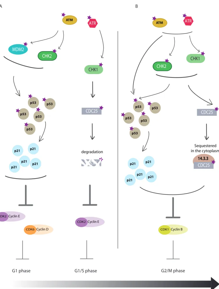
ATM and ATR kinase activity promote cell cycle arrest as well (Figure 9). This step is required in order to protect from damages transmission to the daughter cells.
• G1 block (Figure 9A): CHK2 (Checkpoint Kinase 2) (Ahn et al., 2000) and ubiquitin ligase MDM2 (Maya et al., 2001) phosphorylation by ATM limits p53 degradation, promotes its accumulation and activates its targets such as p21 transcription. Finally,
ATM ATR CHK2 MDM2 p21 p21 p21 p21 p21 Cyclin E CDK2 Cyclin D CDK6 CHK1 CDC25 Cyclin E CDK2 CDC25 degradation ATM ATR p53 p53 p53 p53 p53 p21 p21 p21 p21 p21 CHK1 CDC25 Cyclin B CDK1 CDC25 14.3.3 Sequestered in the cytoplasm CHK2
G1 phase G1/S phase G2/M phase
A B p53 p53 p53 p53 p53
Figure 9: ATM and ATR activity on cell cycle arrest
A. G1/S phase arrest: G1 arrest can be achieved by p53 stabilization. P53 can then induce p21 production and therefore inhibits Cdk/Cyclins involved in G1 to S phase transition. Cdc25a degradation can also take part in cell cycle arrest by blocking the activation of Cdk2/Cyclin E involved in this transition. (phosphorylations: purple star)
B. G2/M phase arrest: As for G1/S phase arrest, p21 accumulation inhibits Cdk1/Cyclin B. This arrest can also be achived by cdc25a sequestration in the cytoplasm by the 14.3.3 complex, hence blocking the activation of Cdk1/Cyclin B.
p21 inhibits Cdk (Cyclin dependent kinase) 2/cyclin E and Cdk4-6/cyclin D activity and blocks cell cycle in G1.
• G1/S transition block (Figure 9A): CHK1 and CHK2 phosphorylated by ATR and ATM respectively, can phosphorylate cdc25 phosphatase which leads to its ubiquitinylation and degradation. Cdk2/cyclin E is finally inhibited and the cell cycle is blocked (Deckbar et al., 2011; Warmerdam and Kanaar, 2010).
• G2/M transition block (Figure 9B): similarly to G1/S transition block, CHK1 and CHK2 phosphorylation by ATM and ATR leads to cdc25a phosphorylation. Cdc25a is then exported and sequestered in the cytoplasm by the protein 14-3-3. The complex cdk1/cyclin B is inhibited and the cell cycle blocked. Another possibility is the direct phosphorylation of p53 by ATM/ATR (or via CHK1/CHK2) leading to p21 expression and cdk1/cyclin B inhibition (Deckbar et al., 2011; Warmerdam and Kanaar, 2010).
B. Mechanisms
There are 2 major repair mechanisms, one requiring only the ligation or “joining” of both extremities together or one more complex, requiring a homologous sequence intact as template to repair the DSB.
Non-Homologous End Joining
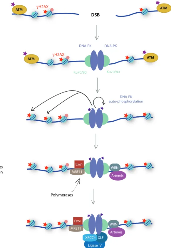
Non-homologous end joining (NHEJ) is the major DSB repair mechanism in human cells has it occurs all across the cell cycle. It consists only on the ligation of both extremities (Figure 10).
First step is Ku70 and Ku80 fixation at break (Walker et al., 2001). Those 2 proteins form a complex interacting with PI3K-like protein DNAPK extremities (Weterings et al., 2003). DNAPK catalyzes the phosphorylation of targets such as H2AX serine 139 but also its auto-phosphorylation (Costantini et al., 2007).
Some damaging agents can lead to bases modification making the direct ligation between extremities impossible. Therefore, NHEJ can require extremities maturation by various enzymes (phosphatases, nucleases or kinases) such as EXO1, MRE11, Artemis and WRN (Kusumoto et al., 2008; Weterings et al., 2009; Xie et al., 2009). Degradation by nucleases can sometimes lead to DNA synthesis of few nucleotides by DNA polymerase λ and μ to restore extremities (Capp et al., 2007; Lee et al., 2004). All those modifications can generate loss of fidelity.
The final step of ligation is achieved by XRCC4/XLF/DNA ligase IV complex (Ahnesorg et al., 2006). XRCC4 itself does not possess a catalytic activity however it is essential for DNA ligase IV recruitment at the break.
MRE11 Exo1 Artemis WRN Polymerases XLF XRCC4 MRE11 Exo1 Artemis WRN Ligase IV DNA-PK auto-phosphorylation DNA-PK DNA-PK γH2AX Ku70/80 Ku70/80 Extremities maturation Ligation DSB γH2AX ATM ATM ATM ATM
Figure 10: Non-Homologous End Joining pathway
Ku70/80 early recruitment at DSB promotes DNA-PK binding which maintain extremities together. DNA-PK phosphorylate H2AX and itself which promote the recruitment various enzymes (polymerases, phosphatases, nucleases or kinases) such as EXO1, MRE11, Artemis and WRN, hence leading to extremities maturation. Extremities ligation is achieved by
Homologous Recombination
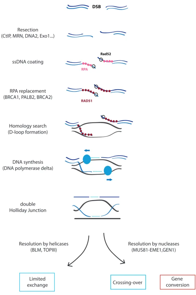
Secondary repair mechanism in human cells is Homologous Recombination (HR). It is known to be a more precise mechanism than NHEJ (Figure 11) as it requires a homologous sequence as template for repair.
First and crucial step for HR is resection by nucleases. Resection of the 5’ strand generates a 3’ tail ssDNA. It is initiated by MRE11 (Lavin, 2004) and CTIP (Sartori et al., 2007) and can be resumed by 2 mechanisms. First one requires the unwinding of ssDNA by RecQ helicases such as BLM or WRN followed by DNA2 nuclease activity (Gravel et al., 2008). Second mechanism requires simply EXO1 direct degradation of dsDNA in ssDNA (Nimonkar et al., 2011).
Resection is followed by coating and stabilization of ssDNA by RPA (Wold, 1997). RPA is then displaced by BRCA2 (Pellegrini et al., 2002), recruited by BRCA1 and PALB2 (Zhang et al., 2009), and RAD52. This displacement allows RAD51 recruitment on ssDNA (Conway et al., 2004) in order to achieve homology search: the search for a homolog sequence to the damaged one. It is followed by Rad51-ssDNA filament invasion of the dsDNA. This invasion generates a D-loop with the 3’ end as start for DNA synthesis by DNA polymerase, such as DNA polymerase δ, followed by ligation. The second damaged extremity is either engaged in the D-loop by independent invasion or captured by RAD52.
Those steps lead to the formation of a double Holliday Junction (dHJ) that can be resolved by 2 mechanisms.
• Resolution by nucleases: MUS81-EME1, GEN1 or resolvase A which can lead to crossing-over (CO, with important exchange information) or gene conversion (GC, limited exchange) (Heyer, 2004).
• Resolution by helicases: major mechanism, BLM and TOPIII-α activity leading to limited information exchange (neither CO or GC) (Raynard et al., 2006).
Alternative End-joining/Microhomology Mediated End-joining
In cases where NHEJ factors are missing or HR is blocked after the resection step, making it impossible to use NHEJ, DSB repair can be achieved by a rescue mechanism: alternative end-joining (Alt-NHEJ) or microhomology mediated end-joining (MMEJ). This mechanism seems to occur preferentially in S-phase (Truong et al., 2013) and is based on the ligation of microhomology sequences at each sides of the break. As for the other mechanisms, the DSB is detected and signaled at first, then it is followed by a 5’ ends resection as for HR leaving 3’ single strand exposed. As for HR, it appears that CtIP and MRN complex are required for the resection step (Rass et al., 2009). Those 3’ ends are 5-25 bp microhomologies are able to anneal together. Non-annealed DNA ends are eventually removed and it can be
DSB Rad52 PALB2 RPA RAD51 Gene conversion Crossing-over Limited exchange Resolution by nucleases (MUS81-EME1,GEN1) Resolution by helicases (BLM, TOPIII) double Holliday Junction Resection
(CtIP, MRN, DNA2, Exo1...)
ssDNA coating
RPA replacement (BRCA1, PALB2, BRCA2)
Homology search (D-loop formation)
DNA synthesis (DNA polymerase delta)
Figure 11: Homologous Recombination pathway
First step of HR is resection, then single stranded DNA is coated by RPA. RPA is replaced by RAD51 thanks to BRCA2. It is followed by Rad51-ssDNA filament invasion of the dsDNA for homology search. This invasion generates a D-loop with the 3’ end as start for DNA synthesis by DNA polymerase δ followed by ligation. Depending of the resolution of double Holliday junction, HR can lead to limited information exchange, crossing-over or gene conversion.