HAL Id: tel-01617631
https://pastel.archives-ouvertes.fr/tel-01617631
Submitted on 16 Oct 2017
Chiara Galdi. Design and development of multi-biometric systems. Signal and Image Processing.
Télécom ParisTech; Università degli studi (Salerne, Italie), 2016. English. �NNT : 2016ENST0015�.
�tel-01617631�
2016-ENST-0015 Università degli Studi di Salerno EDITE - ED 130
Doctorat ParisTech
Cotutelle de thèse entreTélécom ParisTech (France) et
L’Université de Salerno (Italie)
T H È S E
pour obtenir le grade de docteur délivré par
TELECOM ParisTech
Spécialité « Traitement du signal et des images »
présentée et soutenue publiquement par
Chiara GALDI
le 22 Fevrier 2016
Conception et développement de systémes
biométriques multimodaux
Directeur de thèse (France) : Jean-Luc DUGELAY Directeur de thèse (Italie) : Genoveffa TORTORA
Jury
M. Jean-Luc DUGELAY,Professeur, Eurécom Directeur de thèse
Università degli Studi di Salerno
Corso di Dottorato in Economia e Direzione delle Aziende Pubbliche - XIV ciclo
in cotutela con Télécom ParisTech Ecole doctorale EDITE - ED 130
Tesi di Dottorato
Design and Development of multi-biometric systems
Candidata Chiara Galdi
Tutors Coordinatore
Prof.ssa Genoveffa Tortora Prof.ssa Paola Adinolfi
Università degli Studi di Salerno Prof. Jean-Luc Dugelay
Télécom ParisTech
Contents
1 Introduction 11
Biometric recognition on mobile devices . . . 11
Thesis outline . . . 13
1.1 Authentication overview . . . 13
1.2 Biometrics overview . . . 14
1.3 Iris recognition overview . . . 16
1.4 Challenges . . . 20
1.4.1 ICE . . . 20
1.4.2 NICE . . . 20
1.4.3 MICHE . . . 26
MICHE I - Evaluation protocol . . . 27
MICHE II - Challenge protocol . . . 29
1.5 Commercial applications . . . 31
2 State of the Art 33 Behavioural biometric recognition on mobile devices . . . 33
Physical biometric recognition on mobile devices . . . 34
Iris detection and recognition on mobile devices . . . 35 5
6 CONTENTS
3 MICHE dataset 37
3.1 Acquisition Protocol . . . 38
3.2 Database composition . . . 39
3.2.1 MICHE Iris . . . 40
3.2.2 MICHE Iris - Fake . . . 41
3.2.3 MICHE Iris - Video . . . 41
3.2.4 MICHE Face . . . 42
3.2.5 MICHE Eyes . . . 43
3.3 Metadata . . . 43
3.4 Noise Factors . . . 44
4 Iris segmentation on mobile devices 47 4.1 ISIS . . . 48 4.1.1 Preprocessing . . . 48 4.1.2 Pupil location . . . 48 4.1.3 Linearisation . . . 50 4.1.4 Limbus location . . . 50 4.2 BIRD . . . 51 4.2.1 Pre-processing . . . 52
4.2.2 Watershed transform and binarization . . . 54
Watershed transform, region merging and colour quanti-zation . . . 55
Binarization of watershed transform . . . 56
4.2.3 Iris Detection . . . 57
Circle detection . . . 57
Limbus boundary refinement . . . 59
4.2.4 Experimental results . . . 61
Tan et al. technique . . . 62
4.3 Conclusive remarks . . . 67
5 Multi-biometric and multi-modal authentication on mobile de-vices 69 5.1 Combining iris and periocular area . . . 70
CONTENTS 7
5.1.1 Periocular region segmentation . . . 70
5.1.2 Iris and periocular area feature extraction . . . 71
CSUM algorithm . . . 72
5.1.3 Experimental results . . . 73
5.2 Combining Iris and Face . . . 77
5.2.1 Face recognition . . . 79
Acquisition and segmentation . . . 79
Spoofing detection . . . 81
Best template selection . . . 82
Feature extraction and matching . . . 84
5.2.2 Iris recognition . . . 85
Acquisition and segmentation . . . 85
5.2.3 Score level fusion of face and iris biometrics . . . 86
Confidence values . . . 87
Fusion schema . . . 89
5.2.4 Experimental results . . . 90
Data acquisition and preprocessing . . . 90
Performance of single biometrics . . . 90
Performance of FIRME with different confidence functions 92 5.3 Combining Biometry and Hardwaremetry . . . 94
5.3.1 Hardwaremetry . . . 95
5.3.2 Score Normalization . . . 99
Max-Min normalization technique . . . 99
Z-score normalization technique . . . 100
Median/MAD normalization technique . . . 100
TanH normalization technique . . . 100
Sigmoidal normalization technique . . . 101
5.3.3 Experimental results . . . 101
Data acquisition and preprocessing . . . 101
Sensor recognition . . . 101
Different sensors of the same model . . . 102
8 CONTENTS
Iris recognition . . . 104
Fusion at feature level . . . 106
Fusion at score level . . . 107
Noise response . . . 107
5.3.4 Conclusive Remarks . . . 108
Future Implications and Open Issues . . . 111
6 Gaze Analysis 113 6.1 Method . . . 114
6.1.1 Data acquisition . . . 114
6.1.2 Data normalization . . . 115
Face fiducial points detection . . . 116
6.1.3 Feature extraction . . . 117 Features graph . . . 119 6.1.4 Comparison . . . 123 6.2 Experimental results . . . 124 6.2.1 Experimental protocol . . . 124 6.2.2 Results . . . 125
6.2.3 Single feature experiments . . . 128
6.2.4 Combined features experiments . . . 128
6.2.5 Weighted combined features experiments . . . 130
6.2.6 Comparison with previous experiments . . . 130
6.3 Conclusive remarks . . . 131
7 Conclusions 135 7.1 Future perspectives . . . 136
A Résumé en français 139 A.1 Introduction . . . 139
A.1.1 Reconnaissance biométrique sur les appareils mobiles . . . 140
A.1.2 L’authentification . . . 141
A.1.3 La biométrie . . . 142
CONTENTS 9
A.2 La base de données biométrique MICHE . . . 146
A.2.1 Protocole d’acquisition . . . 147
A.3 Segmentation de l’iris sur les appareils mobiles . . . 149
A.3.1 ISIS . . . 149
A.3.2 BIRD . . . 150
A.4 Authentification multi-biométrique et multi-modale sur les ap-pareils mobiles . . . 152
A.4.1 Combinaison de l’iris avec la zone périoculaire . . . 152
A.4.2 Combinaison de l’iris avec le Visage . . . 153
A.4.3 Combinaison de la biométrie avec l’hardwaremétrie . . . . 153
A.5 Analyse du regard . . . 155
Chapter
1
Introduction
Biometrics can provide a higher level of security compared to other authentica-tion systems based on passwords or cards, however there are some issues related to the characteristics of biometrics themselves (some change substantially over time) or devices used to capture them (some can be fooled or they may have difficulties in acquiring the biometric trait) that deter their spread. Biometrics mostly used for the automatic recognition of people are fingerprints and face. The first is highly reliable but computationally expensive, while the second re-quires a well-controlled setting. We will see that the iris lends itself much better than other biometrics to reliable identification, but that applications on the market until today have been limited by the need to acquire the iris at a close distance and with cooperation from the user. For this reason, recent research investigates the use of iris recognition systems in the presence of noise in order to develop reliable systems that can acquire the iris at a distance and with little cooperation from the user. [27]
Biometric recognition on mobile devices
Biometric recognition for a long time has been used in confined spaces, usually indoor, where security-critical operations required high accuracy recognition
12 1. INTRODUCTION
systems, e.g. in police stations, banks, companies, airports (usually for fre-quent flyers, so just for a limited number of voyagers). Field activities, on the contrary, required more portability and flexibility leading to the development of devices for less constrained biometric traits acquisition and consequently of robust algorithms for biometric recognition in less constrained conditions [10]. However, the application of "portable" biometric recognition, was still limited in specific fields e.g. for immigration control, and still required dedicated devices. A further step would be to spread the use of biometric recognition on personal devices, as personal computers, tablets and smartphones.
Some attempts in this direction were made embedding fingerprint scanners in laptops or smartphones1. However, so far biometric recognition on personal
devices has been employed just for a limited set of tasks, as to unlock the screen using fingerprints instead of passwords, PINs, or patterns. One of the reasons is that systems presented so far can be easily spoofed, as the well-known hacking of the Touch ID on iPhone6.
In this thesis, the results of the study of new techniques for biometric recog-nition on mobile devices are presented. In particular, because of the background knowledge of the PhD candidate, the use of iris recognition has been investi-gated. Many aspects of iris recognition on mobile devices have been analysed, starting from the study of the issues related to the iris images acquisition process using mobile devices, that has led to the formulation of an acquisition protocol and the collection of an iris image database, namely the MICHE dataset, then analysing the challenges of iris segmentation on mobile devices, exploring the benefits of combining iris with other biometric traits or authentication items, and finally investigating its possible combination with a relative new biometric trait, i.e. the gaze.
It is worth noticing that each analysed aspect, has been then employed in the development of the others, e.g. the database has been used to test all the techniques developed, and the iris segmentation methods have been used as part of the recognition systems presented.
1.1. AUTHENTICATION OVERVIEW 13
Thesis outline
In chapter 2 "State of the art", an overview of the State of the Art in the field of the biometric recognition on mobile devices is presented; in chapter 3 "MICHE dataset", the MICHE dataset is presented, and its collection building process is described; in chapter 4 "Iris segmentation on mobile devices", two iris segmentation techniques, namely ISIS and BIRD, are presented; the works presented in chapter 5 "Multi-biometric and multi-modal authentication on mo-bile devices" illustrate two different approaches to combine iris recognition with another biometric trait or to combine iris recognition with sensor recognition; in chapter 6 "Gaze Analysis" the use of Gaze analysis for human recognition and for gender/age classification in order to verify its possible fusion with iris recognition has been investigated, since both biometric traits, iris pattern and gaze, come from the analysis of the eyes and thus can be captured at the same time.
The Thesis ends in chapter 7 with the conclusions and future perspectives.
1.1
Authentication overview
Authentication can be performed based on one or a combination of the following items:
• Something the user knows (e.g., password, personal identification number (PIN), secret answer, pattern);
• Something the user has (e.g., smart card, ID card, security token, software token);
• Something the user is or does (e.g. fingerprint, face, gait).
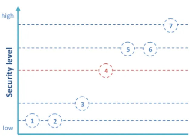
The last are known as biometrics and will be discussed in more detail later. For now we want to briefly analyse the security level associated with each type of authentication item or to combinations as well. As a premise, it is worth considering that passwords can be forgotten or snatched by malicious people,
14 1. INTRODUCTION
physical objects such as badges and ID documents can be lost or stolen, while biometrics can hardly be stolen and also the process of falsification is much more complicated (e.g. plastic surgery). The most recent biometric recognition systems also embed mechanisms to recognize live biometrics (liveness detection) and fakes (spoofing detection). If we consider all possible combinations of the three factors of authentication, we obtain the following ranking, from lower to higher security:
1. Something the user knows; 2. Something the user has;
3. Something the user knows + something the user has (e.g. ATM card + PIN);
4. Something the user is or does;
5. Something the user has + something the user is or does (e.g. biometric passport);
6. Something the user knows + something the user is or does;
7. Something the user knows + Something the user has + Something the user is or does.
Figure 1.1 shows the relative degrees of security. Biometrics by itself ensures an adequate level of security which may be increased by combining it with the other factors.
1.2
Biometrics overview
Biometric authentication is the process of human identification by their physio-logical or behavioural characteristics. These characteristic have to be distinctive and measurable in order to perform recognition.
Recognition can be performed in verification mode (1:1 matching, when the subject claims an identity that must be verified), or in identification mode (1:N
1.2. BIOMETRICS OVERVIEW 15
Figure 1.1: Security levels. (1) Something the user knows; (2) Something the user has; (3) Something the user knows + something the user has; (4) Something the user is or does; (5) Something the user has + something the user is or does; (6) Something the user knows + something the user is or does; (7) Something the user knows + something the user has + something the user is or does.
matching, or one against all, when there is no preliminary claim and the system must return the identity of a probe subject). Physiological biometrics include fingerprints, face, hand geometry, retina, DNA, veins pattern, ear and iris. Be-havioural biometrics are related to the particular behaviour of a person and may be influenced by one’s mood; they includes signature, speech, keystroke and gait. A good biometrics has to satisfy the following features: uniqueness, permanence, ease of use, good performance, accuracy, low cost, positive public perception. Iris optimally satisfies almost all of them.
Uniqueness: iris complex pattern can contain many distinctive features such as arching ligaments, furrows, ridges, crypts, rings, corona, freckles, and a zigzag collarette. It is proved statistically that iris is more accurate than even DNA matching as the probability of two irises being identical is 1 in 10 to the power of 78.
Permanence: the iris begins to form in the third month of gestation and the structures creating its pattern are largely complete by the eighth month, although pigment accretion can continue into the first postnatal years. Then it
16 1. INTRODUCTION
remains almost unchanged lifelong. Its position behind the cornea protects it from the environment.
Ease of use: iris is externally visible and automatic eyes detection is a relatively simple operation.
Performance: template obtained from iris are small and feature extraction and matching on iris are very fast operations.
Accuracy: the iris has the great mathematical advantage that its pattern variability among different persons is enormous.
Low cost: iris recognition devices may have limited costs and new noisy iris recognition systems that use simple camera devices will have even lower prices. Positive public perception: although iris image can be acquired without direct contact, iris based recognition systems are still perceived as intrusive [37].
1.3
Iris recognition overview
The identification process can be seen as a classification problem. A biometric trait can be reliably classified only if the variability among different instances of a given class is less than the variability between different classes. For example images of the same face have a high variability (intra-class variability) due to expressions, as well as being an active three-dimensional (3-D) object whose image varies with viewing angle, pose, illumination, accoutrements, and age. On the other hand face has a limited inter-class variability because different faces possess the same basic set of features, in the same canonical geometry. On the opposite iris inter-class variability is enormous and intra-class variability is low: as a planar object its image is relatively insensitive to angle of illumination, and changes in viewing angle cause only affine transformations. Even the non-affine pattern distortion caused by pupillary dilation is readily reversible [11].
The weakest element of iris recognition is the relatively low public accep-tance. Although iris acquisition is performed in a contactless way, related ap-plications are perceived as intrusive. Many system use NIR (Near Infra Red) illumination. Such kind of illumination is used because it is not visible and al-lows to light eyes up without annoying the users. However, even though studies
1.3. IRIS RECOGNITION OVERVIEW 17
confirm that a few seconds exposure to NIR ray does not damage eyes in normal conditions, it is not clear what could happen to eyes or skin with pre-existing pathologies, or what if a subject is accidentally exposed to NIR ray for a long time.
Almost all commercial systems, visible or NIR light-based, require the users to stand at a distance of up to 1 m (usually much less), in order to capture an high quality iris image. The need of standard conditions and cooperation of users, still limits the application fields for iris-based recognition. Therefore, new techniques for noisy iris recognition have been proposed. "Noisy Iris" relates to the quality of iris images on which recognition is performed [43]. They could present the following problems:
• Occlusions: eyelids, eyelashes, glasses, hair, etc.; • Reflections;
• Different size;
• Low resolution (due to device or distance);
• Different dominant colours in images of the same iris (due to different conditions during acquisition).
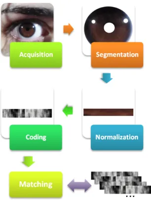
Such problems may especially arise if recognition is performed on subjects at a distance, on the move, unconscious of ongoing acquisition, as well as if there is not a standard illumination, or simply if a lower level of user’s cooperation is desired in order to speed up the identification process. Noisy iris recognition phases are the same used in controlled conditions, and therefore in "traditional" systems, even if they require different approaches due to image characteristics mentioned before. Such phases are: acquisition; segmentation; normalization; coding; matching.
Acquisition: with respect to traditional systems, acquisition is not nec-essarily performed with dedicated devices or high quality video camera. Iris images may be obtained from simple cameras, or standard acquisition equip-ment built in computers or mobile devices. Acquisition conditions
(illumina-18 1. INTRODUCTION
tion, distance, pose, etc.) are not strictly controlled, contrarily to traditional systems.
Segmentation: it is the process of identification of iris boundaries in order to extract only iris information from eye images. In traditional systems this is a relatively simple operation consisting in finding two circles matching with pupillary-iris and pupillary-sclera boundaries. With noisy iris, segmentation is much more complicated. It has to take into account the possible presence of occlusions or reflections, which must be discarded, in the sense that the cor-responding area has not to be considered for coding and matching. The iden-tification of the boundaries is further hindered by the low resolution or noise presence, which make boundaries less clear. For this reason noisy iris segmen-tation methods usually implement a preprocessing phase in which smoothing filter (to reduce noise) and/or enhance filter (to enhance feature such as iris boundaries) are applied [36][28].
Normalization: in traditional systems, due to the controlled acquisition condition, it is only needed to normalize the segmented iris form. Usual nor-malization implies transforming Cartesian coordinates into Polar ones. If colour information is taken into account, colour correction, histogram normalization or similar operations may be also useful.
Coding: this phase produces a template or feature vector, i.e., a compact representation of an iris image. Differences in the feature extraction algorithms when noisy irises are processed depend on the fact that in high quality images even tiny iris texture details are easily visible. On the contrary, noisy images may present altered or less characteristics to observe. The adopted methods for feature extraction in noisy iris images mainly analyse iris texture e.g. colour distribution, presence of lighter or darker region and can also combine a number of operator each computing a particular feature [29].
Matching: matching phase only depends on the kind of templates used. Figure 1.2 is an illustration of the five phases described above.
In the following we will describe some research initiatives aimed at evaluating results of current research on iris recognition.
1.3. IRIS RECOGNITION OVERVIEW 19
20 1. INTRODUCTION
1.4
Challenges
1.4.1
ICE
The National Institute of Standards and Technology (NIST) conducted and managed the Iris Challenge Evaluation (ICE) projects. ICE 2005 was the first public iris recognition contest. The goals were to promote the development of iris recognition algorithms and to evaluate the submitted solutions with a standard protocol in order to obtain a meaningful comparison of their performance [39]. For ICE 2005 a standard set of items were provided:
• a publicly-available dataset for algorithm development; • an experimental protocol for measuring performance; • the irisBEE baseline algorithm.
The database provided by ICE, including 2953 iris images from 132 subjects, was one of the largest publicly-available iris image database at that time. How-ever its images were captured with the aim of obtaining high quality samples and simulating the users’ cooperation in the image capturing process. There-fore, the noise factors in the ICE database are almost exclusively occlusions and poor focused images [39]. For ICE 2006 the performance evaluation of iris recognition algorithms was performed on sequestered data (data not previously seen by the researchers or developers).
1.4.2
NICE
NICE (Noisy Iris Challenge Evaluation) was born to promote the development of noisy iris recognition solutions. This iris biometric evaluation initiative of the SOCIA Lab. (Soft Computing and Image Analysis Group) of the Univer-sity of Beira Interior (Portugal), received worldwide participations [44]. The competition was performed in two phases:
• NICE.I (2007-2009): evaluated iris segmentation and noise detection tech-niques;
1.4. CHALLENGES 21
• NICE.II (2009-2011): evaluated encoding and matching strategies for bio-metric signatures.
The proposed methods were tested on a database provided by NICE itself: UBIRIS.v2 [42]. The UBIRIS database is one of the few iris image databases that contains realistic noise factors that make it suitable for the evaluation of ro-bust iris recognition methods [39]. It was developed within the SOCIA Lab. and released in September, 2004. The main feature of UBIRIS.v2 database is that eye images contain an high level of noise in order to simulate less constrained capturing conditions, e.g., acquisition at-a-distance, on-the-move, with minor cooperation or within dynamic imaging environments. Another important as-pect of this database is that iris images are taken on the visible wavelength in spite of the controlled databases in which acquisition is usually performed under controlled NIR illumination. NICE iris image database contains:
1. Out-of-focus iris images. Due to the limited depth-of-field of the camera. 2. Off-angle iris images. Obtained when the subject is not looking straight at the acquisition device. In this kind of images pupil and iris have an elliptical shape that needs to be taken into account during the detection of pupil and iris boundaries.
3. Rotated iris images. When the subject’s body/head is not in the vertical (natural) position.
4. Motion blurred iris images. Due to on-the-move acquisition or eyelids movements.
5. Iris occlusion due to eyelashes. 6. Iris occlusion due to eyelids.
7. Iris occlusion due to glasses, in particular to eyeglass frames and/or to reflections on the lenses.
8. Iris occlusion due to contact lenses. Contact lenses with high optical power can cause non-linear deformations of the iris texture.
22 1. INTRODUCTION
9. Iris with specular reflections. These reflections appear as small spots that obstruct the iris texture and are relatively easy to remove in the segmen-tation phase because they are usually much lighter than the iris.
10. Iris with diffuse reflections. These reflections are due to reflected informa-tion from the environment where the subject is located or is looking at. They can obstruct a large part of the iris.
11. Partial captured iris. The acquisition at-a-distance and on-the-move does not guarantee to capture the entire iris.
12. Out-of-iris images. In this case the system failed to capture the iris. How-ever the recognition process has to be able also to understand that in the image taken there is not an iris and for example require to repeat the acquisition.
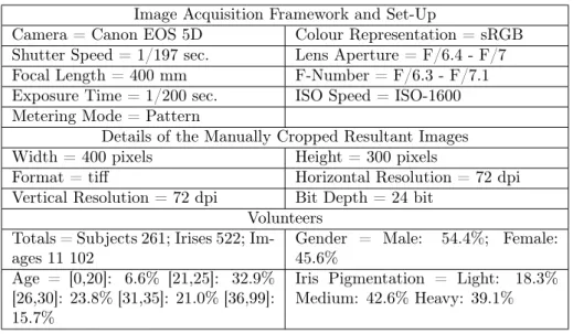
This database was downloaded by over 500 users (individuals and academic, researchers and commercial institutions) from over 70 different countries of the world [41]. The acquisition device used in capturing eye images of the UBIRIS database is a simple camera. Details of the imaging framework set-up are given in Table 1. It was installed in a lounge under both natural and artificial lighting sources. Volunteers were of different ethnicities:
• Latin Caucasian (approximately 90%); • Black (8%);
• Asian (2%).
They were only required to walk at a speed slightly slower than normal, in an area comprised between three and ten meters away from the acquisition device, and to look at few marks, located laterally with respect to the field of view of the camera, to simulate the behaviour of a non-cooperative subject that does not look straight at the camera. The large distance between the subject and the acquisition device is one of the main differences between the UBIRIS.v2 database and most of the remaining ones.
1.4. CHALLENGES 23
Two distinct acquisition sessions were performed, each lasting two weeks and separated by an interval of one week. From the first to the second session, both the location and orientation of the acquisition device and artificial light sources were changed. Approximately 60% of the volunteers participated in both imaging sessions, whereas 40% participated exclusively in one or the other.
Image Acquisition Framework and Set-Up
Camera = Canon EOS 5D Colour Representation = sRGB
Shutter Speed = 1/197 sec. Lens Aperture = F/6.4 - F/7
Focal Length = 400 mm F-Number = F/6.3 - F/7.1
Exposure Time = 1/200 sec. ISO Speed = ISO-1600 Metering Mode = Pattern
Details of the Manually Cropped Resultant Images
Width = 400 pixels Height = 300 pixels
Format = tiff Horizontal Resolution = 72 dpi
Vertical Resolution = 72 dpi Bit Depth = 24 bit Volunteers
Totals = Subjects 261; Irises 522; Im-ages 11 102
Gender = Male: 54.4%; Female: 45.6%
Age = [0,20]: 6.6% [21,25]: 32.9% [26,30]: 23.8% [31,35]: 21.0% [36,99]: 15.7%
Iris Pigmentation = Light: 18.3% Medium: 42.6% Heavy: 39.1%
Table 1.1: UBIRIS.v2 imaging framework
Both in NICE.I and NICE.II participants were required to submit an applica-tion executable written in any programming language and running in standalone mode. Evaluation for NICE.I (segmentation and noise detection) was performed using the following sets:
1. Alg denoted the submitted executable, which performs the segmentation of the noise free regions of the iris.
2. I = {I1, ..., In} was the data set containing the input close-up iris images.
3. O = {O1, ..., On} were the output images corresponding to the above
24 1. INTRODUCTION
4. C = {C1, ..., Cn} were the manually classified binary iris images, given
by the NICE.I Organizing Committee. It must be assumed that each Ci
contains the perfect iris segmentation and noise detection result for the input image Ii.
All the images of I, O and C had the same dimensions: c columns and r rows. Two measures of evaluation were used:
• The classification error rate (E1);
• The type-I and type-II error rate (E2).
The classification error rate (E1) of the Alg on the input image Ii(Ei) is
given by the proportion of correspondent disagreeing pixels (through the logical exclusive-or operator) over the whole image:
Ei= 1 (c × r) X c0 X r0 O(c0, r0) ⊗ C(c0, r0)
where O(c0, r0) and C(c0, r0) are, respectively, pixels of the output and class images. The classification error rate (E1) of the Alg is given by the average of the errors on the input images Ei:
E = 1 n
X
i
Ei
The value of (E1) belongs to the [0, 1] interval and was the measure of
evaluation and classification of the NICE.I participants. In this context, "1" and "0" will be respectively the worst and optimal values.
The second error measure aims to compensate the disproportion between the a-priori probabilities of "iris" and "non-iris" pixels in the images. The type-I and type-II error rate (E2) of the image Eiis given by the average between the
false-positives (FPR) and false-negatives (FNR) rates: Ei= 0.5 ∗ F P R + 0.5F N R
1.4. CHALLENGES 25
Similarly to the E1error rate, the final E2error rate is given by the average of
the errors (Ei) on the input images. The best 8 participants, that achieved the
lowest test error rates, were invited to publish their approach in a Special Issue on the Segmentation of visible Wavelength Iris Images Captured At-a-distance and On-the-move, Elsevier Image and Vision Computing 28 (2010).
The evaluation procedure for NICE.II (encoding and matching strategy) was the following:
• Let P denote the submitted application, which gives the dissimilarity be-tween segmented iris images.
• Let I = {I1, ..., In} be the data set containing the input iris images and
let M = {M1, ..., Mn} be the corresponding binary maps that give the
segmentation of the noise-free iris region.
1. P receives two iris images (and the corresponding binary maps) and outputs the dissimilarity value between the corresponding irises: P (Ii, MiIj, Mj) → D.D should be a real positive value.
2. Performing a "one-against-all" comparison scheme for each image of I gives a set of intra-class dissimilarity values DI = {DI
1, ..., DIk} and
a set of inter-class dissimilarity values DE = {DE
1, ..., DEm}, whether
the captured images are from the same or from different irises. 3. The decidability value d0(DI
1, ..., DIk, D E
1, ..., DEm) → [0, ∞[ was used
as evaluation measure:
d0= |avg(DI) − avg(DE)|/sqrt(0.5 ∗ (std(DI)2+ std(DE)2))
where avg(DI) and avg(DE) denote the average values of the
intra-class and inter-intra-class comparisons and std(DI) and std(DE) the
cor-responding standard deviation values.
Participants of the NICE:II contest were ranked from the highest (best) to the lowest (worst) decidability values [45].
26 1. INTRODUCTION
1.4.3
MICHE
A more challenging problem is faced by MICHE (Mobile Iris CHallenge Evalua-tion). As the name suggests, MICHE is an iris recognition technology evaluation that requires all the steps of the iris recognition algorithm to be performed on a mobile device (smartphones or tablets).
Performing biometric recognition on mobile devices is an important issue due to the need of a secure use of critical services (e.g. home banking) and to protect sensitive data that nowadays are mostly stored on our personal smartphones or tablets.
Iris is a natural candidate for mobile biometric recognition for two main reasons: iris acquisition is little intrusive, and iris codes are among the less expensive templates from the storage point of view.
MICHE contest, is the result of the collaboration of the BIPLab (Biomet-ric and Image Processing Lab) from the University of Salerno (Italy) and the SOCIA Lab. (Soft Computing and Image Analysis Group) of the University of Beira Interior (Portugal). MICHE will include two phases:
- MICHE I (2014-2015): participants are required to provide both their executable programs and the dataset they used for their experiments, as well as the characteristics of the devices used for acquisition and testing. They can submit their results related to one or all of the steps of an iris recognition system performed on a mobile device (detection, segmentation, recognition) as well as applications of iris biometrics on mobile devices; - MICHE II (2015-2016): the collected dataset is used to as test-bed for
a challenge which is accessible for both original authors and new groups [46].
Performing iris recognition on mobile devices may introduce many noise factors while performing the acquisition due to the fact that:
• the user may need to get authenticated in any moment and in any place, with different illumination conditions, while walking, standing or sitting;
1.4. CHALLENGES 27
• the user holds the mobile device by his hand and may involuntarily move the device;
• the acquisition device characteristics may influence the acquisition: res-olution of the sensor, presence of the frontal camera, possibility of using voice control to take the picture, etc.
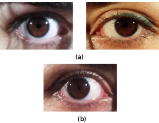
In order to develop a robust solution for iris recognition on mobile devices, the database used for testing should simulate the uncontrolled acquisition con-ditions described above. An example of such images acquired by mobile devices is shown in Figure 1.3.
Figure 1.3: MICHE I iris images example. (a) Images acquired with Samsung Galaxy S4 rear camera (left) and front camera (right); (b) Image captured with Samsung Galaxy Tab 2 front camera.
MICHE I - Evaluation protocol
The algorithms presented to MICHE I deal with both iris segmentation and iris recognition. Two evaluations were carried out in order to compare the algo-rithms and to identify the best segmentation and the best recognition methods. Four iris segmentation algorithms have been tested on a subset of MICHE database. Here are summarized the metrics used to evaluate the segmentation
28 1. INTRODUCTION
quality of the methods submitted to MICHE I. Each method was evaluated on a subset of MICHE database for which ground-truth data have been made available:
• PRATT: This metric is a formulated function of the distance between correct and measured edge positions, but it is also indirectly related to the false positive and false negative edges.
• F1 Score: It is a measure of a test’s accuracy. It considers both the precision p and the recall r of the test to compute the score. The F 1score can be interpreted as a weighted average of the precision and recall, where an F 1 score reaches its best value at 1 and worst at 0.
• Rand Index: RI counts the fraction of pairs of pixels whose labelling are consistent between the computed segmentation and the ground truth. • Global Consistency Error: The Global Consistency Error (GCE) [Mar-tin2001] measures the extent to which one segmentation can be viewed as a refinement of the other. Segmentations which are related in this man-ner are considered to be consistent, since they could represent the same natural image segmented at different scales.
• E1 Score: The classification error rate (E1) of the algorithm on the input image is given by the proportion of correspondent disagreeing pixels (through the logical exclusive-or operator) over the whole image.
• Pearson Correlation Coefficient: It is a measure of the linear correla-tion between two variables X and Y , giving a value between +1 and −1 inclusive, where 1 is total positive correlation, 0 is no correlation, and −1 is total negative correlation.
The iris recognition methods submitted to MICHE I have been evaluated in terms of decidability, area under ROC curve and equal error rate. The algo-rithms have been tested on iris images segmented by the segmentation methods proposed to MICHE I and discussed above.
1.4. CHALLENGES 29
MICHE II - Challenge protocol
MICHE II 2 is a challenge for iris recognition on the MICHE database (see 3
for details). Each executable should therefore be able to receive from command line a pair of images from the dataset and a pair of corresponding segmentation masks and should produce a score in terms of dissimilarity between the two irises.
The order of inputs is strictly defined. Let:
I1 = image1Filename.ext be the first RGB image containing an iris; M1 = mask1Filename.ext be the binary mask of I1;
I2 = image2Filename.ext be the second RGB image containing an iris;
M2 = mask2Filename.ext be the binary mask of I2; path be the directory for matching results;
Let APP be the executable application, then by running: APP I1 M1 I2 M2 path
a TXT file containing the dissimilarity_score is created. Such TXT file must have the following properties:
a. it is saved in path (preferably something like "./results");
b. its filename is image1Filename_image2Filename.txt (NOTE. filenames without file extensions);
c. its content is image1Filename [whitespace] image2Filename
[whitespace] dissimilarity_score (NOTE. filenames without file ex-tensions).
The dissimilarity score d : {d ∈ < : 0 ≤ d ≤ 1} is meant as the probability that two irises are from two different subjects. The higher is the dissimilarity the higher is the probability that the two irises are not from the same person. Let I be set of images from MICHE database, the dissimilarity function D is defined as:
30 1. INTRODUCTION
D : Ia× Ib→ [0, 1] ⊂ <
where Ia, Ib∈ I
and satisfies the following properties: a. D(Ia, Ia) = 0;
b. D(Ia, Ib) = 0 → Ia = Ib;
c. D(Ia, Ib) = D(Ib, Ia).
The participants can use the whole MICHE database for developing and per-forming experimentations of their proposed algorithm. The participants should take into account that the dataset is going to be extended with new acquisitions by new mobiles and of new subjects according to the same acquisition protocol applied to the current version of the database. The challenge will be run on a subset of the new version of the MICHE database that will be revealed together with the final ranking.
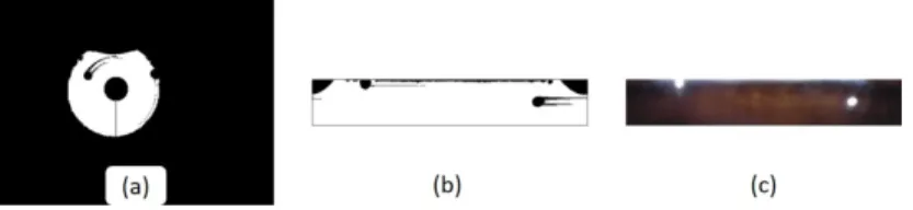
Participants must consider that the best segmentation algorithm submitted to MICHE I (The "Unsupervised detection of non-iris occlusions", by Haindl et al., is available for download on MICHE II website) will be used to generate the binary masks. Since it will be used also for the final ranking of submitted algorithms, participants are invited to use it for testing their proposal. Given an RGB image in input, the segmentation algorithm gives in output (a) the binary mask; (b) the normalised mask of the iris region; (c) the normalised RGB iris extracted from the image (see figure 1.4 below).
Each executable is supposed to be self-contained and it will not have access to the Internet. No any additional download has to be expected to run the application. The submitted proposal must therefore contain all supporting files (dlls, libraries and so on) useful to its proper running.
The executable can be written in any programming language and should run on one of the following operating systems: (1) Windows 7 64/32 bit, (2) Linux Ubuntu 14.04. Code written in Matlab is also acceptable at condition that
1.5. COMMERCIAL APPLICATIONS 31
Figure 1.4: MICHE II : (a) the binary mask; (b) the normalised mask of the iris region; (c) the normalised RGB iris extracted from the image.
it runs on Matlab 2013. In case of any special setting needed for the proper running of the algorithm, a README file is expected.
Executables that do not match the requirements above could be discarded from the contest at the discretion of the Evaluating Committee.
1.5
Commercial applications
Current commercial applications mostly address access control to restricted ar-eas. Iris recognition systems in less controlled situation were installed in some major international airports, especially in the UK and the Arab Emirates. For example both Gatwick airport and Dubai airport adopted an AOptix solution. AOptix InSight VM iris recognition system was integrated into 34 auto-R
mated e-Gates at the Gatwick Airport South Terminal in order to speed up the process of passport control, formerly performed manually. The iris recognition system allows passengers to be acquired at a distance of two meters, and they are only required to look at a specific point indicated on the device. A monitor gives passengers a textual feedback indicating them where to look at the device and eventually to open their eyes if there is an occlusion problem. The recogni-tion process lasts few seconds. The illuminarecogni-tion employed is a NIR lamp. The system can perform recognition whether passenger is in a wheelchair or above 2.15 meter tall.
AOptix InSight Duo maintains the same characteristics of AOptix InSightR VMR
32 1. INTRODUCTION
Dubai airport and probably will be soon integrated also at Gatwick3.
It is interesting to know that before using AOptix products, Gatwick and many UK airports adopted an iris recognition system in controlled condition. However because of the high percentage of false rejections and the difficulties for the passengers in lining up eyes with the iris recognition equipment, the identification process took a lot longer than it was supposed to and the system was abandoned. This demonstrates the need to develop iris recognition systems requiring less and less users’ collaboration and that are suitable to any type of environment in order to be a valid and faster but also more secure tool than existing authentication systems.
Iris can be used also in multi-biometric systems, as the already mentioned AOptix InSight Duo which combines face and iris biometric traits, but noisyR
iris recognition may be also combined with other biometrics or soft-biometrics, for example periocular information, gaze analysis, etc. [47][6]. Finally, thanks to the ever increasing technological development in a near future iris may be captured at a considerable distance, we can then imagine to integrate the iris biometric in video surveillance systems for people recognition or re-identification [38].
Chapter
2
State of the Art
Among the issues linked to biometric recognition on mobile devices, there are undoubtedly the technological limits of nowadays devices: the classic configu-ration for a biometric recognition system, for example for face recognition, is composed by one or more high-quality cameras placed in fixed positions, pos-sibly in an environment with controlled lighting, and connected to a processor. The use of a mobile device, e.g. a smartphone, for its nature, implies very differ-ent usage scenarios, limitations on the quality of the hardware, nonconformity with the software requirements of a biometric recognition system.
However, the opportunity offered by these devices is to make biometric recog-nition portable, replacing or strengthening the existing authentication systems, a good reason to further investigate in this direction.
In the following a quick overview on the biometric traits employed for the recognition on mobile devices is given.
Behavioural biometric recognition on mobile devices
Among the behavioural biometrics being studied, there are the user’s gait mod-elled using the smartphone embedded accelerometer, as described in [12], the
34 2. STATE OF THE ART
recognition of the typing style on virtual keyboards, presented in [13], and the recognition of the arm movement, recorded by the embedded accelerometer when answering a call, proposed in [14], where the idea is to minimize user involvement in the authentication process, making it transparent to the user by using the arm movement that instinctively the user does to answer his/her phone.
Physical biometric recognition on mobile devices
For what concerns recognition on mobile devices based on physical biometrics, there are many examples, exploiting in particular the mobile devices embedded cameras and microphone. In [15], another approach for transparent user au-thentication is presented, but in this case, the ear is used and captured when the user answers (or places) a call. A multi-modal framework employing ear and speaker recognition is proposed in [16], based on the use of a hybridization of DWT (Discrete Wavelet Transform, using haar wavelet) and GLCM (Gray-Level Co-Occurrence Matrix) to extract both shape and texture information from ear images, and MFCC (Mel Frequency Cepstral Coefficient) technique to extract features from speech signal.
In [17] the system presented is based on creating a 3d face model by recon-structing depth information from videos recorded by the phone camera. One of the most recent works on mobile biometrics presents the MOBIO system, combining face and voice biometrics [18]. It is preferable to combine face with iris to simplify capture and processing, while at the same time we avoid heavy normalization procedures, which are performed in [18].In general the quality of captured signals, both audio and video, depends on factors, which are internal (device dependent) and external (environment dependent). Even if the concept of quality is often quite vague, we can assume for sure that resolution is one of the internal factors, which concur to it. The image resolution depends on the size and technology of the sensor, while the resolution of captured audio signals depends on the precision of the sensor and on sampling frequency. It is well known that during interaction, low quality in images is better tolerated
35
than low quality in sound [19], because the human visual system is able to cope with more interferences than the auditory one, and this often dictates the compression parameters, which are used during signal processing.
Iris detection and recognition on mobile devices
For what concerns iris detection, in [20] and [21] methods for pupil and iris boundaries detection are presented; in these two works however, the databases employed were collected respectively with a Samsung SPH-S2300 and Samsung SPH-23001 (in [20] only 132 images were captured with the mobile phone and the others were from CASIA database2) which embed a 3.2 megapixel digital
camera with a 3X optical zoom, which is a very specific imaging sensor that cannot commonly be found in the most popular smartphones.
One of the first works investigating the possibility of optimizing iris segmen-tation and recognition on mobile phones is [22], Jeong et al. propose a method for computing the iris code based on Adaptive Gabor Filter. In [23], Park et al. present a recognition method based on corneal specular reflections, while Kang in [24] presents a method to pre-process iris in order to remove the noise related to occlusions of eyelids and improve system performances. In [25][26] an iris recognition system based on Spatial Histograms is presented.
1Samsung. http://www.samsung.com/
Chapter
3
MICHE dataset
In order to reliably test an approach, it is necessary to have a dataset that replicates the real conditions in which the approach should be applied. The database described in this chapter simulates the acquisition of iris and face on mobile devices.
The MICHE dataset [30] was collected on the occasion of the Mobile Iris CHallenge Evaluation (see paragraph 1.4.3) at the University of Salerno, by Chiara Galdi (at that time Ph.D. candidate at University of Salerno, Italy) and Silvio Barra (at that time Ph.D. candidate at University of Cagliari, Italy), members of the BIPLab (Biometric and Image Processing Lab) that promoted the Challenge and the collection of the MICHE biometric database.
The images were acquired with three different mobile devices, representative of the current top market category:
• iPhone 5 (hereafter IP5)
Operating System: Apple iOS;
Front Camera: FaceTime HD Camera with 1.2 Megapixels; Rear Camera: iSight with 8 Megapixels.
• Samsung Galaxy S4 (hereafter GS4) 37
38 3. MICHE DATASET
Operating System: Google Android; Front Camera: CMOS with 2 Megapixel; Rear Camera: CMOS with 13 Megapixel. • Samsung Galaxy Tablet II (hereafter GT2)
Operating System: Google Android; Front Camera: VGA for Video Call; Rear Camera: 3 Megapixel Camera.
3.1
Acquisition Protocol
Biometry is very suitable for human recognition on mobile devices in fact the users are used to employ the frontal camera of their personal mobile devices to capture pictures of themselves, the so called "selfie". The subjects involved in the database acquisition process are only asked to take self-pictures of their face, eyes, and single iris, sometimes with both frontal and rear camera and sometimes with the frontal camera only. Further details on the procedure to acquire each sub-database are given later in this chapter in the respective paragraphs.
Since the goal of the acquisition process is to achieve a realistic simulation of the data capture process, users are left free to hold the devices (smartphones or tablet) in their hands, to use the voice commands (when available) and, for the subjects wearing eyeglasses, to decide if wear them or not during the acquisition according to what they would have done during a real use of such an application.
The standard acquisition process was the following: 1. Smartphones (GS4 and IP5):
(a) indoor:
- 4 shots of the face taken with the rear camera;
- 4 shots of the two eyes (landscape format) taken with the rear camera; - 4 shots of the iris (left or right, the chosen one will be used for the
3.2. DATABASE COMPOSITION 39
- 4 shots of the iris taken with the frontal camera. (b) outdoor:
- 4 shots of the iris taken with the rear camera; - 4 shots of the iris taken with the frontal camera. 2. Tablet (GT2):
(a) indoor:
- 4 shots of the iris taken with the frontal camera. (b) outdoor:
- 4 shots of the iris taken with the frontal camera.
For a total of at least 56 pictures per person (there were few exceptions). During the indoor acquisition mode various sources of artificial light, sometimes combined with natural light sources, are used, while during the outdoor acqui-sition mode data capture takes place using natural light only. The resulting captured images are affected by different noise factors that we will discuss later in paragraph 3.4.
3.2
Database composition
The database currently consists of 92 different subjects with age ranging between 20 and 60 years, among them 66 are males and 26 are females and all of them are of Caucasian ethnicity. MICHE is composed by different sections:
1. MICHE Iris; 2. MICHE Iris - Fake; 3. MICHE Iris - Video. 4. MICHE Face; 5. MICHE Eyes.
40 3. MICHE DATASET
Part of the database, a subset of MICHE Iris containing the subjects from ID number 1 to 75, the MICHE Iris - Fake, and MICHE Iris - Video, are available on-line for research purposes1.
3.2.1
MICHE Iris
MICHE Iris is the main database section, in its current version it contains more than 3500 pictures of irises of 92 different subjects, around 40 pictures per subject.
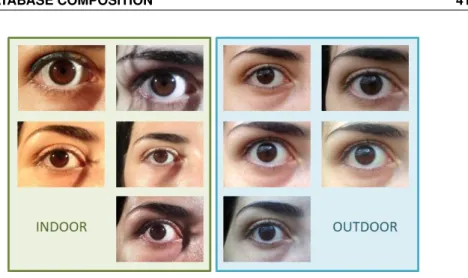
Only one iris per subject has been captured but in different modalities, i.e. indoor, outdoor, and with all the three different mobile devices mentioned before. Some examples of iris images contained in this database are showed in figure 3.1. Due to the fact that users were let free to take a self-picture and because of the use of different devices, the resulting images are very different from each other.
Figure 3.1: MICHE Iris images example. First row: pictures acquired by GS4; Second row: pictures acquired by IP5; Third row: pictures acquired by GT2
3.2. DATABASE COMPOSITION 41
Figure 3.2: MICHE Iris acquisition modalities.
In figure 3.2 some pictures of the same eye captured in indoor and outdoor modalities are given.
3.2.2
MICHE Iris - Fake
This database is suitable for the testing of anti-spoofing techniques, i.e. tech-niques to identify if the iris in front of the camera is a real iris or an artefact reproducing the real iris (e.g. a photo, a video, etc.).
This small database contains 40 photos of grey-scale printed eye images (by a LaserJet Printer) of 10 different subjects. Photos were taken by the Samsung Galaxy S4 rear camera. For each of the 10 subjects, 4 indoor images were selected from the MICHE Iris database, 2 pictures acquired by the GS4 and 2 acquired by the IP5 smartphone. Figure 3.3 shows some samples from MICHE Iris - Fake database.
3.2.3
MICHE Iris - Video
MICHE Iris - Video is made up of 113 videos of about 15 seconds, recording the eye of 10 subjects both in indoor and outdoor mode. Also this database is
42 3. MICHE DATASET
Figure 3.3: MICHE Fake images example.
suitable to test anti-spoofing techniques and in particular those aimed to detect iris liveness.
3.2.4
MICHE Face
Although the MICHE Face, as the name suggests, is a face database, it was acquired with the purpose of providing a database suitable for iris and face recognition at once. In fact, the photos were taken in the best illumination conditions and with the highest-resolution cameras (smartphones rear cameras) in order to provide an adequate iris resolution for analysing its features. Some images contained in MICHE Face database are shown in figure 3.4.
3.3. METADATA 43
3.2.5
MICHE Eyes
The idea behind the collection of this database is to provide images that contain both irises captured in one shot. This allows the study of advantages/disadvan-tages behind the use of both irises in a human recognition system. This kind of photos were captured only in indoor modality, by only the two smartphones and with their rear camera. The reason behind this choice is that since the relative iris size in these pictures is small an adequate resolution has to be assured, that is why the irises were collected in the most controlled scenario (indoor) and by the cameras with best resolution. Examples of MICHE Eyes are given in figure 3.5.
Figure 3.5: MICHE Eyes images example.
3.3
Metadata
MICHE database, at the best of our knowledge, is the first database that along with the images provides metadata files with full information about the subjects acquired, the acquisition device characteristics, the acquisition conditions. An example of the metadata structure (xml file) is given in the following:
<img>
<filename>img.jpg</filename> <img_type>[iris, face]</img_type> <iris>[left, right, both]</iris>
<distance_from_the_device>[100cm, 10cm]
</distance_from_the_device> (in centimetres) <session_number>[01, 02, 03, ...]</session_number>
44 3. MICHE DATASET
<image_number>[1, 2, 3, 4, ...]</image_number> <user id="[001, 002, ..., 022, ...]">
<age>[20, 32, 55, ...]</age> <gender>[M, F]</gender>
<ethnicity>[Afro American, Asian, Caucasian, Indians, Latinos]</ethnicity>
</user> <device>
<type>[Smartphone, Tablet]</type>
<name>[IPhone5, Galaxy S4, Galaxy Tab 2, ...]</name> <camera type="[front, rear]">
<name>[VGA, CMOS, iSight, ...]</name> <resolution>[0.3MP, ...]</resolution> <dpi>[72, ...]</dpi> </camera> </device> <condition> <location>[indoor, outdoor]</location>
<illumination>[artificial, natural, both]</illumination> </condition>
<author>[BIPLab, University of ...]</author> </img>
3.4
Noise Factors
The noise factors have been in part previously presented in section 1.4.2: out-of-focus iris images; off-angle iris images; rotated iris images; motion blurred iris images; iris occlusion due to eyelashes; iris occlusion due to eyelids; iris oc-clusion due to glasses; iris ococ-clusion due to contact lenses; iris ococ-clusion due to hairs; occlusion due to shadows; iris with specular reflections; iris with diffuse reflections; partial captured iris; out-of-iris images. Another factor that can af-fect the iris segmentation phase is the presence of strong make-up in the picture,
3.4. NOISE FACTORS 45
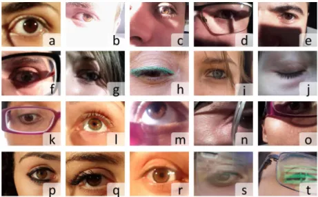
since dark areas can be confused with the pupillary and a wrong detection of the pupillary boundary can lead to a completely wrong iris segmentation. Some examples of iris images affected by the noise factors listed above, are given in figure 3.6.
Figure 3.6: MICHE I iris noise factors examples: (a) out-of-focus iris image; (b-g) examples of images affected by strong light/shadows; (h) eyelids and eyelashes occlusion; (i) hairs occlusion; (j) out-of-iris image; (k-m) off-angle iris image; (m-o) partial captured iris; (p-q) strong make-up; (r) specular reflections; (s-t) diffuse reflection on eyeglasses.
Chapter
4
Iris segmentation on mobile
devices
Iris recognition on mobile devices is a challenging task, in fact, with respect to other dedicated iris acquisition devices, usually fixed on a desk or on a stand, the use of the smartphone embedded sensors introduce a number of noisy fac-tors during the iris acquisition process [41] due to the fact that the device is hand-held by the users: out-of-focus, off-angle iris, rotated iris images, motion blurring, occlusions due to eyelashes, occlusions due to eyelids, occlusions due to eyeglasses, occlusions due to contact lenses, specular reflections, diffuse re-flections, partially captured iris. In off-angle iris images, the iris aspect ratio is distorted. All these aspects have to be taken into account when designing an iris segmentation algorithm. In the following, two methods for iris segmentation are presented, specifically designed for iris segmentation in adverse conditions and on mobile devices. These two methods, namely ISIS and BIRD, are then em-ployed in the systems presented in chapter 5 "Multi-biometric and multi-modal authentication on mobile devices".
48 4. IRIS SEGMENTATION ON MOBILE DEVICES
4.1
ISIS
ISIS (Iris Segmentation for Identification Systems) is an iris segmentation algo-rithm proposed by the BIPLab - Biometric and Image Processing Lab [31], of the University of Salerno. It was especially devised and implemented to address under-controlled acquisition conditions, therefore it is well suited to be used on mobile devices. It is robust to the presence of reflections and requires a limited computational time. It has four main phases:
• Pre-processing; • Pupil location; • Linearisation; • Limbus location.
ISIS algorithm uses operative (e.g. image window sizes) and decision param-eters (e.g. thresholds) that have been experimentally tuned by using a training set of images.
4.1.1
Preprocessing
Eye image contains many disturbing details such as sclera vessels, skin pores, or eyelashes shape; these are complex patterns that can negatively interfere with edge detection. Moreover, the same internal characteristic patterns, which are fundamental for recognition, may hinder a correct segmentation. To avoid this problem, a posterization filter F E (Enhance) is applied: a square window W is moved over the whole image, pixel by pixel, a histogram is computed for the region contained in W , and the value with the maximum frequency is substituted for the central position.
4.1.2
Pupil location
In this phase, a first step implies to apply Canny filtering on the preprocessed image with ten different thresholds th = 0.05, 0.10, 0.15, ..., 0.55. We start from
4.1. ISIS 49
the assumption that relevant circles can be detected at different thresholds (e.g. at 0.15, 0.20, and 0.25), while circles detected only for a single value of the threshold may be artefacts. Choosing a fixed step (0.05) for the threshold value allows to explore uniformly its overall admissible range, which was experimen-tally found. Processing the image (Canny filtering, circle fitting) with different thresholds has a little impact on the computational burden of the overall tech-nique, but significantly increases the accuracy of the segmented result. On the other hand, an adaptive threshold technique may concentrate only on specific parts of this range; however, it is important to explore it completely, because we don’t know in advance the dominant grey level of the pupil. The connected components are identified in each resulting image, and those containing a num-ber of pixels greater than a threshold T hC are all included in a unique list L
of starting candidates. Since the pupil is not a perfect circle, many approaches search elliptical shapes possibly representing it. However the presence of noise (e.g., spurious branches by Canny filter) may cause erroneous results from ellipse fitting algorithms. Therefore, ISIS detects circular objects within the image by using a precise and fast circle detection procedure presented by Taubin in [72]. Taubin’s algorithm is applied to each element in L to compute the approximat-ing circle. All the components whose correspondapproximat-ing circles are not completely contained inside the image are removed from L. To extract the real pupil bound-ary from all candidates in the final list, each remaining circle undergoes a voting procedure, according to the sum of two ranking criteria:
Homogeneity: pupil is a darker, homogeneous area with respect to iris. Separability: both limbus and pupil contour represent a boundary region with a pronounced step from a darker to a lighter zone.
To calculate the separability score, for each candidate circle a slightly smaller and a little larger circles are considered: let CL be a circle with radius r and let CL1 and CL2 be two additional circles having the same centre as CL and
radii 0.9r and 1.1r, respectively. For each point in CL having polar coordinates (ρ, θ), two pixels, p1 and p2, located at the same angle θ on CL1 and CL2
are considered. The score for CL is computed as the sum of the differences between each pair of pixels corresponding to p1and p2. The circle with highest
50 4. IRIS SEGMENTATION ON MOBILE DEVICES
homogeneity and separability scores is considered as the circular shape which better approximates the pupil.
4.1.3
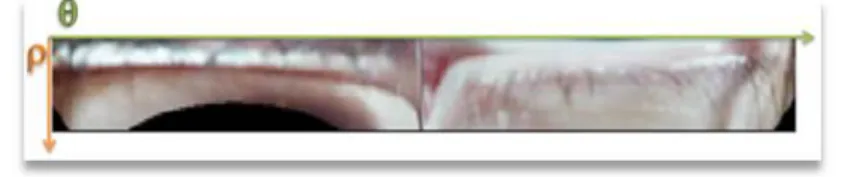
Linearisation
In order to perform limbus location, the image is first transformed from Carte-sian coordinates to polar coordinates. The pixel with the greatest distance ρmax from the centre of the identified pupil circle is selected as the starting point for the image transformation. See figure 4.1.
4.1.4
Limbus location
A median filter is applied to the polar image. For each column corresponding to a position θi on the horizontal axis, ranging over ρj on the vertical axis, the
following weighted difference is calculated:
∆(ρj, θi) = φ( ˙I, ρj, θi) · ( ˙I(ρj+ δ, θi) − ˙I(ρj− δ, θi))
where ˙I is the image in polar coordinates, and
φ( ˙I, ρj, θi) = 1 if ˙I(ρj+ δ, θi) − ˙I(ρj− δ, θi) > 0
and min ( ˙I(ρj− δ, θi), ˙I(ρj+ δ, θi)) > εG
0 otherwise
This procedure allows to identify the points with the higher positive vari-ation,which indicates the transition from a darker zone (the iris) to a lighter one (the sclera). The first inequality just selects points with a positive gradient, while the second one rules out those points between pupil and iris by prescribing that the darker pixel in the pair at hand must exceed a threshold εG ∈ [0, 255]
(here εG = 50). Points which maximize (2) for each column θi in ˙I make up
the limbus that we will call F . After this, it is possible to discard outliers by considering that, in the polar space, point of the limbus must lie approximately on a line, and therefore have an almost constant ρ component. To this aim,
4.2. BIRD 51
points with a ρi producing a relative error above a threshold ε are cancelled
(here ε = 0.4). The relative error err is calculated as follows:
err = |ρi− ρmed| maxi|ρi− ρmed|
where ρmed is the median value over F .
Figure 4.1: Illustration of ISIS algorithm.
At the end of acquisition and segmentation procedures, we obtain an image in polar coordinates where most useless information has been discarded.
4.2
BIRD
The iris segmentation method BIRD (watershed Based IRis Detection), has been presented in [2] along with a periocular area segmentation technique and a recognition approach based on the fusion of the iris and the periocular area. In this section only the iris segmentation method is presented, while the periocular segmentation and fusion approaches are presented in chapter 5 "Multi-biometric and multi-modal authentication on mobile devices" as one of the multi-biometric system proposed in this thesis.
BIRD[2] is a technique for smart mobile devices, which is the follow up of a technique presented in [1]. BIRD exploits the use of the watershed transform to identify more precisely the iris boundary and, hence, to obtain a more accurately computed code for iris recognition.
delimit-52 4. IRIS SEGMENTATION ON MOBILE DEVICES
ing the regions into which an image is divided are mostly placed where human observers perceive them. In fact, the watershed transformation is a growing pro-cess performed generally on the gradient image, where the edges are enhanced. This feature should allow to correctly detect the limbus boundary. In turn, a negative feature is over-segmentation, i.e., the image may be partitioned into a number of parts that is remarkably larger than expected. Over-segmentation is particularly evident when all the regional minima in the gradient image are considered as seeds for the growing process. A common strategy to overcome this drawback is to adopt region merging and/or seed selection to reduce the number of watershed regions. However, in the case of eye images, processes for over-segmentation reduction cannot be stressed. Otherwise, some weak bound-aries between sclera and limbus (light eye case) or between eyelashes and limbus (dark eye case) might be no longer present in the segmented image.
BIRD performs a binarization of the watershed transform to obtain an im-age where large portions of the limbus boundary are better enhanced. In this way BIRD is able to exploit the positive features of the watershed transform independently of over-segmentation problem. The boundaries of the foreground region are then given as input to a circle detection process, which aims at finding the circle that best approximates the limbus boundary (limbus circle).
4.2.1
Pre-processing
Uncontrolled iris acquisition may produce an image with local distortions due for example to shadows and different colour temperature. A colour/illumination correction is performed to reduce such local distortions, by processing separately the three RGB components of the eye image as grey level images. For each grey level image, a Gaussian filtered version is computed. A new image is built, where each pixel is set to the ratio between the value of the homologous pixels in the grey level image and in its filtered version. This ratio has the side effect to bring out the details in the image, so the kernel parameters of adopted Gaussian filter play a fundamental role. The parameters are the kernel size gk,
the average mkand variance σk(they are mainly related to the resolution of the
4.2. BIRD 53
the colours within the image, while one that is too large will not produce any substantial correction on lighting and colour distortions in it. In order to find a viable relationship between Gaussian kernel parameters to be adopted and the resolution of the image, was considered a set of pictures of irises at different resolutions wk× hk where k = 1, 2, ..., n, wk+ 1 > wk and hk + 1 > hk. The
image resolution was represented by considering the value of the diagonal dk =
pw2 k+ h
2
k. The optimal parameters for the Gaussian kernel were determined in
terms of segmentation and recognition accuracy obtained on the set of images. It was observed that the relationship between gk and dk is quadratic, i.e. gk =
α2d2k+ α1dk+ α0, while mk = gk and σk = 0.1 · gk. In this case, it was found
that α2= −0.0001, α1= 0.3064 and α0= 11.1351.
Figure 4.2: Colour correction.
A normalization process of pixel values is performed to map the values in the range [0, 255]. The combination of three obtained grey level images originates the colour/illumination corrected image. The left image in figure 4.2 shows the original image, while the right one corresponds to the colour/illumination corrected image. As BIRD is able to work even on low resolution images, it is possible to limit the computational cost of the method. The colour/illumina-tion corrected image is resized by using a linear interpolacolour/illumina-tion method without changing the aspect ratio, in order to get an image of the eye in the foreground with a horizontal resolution of 200 pixels (vertical resolution depends on aspect ratio). As previously, the process of correcting lighting/colour enhances the de-tails in the image and these dede-tails are irrelevant for the segmentation. Thus a median filter is applied with a fixed-size window 7 × 7. The window size can be