Differential functionalities of amphiphilic peptide segments of the
cell-septation penicillin-binding protein 3 of Escherichia coli
Monique Marrec-Fairley,1 André Piette,1 Xavier Gallet,2 Robert Brasseur,2 Hiroshi Hara,3 Claudine Fraipont,1 J. M. Ghuysen1 and M. Nguyen-Distèche1
1Centre d'Ingénierie des Protéines, Université de Liège, Institut de Chimie, B6, Sart Tilman, B-4000 Liège, Belgium.
2Centre de Biophysique Moléculaire Numérique, Faculté Universitaire des Sciences Agronomiques, B-5030 Gembloux, Belgium. 3
Department of Biochemistry and Molecular Biology, Saitama University Faculty of Science, 255 Shimo- Ohkubo, Urawa, Saitama 338-8570, Japan.
Summary
The class B M1-V577 penicillin-binding protein (PBP) 3 of Escherichia coli consists of a M1-L39 membrane anchor (bearing a cytosolic tail) that is linked via a G40-S70 intervening peptide to an R71-I236 non-catalytic module (containing the conserved motifs 1-3) itself linked via motif 4 to a D237-V577 catalytic module
(containing the conserved motifs 5-7 of the penicilloyl serine transferases superfamily). It has been proposed that during cell septation the peptidoglycan crosslinking activity of the acyl transferase module of PBP3 is regulated by the associated M1-I236 polypeptide itself in interaction with other components of the divisome. The fold adopted by the R71-V577 polypeptide of PBP3 has been modelled by reference to the corresponding R76-S634 polypeptide of the class B Streptococcus pneumoniae PBP2x. Based on these data and the results of site-directed mutagenesis of motifs 1-3 and of peptide segments of high amphiphilicity (identified from hydrophobic moment plots), the M1-1236 polypeptide of PBP3 appears to be precisely designed to work in the way proposed. The membrane anchor and the G40-S70 sequence (containing the G57-Q66 peptide segment) upstream from the non-catalytic module have the information ensuring that PBP3 undergoes proper insertion within the divisome at the cell septation site. Motif 1 and the I74-L82 overlapping peptide segment, motif 2 and the H160-G172
overlapping peptide segment, and the G188-D197 motif 3 are located at or close to the intermodule junction. They contain the information ensuring that PBP3 folds correctly and the acyl transferase catalytic centre adopts the active configuration. The E206-V217 peptide segment is exposed at the surface of the non-catalytic module. It has the information ensuring that PBP3 fulfils its cell septation activity within the fully complemented divisome.
Introduction
The bacterial cell wall peptidoglycan is a covalently closed net-like polymer in which glycan chains made of alternating β-1,4-linked N-acetylglucosamine and N-acetylmuramic acid residues are crosslinked by short peptides. In Escherichia coli, the carboxyl groups of the N-acetylmuramic acid residues are amide-linked to L -alanyl-γ-D-glutamyl-(L)-meso-diaminopimelyl-(L)-D-alanine peptides and the peptide crosslinkages are, mainly, direct D-alanyl-(D)-meso-diaminopimelic acid peptide bonds. The immediate biosynthetic precursor of the peptidoglycan is lipid II. A disaccharide pentapeptide [L-alanyl-γ-D-glutamyl-(L)-meso-diaminopimelyl-(L)-D -alanyl-D-alanine] is linked to a C55H89 undecaprenyl carrier via a pyrophosphate bridge involving C1 of
N-acetylmuramic acid.
From this precursor, the class A multimodular penicillinbinding protein (PBP) 1a and PBP1b (Goffin and Ghuysen, 1998) catalyse the formation of polymeric peptidoglycan (Nakagawa et al., 1984; van Heijenoort, 1996; Terrak et al., 1999). These PBPs consist of a membrane anchor which is linked to the amino end of a catalytic glutamic acid glycosyl transferase module (which catalyses glycan chain elongation), itself linked to the amino end of a catalytic serine acyl transferase module (which catalyses peptide crosslinking). Penicillin is a suicide substrate of the acyl transferase. The reaction produces a stable serine ester-linked penicilloyl enzyme such that the inactivated enzymes behave as PBPs.
Shaping the raw peptidoglycan material made by PBP1a and PBP1b into a sacculus in a cell cycle-dependent fashion relies on the class B multimodular PBP2 and PBP3 and on a set of non-penicillin-binding proteins
(Wientjes and Nanninga, 1991). PBP2 and RodA, whose encoding genes are located at the 14 min region of the chromosome, control wall expansion and shape maintenance (Spratt, 1975; Begg et al., 1990). PBP3, FtsL, FtsW, FtsQ, FtsA, FtsZ, whose encoding genes are located at the 2 min region, and FtsK, ZipA and FtsN, whose encoding genes are located at 21 min, 52 min and 88 min, respectively, form a multiprotein complex, the divisome, which encompasses the cytoplasm, the plasma membrane and the periplasm (Hale and de Boer, 1997; Nanninga, 1998; Wang and Lutkenhaus, 1998; Wang et al., 1998; Weiss et al., 1999). FtsZ is a GTPase related to eukaryotic tubulins (de Boer et al., 1992; RayChaudhuri and Park, 1992; Lowe and Amos, 1998). FtsZ polymerizes into a ring-shaped structure at the cell division site, recruits the other cell division proteins and provides the driving force for cytokinesis (Bi and Lutkenhaus, 1990; Lutkenhaus and Addinall, 1997).
The class B PBPs have a multimodular design similar to that of the class A PBPs (Goffin and Ghuysen, 1998). However, the polypeptide upstream from the acyl transferase module is not a glycosyl transferase. The role that this module plays in the spatiotemporal control of peptidoglycan synthesis remains contentious. There is evidence that E. coli PBP3 performs peptidoglycan crosslinking (the acyl transferase module) and that this activity is regulated by the associated non-catalytic module itself in interaction with other components of the divisome (Adam et al., 1997; Goffin and Ghuysen, 1998; Nguyen-Distèche et al., 1998).
The notion that peptide segments of relatively great amphiphilicity have protein-protein interaction potentials was formulated by De Loof et al. (1986) in their studies on apolipoprotein E. In an attempt to identify regions of the class B PBP3 possibly involved in intra- and intermolecular interactions, the mean α-helical hydrophobic moment and the mean hydrophobicity were calculated along the amino acid sequence of the protein. The roles of amphiphilic segments detected on the basis of these hydrophobic moment plots were probed experimentally. By combining the results thus obtained and previously published data, it was possible to identify three regions of the non-catalytic module of PBP3, each performing distinct specific functions.
Results
Predictive studies: detection and spatial disposition of amphiphilic segments of E. coli PBP3
Following the normalized scale of Eisenberg, hydrophobic residues have a positive value (IIe, 1.4; Pro, 0.12) and hydrophilic residues have a negative value (Thr, -0.05; Arg, -2.5). The asymmetry of hydrophobicity of a periodic peptide structure can be represented as a two-dimensional helical wheel, whose projection down the helix shows the relative orientations of residues (Eisenberg, 1984). The hydrophobic moment, which is a quantitative measure of the amphiphilicity perpendicular to the axis, is calculated from the sum of the mean hydrophobicity values over all amino acid residues and the angles at which successive side-chains emerge from the backbone (Eisenberg et al., 1982). The hydrophobic moment is small when the residues are evenly
distributed along the helix. It is large when most of the hydrophobic residues occur on one side of the helix and most of the hydrophilic residues on the other side. The hydrophobic moment concept is not restricted to α-helices. β-Turns consist of four residues whose side-chains protrude outside the peptide fold and the value of the angle between two linked residues is 90°, close to the 100° angle in an α-helix. Moreover, molecular modelling of proteins assumed to adopt a complete α-helix form gives valuable information on the helix and β structures (Poupon and Mornon, 1998).
The 588-amino-acid residue PBP3 of E. coli is processed into mature M1-V577 PBP3 (Nagasawa et al., 1989). The hydrophobic moment plot of the mature protein was calculated by moving a seven-residue window through the sequence, calculating the mean α-helical hydrophobic moment <µ> and mean hydrophobicity <H> per segment and placing the central amino acid residue as a point on a plot <µ> vs. <H> (Eisenberg et al., 1982; De Loof et al., 1986).
As shown in Fig. 1, nine peptide segments, A-I, were plotted in a region whose <H> values were equal to or less than -0.5 and <µ> values were equal to or greater than 0.25. Individual amino acid residues affected by <µ> values between 0.25 and 0.5 are in black on a grey background, those affected by <µ> values larger than 0.5 are in white on a black background and the three flanking residues which fall outside the cut-off region and were used for the calculations are also identified. On the basis of the data shown in Fig. 1, segments A-I fall roughly in the following decreasing order of amphiphilicity and, presumably, of interaction potential: [E =~ F] > C > [B
=
~ H] > [G =~ I] > [A =~ D]. Figure 2 shows the effects of selected mutations on the hydrophobic moment plots of segments A-I. Finally, Fig. 3 shows the distribution of segments A-I along the amino acid sequence of PBP3 by reference to the conserved motifs of the non-catalytic module (motifs 1, 2 and 3), the intermodule junction site (motif 4) and the acyl transferase module (motifs 4b, 5 containing the catalytic serine 307, 6 and 7).
The 750-amino-acid residue PBP2x of Streptococcus pneumoniae, also of class B, has the same motifs and modular organization as PBP3 of E. coli except that PBP2x possesses a Pro635-Asp750 carboxy-terminal module that is not present in PBP3. The Cα co-ordinates (at 3.5 Å resolution; Pares et al., 1996) of the membrane
anchor-free Arg76-Asp750 polypeptide of PBP2x are available. On the basis of these data, the Arg76-IIe265 non-catalytic module of PBP2x is shaped like a pair of sugar tongs; the core, which comprises motifs 1, 2 and 3, is a cluster of peptide segments that fit in or are located close to a non-catalytic groove of the acyl transferase module. And, the two sugar tongs each possess helices exposed at the surface of the protein. In turn, the associated Ser266-Ser634 acyl transferase module has the characteristic all-α and α/β domains of the penicilloyl serine transferases superfamily. Finally, the Pro635-Asp750 carboxy-terminal module, which is PBP2x specific, consists of two small α/β/β/β domains.
Fig. 1. Hydrophobic moment <µ> vs. hydrophobicity <H> plots of segments A-l of E. coli PBP3. The mutated
Fig. 2. Alterations of the hydrophobic moment plots of segment A caused by the R12S mutation (A), segment B
caused by the D58V mutation (B), segment C caused by the DF6N mutation (C), segment D caused by the K133S mutation (D), segment E caused by the R166Q;R167Q double mutation (E), segment F caused by the
R210Q;R213Q double mutation (F), segment G caused by the K291S mutation (G), segment H caused by the R234Q mutation (H) and segment I caused by the D409A mutation (I).
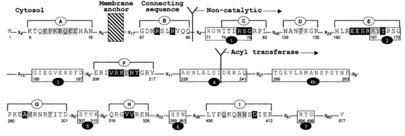
Fig. 3. Amino acid sequence analysis of E. coli PBP3. Conserved motifs 1-7 and segments A-l are shown.
Hatched area, plasma membrane; x, variable amino acid residue. The amino acid residues shown in black on a grey background are characterized by a hydrophobic moment ranging between 0.25 and 0.5. The amino acid residues shown in white on a black background are characterized by a hydrophobic moment >0.5.
Fig. 4. Spatial disposition of motifs 1, 2 and 3 and segments A-l along the polypeptide chain of E. coli PBP3.
The peptide segment of unknown structure that connects the membrane anchor and the non-catalytic module is represented as a dotted grey line. The non-catalytic module is in red. The acyl transferase module is in yellow. Amphiphilic segments are identified by the amino acid residues (in CPK; Koltun, 1965) falling within the region <H> < -0.5; <µ> >0.25 of the hydrophobic moment plots. Motif 1 is in green. Motifs 2 and 3 are in blue. Motif 4 (not shown) overlaps the junction between the non-catalytic (red) and acyl transferase (yellow) modules. The structure was modelled as described in the Experimental procedures.
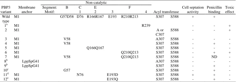
Table 1. Structural requirements for E. coli PBP3 activities. Non-catalytic PBP3 variant Membrane anchor Segment: Motif: B C 1 E 2 3 F 4 Acyl transferase Cell septation activity Penicillin binding Toxic effect Wild type M1 G57D58 D76 R166R167 E193 R210R213 S307 S588 + + 1a M1 R239 - - + 2 M1 A or C307 S588 - - + 3 M1 V58 A307 S588 - - - 4 M1 V58 S307 S588 - + - 5 M1 Q166Q167 S307 S588 - - - 6 M1 Q210Q213 S307 S588 - + + 7 M1 V58 Q210Q213 S307 S588 - ND - 8b LppSpG41 A307 S588 - - - 9b LppSpG41 S307 S588 - + - 10c G57 S307 S588 - + - 11d M1 N76 E193D S307 S588 + + - 12e M1 E193Q S307 S588 - + -
a. Variant 1 contains an eight-amino-acid residue extension downstream from R239. See Materials and Methods. b. LppSp, lipoprotein signal peptide. For more details on variants 7 and 8, see Houba-Hérin et al. (1985). c. Variant 9 lacks the M1-E56 peptide segment of the native PBP (Fraipont et al., 1994).
d. Variant 10 has decreased thermostability (Goffin et al., 1996).
e. The E193-D variant can be detected in very small amounts, whereas the E193-Q variant is not detectable by fluorography and immunoblotting (Goffin et al., 1996).
ND, not determined.
The Arg71 -Val577 polypeptide of E. coli PBP3 and the corresponding Arg76-Ser634 polypeptide of S. pneumoniae PBP2x share about 22% identities in the amino acid sequences. The fold adopted by the Arg71-Val577 polypeptide of E. coli PBP3 was modelled (see Experimental procedures). Although the structure shown in Fig. 4 remains short of reality, several observations can be safely made. Segment A is in the cytosol; it has low amphiphilicity. Segment B is located in the peptide that connectsthe membrane anchor to the non-catalytic module; it has moderate amphiphilicity. Segments C and E are within the core, and segment F is on one sugar tong of the non-catalytic module; they each have strong amphiphilicity. Segment D, on the other sugar tong, has weak amphiphilicity. Segments G-l are located within the acyltransferase module; they have low amphiphilicity except segment H, whose amphiphilicity is moderate.
Based on published data and the results of site-directed mutagenesis experiments described below, several regions of PBP3 endowed with different functionalities were identified.
The K6-N18 segment A and G57-Q66 segment B
PBP3 occurs in a small copy number, about 50-100 per cell, and its overproduction to a few thousands is harmless. The acyl transferase moduleless M1-R239 polypeptide of PBP3 (variant 1 in Table 1) and, likewise, the full-size PBP3 mutants in which an alanine or cysteine residue substitutes for the catalytic serine 307 (variant 2 in Table 1; Broome-Smith et al., 1985; Houba-Hérin et al., 1985) each lack penicillin-binding and cell
septation activities, as expected, and they each are toxic. E. coli strains co-producing the wild-type PBP3 and the truncated M1-R239 PBP3 or the wild-type PBP3 and the full-size Ser307Ala or Ser307Cys PBP3 mutant grew as filaments. This toxic phenotype indicates that these variants each compete with the resident native PBP3 of the E. coli transformants in which they are overproduced and prevent it from being functional.
The substitution of Val for Asp58 of segment B of the Ser307Ala PBP3 mutant gave rise to a Asp58Val; Ser307Ala double mutant (variant 3 in Table 1), which was no longer toxic. In turn, the substitution of Val for Asp58 of segment B of the wild-type PBP3 gave rise to a Asp58Val single mutant (variant 4 in Table 1), which bound penicillin but lacked complementation activity in an E. coli ftsl null mutant. This was examined by introducing a plasmid carrying the modified ftsl gene into the ftsl-deletion JE7947 strain harbouring plasmid pHR295 in which the wild-type ftsl was under the control of the lac promoter. At 37°C and in Luria-Bertani medium containing 10 mM glucose and appropriate antibiotics, the transformant was completely dependent on the presence of 0.5 mM isopropyl-β-D-thiogalactopyranoside (IPTG) for growth. Hence, Asp58 of segment B was important for the in vivo functionality of the wild-type PBP3 and the substitution of Val for Asp58 abolished the toxic effect of the S307A mutation. One may note that segment B had two amino acid residues, Arg60 and
Arg63, whose <µ> values were greater than 0.5 (Fig. 1 B). Substitution of Val for Asp58 (Fig. 2B) caused a displacement of the hydrophobic moment plot towards a greater hydrophobic moment but also a greater hydrophobicity so that one single residue, Arg63, had a <µ> value greater than 0.5.
Segment A (K6-N18) has low amphiphilicity (Fig. 1A) and, presumably, low interaction potential. The hydrophobic moment plot of the mutated segment A in which Ser substitutes for Arg12 falls outside the cut-off region (Fig. 2A). The R12S mutation did not affect the in vivo functionality of PBP3 (as examined in the ftsl null E. coli strain EC548).
The H160-G172 segment E and overlapping R167-G172 motif 2
These sequences fall within the core of the non-catalytic module. Arg166 and Arg167 of segment E (HLREESR166R167YYPSG) were changed into Gln (variant 5 in Table 1), resulting in a much decreased amphiphilicity of the segment (Figs 1E and 2E). E. coli RP41 and E. coli SP1097, which each produce a thermosensitive (42°C) PBP3, were used as hosts to produce the wild-type PBP3 and the R166Q;R167Q PBP mutant from properly engineered pUCBM20/ftsl vectors (see Experimental procedures). The transformants harbouring the gene encoding the wild-type PBP3 grew as rods at 30°C and at 42°C in Luria-Bertani (LB) medium supplemented with 50 µg ml-1 ampicillin. The transformants harbouring the gene encoding the R166Q;R167Q PBP3 mutant grew as rods at an unchanged rate at 30°C. At 42°C, they grew as filaments for about 2 h (Fig. 5A). Hence, decreasing the amphiphilicity of segment E yielded a protein mutant which was no longer functional in vivo.
E. coli LMG194/pLD139 transformants were then constructed in which the genes encoding the wild-type PBP3 and the R166Q;R167Q PBP mutant were under the control of the PBADarabinose promoter (see Experimental
procedures; Guzman et al., 1997), thus allowing the desired proteins to be overproduced in a controlled manner. The transformants each grew at a normal rate and with a normal morphology at 37°C in LB medium containing 0.2% glucose and 50 µg ml-1 kanamycin. When the cultures reached an optical density of 0.8 at 600 nm, the cells were collected and then cultured in LB medium containing 0.2% arabinose for 1 h at 37°C. The transformants overproducing the wild-type PBP3 and those overproducing the R166Q;R167Q protein mutant grew normally. Occasionally, transformants producing the protein mutant occurred in the form of small chains. Hence, the overproduction of the R166Q;R167Q protein mutant did not prevent the resident native PBP3 from being functional.
To translate these observations into biochemical terms, the transformants grown in the presence of arabinose for 1 h at 37°C were spheroplasted and the isolated plasma membranes were analysed by Western blot with
polyclonal anti-PBP3 antibodies. The R166Q;R167Q protein mutant was overproduced in similar amounts to the wild-type PBP3 (Fig. 6A), showing that this double mutation did not cause proteolytic susceptibility. The membrane-bound R166Q;R167Q protein mutant was then examined with respect to its ability to bind [3 H]-benzylpenicillin. It failed to bind penicillin (Fig. 6B).
The N129-R135 segment D and N206-V217 segment F
Segment F is located at the surface of one sugar tong of the non-catalytic module. It has strong amphiphilicity (Fig. 1F). Arg210 and Arg213 of segment F (ERIVR210K-DR213YGRV) were changed into Gin (variant 6 in Table 1), causing a much decreased amphiphilicity (Fig. 2F). E. coli SP1097 and RP41 were used as hosts to produce the R210Q;R213Q PBP mutant, as described above for the R166Q;R167Q PBP mutant. The
transformants harbouring the gene encoding the R210Q;R213Q PBP3 mutant grew at 30°C at a reduced rate as a mixture of rods and filaments. At 42°C, they stopped growing and they underwent conversion into long
autolysing filaments (Fig. 5B).
E. coli LMG194/pLD139 transformants overproducing the R210Q;R213Q PBP mutant under the control of the PBADarabinose promoter grew at a normal rate with normal morphology at 37°C in LB medium containing 0.2%
glucose. Upon transfer of the cells into LB medium containing 0.2% arabinose, the transformant stopped growing and the cells underwent conversion into long filaments, indicating that the R210Q;R213Q protein mutant prevented the resident wild-type PBP3 from being functional.
As derived from Western blot analysis of the plasma membranes isolated from transformants grown in the presence of 0.2% arabinose for 1 h at 37°C, the R210Q;R213Q protein mutant was produced in amounts nearly similar to that obtained with the wild-type PBP3 (Fig. 6A). The protein mutant bound penicillin (Fig. 6B), and its thermostability and affinity for penicillin were similar to those of the wild-type PBP3 (second-order rate constant
of penicilloylation 500 M-1s-1 at 30°C).
The substitution of Val for Asp58 in segment B of the R210Q;R213Q PBP3 mutant gave rise to a PBP3 mutant (variant 7 in Table 1) which was no longer toxic. This was analysed after transformation of E. coli XL1 blue with the proper pUCBM20/ftsl recombinants (see Experimental procedures). At 37°C, in LB medium, the transformants producing the double R210Q;R213Q PBP3 mutant grew as a mixture of rods and filaments, whereas the transformants producing the triple D58V;R210Q;R213Q PBP3 mutant grew as rods.
Segment D is located at the surface of the other sugar tong of the non-catalytic module of PBP3. At variance with segment F, segment D has low amphiphilicity (Fig. 1D) and, presumably, low interaction potential. The Lys133-Ser mutation did not affect the in vivo functionality of PBP3, although the hydrophobic moment plot of the mutated Lys133Ser segment D fell outside the cut-off region (Fig. 2D).
The P290-D301 segment G, Q319-N326 segment H and L400-R412 segment I
Segment G, H and I are located within the acyl transferase module. The K291S mutation of segment G, the Arg324Gln mutation of segment H and the Asp409Ala mutation of segment I resulted in a displacement of the hydrophobic moment plot outside the cut-off region (Figs 1G-I and 2G-I). The mutations Lys291Ser and Arg324Gln did not affect the in vivo activity of PBP3 (as measured in ftsl null E. coli strain EC548). Likewise, and as shown by Goffin et al. (1993), the mutation Asp409Ala was neutral.
Fig. 5. Complementation activities of (pUCBM20/ftsl) plasmids encoding the wild-type PBP3 (WT), the
R166Q;R167Q mutant (A) and the R210Q;R213Q;PBP3 mutant (B). The strain SP1097 was used as host (see Experimental procedures). The cells were observed under phase-contrast microscopy.
Fig. 6. SDS-PAGE analysis of the purified G57-V577 PBP3, the membrane-bound wild-type PBP3 (WT) and the
R166Q;R167Q and R210Q;R213Q PBP mutants isolated from E. coli LMG194/pLD139 transformants. The samples were labelled with 100 µM [3H]-benzylpenicillin before fluorography. Polyclonal antibodies directed against PBP3 were used for immunoblotting.
Discussion
The divisome encompasses the cytosol, the plasma membrane and the periplasm and its global function is to build the septum at mid-cell during cell division. The M1-V577 class B PBP3 of E. coli is an essential
component of the divisome. It was proposed that the peptidoglycan crosslinking activity of the D237-V577 acyl transferase module of PBP3 might be regulated by the associated amino terminal polypeptide itself in interaction with other components of the morphogenetic complex. The main result of the experiments presented above is that the M1-I236 polypeptide chain of PBP3 is precisely designed to work in the way that was suggested. Three regions of this polypeptide are endowed with distinct specific functionalities.
Segment B is located on the peptide chain that links the transmembrane anchor and the non-catalytic module. Substitution of Val for Asp58 of segment B gives rise to a PBP3 mutant (variant 4 in Table 1) which binds penicillin but is not functional, and the same substitution abolishes the toxic effect of the S307A mutation (variant 3 in Table 1). Segment B, however, is not the only structural determinant involved in this detoxication process. The toxic effect of the S307A mutation can also be abolished by substituting the uncleavable lipoprotein signal peptide for the genuine M1-G40 sequence of the S307A PBP3 mutant (variant 8 in Table 1) (Houba-Herin et al., 1985). Moreover, the PBP3 mutant obtained by substituting the Lpp signal peptide for the M1-G40 segment of the wild-type PBP3 (variant 9 in Table 1) (Houba-Herin et al., 1985) and the OmpA signal peptide-transported, membrane anchor-less and water-soluble G57-V577 polypeptide chain (variant 10 in Table 1) (Fraipont et al., 1994; Goffin et al., 1996) each bind penicillin, lack cell septation activity and can be
overproduced without causing detectable toxic effects. Hence, elimination of the membrane anchor of PBP3 and proper alteration of segment B (the D58V mutation) each prevent the corresponding, non-functional protein variants from competing with the wild-type PBP.
A straightforward interpretation of these results is that the F24-Q66 sequence, which comprises the membrane anchor and segment B, plays an important role in the proper orientation and positioning of PBP3 during its insertion within the divisome at the cell septation site. In previous studies, the F24-G40 transmembrane segment was identified as an essential determinant for the cell septation activity and proper localization of PBP3 (Weiss et al., 1999), and the M1-D56 sequence was shown not to be required for proper folding of the acyl transferase module (Goffin et al., 1996). One may note that the role that the cytosolic tail M1-R23 and segment A may play in PBP localization remains uncertain (Weiss et al., 1999).
The R71-G75 motif 1 and overlapping I74-L82 segment C, the R167-G172 motif 2 and overlapping H160-G172 segment E and the G188-D195 motif 3 each fall within the core of the non-catalytic module of PBP3, at the intermodule junction. Altering the H160-G172 segment E (and overlapping motif 2) in a way that decreases the amphiphilicity of the segment gives rise to a R166Q;R167Q protein mutant (variant 5 in Table 1) which lacks cell division activity, lacks penicillin-binding capacity and lacks toxicity, i.e. to a protein mutant whose F24-Q66 peptide segment is no longer capable of targeting the PBP within the divisome and D237-S588 acyl transferase
module is no longer capable of binding penicillin. Hence, segment E and the overlapping motif 2 play an important role in module-module interaction and/or contain an essential folding information for the entire polypeptide chain. Similar roles were previously attributed to the I74-L82 segment C and overlapping motif 1 and the G188-D197 motif 3. As shown by Goffin et al. (1996), substituting Asn for Asp76 of segment C (variant 11 in Table 1) yielded a protein mutant which bound penicillin and had cell septation activity. This mutation did not modify the hydrophobic moment plot of the segment (Fig. 2C). Yet, the D76N PBP3 mutant had a decreased thermostability. In turn, substituting Asp or Asn for Glu193 of motif 3 yielded protein mutants (variants 12 in Table 1) that were so unstable that they could not be isolated from the producing transformants (Goffin et al., 1996).
A straightforward interpretation of these results is that segment C and overlapping motif 1, segment E and overlapping motif 2 and motif 3 form, within the core of the non-catalytic module of PBP3, an integrated structure-folding information ensuring that PBP3 acquires its proper conformation for cell septation activity. The E206-V217 segment F is located at the surface of one of the sugar tongs of the non-catalytic module of PBP3. Altering segment F in a way that decreases the amphiphilicity gives rise to a R210Q;R213Q PBP3 mutant which has the same in vitro activities as the wild-type PBP3 (variant 6 in Table 1). However, it lacks cell division activity and, in addition, it has the same toxic phenotype as the S307C and S307A protein mutants. This toxic phenotype indicates that the F24-Q66 peptide segment still targets the R210Q;R213Q PBP mutant to the divisome. Consistently, the D58V mutation in segment B abolishes the toxicity of the R210Q;R213Q double mutation. Hence, segment F is not involved in the folding of PBP3 nor in its positioning within the divisome. Rather, segment F plays a key role in the functioning of the PBP within the fully complemented divisome, presumably by providing an essential site for interaction with other components of the cellular machine. The partner(s) possibly involved in these interactions remain(s) unknown. FtsW (an integral membrane protein), FtsL, FtsN, FtsQ, PBP1a and/or PBP1b are plausible candidates.
The results of the experiments presented above also deserve additional comments: Segments B, C, E and F of the non-catalytic module of the E. coli PBP3 are involved, one way or another, in intra-and/or interprotein
interactions. They are regions of the protein in which the hydrophobic residues align mainly at one side of the structure, either α or β, and, at the same time, show a low hydrophobicity. Their mean hydrophobicities are smaller than -0.5 and they contain two (segment B), three (segment C) and five (segments E and F) amino acid residues whose mean hydrophobic moments are greater than 0.5. In contrast, segment A in the cytosol, segment D of the catalytic module and segments G-I of the acyl transferase module of PBP3 are functionally non-important. Their mean hydrophobicities are also smaller than -0.5. Segments A and D have no amino acid residues and segments G and I have only one amino acid residue whose mean hydrophobic moment is greater than 0.5. Segment H, however, has two amino acid residues whose mean hydrophobic moments are greater than 0.5. Amphiphilicity is not the sole parameter governing protein-protein interaction; shape and accessibility are also involved. Moreover, the <µ> vs. <H> predictive method does not take into account possible hydrophobic interactions.
E. coli PBP3 has no transglycosylase activity (Adam et al., 1997). One has to assume that during synthesis of the septal peptidoglycan, PBP3 works in conjunction with one class A PBP, PBP1a or PBP1b or both. The class A PBP(s) would manufacture and, to some extent, crosslink peptidoglycan primers that would be used by the acyl transferase module of PBP3. Inactivation of the cytoskeletal FtsZ in E. coli is associated with a significant change in the length distribution of glycan strands in newly synthesized peptidoglycan, with a shift from long to short chain lengths, indicating a link between FtsZ and at least one class A PBP (Ishidate et al., 1998). One may also note that the class A PBP1 of Bacillus subtilis, which is an orthologue of PBP1 a or PBP1 b of E. coli, localizes at the division site and is a component of the divisome in this organism (Pedersen et al., 1999). The class B PBPs are endowed with multiple functionalities: in E. coli, PBP2, a paralogue of PBP3, is involved in wall expansion and cell shape maintenance. Motif 1 and overlapping segment C, motif 2, and motif 3 and overlapping segment E of PBP3 are conserved features of the class B PBPs, suggesting that these structural features have similar functional properties in all class B PBPs, irrespective of the roles that these PBPs play in cell morphogenesis. In contrast, the membrane anchor, segment B and segment F of PBP3 are not associated with conserved motifs, suggesting that they are specific to PBP3 and, by extension, to other cell septation class B PBPs.
Experimental procedures
Bacterial strains, vectors and growth media
The bacterial strains and vectors used in this study are E. coli K12 derivatives. They are listed in Table 2. The bacteria were grown in Luria-Bertani (LB) medium supplemented with ampicillin (100 µg ml-1) or kanamycin (50 µg ml-1 for multicopy plasmids; 20 µg ml-1 for a mini-F plasmid), chloramphenicol (10 µg ml-1) and/or IPTG (0.5 mM), when appropriate.
Constructions of PBP3 variants
The ftsl gene cloned in the pUCBM20 vector in the opposite direction to the lac promoter (Goffin et al., 1996) was used as a template for mutagenesis using the Quick Change Site-directed Mutagenesis kit from Stratagene. The following oligonucleotides were used: 5'-CGCAGAAACCAAAATCCCAGGAAGAACATGCC-3' and its complementary strand (R12S mutation); TAACGCCAACCCGTCCGGGCGCTTTATTTATCTGG-3' and 5'-AATAAAGCGCCCGGACGGGTTGGCGTTAATGC-3' (K133S mutation);
5'-ATC-TGCGTGAAGAGTCTCAACAATACTATCCGTCCGG-3' and its complementary strand (double mutation R166Q;R167Q); 5'-GAGCGCATTGTGCAAAAAGACCAATATGGT-CGCGT-AA-3' and its complementary strand (double mutation R210Q;R213Q); 5'-CTGAGCGGCACGCCGTCCGAGGCGATGCGTAACC-3' and its complementary strand (K291S mutation); 5'-CGTGGCGTGGTGCAGGAAAACTEGG-3' and its
complementary strand (R234Q mutation); 5'-GGTGAAAGAGGGCGTCATGCGTTCTCTTCGCG-3' and its complementary strand (triple mutation D58V;R210Q; R213Q). After mutagenesis, plasmids were prepared from randomly picked transformants. The mutated clones were identified by sequencing the ftsl genes. The NcoI-SacII fragment containing the R12S mutation was excised from pUCBM20/ftsl recombinants and inserted into the corresponding sites of pUCBM20/ftsl. The SacII-PstI fragment containing the K133S mutation was isolated from the pUCBM20/ftsl recombinant and inserted into the sites of pUCBM20/ftsl. The PstI-EcoRI fragment containing the K291S or R324Q mutation was excised from the pUCBM20/ ftsl recombinant and introduced between the corresponding sites of pUCBM20/ftsl. The SacII-PstI or SacII-KpnI fragments containing double mutations were then excised from pUCBM20/ftsl recombinants and inserted into the SacII-PstI sites of pUCBM20/ftsl or SacII-KpnI site of pLD139, a plasmid carrying ftsl gene under the control of the arabinose promoter (Guzman et al., 1997). The pUCBM20/ftsl recombinants were used for complementation assays, the pLD139 recombinants for protein overproduction and analysis.
Mutant D58V;S307A (variant 3 in Table 1) and mutant D58V (variant 4 in Table 1) were isolated as follows: (i) the N-terminal module of PBP3 was mutagenized in a mutD mutator strain KD1067 and then combined with the S307A acyl transferase module under the lac promoter of pUC19; (ii) the mutation that reversed the dominant lethal effect of the S307A mutation in the presence of 0.5 mM IPTG was selected, and the D58V mutation was found among such intragenic suppressors (variant 3 in Table 1); (iii) the N-terminal module containing D58V was then combined with the wild-type acyl transferase module on pGB2 vector (variant 4 in Table 1).
Variant 1 (Table 1) was constructed by cleaving the ftsl gene on pIN-II-B2 vector with PstI, filled with T4 DNA polymerase and religated. The resultant ftsl gene codes for a polypeptide M1-R239, followed by eight residues of unnatural sequence.
Molecular biological procedures
Standard techniques were used for cloning, DNA analysis, PCR and transformation (Sambrook et al., 1989). Overproduction of the PBP3 derivatives
E. coli LMG194 harbouring pBAD18, pLD139 or pLD139 derivatives carrying various ftsl mutants were grown at 37°C in LB medium containing 50 µg ml-1 kanamycin and 0.2% D-glucose to an optical density of 0.8 at 600 nm. To induce the production of PBP3 derivatives, the culture (5 ml) was centrifuged at 3000 g for 10 min and the cells were suspended into 10 ml of LB/kanamycin medium supplemented with 0.2% of L-arabinose. The growth was maintained for an additional hour at 37°C. The cells were spheroplasted (Fraipont et al., 1994) and the isolated membranes were stored at -20°C in 10 mM tris-HCl, pH 8, 10% glycerol.
Table 2. Bacterial strains and plasmids.
Strains or plasmids Relevant genetic marker(s) or feature(s) Reference
Strains
LMG194 ∆ara714 Guzman et al. (1995)
SP1097 ftslr7 (Ts) Taschner et al. (1988)
RP41 ftsl2158(Ts) del Portillo and de Pedro
(1990)
KD1067 mutD Degnen and Cox (1974)
JE7947 ∆ftsl::cat recA1 Hara et al. (1997)
JE7611 ftsl (Ts) recA1 Houba-Hérin et al. (1985)
EC548 ∆lacX74 galE galK thi rpsL ∆phoA PvuII ftsl::Tn phoAI173 ∆IS50R
(Kanr)/pDSW262
Weiss et al. (1999)
Plasmids
pUCBM20/ftsl Ampr Goffin et al. (1996)
pBAD18-Kan Arabinose PBAD promoter, Kan r
Guzman et al. (1995) pLD139 ftsl under the control of arabinose
PBAD promoter
Guzman et al. (1997)
pGB2 Spcr, pSC101 replicon Churchward et al. (1984)
PINII-B2 PLPP-PLAC,Amp r
Masui et al. (1983) pHR295 laclq ftsl under the control of P lac promoter operator, Kanr, F
replicon
Hara et al. (1997)
Complementation assay
The pUCBM20/ftsl recombinants (producing variants 5 and 6) and the control plasmids were used to transform the thermosensitive PBP3-producing E. coli RP41 or SP1097. Transformants selected for ampicillin resistance at 30°C (Goffin et al., 1996) were grown at 30°C or 42°C in liquid LB medium supplemented with 50 µg ml-1 ampicillin.
The in vivo activity of R12S, K133S, K291S and R234Q PBP3 mutants was tested in strain EC548 that has a transposon insertion in the chromosomal ftsl gene and a wild-type copy under the control of the arabinose promoter in plasmid pDSW262 (Weiss et al., 1999). The complementation was made in liquid LB medium containing 0.2% glucose, kanamycin, chloramphenicol and ampicillin at 37°C.
Cell septation activity of the D58V mutant (variant 4 in Table 1) was examined by introducing the plasmid into an ftsl deletion strain JE7947 that carries wild-type ftsl under the control of the lactose promoter on plasmid pHR295. The M1-R239 truncated PBP3 (variant 1) was produced into an ftsl (Ts) strain JE7611 harbouring F' laclq and examined for in vivo activity at 42°C.
Penicillin binding, thermostability and Western blotting
The level of production of the wild-type PBP3 and (R210;R213Q) PBP3 mutant was estimated by sodium dodecyl sulphate-polyacrylamide gel electrophoresis (SDS-PAGE) of membrane samples labelled with 10-4 M [3H]-benzylpenicillin (18 Ci mmol-1; Radiochemical Centre, Amersham, UK) and then by Coomassie blue staining and fluorography of the gels (Fraipont et al., 1994). The values of the second-order rate constant k2/K of
penicilloylation were determined as described previously (Adam et al., 1997). The thermostability of PBP3 mutant was analysed by maintaining the membranes for 10 min at various temperatures from 30°C to 52°C. The amount of PBP3 left in active form was estimated by [3H]-benzylpenicillin labelling at 30°C, as measured after SDS-PAGE and fluorography. The level of production of the R166Q;R167Q PBP3 mutant was estimated by immunoblot after SDS-PAGE. Immunodetection was performed with polyclonal antibodies directed against PBP3 according to the protocol of Bio-Rad. The antigen-antibody complexes were detected with alkaline phosphatase coupled to goat anti-rabbit IgG.
Prediction of interaction sites
Interaction sites were predicted on the basis of plots of mean hydrophobic moment <µ> vs. mean hydrophobicity <H>, constructed according to the algorithm of Eisenberg et al. (1982). These values were calculated with a seven-residue window and a gyration angle of 100° (i.e. the angle spacing two consecutive residues observed in
the direction of the helix axis), corresponding to an α-helix conformation. The consensus hydrophobicity scale (Eisenberg, 1984) was used for the calculations.
Structure of S. pneumoniae PBP2x and E. coli PBP3
The PDB file of PBP2x protein structure (PDB 1 pmd file) contains only the Cα co-ordinates (from Arg76 to Asp750). A model including all atoms of the protein was calculated using the MODELLER 4 program (Sali and Blundell, 1993) and the Cα atom co-ordinates. This software uses homology modelling to predict three-dimensional structure by satisfaction of spatial restraints. The calculated structure had only 53% of the (Φ,Ψ) angle pairs in most favoured and additional allowed regions of the Ramachandran map, as plotted with PROCHECK (Laskowski et al., 1993). The P-SEA program (Labesse et al., 1997) was used to predict secondary structures from the Cα co-ordinates. These data were introduced as constraints in a new computation with MODELLER.The final model contained 74% of (Φ,Ψ) angle pairs in most favoured and additional allowed regions of the Ramachandran plot. The root mean square deviation (RMSd) between the Cα atoms of the PDB file and those of the calculated structure by MODELLER was 1.6 Å. This supports that the calculated model of PBP2x is very similar to the crystal structure. Comparison of this model of PBP2x with a model of PBP2x that was computed using the MAX SPROUT program (Holm and Sander, 1991, 1992; www2.ebi.ac.uk/dali/maxsprout) provides a RMSd value of 1.2 Å, taking into account the atoms of both backbones.
From MODELLER 4, the computed structure of PBP2x (from Arg76 to Ser634) was used as template to predict a three-dimensional model of PBP3 (from Arg71 to Val577). The PBP3 and PBP2x sequences were aligned with CLUSTALW (Higgins and Sharp, 1988). The calculated structure of PBP3 had 82% of its (Φ,Ψ) angle pairs in most favoured and additional allowed regions of the Ramachandran map, as plotted with PROCHECK.The overall conformation was very close to that of the PBP2x structure (except that PBP3 has no carboxy-terminal module). All graphic visualizations were performed using WINMGM software (Rahman and Brasseur, 1994) from ab initio Technology.
Acknowledgements
This work was supported in part by the Belgian programme on Interuniversity Poles of Attraction initiated by the Belgian State, Prime Minister's Office, Services fédéraux des affaires scientifiques, techniques et culturelles (PAI no. P4/03), the Fonds de la Recherche Fondamentale Collective (contract no. 2.4534.95), the Fonds de la Recherche Scientifique Médicale (contract no. 3.4589.96), a Grant-in-Aid for Scientific Research (C) from the Ministry of Education, Science, Sports and Culture of Japan (no. 05680601), and Hoechst Marion Roussel, Romainville, France. R.B. is Research Director at the Fonds National de la Recherche Scientifique, Brussels. A.P. is fellow of the Fonds pour la formation à la Recherche dans l'lndustrie et dans l'agriculture, Brussels. We thank S. Blacher (Liège University) for her help in microscopy.
References
Adam, M., Fraipont, C., Rhazi, N., Nguyen-Distèche, M., Lakaye, B., Frère, J.M., et al. (1997) The bimodular G57-V577 polypeptide chain of the class B penicillin-binding protein 3 of Escherichia coli catalyzes peptide bond formation from thiolesters and does not catalyze glycan chain polymerization from the lipid II intermediate. J Bacteriol 179: 6005-6009.
Begg, K.J., Takasuga, A., Edwards, D.H., Dewar, S.J., Spratt, B.G., Adachi, H., et al. (1990) The balance between different peptidoglycan precursors determines whether Escherichia coli cells will elongate or divide. J Bacteriol 172: 6697-6703.
Bi, E., and Lutkenhaus, J. (1990) FtsZ regulates frequency of cell division in Escherichia coli. J Bacteriol 172: 2765-2768.
de Boer, P.A.J., Crossley, R.E., and Rothfield, LI. (1992) The essential bacterial cell division protein FtsZ is a GTPase. Nature 359: 254-256. Broome-Smith, J.K., Hedge, P.J., and Spratt, B.G. (1985) Production of thiol-penicillin-binding protein 3 of Escherichia coli using a two primer method of site-directed mutagenesis. EMBO J 4: 231-235.
Churchward, G., Belin, D., and Nagamine, Y. (1984) pSC101-derived plasmid which shows no sequence homology to other commonly used cloning vectors. Gene 31: 165-171.
Degnen, G.E., and Cox, E.C. (1974) A conditional mutator gene in Escherichia coli: isolation, mapping and effector studies. J Bacteriol 117: 477-487.
De Loot, H., Rosseneu, M., Brasseur, R., and Ruysschaert, J.M. (1986) Use of hydrophobicity profiles to predict receptor binding domains on apolipoprotein E and the low density lipoprotein apolipoprotein B-E receptor. Proc Natl Acad Sci USA 83: 2295-2299.
Eisenberg, D. (1984) Three-dimensional structure of membrane and surface proteins. Annu Rev Biochem 53: 595-623.
Eisenberg, D., Weiss, R.M., and Terwilliger, T.C. (1982) The helical hydrophobic moment: a measure of the amphiphilicity of a helix. Nature 299: 371-374.
Fraipont, C., Adam, M., Nguyen-Distèche, M., Keck, W., Van Beeumen, J., Ayala, J., et al. (1994) Engineering and overexpression of periplasmic forms of the penicillin-binding protein 3 of E. coli. Biochem J 298: 189-195.
Goffin, C., and Ghuysen, J.M. (1998) Multimodular penicillin-binding proteins: an enigmatic family of orthologs and paralogs. Microbiol Mol Biol Rev 62: 1079-1093.
Goffin, C, Ayala, J.A., Nguyen-Distèche, M., and Ghuysen, J.M. (1993) Site-directed mutagenesis of dicarboxylic acid residues of the penicillin-binding module of the Escherichia coli penicillin-binding prodein 3. FEMS Microbiol Lett 113: 247-252.
Goffin, C., Fraipont, C., Ayala, J., Terrak, M., Nguyen-Distèche, M., and Ghuysen, J.M. (1996) The non-penicillin-binding module of the tripartite penicillin-binding protein 3 of Escherichia coli is required for folding and/or stability of the penicillin-binding module and the membrane-anchoring module confers cell septation activity on the folded structure. J Bacteriol 178: 5402-5409.
Guzman, L.M., Belin, D., Carson, M.J., and Beckwith, J. (1995) Tight regulation, modulation, and high-level expression by vectors containing the arabinose PBAD promoter. J Bacteriol 177: 4121-4130.
Guzman, L.M., Weiss, D.S., and Beckwith, J. (1997) Domain-swapping analysis of Ftsl, FtsL, and FtsQ, bitopic membrane proteins essential for cell division in Escherichia coli. J Bacteriol 179: 5094-5103.
Hale, C.A., and de Boer, P.A.J. (1997) Direct binding of FtsZ to ZipA, an essential component of the septal ring structure that mediates cell division in Escherichia coli. Cell 88: 1-20.
Hara, H., Yasuda, S., Horiuchi, K., and Park, J.T. (1997) A promoter for the first nine genes of the Escherichia coli mra cluster of cell division and cell envelope biosynthesis genes, including ftsl and ftsW. J Bacteriol 179: 5802-5811.
van Heijenoort, J. (1996) Murein synthesis. In Escherichia coli and Salmonella. Neidhardt, F.C. (ed. ). Washington, DC: American Society for Microbiology Press, pp. 1025-1034.
Higgins, D.G., and Sharp, P.M. (1988) CLUSTAL: a package for performing multiple sequence alignment on a microcomputer. Gene 73: 237-244.
Holm, L., and Sander, C. (1991) Database algorithm for generating protein backbone and side chain co-ordinates from a C (α) trace. J Mol Biol 218: 183-194.
Holm, L., and Sander, C. (1992) Fast and simple Monte Carlo algorithm for side chain optimization in proteins. Proteins 14: 213-223. Houba-Hérin, N., Hara, H., Inouye, M., and Hirota, Y. (1985) Binding of penicillin to thiol-penicillin-binding protein 3 of Escherichia coli: identification of its active site. Mol Gen Genet 201: 499-504.
Ishidate, K., Ursinus, A., Höltje, J.V., and Rothfield, L. (1998) Analysis of the length distribution of murein glycan strands in ftsZ and ftsl mutants of Escherichia coli. FEMS Microbiol Lett 168: 71-75.
Koltun, W.L. (1965) Precision space-filling atomic models. Biopolymers 3: 665-679.
Labesse, G., Colloc'h, N., Pothier, J., and Mornon, J.P. (1997) P-SEA: a new efficient assignment of secondary structure from Cα trace of proteins. Comput Appl Biosci 13: 291-295.
Laskowski, R.A., MacArthur, M.W., Moss, D.S., and Thornton, J.M. (1993) PROCHECK: a program to check the stereochemical quality of protein structures. J Appl Crystallogr 26: 283-291.
Lowe, J., and Amos, L.A. (1998) Crystal structure of the bacterial cell-division protein FtsZ. Nature 391: 203-206.
Lutkenhaus, J., and Addinall, S.G. (1997) A comprehensive review of the role of FtsZ in cell division in E. coli and other bacteria. Annu Rev Biochem 66: 93-116.
Masui, Y., Coleman, J., and Inouye, M. (1983) Multipurpose expression cloning vehicles in Escherichia coli. In Experimental Manipulation of Gene Expression. Inouye, M. (ed.). New York: Academic Press, pp. 15-32.
Nagasawa, H., Sakagami, Y., Suzuki, A., Suzuki, H., Hara, H., and Hirota, Y. (1989) Determination of the cleavage site involved in C-terminal processing of penicillin-binding protein 3 of Escherichia coli. J Bacteriol 171: 5890-5893.
Nakagawa, J., Tamaki, S., Tomioka, S., and Matsuhashi, M. (1984) Functional biosynthesis of cell wall peptidoglycan by polymorphic bifunctional polypeptides. J Biol Chem 259: 13937-13946.
Nanninga, N. (1998) Morphogenesis of Escherichia coli. Microbiol Mol Biol Rev 62: 110-129.
Nguyen-Distèche, M., Fraipont, C., Buddelmeijer, N., and Nanninga, N. (1998) The structure and function of Escherichia coli penicillin-binding protein 3. Cell Mol Life Sci 54: 309-316.
Pares, S., Mouz, N., Pétillot, Y., Hakenbeck, R., and Dideberg, O. (1996) X-ray structure of Streptococcus pneumoniae PBP2x, a primary penicillin target enzyme. Nature Struct Biol 3: 284-289.
Pedersen, L.B., Angert, E.R., and Setlow, P. (1999) Septal localization of penicillin-binding protein 1 in Bacillus subtilis. J Bacteriol 181: 3201-3211.
del Portillo, F.G., and de Pedro, M.A. (1990) Differential effect of mutational impairment of penicillin-binding proteins 1A and 1B on Escherichia coli strains harboring thermosensitive mutations in the cell division genes ftsA, ftsQ, ftsZ, and pbpB. J Bacteriol 172: 5863-5870. Poupon, A., and Mornon, J.P. (1998) Populations of hydrophobic amino acids within protein globular domains: identification of conserved 'topohydrophobic' positions. Proteins Struct Funct Genet 33: 329-342.
Rahman, M., and Brasseur, R. (1994) WinMGM: a fast CPK molecular graphics program for analyzing molecular structure. J Mol Graphics 12: 212-218.
RayChaudhuri, D., and Park, J.T. (1992) Escherichia coli cell division gene ftsZ encodes a novel GTP-binding protein. Nature 359: 251-254. Sali, A., and Blundell, T.L. (1993) Comparative protein modelling by satisfaction spatial restraints. J Mol Biol 234: 779-815.
Sambrook, J., Fritsch, E.F., and Maniatis, T. (1989) Molecular Cloning: a Laboratory Manual. Cold Spring Harbor, NY: Cold Spring Harbor Laboratory Press.
Spratt, B.G. (1975) Distinct penicillin binding proteins involved in the division, elongation, and shape of Escherichia coli K12. Proc Natl Acad Sci USA 72: 2999-3003.
Taschner, P.E.M., Ypenburg, N., Spratt, B.G., and Woldringh, C.L. (1988) An amino acid substitution in penicillin-binding protein 3 creates pointed polar caps in Escherichia coli. J Bacteriol 170: 4828-4837.
Terrak, M., Ghosh, T.K., van Heijenoort, J., Van Beeumen, J., Lampilas, M., Aszodi, J., et al. (1999) The catalytic, glycosyl transferase and acyl transferase modules of the cell wall peptidoglycan-polymerizing penicillin-binding protein 1 b of Escherichia coli. Mol Microbiol 34: 350-364.
Wang, L., and Lutkenhaus, J. (1998) FtsK is an essential cell division protein that is localized to the septum and induced as part of the SOS response. Mol Microbiol 29: 731-740.
Wang, L., Khattar, M.K., Donachie, W.D., and Lutkenhaus, J. (1998) Ftsl and FtsW are localized to the septum in Escherichia coli. J Bacteriol 180: 2810-2816.
Weiss, D.S., Chen, J.C., Ghigo, J.M., Boyd, D., and Beckwith, J. (1999) Localization of Ftsl (PBP3) to the septal ring requires its membrane anchor, the Z ring, FtsA, FtsQ, and FtsL. J Bacteriol 181: 508-520.
Wientjes, F.B., and Nanninga, N. (1991) On the role of the high molecular weight penicillin-binding proteins in the cell cycle of Escherichia coli. Res Microbiol 142: 333-344.