The Disulphide Mapping, Folding and Characterisation
of Recombinant Ber e 1, an Allergenic Protein, and
SFA8, Two Sulphur-rich 2 S Plant Albumins
Marcos J. C. Alcocer
1*, Gareth J. Murtagh
1, Kevin Bailey
2Mireille Dumoulin
3, Amparo Sarabia Meseguer
1, Martin J. Parker
4and
David B. Archer
11School of Life and
Environmental Sciences University of Nottingham University Park, Nottingham NG7 2RD, UK
2School of Biomedical Sciences
Queens Medical Centre Nottingham NG7 2UH, UK
3Department of Chemistry
University of Cambridge Lensfield Road, Cambridge CB2 1EW, UK
4Department of Molecular
Biology and Biotechnology. University of Sheffield, Firth Court, Western Bank, Sheffield S10 2TN, UK
We have cloned and expressed genes encoding the allergenic brazil nut 2 S albumin (Ber e 1) and the sunflower albumin 8 (SFA8) in the methyl-otrophic yeast Pichia pastoris. We show that both proteins were secreted at high levels and that the purified proteins were properly folded. We also showed that Ber e 1 is glycosylated during secretion and that the glycan does not interfere with the folding or immunoreactivity. The disulphide map of the Ber e 1 protein was experimentally established and is in agreement with the conserved disulphide structure of other members of the 2 S albumin family. A model three-dimensional structure of the allergen was generated. During the expression studies and through mutation we have also shown that alteration of the sequences around the Kex2 endoproteolytic processing site in the expressed fusion protein can compromise the secretion by targeting part of the protein for possible degradation. The secreted production of these properly folded sulphur-rich plant albumins presents an opportunity to delineate the attributes that make an allergen and to facilitate the diagnosis and therapy of type I allergy.
q2002 Elsevier Science Ltd. All rights reserved
Keywords: recombinant allergens; plant allergen; brazil nut 2 S; sunflower 2 S; Kex2 processing
*Corresponding author
Introduction
It has been reported, since the late 1920s that manual workers in the oil milling industry (castor and cotton seed) or in the baking industry (“baker’s asthma”) became sensitised by inhalation
of air-borne particles.1 – 3During the 1970 – 1980s the
major allergens implicated in those allergies were characterised as belonging to the family of the 2 S
albumin/amylase –protease inhibitor proteins
(Spies2 Youle & Huang4 and references therein).
Subsequently, the so-called prolamin superfamily was expanded to include other proteins of similar structure, such as lipid transfer proteins (LTP), some prolamins, and indolins, and some members of the superfamily have been implicated as major allergens in a great variety of seeds (wheat,
mustard, sesame, pepper, hazelnut, walnut, pea-nuts, brazil nut) and fruits (peach, apple, apricot
amongst others).5 – 7
The 2 S albumins are water-soluble proteins present in seeds of a wide range of dicotyledonous
and monocotyledonous species.8 They are
hetero-dimeric proteins, product of a multigene family comprising subunits of about 30 – 40 and 60 –90 amino acid residues and are synthesised as precur-sor proteins that are co-translationally transported
into the lumen of the endoplasmic reticulum.8
After the formation of the four intra-chain disul-phide bonds, involving the eight conserved cysteine residues, the folded proteins are trans-ported to the Golgi apparatus, where they are sorted into vesicles for further transport to the
vacuole.9 The vacuolar targeting signal of brazil
nut 2 S albumin in particular (Ber e 1) has been mapped and resides at the C-terminal propeptide
of four amino acid residues (IAGF).9 In plant
seeds, during their transport to the vacuole, the 0022-2836/02/$ - see front matter q 2002 Elsevier Science Ltd. All rights reserved
E-mail address of the corresponding author: marcos.alcocer@nottingham.ac.uk
2 S pre-proteins are processed10to two subunits, in
the case of Ber e 1 to units of 3628 Da and 8500 Da, with loss of short linker and flanking sequences in at least three stepwise cleavages from the
precur-sor protein.11 – 13 Variant types of 2 S albumins,
such as the sunflower 8 2 S albumin (SFA8) described here, are not cleaved in planta into large and small subunits but remain as a single
mono-meric subunit.14,15 Although some functions have
been assigned to some 2 S albumins, their specific
biological roles are mostly unknown.8
The allergenicity of Ber e 1 is well-established.16
Historically, Ber e 1 was the first food allergen transferred by transgenic techniques from one plant to another, triggering strong international
debate.17 In contrast, the allergenicity of SFA8 is
disputed. While one group has described allergic patients that specifically recognised 2 S protein
from the sunflower seed,18 the large consumption
of this seed in countries like Spain, Germany and Greece, and the low incidence of reported specific cases of allergic individuals, suggest otherwise.
In type I allergy there appears to be a breakdown of tolerance to innocuous proteins that is mani-fested by an immune response that causes a direct hypersensitivity reaction. The cross-linking of mast cell-bound immunoglobulin E (IgE) anti-bodies by allergens represents the signal for the release of inflammatory mediators. This cascade of events relies fundamentally on antibody recog-nition of the allergen and the specific “help” of T lymphocytes that are stimulated by linear
frag-ments of the antigen.19Although several linear IgE
binding epitopes obtained by overlapping peptides
are known, including some from 2 S albumins,20,21
antibodies generally recognise conformational epi-topes brought together by folding of the peptide
chain.22 These complementary functions,
confor-mation specificity of the allergen– antibody and the recognition of the linear peptides by T cells, have been successfully exploited in the
manipu-lation of allergens in the specific immunotherapy treatment (SIT) of patients suffering from type I
allergy.23
In order to be able to manipulate and study the three-dimensional structure of Ber e 1 and SFA8 as representatives of the 2 S family of allergens, and maintain the conserved pattern of cysteine found in the native seed protein, we directed the expression of both 2 S albumins as fusion products to the secretion pathway of the methylotrophic yeast Pichia pastoris. Here we show that both proteins were secreted at high levels and that both proteins were properly folded. The post-trans-lational processing and disulphide map of the brazil nut protein were established and a model three-dimensional structure of the allergen was generated. Through mutation we have also shown that alteration of the sequence around the Kex2 endoproteolytic processing site encoded by the expression construct can compromise the secretion by targeting part of the protein to possible destruction.
Results and Discussion
Design and cloning
Aiming at directing the product proteins to the
secretion pathway of P. pastoris, the mature
sequences of the 2 S albumin genes were fused to the Saccharomyces cerevisiae a-mating sequence during the construction of the expression cassettes.
As shown in Figure 1, the bases encoding the
amino acids EAEA after the processing site (KR) were maintained to avoid inaccuracy in the final separation of the fusion a-mating peptide:albumin by the Kex2 peptidase. The shuttle pPIC9 vector containing the albumin sequences was trans-formed into P. pastoris and selected for histidine auxotrophic mutants. In pPIC9 the a-mating gene
Figure 1. Expressed sequences. Expressed 2 S albumin sequences showing: mature expressed protein (capital letters), S. cerevisae a-mating factor peptide (italic lower case), Kex2 processing site (bold italic lower case), non-secreted peptide (italic lower case underlined), main (P) and secondary (L) proteolytic processing sites, experimen-tally determined N-terminal sequence (underlined) and amino acids not present on the mature plant native protein (italic capital). The serine residue underlined by p was mutated to methionine in the construct S- . M.
is under the control of the AOX1 promoter, provid-ing induction of expression by the presence of methanol in the media.
All clones produced recombinant 2 S proteins and higher than average protein-producer clones (4– 8% of all transformants) were identified by dot-blot screening. In order to avoid complications during protein purification and to facilitate future labelling experiments, only minimal medium was used throughout expression studies. Under these conditions, the clones best expressing Ber e 1 and SFA8 yielded around 200 mg and 80 mg of the specific albumin per litre respectively, after 72 hours of induction at 30 8C.
Protein concentration and purification
After the cell separation and concentration of proteins, albumins from the supernatant were purified by a single passage through a heparin-Sepharose column and size fractionation as
described below. The heparin column was
unusually specific for the purification of the 2 S albumins. Under the conditions described, it was possible to concentrate and purify both 2 S albumins described here and also a variety of other 2 S proteins derived from plant seeds or expressed in Pichia (unpublished results).
Antibody recognition
Both recombinant proteins were specifically recognised by rabbit polyclonal antibodies raised
against the native seed protein as shown inFigure
2. Moreover, both recombinant proteins showed
the same pattern of cross-reactivity of the native seed protein in an ELISA format when sera from nut-allergic subjects were used (unpublished results).
Kex2 processing
Kex2 and related proteases are the main enzymes responsible for the processing of secreted proteins in eukaryotic systems. They are located within the secretory system and cleave following
exposed dibasic amino acid residues (e.g. Lys-Arg) during the transit of proteins to the cell exterior.
As shown in Figure 2, both Ber e 1 and SFA8
pro-teins were produced as main products with a nick in approximately 20% of the proteins at a second-ary processing site downstream of the principal protease processing site. The amount of nicked product was variable amongst different batches of purified proteins. The N-terminal amino acid
sequence results (Figure 1) showed that the
engineered processing site (KR) was mainly recog-nised and cleaved in P. pastoris and that the presence downstream of charged residues PRR and SPR in Ber e 1 and SFA8, respectively, acted as secondary processing sites. The same pattern of cleavage was observed in early stages of secretion (, five hours) when proteins were synthesised in
the presence of [35S]methionine (unpublished
results), suggesting that processing occurred intra-cellularly, possibly at later stages of the secretion, and is not due to the degradation of the proteins by secreted proteases in the medium.
An attempt to improve the trimming of the SFA8 protein by shortening the N-terminal amino acid sequence added to SFA8 by three residues
(Kex-SFA8,Figure 1) unexpectedly resulted in the
exten-sive deletion of the N-terminal subunit sequence (Figure 2).
The sequence around the dibasic amino acid Kex2 cleavage site is known to influence the extent and fidelity of cleavage in other fungi, including
yeasts.24 – 27Interestingly though, only trace amount
of the intact SFA8 protein and none of the fused product alpha mating type < SFA8 N-terminal sequence were detected in the extract when the anti-SFA8 polyclonal antibody was used in a Western format. Although the specificity of the polyclonal antibody for the SFA8 N-terminal sequence might explain this result, the absence of any band in SDS-PAGE suggests that the
N-termi-nal alpha-mating peptide < SFA-8 fusion is
degraded intracellularly and not secreted. Whilst unusual, similar results were observed during the processing of the prepro-a-factor in S. cerevisiae, which the signal and glycosylated prepro sequence were not removed into the ER and possibly inter-nalised after the processing in the secretory
vesicles.28Our studies with the plant albumins
con-firm the importance of context for efficient cleavage of the Kex2 processing site.
Glycosylation
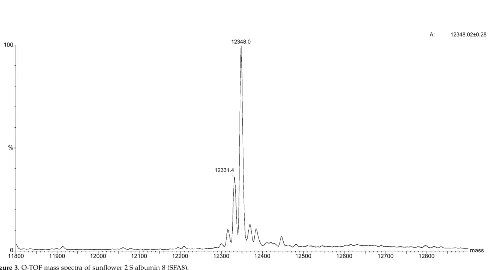
Possibly due to their compact structure and dis-ulphide cross-linking, the 2 S albumin proteins have shown a characteristic resilience to ionisation in mass spectra (MS).
When the recombinant SFA8 protein was analysed by electro-spray (Q-ToF), there was no evidence of attached sugars as the main peak
possessed exactly the predicted mass value (Figure
3) when deconvoluted to singly charged ions.
Longer (. 72 hours) expression times, however,
Figure 2. SDS-PAGE-Western blot. Recombinant Ber e 1, recombinant SFA8 and the product of Kex-SFA8-con-struct protein expression, in which the Kex2 site was mutated, separated by SDS-PAGE and developed using specific rabbit polyclonal antibody anti-native Ber e 1 or anti-native SFA8 protein.
resulted in extra peaks with added mass of 16 Da
(not shown), suggesting that oxidation of
methionine residues was occurring during the
high aeration conditions employed during
expression. Due to the high methionine content of some 2 S albumins, oxidation of methionine to methionine sulphoxide was expected and also reported in early studies as artefacts during
extraction from plant seeds.11
After several unsuccessful attempts with the electro-spray, the mass of the Ber e 1 protein was finally ascertained using a MALDI-TOF system. Instead of the sharp peaks observed for SFA8, Ber e 1 showed a pattern typical of a glycosylated protein with a much broader mass distribution (Figure 4). The presence of attached sugars was also established by 1D NMR (M. Williamson, personal communication) and further confirmed
by the binding of Galanthus nivalis agglutinin to terminal mannose (data not shown). No change in mobility of the recombinant Ber e 1 protein was observed in SDS-PAGE following treatment with either N-glycosidases H or F, or O-glycosidase.
The mass results and the undetectable gel-shifts following treatment of the recombinant Ber e 1 with N and O-glycanases indicate that the sacchar-ides attached to the protein are small. Mutation of
the serine to methionine (S ! M, Figure 1) at the
potential N-glycosylation site (N-X-S/T, where X represents any amino acid residue) in the Ber e 1 did not abolish binding of the G. nivalis agglutinin
or alter the overall MALDI-TOF spectrum,
suggesting that O-type glycans might be involved. The absence of N-glycosylation in the recombinant protein is consistent with the lack of evidence for
the attachment of N-linked glycans to the native
protein derived from plant seed.11
Different degrees of glycosylation of allergenic proteins produced from P. pastoris have been
reported29,30 and reviewed elsewhere.31,32 In
par-ticular, short O-linked saccharides of mannose con-taining alpha 1,2 glycosidic or phosphorylated
mannose linkages have been characterised.31,33The
published results indicate that the short glycans
added by P. pastoris did not alter the 3D structures31
or the immune reactivity34,35 of expressed proteins
but did reduce the authenticity of product.36
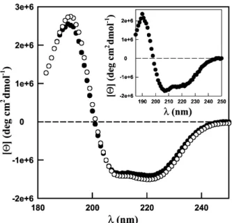
Circular dichroism
In contrast to the recombinant Ber e 1, the recom-binant SFA8 protein had a molecular mass identi-cal with the expected value, indicating that glycosylation was not a problem. Furthermore, the
CD spectra (Figure 5inset) showed virtually
identi-cal traces to the native protein isolated from seed
and published elsewhere.37Whilst the comparison
of recombinant SFA8 and the published native seed protein was straightforward, the presence of short glycans attached to the recombinant Ber e1 and their influence on protein folding needed to be addressed. The CD spectra of the native seed and the recombinant Ber e 1 protein were
com-pared (Figure 5). Despite processing of the two
subunits and loss of the short linker and flanking sequences in at least three stepwise cleavages in the wild-type protein, the spectra of the native and recombinant Ber e 1 proteins recorded at 25 8C
and pH 6.8, are very similar to each other (Figure
5), suggesting that the recombinant and native
proteins have similar secondary structure.
In order to compare the physical characteristics of the recombinant and native proteins, the stability of Ber e 1 and the recombinant Ber e 1 were further tested against pH, temperature and chemical denaturants. The circular dichroism (CD) spectra of the native and recombinant proteins were very similar to each other at different pHs. For the two proteins, spectra obtained at pH 2.2
and 25 8C (Figure 6) were superimposable to those
recorded at pH 6.8 (Figure 5). Temperatures over
62 8C were necessary (Figure 6 inset) to start
unfolding both Ber e 1 proteins at pH 2.2, further demonstrating the high resistance of these proteins to acidic pH. At 95 8C, both proteins were signifi-cantly destabilised although the transition was not complete. Upon cooling, the denaturation was
Figure 5. CD spectra in the far UV region of Ber e 1 (W), recombinant Ber e 1 (X) and recombinant SFA-8 (inset) at pH 6.8 and 25 8C.
Figure 6. CD spectra of recombi-nant Ber e 1 under various con-ditions in 10 mM glycine buffer pH 2.2. (X) 25 8C upon heating; (O) 95 8C; (W) 25 8C upon cooling; and (A) 6 M guanidinium chloride. The CD spectra of the native Ber e 1 are similar to the recombinant protein under the same conditions and are not shown for clarity. Insets: heat induced unfolding transition of Ber e 1 (W) and rBer e 1 (O) followed by far UV CD measurements at 222 nm. The dotted lines represent the slope of the pre-transition.
largely reversible. The two proteins were also very resistant against chemical-induced unfolding. No changes in the secondary structure were observed up to a concentration of at least 4.5 M guanidinium chloride and the protein exhibited a significant amount of secondary structure in 6 M guanidinium
chloride (Figure 6). A comprehensive comparison
of the proteolytic, thermal, acid and chemical stability of the recombinant and native Ber e 1 pro-tein will be presented elsewhere. These results demonstrate that the recombinant Ber e 1, albeit being glycosylated, has the same properties as the native seed protein.
Disulphide mapping
One of the main features of the 2 S family of plant proteins is the conserved structure of the
disulphide bonds.38 Therefore, determination of
the disulphide connectivities is an essential
experi-mental first step towards modelling and elucidat-ing the full protein structure. In order to confirm the CD results and ascertain whether the Ber e 1, despite being glycosylated, is properly folded, dis-ulphide mapping was undertaken. A number of peaks were resolved from the reverse phase HPLC analysis of Ber e 1 tryptic digests. A relatively abundant peak eluting after 30.81 minutes was collected for further analysis. Mass spectrometry revealed the presence of an apparently abundant ion of 8330.13 mass units when deconvoluted to a singly charged species. N-terminal sequencing suggested that the fragment was composed of a number of peptides, linked by disulphide bridges. Following reduction with DTT, several new ion species were detected. MS – MS fragmentation
results (Figure 7) indicated that the amino acid
sequences present in the complex corresponded to the predicted Ber e 1 disulphide core and are in agreement with the N-terminal sequencing of the large complex prior to reduction. In order to pre-cisely map the position of the disulphide bridges, the 8330.13 mass unit peptide fragment was further digested with cyanogen bromide. Mass spec-trometry revealed the presence of an apparently abundant series of ions of 2848.64, 2866.65, 2876.62 mass units when deconvoluted to singly charged species. The N-terminal sequencing of this peak (Figure 7) agreed with the predicted mass of the cyanogen bromide fragment. These results are in agreement with the disulphide map determined
for other members of the 2 S albumin family38and
Figure 7. Disulphide mapping of the recombinant Ber e 1 protein. Derived monoisotopic mass (M þ ) is shown between brackets. In bold and outlined are the products determined by cyanogen bromide cleavage.
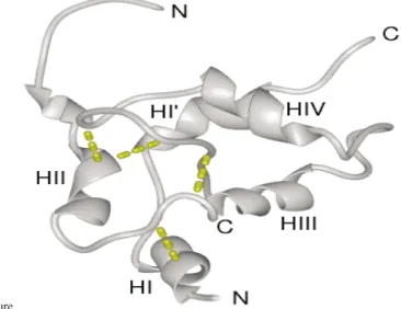
Figure 8. Molecular modelling of Ber e 1 protein. (a) Sequence alignment between 1PNB and Ber e 1 demonstrating the absolute and conservative (þ ) amino acid identities. (b) Ribbon representation of the backbone of the model Ber e 1 structure. The disulphide bonds are represented as broken lines. Picture produced using MidasPlus (UCSF Computer Graphics Laboratory).
together with the CD results strongly suggest that the recombinant Ber e 1 is properly folded.
Modelling
On the basis of the conserved 2 S albumin disulphide connectivity determined for the Ber e 1 protein, together with both sequence and structural alignments with other 2 S albumin proteins, it was possible to construct a model for this protein using the structure of the napin seed storage
protein39(1PNB) as a template. The experimentally
determined structure of 1PNB is that of the mature, proteolytically cleaved protein, hence the model generated here for Ber e 1 corresponds to the proteolytically cleaved form. The sequence align-ment used for building the Ber e 1 model is shown in Figure 8(a). This alignment exhibits ,26% absolute and , 40% conservative amino acid sequence identity. The final, energy-minimised
model for Ber e 1 is shown in Figure 8(b), and
exhibits acceptable geometrical parameters,
assessed using the program Procheck.40The quality
of the alignment and structural model allow us to classify Ber e 1 as a member of the “four helices; folded leaf; right-handed superhelix; disulphide-rich” fold family with reasonable confidence.
In conclusion we have shown that the allergenic Ber e 1 and SFA8 2 S albumins were secreted at high levels from P. pastoris. Through mutation, we also showed that altered sequence around the Kex2 processing site can compromise secretion, probably by targeting the fusion protein to destruc-tion. We also showed that both proteins were properly folded and that the recombinant Ber e 1 is glycosylated, probably by O-linked short sac-charides. The disulphide map of the recombinant Ber e 1 brazil nut protein was established and is in agreement with the map determined for other members of the 2 S albumin family. Using those data a model structure of the allergen was gener-ated and is consistent with the CD spectrum results. The secreted production of these properly folded sulphur-rich plant allergenic albumins will facilitate the determination of the 3D structure of this important family of allergenic proteins and presents an opportunity to delineate the attributes that make an allergen.
Material and Methods
Media composition for P. pastoris
P. pastoris GS115 (Invitrogen) was maintained in YPD broth (10.0 g yeast extract (Difco), 20.0 g peptone, 20.0 g dextrose per litre). MD plates (13.4 g yeast nitrogen base, 10.0 g glucose, 0.4 mg biotin, 100 ml 1 M K2HPO4/
KH2PO4(pH 6.0), 20 g agar per litre) were used for
plas-mid selection. BMG broth (13.4 g yeast nitrogen base, 0.4 mg biotin, 10 ml glycerol, 100 ml 1 M K2HPO4/
KH2PO4 (pH 6.0) per litre) was used for enrichment.
BMM broth (13.4 g yeast nitrogen base, 0.4 mg biotin, 5 ml methanol, 100 ml 1 M K2HPO4/KH2PO4 (pH 6.0)
per litre) was used for induction. Unless otherwise stated, all the chemicals were supplied by Sigma.
Construction of expression plasmids
A plasmid containing the cDNA sequence of sun-flower albumin 8 (Genbank X56686) was kindly donated by T. J. Higgins (CSIRO Plant Industry, Australia). The brazil nut genomic DNA was isolated from seed using a plant DNA extraction kit (Nucleon Phytopure, Amer-sham) according to the manufacturer’s instructions. The 2 S albumin genes were amplified by PCR using the pri-mers: 50
-TCTCTCGAGAAAAGAGAGGCTGAAGCTCC-CTATGGCCGAGGTAGAACTGA and 50
-CTGAATTCT-CATTACATTTGGC ATGGTTGGGACAT; 50
-GAAGGG-GTATCTCTCGAGAAAAGAG AGGCTGAAGCTATGG-GTGT CTACACATTTGAAAACGA and 50
-ATTGAATT-CTCA GAACCCGGCAATGGAGCCAC; for SFA8, and Ber e 1, respectively, at 35 cycles of 94 8C 30 seconds, 54 8C 30 seconds, 72 8C 30 seconds, using Amplitaq (two units/100 ml; Applied Biosystems) according to the man-ufacturer’s instructions. The PCR products (ca 400 bp) were gel-purified, digested with Eco RI/Xho I enzymes for four hours at 37 8C and ligated into pPIC9 (Invitro-gen) previously digested with the same enzymes. The resulting plasmids were transformed into E. coli XL1Blue (Stratagene) and selected under ampicillin (100 mg/ml) following standard protocols.41Plasmids containing the
inserted gene were purified (midi kit, Qiagen) and sequenced using the Sanger method with dye termin-ators (Applied Biosystems). The small intron (70 bp) from genomic Ber e 1 (Genbank X54491) was removed by Splice over extension PCR, cloned as described above and re-sequenced.
Transformation and expression in P. pastoris pPIC9-derived plasmids containing the 2 S protein-encoding sequences were linearised at the Sac I site for four hours at 37 8C. The linearised plasmids were then transformed into P. pastoris GS115 (5 mg/transformation) and the transformed strains were selected on MD plates following electroporation according to the manufac-turer’s instructions (Invitrogen). Clones able to grow in the absence of histidine were inoculated into a sterile 96-well plate format containing BMG and incubated overnight at 30 8C. The 96 clones were then expanded to 4 £ 24-well plates containing BMG (1.5 ml/well) and incubated overnight at 30 8C. After centrifugation (750g for 15 minutes), the 24-well plates were emptied by suc-tion and the BMM broth (containing methanol at 0.5% v/v) added. Expression was allowed to take place in an orbital shaker (200 rpm) for 48 hours with addition of methanol (0.5% v/v) every 12 hours.
Dot-blot screening
After induction with methanol, the 4 £ 24-well plates were centrifuged (750g for 15 minutes) and the super-natant (500 ml) transferred under vacuum to a wetted PVDF (Millipore) membrane in a dot-blot format system. The membrane was then blocked by incubation with 10 ml of BSA (bovine serum albumin, 5% (w/v)) in TBS (Tris buffered saline, 20 mM Tris (pH 7.5), 0.9% (w/v) sodium chloride) for one hour. All incubations were per-formed at 37 8C in a standard hybridisation oven. After three washes (five minutes each) with TBST (TBS plus Tween 20, 0.1% w/v), the membranes were incubated
for two hours with the specific rabbit polyclonal anti-body anti-native Ber e 1 or anti-native SFA8 diluted (1:1000) in TBST plus 20% (v/v) of P. pastoris supernatant extract that was previously prepared and did not contain the target protein. The membrane was then washed three times with TBST and incubated for a further one hour with the secondary alkaline phosphatase-labelled anti-rabbit antibody, diluted 1:20,000 in TBST. After the three washes with TBST the membrane was incubated with Nitroblue tetrazolium and 5-bromo-4-cloro-3-indolyl phosphate toluidine substrate. Once the reaction was completed, the blot was washed with distilled water and dried.
Large-scale production
Selected clones were grown at high density (A600 nm3–
6) in five £ 4 l-flasks containing BMG (2 l) at 30 8C. The culture was then centrifuged (6000g for 15 minutes), and the pellet re-suspended in BMM (10 l) and incubated with agitation (200 rpm) at 30 8C for 48–72 hours with addition of methanol (0.5% v/v) every 12 hours. After induction, the supernatant was separated from cells by centrifugation (6000g for 15 minutes) and filtered through a 0.45 mm membrane. The supernatant was then concentrated (50 £ ) and the salt eliminated by buf-fer exchanged (citrate bufbuf-fer, 10 mM, pH 4.3) using a tan-gential flow system (Viva Science, cut-off 5000 Mr). The
concentrated sample was then loaded on to a heparin-Sepharose column (3 £ 5 ml) fitted to a FPLC (Pharma-cia) system and the 2 S proteins eluted at 400–600 mM sodium chloride. The eluted peak was then loaded on to a Sephadex 75 column (Pharmacia, 2.5 cm £ 90 cm) run (2 ml/minute) with phosphate buffer saline (100 mM sodium phosphate (pH 7.5), 1 M NaCl) and the purified 2 S protein eluted after two hours. The eluted peak was dialysed overnight against water (4 8C) and freeze-dried.
SDS-PAGE/Western blot
SDS-PAGE and Western analysis were performed following the manufacturer’s instructions (NOVEX— Invitrogen), using Immobilon PVDF membrane (Millipore) and antibody dilutions and incubations as described above for the dot-blot screening.
Glycan analysis
The glycosylation of the proteins was confirmed by the binding of G. nivalis agglutinin to terminal mannose in a Western blot format following the instructions from the glycan differentiation kit (Boehringer Mannheim Bio-chemica). De-glycosylation was carried out following the manufacturer’s instructions using peptide-N-glycosidase F or endoglycosidase-H, after heat-denaturation and incubation for 18 hours at 25 8C (Oxford GlycoSystems).
Circular dichroism (CD) measurements
Native Ber e 1 protein from Brazil nut seeds was pre-pared essentially as described.13CD measurements were
performed with a Jasco J-810 spectropolarimeter in the far UV region using a protein concentration of 0.03– 0.32 mg/ml and a 0.1 cm cell pathlength. The buffers used were 10 mM phosphate buffer (pH 6.8) and 0.1 M glycine buffer (pH 2.2). The instrument was calibrated with d-10-camphorsulphonic acid. Spectra were acquired using a scan speed of 20 nm/minute, with a 2 mm
band-width and a two seconds integration time. The spectra were measured 20 times, averaged, and corrected by subtraction of the solvent spectrum obtained under iden-tical conditions. Native and recombinant Ber e 1 proteins, as well as recombinant SFA8 were examined by CD.
Heat-induced unfolding
Heat-induced unfolding transitions were monitored at fixed wavelength of 222 nm using the same protein and buffer concentration as above. The temperature was increased monotically from 25 8C to 95 8C at a rate of 0.5 deg.C/minute. A spectrum was recorded at 95 8C, and the reversibility of the phenomenon was assayed by measuring the spectra of the sample cooled down in a single step to 25 8C. Data were acquired with a reading frequency of 1/60 second, eight seconds integration time and 2 nm bandwith. The resulting curves were cor-rected for the contribution of the solvent obtained under identical heating conditions. The protein concentration was determined using the BCA protein assay reagent kit (Pierce, Illinois).
Chemical-induced unfolding
Samples of the protein were incubated overnight at 25 8C in the presence of various concentrations in guanidinium chloride (i.e. 0, 1.5, 3, 4.5 and 6 M). The denaturant concentration was determined from refractive index measurements using a R5000 hand refractometer from Atago. Spectra were recorded using the same parameters as for heat-induced unfolding and were corrected for the contribution of the solvent obtained under identical heating conditions.
Digestion of Ber e 1 and HPLC analysis
Non-reduced protein (200 –500 mg) was digested over-night at 37 8C with (5 mg) sequencing grade-modified trypsin (Promega) in (100 ml) ammonium hydrogen car-bonate buffer (150 mM, pH 8.1). Reverse phase HPLC was carried out on a model 130A (Applied Biosystems) equipped with a Phenomenex Jupiter C18 column (2.1 mm £ 250 mm). Aqueous formic acid (0.1% (v/v) solvent A) and 70% (v/v) acetonitrile in solvent A (sol-vent B) were used in a linear gradient of 0–100% B over 60 minutes following an initial period of five minutes at 0% B. The flow rate was 150 ml/minute and the peptides detected at 220 nm with manual collection. Whenever required, the large peptide fragments collected from HPLC were reduced with DTT (20 ml, 100 mM) and incu-bated at room temperature for one hour. Cyanogen bromide cleavage was carried out with large crystals in 70% (v/v) formic acid and incubated overnight at room temperature in the dark. The resulting fragments were re-run on HPLC as detailed above and peaks collected for subsequent mass and N-terminal sequencing analysis.
Mass spectrometry
Masses of glycosylated proteins were derived by analysis using a MALDI-TOF (matrix-assisted laser desorption ionisation-time of flight) spectrometer on a Voyager-DEe Biospectrometrye Workstation (Applied Biosystems, USA) operating at 20,000 V accelerating potential and in delayed extraction mode. The sample was prepared at 2 pmol/ml in water and mixed with
saturated sinipinic acid solution (in 50% acetronitrile, 0.1% TFA).
Masses of intact peptide complexes and individual peptide fragments were derived by analysis using a Micromass QTOF-2 hybrid (quadrupole time of flight mass spectrometer) utilising MassLynx software. Peptide solutions were introduced into the sample cone as a fine spray from the end of a thin-walled glass capillary using, typically, a capillary voltage of 800 V. The masses of large peptide complexes and other multiply charged ion species were deconvoluted to singly charged masses using the maximum entropy MaxEnt3 software (Micro-mass). Peptides were selected for fragmentation in MS – MS mode in the mass spectrometer and subjected to collision energies ranging from 20–80 eV over a period of time. All data collected from each fragmentation experiment were deconvoluted using MaxEnt3 software. De novo sequence assignments were carried out inter-actively utilising the peptide-sequencing portion of the BioLynx menu included in the MassLynx software package.
Amino acid sequence analysis
N-terminal sequencing of proteins was carried out using blotted PVDF membranes or eluted HPLC peaks onto Bolybrene-coated glass fibre discs and sequenced using an automated protein sequencer Model 473A (Applied Biosystems) equipped with an on-line PTH Chromatograph and model 610A software (Applied Bio-systems).
Homology modelling of Ber e 1 protein
On the basis of the results of 3D-PSSM42comparisons
of the Ber e 1 amino acid sequence, the experimentally determined disulphide connectivity, and a structure-based sequence alignment of eight members of the 2 S albumin “four helices; folded leaf; right-handed super-helix; disulphide-rich” fold family produced previously,37 we used the structure of the napin seed
storage protein (PDB code 1PNB39) as a structural
tem-plate. The appropriate sections of the Ber e 1 amino acid sequence were directly substituted onto the 1PNB struc-ture according to the alignment in Figure 7(a). Side-chain clashes in the resulting model were relieved by altering torsion angles by inspection. The structure was then covered in a 5 A˚ layer of water and energy mini-mised until the absolute derivative of co-ordinates with respect to the energy fell below 0.01 kcal mol21A˚21.
Analysis of the structure in the program Procheck40
demonstrated that the minimised model had acceptable geometry, as shown by the Ramachandran angles and G factors (data not shown). Visualisation, building and manipulation of the structures was carried out using InsightII 2.35, and energy calculations were preformed using Discover 2.95 (MSI) using the consistent valence force field.
Atomic co-ordinates
The model structure has been deposited in the PDB database under the accession number 1GYS.
Acknowledgements
This work was sponsored by the UK Food Standards Agency (T07019) at the University of Nottingham.
References
1. Spies, J. R., Coulson, E. J., Chambers, D. C., Bernton, H. S., Stevens, H. & Shimp, J. H. (1951). The chem-istry of allergens. XI. Properties and composition of natural proteoses isolated from oilseeds and nuts by the CS-1A procedure. J. Am. Chem. Soc. 73, 3995–4001.
2. Spies, J. R. (1974). Allergens. J. Agric. Food Chem. 22, 30–36.
3. Atkins, F. M., Wilson, M. & Bock, S. A. (1988). Cot-tonseed hypersensitivity: new concerns over an old problem. J. Allergy Clin. Immunol. 82, 243–250. 4. Youle, R. & Huang, A. H. C. (1981). Occurrence of
low molecular weight and high cysteine containing albumin storage protein in oilseeds of disperse species. Am. J. Bot. 68, 44–48.
5. Pastorello, E. A., Pompei, C., Pravettoni, V., Brenna, O., Farioli, L., Trambaioli, C. & Conti, A. (2001). Lipid transfer proteins and 2 S albumins as allergens. Allergy, 56, 45–47.
6. Takizawa, T., Arakawa, H., Tokuyama, K. & Morikawa, A. (2001). Identification of allergen frac-tions of wheat flour responsible for anaphylactic reactions to wheat products in infants and young children. Int. Arch. Allergy Immunol. 125, 51–56. 7. Sandiford, C. P., Tatham, A. S., Fido, R., Welch, J. A.,
Jones, M. G., Tee, R. D. et al. (1997). Identification of the major water/salt insoluble wheat proteins involved in cereal hypersensitivity. Clin. Expt. Allergy, 27, 1120–1129.
8. Shewry, P. R. & Pandya, M. J. (1999). The 2 S albumin storage proteins. Seed Proteins, Kluwer Academic Publishers, The Netherlands pp. 619–643.
9. Saalbach, G., Rosso, M. & Schumann, U. (1996). The vacuolar targeting signal of the 2 S albumin from brazil nut resides at the C terminus and involves the C-terminal propeptide as an essential element. Plant Physiol. 112, 975–985.
10. Krebbers, E., Herdies, L., De Clercq, A., Seurinck, J., Leemans, J., van Damme, J. et al. (1988). Determi-nation of the processing sites of an Arabidopsis 2 S albumin and characterisation of the complete gene family. Plant Physiol. 87, 859–866.
11. Ampe, C., van Damme, J., Castro, L. A. B., Sampaio, M. J. A. M., van Montagu, M. & Vandekerckhove, J. (1986). The amino acid sequence of the 2 S sulphur-rich proteins from seeds of brazil nut (Bertholettia excelsa H.B.K.). Eur. J. Biochem. 159, 597–604.
12. Castro, L. A. B., Lacerda, Z., Aramayo, R. A., Sampaio, M. J. A. M. & Gander, E. S. (1987). Evidence for a precursor molecule of brazil nut 2 S seed proteins from biosynthesis and cDNA analysis. Mol. Gen. Genet. 206, 338–343.
13. Sun, S. S. M., Altenbach, S. B. & Leung, F. W. (1987). Properties, biosynthesis and processing of a sulfur-rich protein in brazil nut (Bertholletia excelsa H.B.K.). Eur. J. Biochem. 162, 477–483.
14. Kortt, A. A. & Caldwell, J. B. (1990). Low molecular weight albumins from sunflower seed: identification of a methionine-rich albumin. Phytochemistry, 29, 2805–2810.
15. Kortt, A. A., Caldwell, J. B., Lilley, G. G. & Higgins, T. J. V. (1991). Amino acid and cDNA sequences of a methionine-rich 2 S protein from sunflower seed (Helianthus annuus L.). Eur. J. Biochem. 195, 329–334. 16. Pastorello, E. A., Farioli, L., Pravettoni, V., Ispano,
M., Conti, A., Ansaloni, R. et al. (1998). Sensitization to the major allergen of brazil nut is correlated with clinical expression of allergy. J. Allergy Clin. Immunol. 102, 1021–1027.
17. Nordelee, J. A., Taylor, S. L., Townsend, J. A., Thomas, L. A. & Bush, R. K. (1996). Identification of brazil nut allergen in transgenic soybeans. N Engl. J. Med. 334, 688–692.
18. Kelly, J. D., Hlywka, J. J. & Hefle, S. L. (2000). Identi-fication of sunflower seed IgE-binding proteins. Int. Arch. Allergy Immunol. 121, 19–24.
19. Schwartz, R. H. (1985). T lymphocyte recognition of antigen in association with gene products of the major histocompatibility complex. Annu. Rev. Immunol. 3, 237–255.
20. Robotham, J. M., Teuber, S. S., Sathe, S. K. & Roux, K. H. (2002). Linear IgE epitope mapping of the English walnut (Juglans regia ) major food allergen, Jug r 1. J. Allergy Clin. Immunol. 109, 143–149. 21. Ommen, A. M., Kelly, J. K., Benson, A. K., Hefle, S. L.
& Taylor, S. L. (2000). Characterization of 2 S brazil nut IgE-binding epitopes. J. Allergy Clin. Immunol. 103, pS101 –pS134.
22. van Regenmortel, M. H. V. (1989). Structural and functional approaches to the study of protein anti-genicity. Immunol. Today, 10, 266–272.
23. Ferreira, F., Ebner, C., Kramer, B., Casari, G., Briza, P., Kungl, A. J. et al. (1998). Modulation of IgE reactivity of allergens by site-directed mutagenesis: potential use of hypoallergenic variants for immunotherapy. FASEB J. 12, 231–242.
24. Spencer, J. A., Jeenes, D. J., MacKenzie, D. A., Haynie, D. T. & Archer, D. B. (1998). Determinants of the fidelity of processing glucoamylase –lysozyme fusions by Aspergillus niger. Eur. J. Biochem. 258, 107–112.
25. MacKenzie, D. A., Kraunsoe, J. A. E., Chesshyre, J. A., Lowe, G., Komiyama, T., Fuller, R. S. & Archer, D. B. (1998). Aberrant processing of wild-type and mutant bovine pancreatic trypsin inhibitor secreted by Aspergillus niger. J. Biotechnol. 63, 137–146.
26. Parekh, R., Forrester, K. & Wittrup, D. (1995). Multi-copy overexpression of bovine pancreatic trypsin inhibitor saturates the protein folding and secretory capacity of Saccharomyces cerevisiae. Protein Expr. Purif. 6, 537–545.
27. Ledgerwood, E. C., George, P. M., Peach, R. J. & Brennan, S. O. (1995). Endoproteolytic processing of recombinant proalbumin variant by the yeast Kex2 protease. Biochem. J. 308, 321–325.
28. Julius, D., Schekman, R. & Thorner, J. (1984). Glyco-sylation and processing of prepro-a-factor through the yeast secretory pathway. Cell, 36, 309–318. 29. Takahashi, K., Takai, T., Yasuhara, T., Yokota, T. &
Okumura, Y. (2001). Effects of site-directed mutagen-esis in the cysteine residues and the N-glycosylation
motif in recombinant Der f 1 on secretion and protease activity. Int. Arch. Allergy Immunol. 124, 454 –460.
30. Best, E. A., Stedman, K. E., Bozic, C. M., Hunter, S. W., Vailes, L., Chapman, M. D. & McDermott, M. J. (2000). A recombinant group 1 house dust mite allergen, rDer f 1 with biological activities similar to those of the native allergen. Protein Expr. Purif. 20, 462 –471.
31. Bretthauer, R. K. & Castellino, F. J. (1999). Glycosyla-tion of Pichia pastoris-derived proteins. Biotechnol. Appl. Biochem. 30, 193–200.
32. Gemmill, T. R. & Trimble, R. B. (1999). Overview of N- and O-linked oligosaccharide structures found in various yeast species. Biochim. Biophys. Acta, 1426, 227 –237.
33. Duman, J. G., Miele, R. G., Liang, H., Grella, D. K., Sim, K. L., Castellino, F. J. et al. (1998). O-Mannosyla-tion of Pichia pastoris cellular and recombinant proteins. Appl. Biochem. 28, 39–45.
34. van Oort, E., Heer, P. G., Lerouge, P., Faye, L., Aalberse, R. C. & van Ree, R. (2001). Immunochemi-cal characterisation of two Pichia pastoris-derived recombinant group 5 Dactylis glomerata isoallergens. Int. Arch. Allergy Immunol. 126, 196–205.
35. Vailes, L. D., Kinter, M. T., Arruda, L. K. & Chapman, M. D. (1998). High level expression of cockroach allergen, Bla g 4 in Pichia pastoris. J. Allergy Clin. Immunol. 101, 274–280.
36. Letourneur, O., Gervasi, G., Gaia, S., Pages, J., Watelet, B. & Jolivet, M. (2001). Characterization of Toxoplasma gondii surface antigen I (SAGI) secreted from Pichia pastoris: evidence of hyper-glycosylation. Biotechnol. Appl. Biochem. 33, 35–45.
37. Pandya, M. J., Sessions, R. B., Williams, P. B., Dempsey, C. E., Tatham, A. R., Shewry, P. R. & Clarke, A. R. (2000). Structural characterisation of a methionine-rich, emulsifying protein from sunflower seed. Proteins: Struct. Funct. Genet. 38, 341–349. 38. Egorov, T. A., Odintsova, T. I., Musolyamov, A. Kh.,
Fido, R., Tatham, A. S. & Shewry, P. R. (1996). Disul-phide structure of a sunflower seed albumin: con-served and variant disulphide bonds in the cereal prolamin superfamily. FEBS Letters, 396, 285–288. 39. Rico, M., Bruix, M., Gonza´lez, C., Monsalve, R. I. &
Rodrı´guez, R. (1996).1H NMR assignment and global
fold of napin BnIb, a representative 2 S albumin seed protein. Biochemistry, 35, 15672–15682.
40. Laskowski, R. A., MacArthur, M. W., Moss, D. S. & Thronton, J. M. (1993). Procheck—a program to check the stereochemical quality of protein struc-tures. J. Appl. Crystallog. 26, 283–291.
41. Ausubel, F. M., Brent, R., Kingston, R. E., Moore, D. D., Seidman, J. G., Smith, J. A. & Struhl, K. (2001). Current Protocols in Molecular Biology, Wiley, New York.
42. Kelley, L. A., MacCallum, R. M. & Sternberg, M. J. E. (2000). Enhanced genome annotation using struc-tural profiles in the program 3D-PSSM. J. Mol. Biol. 299, 501–522.
Edited by A. R. Fersht
![[PDF] Formation bureautique gratuite PDF comment ça marche | Cours Bureautique](data:image/gif;base64,R0lGODlhAQABAIAAAP///wAAACH5BAEAAAAALAAAAAABAAEAAAICRAEAOw==)