© FEBS 1987
Primary structure of the Streptomyces R61 extracellular DD-peptidase
2. Amino acid sequence data
Bernard JORIS', Philippe JACQUES', Jean-Marie FRERE', Jean-Marie GHUYSEN' and Jozef VAN BEEUMEN ^ ' Service de Microbiologie appliquee aux sciences pharmaceutiques, Faculte de Medecine, Universite de Liege ^ Laboratoriutn voor Microbiologie, Rijksuniversiteit-Gent
(Received June 16/October 10,1986) - EJB 86 0607
In order to confirm the Streptomyces codon usage, the Streptomyces R61 DD-peptidase was fragmented by (a) cyanogen bromide cleavage of the carboxymethylated protein, (b) trypsin digestion of the carboxymethylated protein and (c) trypsin digestion of the protein treated with /S-iodopenicillinate and endoxo-^^-tetrahydrophthalic acid. The isolated peptides, which altogether represented more than 50% of the polypeptide chain, were sequenced. The data thus obtained were in excellent agreement with the primary structure of the protein as deduced from the nucleotide sequence of the cloned gene. Though a weak acylating agent, ^-iodopenicillanate reacted selectively with the active site of the DD-peptidase and formed an adduct which mas much more stable than that formed with benzylpenicilUn, thus facilitating the isolation and characterization of the active-site peptide.
The Streptomyces R61 chromosomal gene coding for the extracellular DD-peptidase has been cloned and its nucleotide sequence established [1]. In the course of this study, amino acid sequence data became necessary to (a) design a DNA probe as a means to identify the DD-peptidase gene by hybrid-ization experiments and (b) confirm the identification of the open reading frame deduced from the nucleotide sequenee. The present paper describes experiments which were under-taken to eomplement the limited information previously obtained [2—4]. The data obtained confirmed (a) the primary structure of the protein as deduced from the nucleotide se-quence and (b) the validity of the Streptomyces codon usages previously proposed [1, 5 — 9].
MATERIALS AND METHODS
Enzymes and proteins
The DD-peptidase was purified as described [10]. Trypsin (treated with tosylphenylalanylchloromethane) was from Millipore (Freehold, NJ, USA), soybean trypsin inhibitor from Sigma (St. Louis, MO, USA) and carboxypeptidase A from Boehringer (Mannheim, FRG).
Carboxymethylation of the DD-peptidase
An enzyme sample (67 mg dialysed against water for 48 h) was freeze-dried; the residue was dissolved in 4 ml of 0.1 M
Correspondence to B. Joris, Service de Microbiologie, Institut
de Chimie, B6, Universite de Liege au Sart Tilman, B-4000 Liege, Belgium
Abbreviations. ETPA, endoxo-J*-tetrahydrophthalic acid;
DABITC, 4-N,N dimethylaminoazobenzene-4'-isothiocyanate; PITC, phenylisothiocyanate; FPLC, fast protein liquid chromatog-raphy.
Enzymes. D-Alanyl-D-alanine carboxypeptidase or DD-peptidase
(EC 3.4.16.-); trypsin (EC 3.4.21.4); carboxypeptidase A (EC 3.4.17.1).
Tris/HCl buffer pH 8.3 containing 6 M guanidinium chloride, 1 mM EDTA ad 4 mM dithiotreitol, and the mixture left for 4 h at 20 °C under nitrogen. The solution was treated, for 20 min at 20°C and in the dark, with 400 \x\ of a 0.5 mM solution of iodoacetate made in the same buffer as above, and then supplemented with 8 mg iodoacetate dissolved in 80 ^1 of 0.5 M NaOH. After 20 min at 20°C, the solution was dialysed for 36 h against water and freeze-dried. Radioactively derivatized protein was similarly prepared by treatment with iodo['*C]acetate (54 Ci/mol; New England Nuclear, Dreieich, FRG).
Cyanogen bromide cleavage
of the carboxymethylated DD-peptidase
The carboxymethylated protein (50 mg) was dissolved in 7 ml of 70% formic acid. Solid CNBr (140 mg) was added, and the mixture was left for 24 h at 20 °C in the dark and under nitrogen, before being freeze-dried.
Labelling of the DD-peptidase active-site by fi-iodopenicillanate and derivatization of the E-amino group of the lysine residues by endoxo-A*-tetrahydrophthalic acid (ETPA)
The enzyme (100 nmol) and ^-iodopenicillanate (4.5 funol; a gift from Dr. J. Kemp, Pfizer Central Research, Sandwich, Kent, UK) were incubated together for 1 h at 37 °C in 0.65 ml of 10 mM sodium phosphate pH 7.0. Inactivation of the enzyme was complete. The reaction mixture was filtered on Sephadex G-15 in water and the fractions containing the derivatized protein (as detected by measuring the absorbance at 325 nm and 280 nm) were pooled and freeze-dried. The residue in 0.6 ml of 0.25 M borate pH 8.5 was supplemented three times, successively, with 0.7 mg ETPA over a period of 2 h of at 20°C [11]. The pH was maintained at 8.5 by addition of 1 M NaOH and the total amount of ETPA used was 15-fold in excess of the expected number of lysine residues present
in the protein. Complete disappearance of the free amino groups was verified by reacting 60-^1 samples of the reaction mixture with fluorescamine [12]. The excess ETPA was eliminated by filtration on a Sephadex G-15 column in 50 mM NH4HCO3 and the fractions containing the modified protein were freeze-dried.
Purification of the peptides
Paper (Whatman 3 MM) electrophoresis was performed at pH 6.5 (pyridine/aeetic acid/water, 11/4/900, v/v/v) and 3.5 (pyridine/acetic acid/water, 1/10/89, v/v/v). Paper (Whatman 3 MM) descending chromatography was performed in n-butanol/acetic acid/pyridine/water (15/3/12, v/v/v). Peptides were visualized with fluorescamine, ninhydrin or Pauly's re-agent (for peptides containing His or Tyr) [13].
Molecular sieve chromatography was performed on Sephadex G-50, fine, and Sephadex G-75, fine. The solvents were 50 mM formic acid pH 2.6, 50 mM ammonium formate pH 3.5 and 50 mM NH4HCO3 pH 8.0.
Ion-exchange chromatography was performed on a column (1.2 X 9 cm) of DEAE-Sephacel equilibrated against 10 mM NH4HCO3 containing 0.02% dithioglycol; elution was performed using a linear gradient of NH4HCO3 up to 50 mM for a total volume of 100 ml. A Pharmacia FPLC apparatus, equipped with a Pro-RPC HR 5 — 10 column, was also used. Buffer A was 10 mM NH4HCO3 in water and buffer B was 10 mM NH4HCO3 in a mixture of acetonitrile and water (40:60; v/v). The gradient was 0—45% B in 50 min and to 100% B in 10 min (fiow rate 0.3 ml/min).
dodecyl sulphate and 100 nM norleucine as internal standard. The solution was heated at 100°C for 1 min, supplemented with 20 ^g carboxypeptidase A, and the mixture was in-cubated at 37 °C. After 0, 20, 40 and 100 min, 100-^1 samples were removed and the reaction was stopped by addition of 10 nl of 1 M HCl. The precipitate was eliminated by centrifu-gation, the supernatant dry-evaporated, the residue dissolved in 50 \i\ of 0.2 M citrate buffer pH 2.2 and submitted to automatic amino acid analysis.
RESULTS
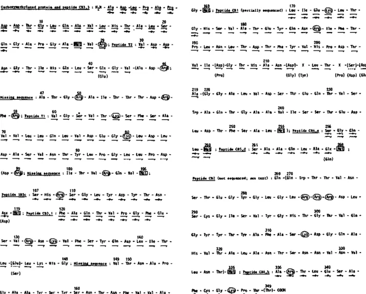
The amino acid sequences of the carboxymethylated DD-p>eptidase and the pieptides obtained by fragmentation of the protein are shown in Fig. 1. The peptides are ordered on the basis of the primary structure of the polypeptide chain as deduced from the nucleotide sequence of the corresponding gene [1].
A'- and C-terminal residues of the protein
Automatic sequencing of 15 nmol of the carboxymethyl-ated protein (dissolved in trifluoroacetic acid) gave the NH2-terminal 29 amino residues (Fig. 1). These data confirmed those reported previously [4] except for Gln-21 (instead of Glu), Ala-26 (instead of Val) and Arg-29 (instead of Arg-28). Threonine was released on treatment of the denatured protein with carboxypeptidase A. The molar Thr/DD-peptidase ratios after 0, 20, 40 and 100 min of treatment were 0.2,1.1,1.1 and 1.1, respectively.
Amino acid analysis
A Dionex D-300 analyser [13] or an adapted version of the Pico-Tag method [14] with a reverse-phase column (ODS C18; Altex, 0.5 x 25 cm) were used.
Amino acid sequencing
Large peptides were sequenced on an Applied Biosystem gas-phase sequenator [15]. The phenylthiohydantoins were analysed off-line from the sequencer by reversed-phase HPLC on an IBM 4.6 x 250 mm cyanopropyl column with 5-^m particles. The protocol was adapted from [16] as suggested by Touchstone (Applied Biosystems users' note no. 3) with the addition of 3.75% tetrahydrofuran to the buffer component of the eluent (solvent A, 0.015 M sodium acetate) and 100% acetonitrile as solvent B. Separation was carried out by gradient elution on Waters equipment comprising two 6000-A pumps as solvent delivery units. Detection was at 254 nm and 313 nm with a 440 dual-channel fixed-wavelength detec-tor. Small peptides were sequenced manually using the double-coupling DABITC-PITC method [17]. The derivatives were identified by two-dimensional chromatography on polyamide plates [16]. When the presence of ['^Qcarboxymethylated cysteine-containing peptides was anticipated, the plates were submitted to autoradiography. In some cases, the classical dansyl-Edman method was used [18]. The N-terminal residues were always identified by dansylation, acid hydrolysis and thin-layer chromatography [18].
Determination of the C-terminal portion of the protein
The enzyme (16.0 nmol) was dissolved in 0.44 ml of 150 mM sodium phosphate pH 7.5 containing 0.1% sodium
Isolation and amino acid sequence of peptides produced by cyanogen bromide cleavage
of the carboxymethylated protein
The carboxymethylated and CNBr-cleaved protein (50 mg) was suspended in 5 ml 50 mM ammdnium formate pH 3.5 containing 4 M guanidinium chloride. After centri-fugation, the solution was filtered through a column (2.5x150 cm) of Sephadex G-50 equilibrated against the formate buffer (without guanidinium chloride). Six fractions (Fig. 2), numbered CBO to CB5 in order of increasing elution volumes, were collected (on the basis of their absorbance at 214 nm). Fraction CBO was excluded from the gel and possessed an N-terminal Ala; most likely, it was some uncleaved protein.
Fractions CBl and CB2 were purified by filtration on a column (1.25 x 150 cm) of Sephadex G-75 in formate buffer. Fragment CB2 had an unique N-terminal residue, Glx (as determined by dansylation), but all further sequencing attempts failed, suggesting cyclisation of an N-terminal Gin into pyrrolidone under acidic conditions; this fragment was not studied. Fraction CB3 was submitted to ion-exchange chromatography on DEAE-Sephacel and gave rise to peptides CB3,1 and CB3,3. Fractions CB4 and CB5 showed complex patterns by paper electrophoresis at pH 6.5. Peptides CB4b, CB4c, CB5c and CB5d were purified by paper electrophoresis at pH 3.5 and paper chromatography. CB4b partitioned into two peptides with R^ values of 0.36 and 0.43, respectively; they had identical amino acid compositions and possessed one N-terminal alanine and one internal cysteine.
All the peptides were submitted to amino acid analysis (Table 1) and manual or automatic sequencing (Fig. 1). On the basis of its amino add composition, peptide CB3,3 consisted of the NH2-terminal 27 amino acids of the protein.
521
C T b o g y . t h y l . f d prot.in Mid p.ptid. CB3.3 : H2N - Ala • Asp -L«u - Pro - Ala - Q
10 20
Asp - Asp - Thr - Gly - L»u - Gin - Ala - Val - Leu - His - Thr - Ala - Leu - Ser • Gin Gly Ala Pro Oly Ala E S Val Q M ) ; p«ptid« T2 : Val Asp Asp
-—* » • I- r r r p r —••» ^ ^ ^ • • • r • » • > »
Asn - 61y - Thr - lie - His - Gl» - Leu - Ser - Glx - Gly - Val -(Ala - Asp -Q (Glu)
Gly i B B j F«ptlJ» CBl ( p a r c U l l ; MciiMiiod) : Leu He Glu { L y S Leo Tiir
G l y -190 Pro -His Leu - Ser - Asii - Val - Leu Ala Thr -Ihr Asp - Glu - Thr T y r Phe Gin Tyr -Asn Va1 -(63)-200 His He Pro -Plw Asp Thr Thr -210
Val - He -[Aspl-Gly - Thr - His - Ala - Asn -lAspl- X - Leu - Thr - X -lS«r)-(Aspl-(Pro) (Gly) (Tyr) (Pro) (Asp) (Glu)
47 50
Mil.jog .equ.Qc. : Ala - Thr - Gly - ( A ^ - Ala - He - Thr - Thr - Thr - Asp -tl Phe @ | ; P.ptU« II : Val Gly Ser Val Thr ( © Ser Phe S«r Ala
Val Val Leu Leu Gin Leu Val Asp Glu Cly ( t y s ) Leu Asp Leu
-219 220 230
Ala (Gly Gly Ala Leu Val Asp Ser Thr Glu Gin Thr Val Ser -240
Trp Ala Gin Thr Gly Ala Ala Val He Ser Ser Thr Glu Asp
-250 255 256
Leu Asp Thr Phe Ser Ala Leu E S ) ; Peptid. CM.. : Ser Gly Gin -L«u - I S S i F.ptid. CBS.J : Ser - Ala - Ala - Gin - Leu - Ala - 61x - ( Asp - Ala - Ser - Val - Asn - Thr - Tyr - Leu - Pro - Gly - Leu - Leu - Pro - Asp - "•-'
» 100 _ ^ 106
(Asp - ^ 3 ) ; Hininn »t(iu«iu!e : He - Thr - Val - ( ^ - Gin - Val - K S 269 270
Ftptid. ct2 (not ..qumccd; u . M i O : Gin (Gin Trp Thr Thr Val Asn -107 n o
ptlile SBSc 1 Ser His (^rg) Ser Gly Leu Tyr Asp Tyr Ihr Asn
-119 120
Asx K B : P«ptld« CB3.1 : Phe Ala Gin Thr Val Pro Gly Phe Glu
-(Asp)
130 , _ ^ 140
> r - Val - @ ) - Asn -Q§- Val - Phe - Ser - Tyr - 61n - Asp - Leu - H e - Thr • 148 149 150
Leu -[Glul- Leu - Lys - His - Gly ; t U n i n t «»iiii«nce : Val - Thr - Asn - Ala - Pro ' (Ser)
280 280
Ser - Thr - Glu - Gly - Tyr - Gly - Leu - Gly - Leu -( ) Asp Leu
-290 300
S«r Cys Gly He Ser Val Tyr Gly His Thr Gly Thr Val Gin -310 - ^
61y Tyr Tyr Thr Tyr Ala Phe Ala Ser ( g s ) Asp Sly Gin Ala
-320 J30
His Val Thr Ala Leu Ala Asn Thr Ser Asn Asn Val Asn Val
-335 336 __ 340
Leu Asn Thr)|SS ; f«ptid. ot.b : Ala ftg) Thr Leu Glu Ser Ala -160
Gly - Ala - Ala - Tyr - Ser - Tyr - Ser - Asn - Thr - Asn - Phe - Val - Val - Ala Phe - Cys - Gly - ( ® - Pro - Thr -(Thr)- COOH
Fig. 1. Amino acid sequences of the carboxymethylated DD-peptidase and ofpeptides obtained by fragmentation of the protein. The peptides are ordered within the primary structure of the protein as deduced from the nucleotide sequence of the cloned gene [1]. Residues and sequences in round brackets, e.g. (Glu) were derived only from gene analysis. The Lys and Arg residues are marked by a circle, the Met residues by a square. The active-site serine* is at position 62. -^ = residues determined with the gas-phase sequenator; - ^ = residues less unambiguously identified but later proven to be exact from the gene sequence; -o^ residues determined by the DABITC-PITC double-coupling method; residues determine by the dansyl-Edman procedure are indicated by a wobbly line. Square brackets, e.g. [Glu], indicate errors; the corresponding residued deduced from gene analysis is shown below in parentheses. The amounts of peptides used were as follows. Manual sequencing: peptides CBl, CB3,1, CB3,3, CB4b, CB5c, CB5d and T2: 15, 4, 34, 29,15, 12 and 10 nmol, respectively. Automatic sequencing: SCM-protein and peptides CB3,1, CBl, CB2 and T,: 15, 11, 7, 10 and 3 nmol, respectively
0.4
CB2
Kinetics of inactivation of the DD-peptidase by fi-iodopenicillanate
As observed with /?-lactamases (19,20], inactivation of the DD-peptidase by /?-iodopenicillanate generated a stable adduct that absorbed at 325 nm (indicating the presence of a dihydrothiazine chromophore). The first-order rate constants for the formation of the adduct at 37 °C, in 10 mM sodium phosphate pH 7.0 and at /S-iodopenicillanate concentration ranging over 1.25 —14 mM, were determined by measuring ^ ' ^ increase of the .4325 values and the decrease of
5 J2s 250 37! vTTi)
Fig. 2. Sephadex G-50 separation of the peptidls obtained after CNBr enzyme activity as a function of time. The data were analysed digestion of the carboxymethylated protein. The column (2.5 x 50 cm) 0 ° ^he basis of the three-step reaction scheme
was equilibrated in 50 mM ammonium formate pH 3.5 containing
4 M guanidinium chloride. The flow rate was 40 ml/h and 5-ml K k k
Table 1. Amino acid composition of the peptides obtained by fragmentation of the DD-peptidase
The underlined figures give the number of residues as deduced from gene sequencing (see text), n.d. = not determined, P = present
Amino acid CBl CB2 CB3,1 CB3,3 CB4b CB4e CB5c CB5d
Lys His Arg Asx Thr Ser Glx Pro Gly Ala Cys or CmCys Val Met or Hse lie Leu Tyr 1.0 2.2 2.5 9.6 10.4 6.1 6.9 3.2 6.1 7.7 0 6.2 P 3.0 8.7 2.5 1 3 1 9 13 6 8 4 6 8 Q 6 1 4 8 3 1.2 1.0 4.2 8.3 7.9 5.2 3.6 0.8 7.4 4.8 P 5.2 P 1.4 5.7 3.8 1 2 3 8 9 5 5 0 8 5 1 6 i 1 5 5 2.6 1.0 2.0 2.0 2.6 2.9 0.9 2.3 1.2 0 2.9 0 0.8 2.8 0.8 2 1 1 2 2 3 3 1 2 1 0 3 0 1 3 1 1.0 3.3 2.2 1.0 2.1 3.1 3.1 5.9 1.2 P 3.6 1 3 2 1 2 3 3 6 1 1 4 0.7 1.0 3.0 1.2 1.0 1.2 1.2 1.9 0.7 1.2 1 i 3 1 1 1 1 2 i i 0.6 0.9 1.0 P 1.1 1 i i 1 1 0.6 1.0 3.0 1 1.5 0.9 P 1.0 1.8 1 1 3 1 2 1 i 1 2 0.8 2.0 3.0 P 1.1 1 2 3 i i 2.1 1.0 6.7 2.7 3.3 2.4 2.5 3.1 2.4 6.2 4.4 1.2 2 1 6 2 4 2 2 3 2 6 8 1 0.6 0.9 4.1 0.8 1.0 2.3 2.0 1.2 2.4 0.8 0.9 1 1 4 1 1 2 2 i 2 1 1 Phe Trp Total N-tenninus 3.0 n.d. i3 .86 Leu 1. n.2d. i1 67 Gin 2.7 n.d. 30 29 Phe n.d. 0 27 Ala 1.0 n.d. 01 14 Ala n.d. 0 5 Ser n.d. 0 13 Ser n.d. 0 8 Ser 0.9 n.d. 01 40 Val n.d. 0 17 Val
where E = DD-peptidase; D =/3-iodopenidllanate; E D = Michaelis complex; E-D* = acyl enzyme; E-D' = rear-ranged, chromophoric adduct; /iT = dissociation constant; k + 2 and ^ + 4 = first-order rate constants. Turnover of /?-iodopenicillanate (that would be represented by the reaction branch E —D* — ^ E + product) and enzyme regeneration did not occur on incubation for 1 h at 37 °C of an inactivated enzjTne sample deprived of inactivator by filtration on Sephadex G-15. Moreover, chromophore formation pro-ceeded without any lag (A:+4 > ^+2). The values of A^ (4 mM), A:4-2 (3 X 10"^s"*) and k + 2lK(0.75 M " * s ~ ' ) were estimated from a plot of [D]/k, vs [D] (not shown).
Isolation and amino acid sequence of the active-site serine-containing peptide produced by trypsin digestion of the DD-peptidase
treated with fi-iodopenicillanate and ETPA
In the course of previous studies [2], derivatization of the active site of the DD-peptidase by )S-iodopenidllanate and trypsin digestion had yielded a small peptide Val-Gly-Ser-Val-Thr-Lys that contained the active-site serine residue. To obtain a peptide of larger size, the e-amino group of the lysine re-sidues of the ^-iodopenicillanate-treated enzyme were derivatized by reaction with ETPA. The modified protein obtained after removal of the excess ETPA by gel filtration (100 nmol) was dissolved in 0.8 ml of 100 mM NH4HCO3 containing 2 M urea and incubated with 0.5 mg trypsin (treated with tosylphenylalanylchloromethane) for 1 h at 37 °C. The reaction was stopped by addition of a stoichio-metric amount of soybean trypsin inhibitor and the reaction mixture was filtered on a column (2 x 140 cm) of Sephadex G-50 in 50 mM NH4HCO3. The non-excluded fraction, which absorbed at 325 nm, was freeze-dried; the residue was dis-solved in 0.5 ml of 10 mM NH4HCO3 and purified by reverse-phase chromatography (Fig. 3). The fractions absorbing at
60 f(min)
Fig. 3. Purification of the active-site serine-containing peptide Ti by reverse-phase chromatography. The chromatography was performed on a Pharmacia FPLC apparatus equipped with a Pro-RPC HR 5 — 10 column. The absorbing at 325 nm fraction is indicated by an asterisk. For conditions, see Materials and Methods
325 nm were pooled, freeze-dried and submitted to a second chromatography under the same conditions as above. The elution profiles as determined at 215 nm and 325 nm were superimposable. The fractions exhibiting the highest absorbance were pooled and freeze-dried and the residue dis-solved in 250 jil water. Table 1 and Fig. 1 give the amino acid composition and the NH2-tenninal 31 residues of peptide T j . The active-site serine residue is at position 3 (position 62 in the protein).
Isolation and amino acid sequence of peptide T2 produced by trypsin digestion
of the carboxymethylated DD-peptidase
The carboxymethylated protein (250 nmol) was digested with trypsin as described in [4]. The reaction mixture was
filtered through a column (1 x 120 cm) of Sephadex G-50 in 50 mM formic acid, pH 3.0. The acidic peptides (detected by submitting 60-|xl samples of the 0.65-ml collected fractions to paper electrophoresis at pH 6.5) were further purified by paper electrophoresis at pH 6.5 and 3.5 and paper chromatog-raphy. Peptide T2, the amino acid composition of which is shown in Table 1, was sequenced up to position 14 (Fig. 1).
Search for free sulphydryl groups
The native enzyme (14 nM, final concentration) was dis-solved in 300 nl 50 mM sodium phosphate buffer pH 7.4 containing 8 M urea, incubated at 37 °C for 60min and supplemented with 25 \i\ of a 6.6 M solution of 5,5'-dithiobis(2-nitrobenzoate) made in the same buffer. After 20 min, the absorbance at 412 nm indicated the presence of less than 0.07 free SH group per protein molecules.
DISCUSSION
The experiments described in this and the preceding [1] papers were complementary. They were carried out in parallel for the purpose of establishing the complete primary structure of the Streptomyces R61 peptidase. The 349-residue DD-peptidase has two half-cysteine residues. Attempts to detect free SH groups failed, suggesting that a disulfide bridge exists between Cys-291 and Cys-344.
Contrary to expectation, peptide CB3,1 (isolated after cyanogen bromide cleavage of the protein) had no methionine residue at the C-terminal position, indicating that a non-specific cleavage must have occurred after Gly-148. Moreover, fragment Val-149 — Met-169 (resulting from Uiis cleavage) and the large fragment Val-28 —Met-106 escaped isolation. The former fragment has an average hydrophybicity index per residue of 0.3 (versus 0.09 for the complete protein) as computed according to Eisenberg's consensus scale [21]. It may have adsorbed on the various column supports during the purification steps. The reason why CNBr fragment Val-28—Val-106, which has a low average hydrophobicity index of 0.01, is missing is unknown. It does contain two very hydrophobic stretches (Ala-69-Val-76 and Tyr-90-Pro-96) but these also occur in peptide Ti which behaved 'normally' during the purification steps. It must be noted that N-terminal analysis of the cyanogen-bromide fragments purified by column chromatography always yielded traces of valine, suggesting the slow and continuous elution of (an) absorbed peptide(s).
In the work carried out previously by Duez et al. [4], several peptides produced by trypsin digestion of the carboxyme-thylated protein had been isolated and analysed for their amino acid content. Most of them are easily positioned in the sequence shown in Fig. 1: peptides A5 (residues 1—29), Ai (residues 30-46, or peptide T2), B4 (residues 47-50), N (residues 51-57), Bg (residues 58-59), B5 residues 60-65), B7 (residues 100-103), A3 (residues 110-131) and B9 (re-sidues 174-186). Peptide Bs (Gly, Leu, Arg) might have originated from residues 283—285 by non-specific cleavage. Peptide Bi (Asx, Gly, Lys, Arg), B2 (Asx2, Ser, Glx, Gly, Val, Met, Lys3, His, Arg) and B3 (Arg4, Lys) cannot be positioned in the sequence and were most likely isolated as mixtures of short peptides. Bi may have been an equimolecular mixture of Asx-132-Lys-133 and Gly-49-Arg-50. As such, B2 may have originated from the comigration of peptides Gln-104—
Arg-109 and Asn-132-Lys-133 present as a threefold excess with Gly as impurity.
Examination of Fig. 1 shows that trypsin should generate a peptide Ile-187-Arg-285 of M^ larger than 10000 and three fragments from the C-terminal region of the protein (Asp-288-Lys-313; Asp-314-Arg-337; And Thr-338-Thr-349). These peptides escaped detection in the work described in [4]. The large peptide neither nugrated nor stained after paper electrophoresis and chromatography. Assimiing that the C-terminal portion escaped trypsin digestion, the C-C-terminal stretch Asp-288-Thr-349 has an M, which is equivalent to that of one of the 'core peptides' described [4].
Finally, Duez et al. [4] could not find any radioactive soluble peptide after digestion of the ['*C]benzylpenicilloyl-DD-peptidase by trypsin though peptide B5 contains the active-site serine 62. The bound penicilloyl moiety must have been lost during trypsin digestion and the ensuing purification steps. /?-Iodopenicillanate has the advantage of forming a specific and Wghly stable adduct with the enzyme, even if it is a weak acylating agent for the R61 DD-peptidase (fc + 2/A^ =
0.75 M " ' s " * versus 14000 M " * s ' ' for benzylpenicillin). Altogether, the peptides that have been sequenced during the present work represent 55% of the Streptomyces DD-peptidase. Except for five residues, the data are in agreement with the primary structure of the protein as deduced from the nucleotide sequence of the gene [1]. Other Streptomyces genes had been sequenced previously but the amino acid sequences of the expressed proteins had not been investigated in detail. The most extensively studied protein was the 313-amino-acid endo-^-A^-acetylglucosaminidase of Streptomyces plicatus [6] for which several fragments representing altogether 43 re-sidues were sequenced.
This work was supported by an Action concertee from the Belgian Government (convention 79/84-11), the Fonds de la Recherche Scientifique Medicate, Brussels (contract 3.4507.83) and a Convention tripartite between the Region wallone. Continental Phanna and the University of Liege. B. J. is Charge de Recherches of the Fonds National de la Recherche Scientifique (FNRS, Brussels) and P. J. is fellow of the Institut pour I'Encouragement de la Recherche Scientifique dans I'Industrie et I'Agriculture (IRSIA, Brussels).
REFERENCES
1. Duez, C , Piron-Fraipont, C , Joris, B., Dusart, J., Urdea, M. S., Martial, J., Frere, J. M. & Ghuysen, J. M. (1987) Eur. J.
Biochem.l62,5(i9-51S.
2. Kelly, J. A., Knox, J. R., Moews, P. C, Hite, G. J., Bartolone,
J. B., Zhao, H., Joris, B., Frere, J. M. & Ghuysen, J. M. (1985) J. Biol. Chem. 260. 6449-6458.
3. Frere, J. M., Duez, C, Ghuysen, J. M. & Vandekerckhove, J. (1976) FEBS Lett. 70, 257-260.
4. Duez, C , Frere, J. M., Ghuysen, J. M., Van Beeumen, J. & Vandekerckhove, J. (1981) Biochim. Biophys. Acta 671, 1 0 9 -116.
5. Thompson, C. J. & Gray, G. S. (1983) Proc. Natl Acad. Sci. USA «0, 5190-5194.
6. Robbins, P. W., Trimble, R. B., Wirth, D. F., Hering, C , Maley, F., Maley, G. F., Das, R., Gibson, B. W., Royal, N. & Biemann, K. (1984) J. Biol. Chem. 259, 7577-7583.
7. Bibb, Me. J., Bibb, Ma. J., Ward, J. M. & Cohen, S. N. (1985) Mol. Gen. Genet. 199, 26-36.
8. Beman, V., Filpula, D., Herber, W., Bibb, M. & Katz, E. (1985) Ge«e 57.101-110.
9. Hopwood, D. A., Bibb, M. J., Chater, K. F., Janssen, G. R., Malpartida, F. & Smith, C. P. (1986) in Regulation of gene expression — 25 years on (Booth, I. R. & Hig^ns, C. F., eds) pp. 251—276, Cambridge University Press, Cambridge.
10. Fossati, P., Saint-Ghislain, M., Sicard, P. J., Frere, J. M., Dusart, J., Klein, D. & Ghuysen, J. M. (1978) Biotechnol. Bioeng. 20, 577-587.
11. Riley, M. & Perham, R. M. (1970) Biochem. J. 118, 733-739. 12. Lai, C. Y. (1977) Methods Enzymol47. 216-237.
13. Joris, B., Van Beeumen, J., Casagrande, F., Gerday, C , Frere, J. M. & Ghuysen, J. M. (1983) Eur. J. Biochem. 130, 53-69. 14. Bidlingmeyer, B. A., Cohen, S. A. & Tarvin, T. K. (1984) J.
Chromatogr. 336, 93-104.
15. Hewick, R. M., Hunkapiller, M. W., Hood, L. E. & Dreyer, W. J. (1981) J. Biol. Chem. 256, 7990-7997.
16. HunkapUler, M. W. & Hood, L. E. (1983) Methods Enzymol. 91, 486-493.
17. Wittmann-Liebold, B. & Lehmann, A. (1980) Methods in peptide and protein sequence analysis (Chr. Bin, ed.) pp. 49—72, Elsevier/North-HoUand Biomedical Press, Amsterdam. 18. Hartley, B. S. (1970) Biochem. J. 119, 805-822.
19. Frere, J. M., Dormans, C , Duyckaerts, C. & De Graeve, J. (1982) Biochem. J. 207, Ail-AAA.
20. Lenzini, M. V. & Frere, J. M. (1985) J Enzyme Inhibition 1, 2 5 -34.