HAL Id: hal-02871577
https://hal.sorbonne-universite.fr/hal-02871577
Submitted on 17 Jun 2020
HAL is a multi-disciplinary open access archive for the deposit and dissemination of sci-entific research documents, whether they are pub-lished or not. The documents may come from teaching and research institutions in France or abroad, or from public or private research centers.
L’archive ouverte pluridisciplinaire HAL, est destinée au dépôt et à la diffusion de documents scientifiques de niveau recherche, publiés ou non, émanant des établissements d’enseignement et de recherche français ou étrangers, des laboratoires publics ou privés.
discriminate clusters of severity in Fabry disease
Wladimir Mauhin, Olivier Benveniste, Damien Amelin, Clémence Montagner,
Foudil Lamari, Catherine Caillaud, Claire Douillard, Bertrand Dussol,
Vanessa Leguy-Seguin, Pauline d’Halluin, et al.
To cite this version:
Wladimir Mauhin, Olivier Benveniste, Damien Amelin, Clémence Montagner, Foudil Lamari, et al.. Cornea verticillata and acroparesthesia efficiently discriminate clusters of severity in Fabry disease. PLoS ONE, Public Library of Science, 2020, 15 (5), pp.e0233460. �10.1371/journal.pone.0233460�. �hal-02871577�
RESEARCH ARTICLE
Cornea verticillata and acroparesthesia
efficiently discriminate clusters of severity in
Fabry disease
Wladimir MauhinID1,2*, Olivier Benveniste2,3, Damien Amelin2, Cle´mence Montagner1,
Foudil Lamari4,5, Catherine Caillaud6,7, Claire Douillard8, Bertrand Dussol9,10, Vanessa Leguy-Seguin11, Pauline D’Halluin12, Esther Noel13, Thierry Zenone14, Marie Matignon15,16, Franc¸ois Maillot17,18, Kim-Heang Ly19, Ge´rard Besson20,
Marjolaine Willems21, Fabien Labombarda22, Agathe Masseau23, Christian Lavigne24, Didier Lacombe25,26, He´lène Maillard27, Olivier Lidove1,2
1 Internal Medicine Department, Reference Center for Lysosomal Storage Disorders, Groupe Hospitalier Diaconesses Croix Saint-Simon, Paris, France, 2 UMRS 974, INSERM, Sorbonne Universite´ , Paris, France, 3 Internal Medicine Department, Pitie´ Salpêtrière University Hospital, Assistance Publique Hoˆpitaux de Paris, Paris, France, 4 Metabolic Biochemistry Department, Pitie´ Salpêtrière University Hospital, Assistance Publique Hoˆpitaux de Paris, Paris, France, 5 Groupe de Recherche Clinique 13 Neurome´ tabolisme, Sorbonne Universite´ , Paris, France, 6 Biochemistry, Metabolomic and Proteomic Department, Necker Enfants Malades University Hospital, Assistance Publique Hoˆpitaux de Paris, Paris, France, 7 UMRS 1151, INSERM, Institute Necker Enfants Malades, Paris Descartes University, Paris, France, 8 Reference Center for Inborn Metabolic Diseases, Jeanne de Flandres Hospital, Lille, France, 9 Nephrology Department, Assistance Publique Hoˆpitaux de Marseille, Marseille, France, 10 Centre d’Investigation Clinique 1409, INSERM, Aix Marseille Universite´ , Marseille, France, 11 Internal Medicine and Clinical Immunology Department, Francois Mitterrand Hospital, Dijon, France, 12 Nephrology and Haemodialysis Department, Centre Hospitalier Coˆte Basque, Bayonne, France, 13 Internal Medicine Department, Strasbourg University Hospital, Strasbourg, France, 14 Internal Medicine Department, Valence Hospital, Valence, France, 15 Nephrology and Renal Transplantation Department, Institut Francilien de Recherche en Ne´phrologie et Transplantation (IFRNT), Henri-Mondor/Albert-Chenevier University Hospital, Assistance Publique Hoˆpitaux de Paris, Cre´ teil, France, 16 UMRS 955, Institut Mondor de Recherche Biome´dicale, INSERM, University of Paris-Est-Cre´teil, Cre´teil, France, 17 Internal Medicine Department, Tours University Hospital, Tours, France, 18 UMRS 1253, University of Tours, Tours, France, 19 Internal Medicine Department, Dupuytren University Hospital, Limoges, France, 20 Neurology Department, Grenoble University Hospital, Grenoble, France, 21 Medical Genetics and Rare Diseases Department, Montpellier University Hospital, Montpellier, France, 22 Cardiology Department, Caen University Hospital, Caen, France, 23 Internal Medicine Department, Hoˆtel-Dieu University Hospital, Nantes, France, 24 Internal Medicine and Vascular Diseases Department, Angers University Hospital, Angers, France, 25 Medical Genetics Department, Bordeaux University Hospital, Bordeaux, France, 26 INSERM U1211, Bordeaux University, Bordeaux, France, 27 Internal Medicine Department, Huriez Hospital, University of Lille, Lille, France
*wmauhin@hopital-dcss.org
Abstract
Backgroud
Fabry disease (OMIM #301 500), the most prevalent lysosomal storage disease, is caused by enzymatic defects in alpha-galactosidase A (GLA gene; Xq22.1). Fabry disease has his-torically been characterized by progressive renal failure, early stroke and hypertrophic car-diomyopathy, with a diminished life expectancy. A nonclassical phenotype has been described with an almost exclusive cardiac involvement. Specific therapies with enzyme substitution or chaperone molecules are now available depending on the mutation carried.
a1111111111 a1111111111 a1111111111 a1111111111 a1111111111 OPEN ACCESS
Citation: Mauhin W, Benveniste O, Amelin D,
Montagner C, Lamari F, Caillaud C, et al. (2020) Cornea verticillata and acroparesthesia efficiently discriminate clusters of severity in Fabry disease. PLoS ONE 15(5): e0233460.https://doi.org/ 10.1371/journal.pone.0233460
Editor: Maria Vittoria Cubellis, University of Naples
Federico II, ITALY
Received: December 31, 2019 Accepted: May 5, 2020 Published: May 22, 2020
Peer Review History: PLOS recognizes the
benefits of transparency in the peer review process; therefore, we enable the publication of all of the content of peer review and author responses alongside final, published articles. The editorial history of this article is available here:
https://doi.org/10.1371/journal.pone.0233460
Copyright:© 2020 Mauhin et al. This is an open access article distributed under the terms of the
Creative Commons Attribution License, which permits unrestricted use, distribution, and reproduction in any medium, provided the original author and source are credited.
Data Availability Statement: All relevant data are
within the manuscript and its Supporting Information files.
Numerous clinical and fundamental studies have been conducted without stratifying patients by phenotype or severity, despite different prognoses and possible different pathophysiolo-gies. We aimed to identify a simple and clinically relevant way to classify and stratify patients according to their disease severity.
Methods
Based on data from the French Fabry Biobank and Registry (FFABRY; n = 104; 54 males), we applied unsupervised multivariate statistics to determine clusters of patients and identify clinical criteria that would allow an effective classification of adult patients. Thanks to these criteria and empirical clinical considerations we secondly elaborate a new score that allow the severity stratification of patients.
Results
We observed that the absence of acroparesthesia or cornea verticillata is sufficient to clas-sify males as having the nonclassical phenotype. We did not identify criteria that significantly cluster female patients. The classical phenotype was associated with a higher risk of severe renal (HR = 35.1; p<10−3) and cardiac events (HR = 4.8; p = 0.008) and a trend toward a higher risk of severe neurological events (HR = 7.7; p = 0.08) compared to nonclassical males. Our simple, rapid and clinically-relevant FFABRY score gave concordant results with the validated MSSI.
Conclusion
Acroparesthesia and cornea verticillata are simple clinical criteria that efficiently stratify Fabry patients, defining 3 different groups: females and males with nonclassical and classi-cal phenotypes of significantly different severity. The FFABRY score allows severity stratifi-cation of Fabry patients.
Introduction
Fabry disease (FD; OMIM #301 500) is an X-linked lysosomal storage disease caused by an enzymatic defect of the hydrolase alpha-galactosidase A (AGAL-A), resulting in the accumula-tion of glycosphingolipids, mainly globotriaosylceramide (Gb3) and its deacetylated form glo-botriaosylsphingosine (lysoGb3), the latter being commonly used as a surrogate biomarker [1– 3]. FD has historically been characterized by acral pain, angiokeratoma, cerebral strokes, pro-gressive renal failure and cardiomyopathy, with a diminished life-expectancy [4]. However, the clinical presentation and incidence of FD are changing as the diagnostic approach is mov-ing from clinicobiochemical algorithms to genetic screenmov-ings. Indeed, the first estimations based on clinical ascertainment before 2000 evaluated the incidence of FD between 1:40,000– 117,000 live births [5,6], whereas three recent newborn screening studies observed incidences greater than 1:10,000 [7–10]. Since 1990, a nonclassical or late-onset phenotype of FD has been described, with higher residual AGAL-A activity and predominant, if not isolated, car-diac manifestations [11]. The majority of the individuals detected by genetic screenings carry galactosidase A alpha (GLA) variants that are usually associated with this nonclassical
pheno-type of FD [7,12]. The classical and nonclassical phenotypes have been empirically determined
Funding: The author(s) received no specific
funding for this work.
Competing interests: WM received honoraria,
congress fees and travel assistance from Shire-Takeda, Amicus and Sanofi- Genzyme. OB, DA, CM declare no conflict of interest. FLam has received travel support from Amicus Therapeutics., Shire and Sanofi-Genzyme. He received lecture fees from Actelion Pharmaceuticals. BD has received honoraria from Amicus (member of the scientific board) and Novartis (lectures) and travel fees from Genzyme-Sanofi. VLS has received travel fees and accommodations from Shire and Sanofi-Genzyme. CC has received consultant honoraria and congress fees from Biomarin and Sanofi-Genzyme and has participated in editorial activity with Takeda-Shire. CD has received travel assistance from Shire, Sanofi-Genzyme, Sobi, Orphan Europe, Nutricia, Lucane Pharma, Amicus, and Ultragenyx and honoraria from Amicus and has participated on boards with Ultragenyx and Sanofi. OB, AD, PDH declare no conflict of interest EN has received travel fees from Shire and Sanofi-Genzyme and an honorarium from Amicus. TZ has received congress fees and travel assistance from Sanofi-Genzyme. MM declare no conflict of interest FM has received honoraria from Shire and travel assistance from Sanofi-Genzyme. KHL declare no conflict of interestGB has received travel assistance from Shire, Genzyme-Sanofi and Amicus. GB has received travel assistance from Shire, Genzyme-Sanofi and Amicus. MW and FLab declare no conflict of interest AM has received travel fees and accommodations from Shire, Sanofi-Genzyme and Amicus. CL has received honoraria from Genzyme and travel assistance from Sanofi-Genzyme and Shire. DL has received honoraria and travel assistance from Sanofi-Genzyme and has participated on boards with Amicus. HM received honoraria and travel assistance from Sanofi-Genzyme and Amicus and has participated on boards with Amicus and Shire. OL has received travel support and lecture fees from Amicus Therapeutics, Shire, and Sanofi- Genzyme. This does not alter our adherence to PLOS ONE policies on sharing data and materials.
on the basis of the presence or absence of characteristic symptoms (usually neuropathic pain, angiokeratoma, and cornea verticillata (CV)),GLA enzyme activity and/or the GLA genetic
variant, though without any consensus [12,13]. As the prognosis of the different phenotypes is markedly different, there is a need to determine reproducible classification criteria to improve the reliability of therapeutic studies and to personalize the bedside management of FD. Some scoring systems already exist, and they have been elaborated with empirical considerations; these scoring systems include many nonobjective criteria with several items that make them difficult to use in a daily practice. Moreover, existing scoring systems do not differentiate non-classical from non-classical phenotypes of the disease whereas a growing literature suggests the need for personalized management [14–16]. In this study, we employed unsupervised multi-variate statistics for clinical data to identify simple and objective criteria that would allow an effective classification of adult patients. Additionally, we propose a new and simple scoring system based on this classification to assess the clinical severity and facilitate the management of FD patients.
Materials and methods
Patients, clinical data and biological samples
We analyzed data from patients prospectively included in the multicenter cohort FFABRY with an enzymatic and/or genetic diagnosis of FD from December 2014 to May 2017. Written consent were obtained after written and verbal information. The present study was approved by the local ethics committee (Comite´ de Protection des Personnes VI—Pitie´ Salpêtrière) and the Comite´ consultatif sur le traitement de l’information en matière de recherche dans le domaine de la sante´, according to the relevant French legislation. Clinical data were prospec-tively collected through a standardized online form. Cardiac hypertrophy was defined as dia-stolic interventricular septum thickness > 13 mm by cardiac echocardiography or magnetic resonance imaging (MRI). Arrhythmia was defined as the presence of cardiac conduction defect or rhythm trouble. Estimation of the glomerular filtration rate (eGFR) was based on the CKD-EPI equation [17]. Glomerular hyperfiltration was defined as eGFR > 135ml/min/ 1.73m2[18]. Proteinuria was positive if above 0.3 g/24 h or if the proteinuria/creatininuria ratio was > 50 mg/ mmol. Cornea verticillata was assessed via slit-lamp examination. If not mentioned in the medical records, the patients were considered to not have a history of the fol-lowing items: cerebral stroke, movement disorder, seizure, renal or cardiac transplantation, dialysis, need of a pacemaker (PM), and cardiac failure. All other items were considered miss-ing if not mentioned. The Mainz Severity Score Index (MSSI) was calculated automatically according to the scoring system established by Whybra et al. [19].
Blood samples were collected at the time of inclusion. Plasma was isolated by centrifugation using BD Vacutainer™ serum tubes with an increased silica act clot activator and BD Vacutai-ner™ heparin tubes before storage at -80˚C. All patients were screened for the presence of anti-agalsidase antibodies, as previously described [20]. LysoGb3 concentrations were measured in available plasma samples (n = 36) by ultra-performance liquid chromatography coupled to tandem mass spectrometry (UPLC-MS/MS), as previously described [20].
Statistical analyses
Males and females were analyzed separately due to the known phenotype differences [12]. We performed ascending hierarchical clustering on principal components (HCPC) after multiple correspondence analysis (MCA) for the following categorical variables: presence or history of CV, angiokeratoma, history of Fabry acral pain, hypertrophic cardiomyopathy (HCM), arrhythmia, eGFR </> 45 ml/min/1.73 m2, renal transplant, ischemic stroke and hearing loss
andGLA variant type (missense vs. others). All the categorical variables were used as active
and included in the clinical clustering except theGLA variant type used as illustrative. Cerebral
MRI abnormalities were excluded due to missing data. Age was considered an illustrative vari-able and was not included in the clustering. MCA and HCPC were performed with R software version 3.4.0 and the package FactoMineR. Patients with missing data were excluded from this analysis. The correlation between variable and dimension was considered significant at p < 0.02. We defined the best algorithm to meet the previous clusters using ROC curves. After verification for normal distribution and equality of variances with Shapiro-Wilk and Levene tests, respectively, we employed parametric tests such as the t-test with and without Welch’s correction for unequal variances, Pearson correlation and linear regression for Gaussian val-ues, or nonparametric tests such as Kruskal-Wallis (KW) and Mann-Whitney (MW) compari-son tests and the Spearman correlation test. We used the log-rank test with Kaplan-Meier to analyze the survival distribution. We applied logistic regression with stepwise selection based on p-values for discrete variables and Fisher’s exact t-test for contingency. The p-value for the alpha-risk in all tests, except for MCA and HCPC, was 0.05. GraphPad Prism 5.0 and the EZR plugin version 1.35v [21] packages for R software were used.
Results
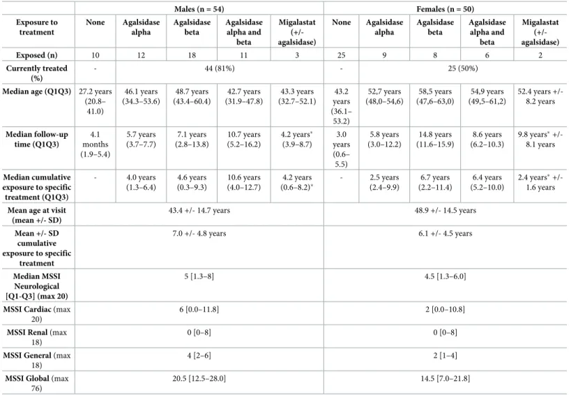
From December 2014 to May 2017, 104 patients (54 males) were prospectively included in the FFABRY cohort. Their general characteristics are described inTable 1.
Clinical clustering in males
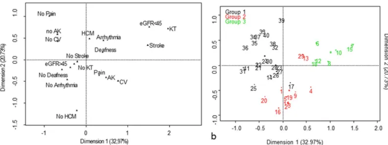
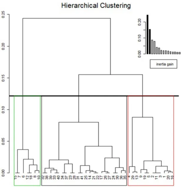
Multiple component analysis (MCA) and hierarchical clustering on principal components (HCPC) were performed with data for 41 male patients who had available complete data (Fig 1). Their mean age was 44.4 years-old. We assume that treatment with ERT or chaperone ther-apy does not modify the overall phenotype of patients. Six patients were untreated at the inclu-sion (mean age 34.6 years-old; min–max 17.1–58.1 y.). Mean duration of treatment was 6.5 years in treated patients (min—max: 0.26–15.5 y.). The first 2 dimensions of MCA expressed 53.7% of the total inertia. An ascending HCPC performed on the first 5 dimensions identified 3 different clusters (Fig 2). Group 1 (mean age = 50.5 +/- 11.2 years; n = 21) was characterized by the absence of CV (p<10−7), the absence of angiokeratoma (p<10−4), the absence of acral pain (p<0.001), a missense mutation (p<0.02), eGFR > 45 ml/min/1.73 m2(p<0.02), and the absence of renal transplant (p = 0.02). Group 2 (mean age = 32.3 +/- 9.9 years; n = 13) was char-acterized by the absence of hypertrophic cardiomyopathy (HCM) (p<10−3), the presence of CV (p = 0.001), the presence of angiokeratoma (p = 0.003), the presence of acral pain (p = 0.007), and eGFR > 45 ml/min/1.73 m2. Group 3 (mean age = 48.8 +/- 9.5 years; n = 7) was character-ized by eGFR < 45 ml/min/1.73 m2(p<10−6), a history of renal transplant (p<10−4) and the presence of CV (p = 0.002). On the basis of these characteristics, we considered group 1 to be the nonclassical phenotype and groups 2 and 3 to be the classical phenotype, with younger and older patients, respectively. When considering the absence of acral pain or CV as criteria for the nonclassical phenotype, we met the previously defined clusters with a sensitivity of 89.5%, a specificity of 91.0%, a positive predictive value of 89.5%, a negative predictive value of 91.0%, and an area under the receiver operating characteristic (ROC) curve of 0.9.
Clinical clustering in females
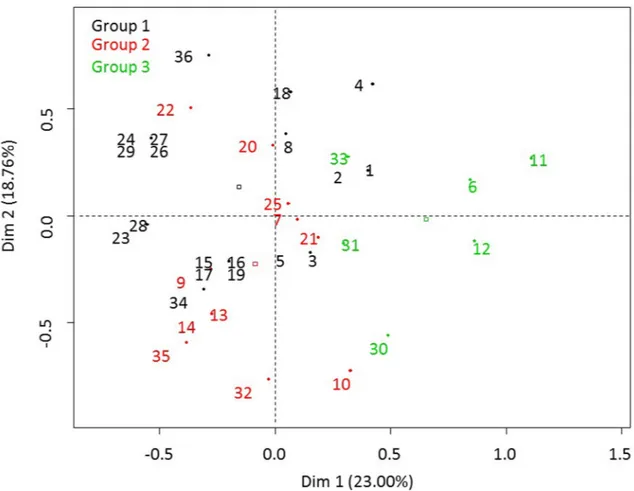
We applied the same approach for females using the same variables for patients with complete data (n = 36). MCA and HCPC revealed 3 different clusters without any clinical significance (Fig 3). Therefore, we considered that clinical clustering was not appropriate for females.
FFABRY score
As already mentioned, the morbidity of FD relies on renal, cardiac and central nervous system involvement. Hence, the prognosis of FD depends on the clinical phenotype of patients. Based on the results of the previous clustering, we introduce the first severity scoring system that takes into account the clinical phenotype of FD. The FFABRY score is therefore constructed with 4 variables: the overall clinical phenotype, the kidney disease score, the heart disease score and the central nervous system score as followed:
Phenotype
• Males with the classical phenotype: past or actual acroparesthesia and cornea verticillata • Males with the nonclassical phenotype: no past or actual acroparesthesia or no cornea
verticillata • Females
Table 1. Characteristics of patients (�time under enzyme replacement therapy included).
Males (n = 54) Females (n = 50) Exposure to treatment None Agalsidase alpha Agalsidase beta Agalsidase alpha and beta Migalastat (+/-agalsidase) None Agalsidase alpha Agalsidase beta Agalsidase alpha and beta Migalastat (+/-agalsidase) Exposed (n) 10 12 18 11 3 25 9 8 6 2 Currently treated (%) - 44 (81%) - 25 (50%)
Median age (Q1Q3) 27.2 years
(20.8– 41.0) 46.1 years (34.3–53.6) 48.7 years (43.4–60.4) 42.7 years (31.9–47.8) 43.3 years (32.7–52.1) 43.2 years (36.1– 53.2) 52,7 years (48,0–54,6) 58,5 years (47,6–63,0) 54,9 years (49,5–61,2) 52.4 years +/-8.2 years Median follow-up time (Q1Q3) 4.1 months (1.9–5.4) 5.7 years (3.7–7.7) 7.1 years (2.8–13.8) 10.7 years (5.2–16.2) 4.2 years� (3.9–8.7) 3.0 years (0.6– 5.5) 5.8 years (3.0–12.2) 14.8 years (11.6–15.9) 8.6 years (6.2–10.3) 9.8 years� +/-8.1 years Median cumulative exposure to specific treatment (Q1Q3) - 4.0 years (1.3–6.4) 4.6 years (0.3–9.3) 10.6 years (4.0–12.7) 4.2 years (0.6–8.2)� - 2.5 years (2.4–9.9) 6.7 years (2.2–11.4) 6.4 years (5.2–10.0) 2.4 years� +/-1.6 years
Mean age at visit (mean +/- SD) 43.4 +/- 14.7 years 48.9 +/- 14.5 years Mean +/- SD cumulative exposure to specific treatment 7.0 +/- 4.8 years 6.1 +/- 4.5 years Median MSSI Neurological [Q1-Q3] (max 20) 5 [1.3–8] 4.5 [1.3–6.0]
MSSI Cardiac (max
20)
6 [0.0–11.8] 2 [0.0–10.8]
MSSI Renal (max
18)
0 [0–8] 0 [0–8]
MSSI General (max
18)
4 [2–6] 2 [1–4]
MSSI Global (max
76)
20.5 [12.5–28.0] 14.5 [7.0–21.8]
Kidney disease: K score from K0 to K5. FD renal involvement is progressive and charac-terized by glomerular hyperfiltration and proteinuria followed by a decrease in glomerular function [22,23]. We propose the scale as follows:
K0: No proteinuria and 90 > eGFR > 135 ml/min/1.73 m2
K1: Hyperfiltration such as eGFR � 135ml/min/1.73m2without proteinuria K2: 60 < eGFR � 90 ml/min/1.73 m2OR proteinuria > 0.3g/24h or 50 mg/ mmol K3: 30 < eGFR � 60 ml/min/1.73 m2+/- proteinuria.
K4: 15 < eGFR � 30 ml/min/1.73 m2+/- proteinuria.
K5: eGFR � 15 ml/min/1.73 m2or dialysis or renal transplant.
Heart disease: H score from H0 to H4. FD is a cause of HCM, progressively leading to diastolic dysfunction, ischemia or obstructive cardiac failure [24]. FD is also characterized by arrhythmia, which has become the leading cause of death [25,26]. An interventricular septum thickness (IST) > 30 mm has been associated with a high risk for sudden death [24]. Addition-ally, we propose the following staging:
H0: No HCM. No cardiac symptomatology.
H1: HCM such as 13 < IST � 30 mm and/or QRS interval on ECG � 200 msec and/or ven-tricular hypertrophy on ECG without cardiac symptoms and no need of antiarrhythmic or beta-blockers.
H2: H1 + need of antiarrhythmic or beta-blockers�.
Fig 1. a: Variable factor map obtained by multiple component analysis using data for 41 males with complete data (AK: angiokeratoma; CV: cornea verticillata; eGFR:
estimated glomerular filtration rate in ml/min/1.73 m2; HCM: hypertrophic cardiomyopathy; KT kidney transplantation). b: Ascending hierarchical classification of
individuals using the first 5 dimensions of multiple component analysis performed using data for 41 males with complete data. Three clusters were identified. Group 1, characterized by the absence of cornea verticillata (p<10−7), the absence of angiokeratoma (p<10−4), the absence of acral pain (p<0.001) and the absence of renal disease, was referred to as the nonclassical cluster. Groups 2 and 3, characterized by the presence of cornea verticillata (p<0.002), were referred to as classical groups with younger (mean age 32.3 +/- 9.9 years) and older (mean age 48.8 +/- 9.5 years) patients, respectively.
H3: 35% < Left ventricular ejection function (LVEF) � 50% and/or a need of pacemaker (PM) implantation and/or angina.
H4: Need of heart transplant or LVEF � 35% or IST > 30 mm.
�
according to guidelines on management of HCM [27]
Central nervous system involvement: N score from N0 to N2. In a logistic regression model, we observed that chronic headaches were associated with a higher risk of stroke in males (OR 16.2, p = 0.01), independent of renal and cardiac diseases. Moreover, a trend toward a higher risk of cerebral stroke was observed in males with cochlear disorder defined by the presence of tinnitus or hearing loss (OR 7.8, p = 0.054), independent of age. Additionally, we propose the following staging:
Fig 2. Hierarchical clustering on the first 5 dimensions of the multiple component analysis performed on data of the 41 males with complete data.
N0: No chronic headache and no cochlear disorder and no history of stroke. N1: Chronic headache and/or tinnitus and/or hearing loss and no history of stroke. N2: History of cerebral stroke.
Total FFABRY score from T0 to T11. The total (T) score was defined as the sum of the K-, H- and N-scores.
Description of the FFABRY cohort clustered by sex, cornea verticillata and
acroparesthesia
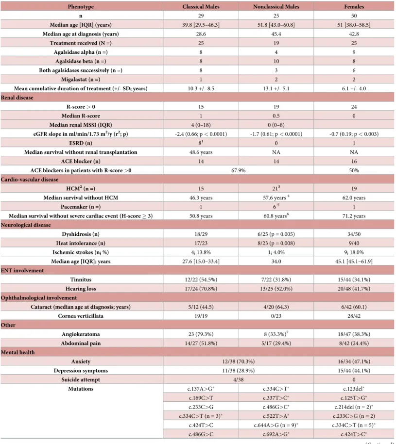
Using our clustering criteria, we distinguished 29 males with the classical phenotype, 25 males with the nonclassical phenotype and 50 females. Their clinical and biochemical characteristics are described extensively inTable 2. Briefly, among males, those with the nonclassical pheno-type were diagnosed at an older age (45.4 vs. 28.6 years; p < 10−3) and had a milder phenotype including a less steep eGFR slope (-1.7 vs. -2.4 ml/min/1.72m2/ years; p < 0.05), a lower risk of renal transplantation (log-rank, HR = 0.07; p < 10−3) and a lower risk of cerebral stroke (1/25 vs. 4/29; non-significant). Surprisingly, ENT involvement was frequent in both male groups, being observed in 70.0% of classical and 52.0% of nonclassical cases (p = ns). Of note, one of the most commonly reported symptoms was anxiety, which was observed in 70.3% of the male
Fig 3. Ascending hierarchical classification of individuals (using the first 5 dimensions of multiple component analysis performed using data for 36 females with complete data) did not reveal any significant groups.
Table 2. Characteristics of patients stratified by clinical phenotype defined with the FFABRY scoring system.
Phenotype Classical Males Nonclassical Males Females
n 29 25 50
Median age [IQR] (years) 39.8 [29.5–46.3] 51.8 [43.0–60.8] 51 [38.0–58.5]
Median age at diagnosis (years) 28.6 45.4 42.8
Treatment received (N =) 25 19 25
Agalsidase alpha (n =) 8 4 9
Agalsidase beta (n =) 8 10 8
Both agalsidases successively (n =) 8 3 6
Migalastat (n =) 1 2 2
Mean cumulative duration of treatment (+/- SD; years) 10.3 +/- 8.5 13.1 +/- 5.1 6.1 +/- 4.0
Renal disease
R-score > 0 15 19 24
Median R-score 1 0.5 0
Median renal MSSI (IQR) 4 (0–18) 0 (0–8)
eGFR slope in ml/min/1.73 m2/y (r2; p) -2.4 (0.66; p < 0.0001) -1.7 (0.61; p < 0.0001) -0.7 (0.19; p < 0.003)
ESRD (n) 81 0 1
Median survival without renal transplantation 48.6 years NA NA
ACE blocker (n) 14 14 16
ACE blockers in patients with R-score >0 67.9% 50%
Cardio-vascular disease
HCM2(n =) 15 213 19
Median survival without HCM 46.3 years 57.6 years4 62.0 years
Pacemaker (n =) 1 65 1
Median survival without severe cardiac event (H-score � 3) 50.8 years 60.8 years6 71.2 years
Neurological disease
Dyshidrosis (n) 18/29 6/25 (p = 0.005) 34/50
Heat intolerance (n) 17/23 8/23 (p = 0.008) 9/40
Ischemic strokes (n; %) 4; 13.8% 1; 4.0% 9; 18.0%
Median age [IQR]; years 27.6 [15.0–33.4] 34.0 45.1 [45.1–61.9]
ENT involvement
Tinnitus 12/22 (54.5%) 7/22 (31.8%) 15/44 (34.1%)
Hearing loss 17/24 (70.8%) 13/25 (52.0%) 20/48 (41.7%)
Ophthalmological involvement
Cataract (median age at diagnosis; years) 5/12 (44.5) 4/20 (64.3) 6/42 (60.1)
Cornea verticillata 19/19 0/23 28/42 Other Angiokeratoma 23 (79.3%) 8 (33.3%)7 18/47 (38.3%) Abdominal pain 14/27 (51.8%) 5/17 (29.4%) 8/42 (24.4%) Mental health Anxiety 12/38 (70.3%) 16/34 (47.1%) Depression symptoms 11/38 (28.9%) 15/44 (44.1%) Suicide attempt 4/38 0
Mutations c.137A>G� c.334C>T� c.123del�
c.169C>T c.337T>C� c.125T>G� c.233C>G c.486G>C� c.214del (n = 2)� c.334C>T (n = 3)� c.522T>A� c.233C>G (n = 2) c.424T>C c.644A>G (n = 9)� c.334C>T (n = 5)� c.486G>C c.692A>G� c.424T>C� (Continued )
patients, with suicide attempts reported for 10.5% of the male cohort. Females exhibited a con-trasting globally milder phenotype (Fig 4andTable 2). However, 18.0% of them had a history of ischemic stroke, with a median age at diagnosis of 45.1 years, and the event was significantly associated with the existence of cardiac rhythm problems (OR: 15.3; p = 0.046). Such an associ-ation with rhythm problems was not observed in men.
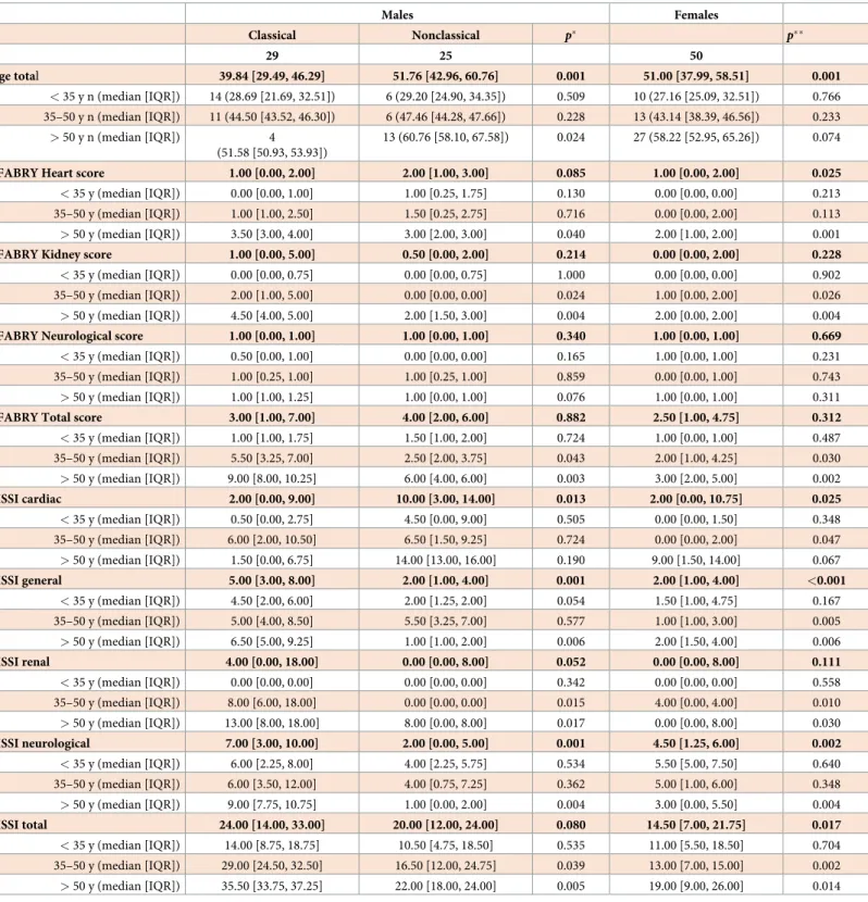
Performances of clustering with MSSI and FFABRY scores in the entire
FFABRY cohort
Clustering appeared clinically relevant using both MSSI and FFABRY scores, with significant differences between groups stratified by classes of ages (Table 3). The classical phenotype was associated with a higher risk of severe renal events (K-score � 3; Cox analysis: HR (classical/ nonclassical) = 35.1; p <10−3; HR (classical/ females) = 100; p < 10−5;Fig 4) and a higher risk of severe cardiac events (H-score � 3; Cox analysis: HR (classical vs. nonclassical) = 4.8;
Table 2. (Continued)
c.539del� c.713G>A(n = 2)� c.427G>A (n = 2)�
c.548G>C� c.758T>C c.486G>C�
c.680G>A c.802-3_802-2del� c.504A>C�
c.729G>C� c.847C>T (n = 2)� c.548G>C�
c.798T>A (n = 2)� c.902G>A� c.655A>C�
c.802-3_802-2del� c.1010T>C� c.680G>A� c.806T>C� c.1016T>G� c.695T>C� c.847C>T� c.1087C>T (n = 2)� c.718_719del� c.875C>T c.729G>C� c.884T>G c.798T>A c.901C>T (n = 2)� c.802-3_802-2del (n = 4)� c.902G>A� c.840A>T c.1010T>C� c.884T>G (n = 3)� c.1069_1079del� c.901C>T (n = 6)� c.1246C>T� c.902G>A� no data (n = 4) c.1277_1278del (n = 2)� c.1021del c.1024C>T c.1087C>T c.1168G>A no data (n = 6)
LysoGb3 (median in ng/ml; IQR; n)
In treated patients 18.9 (11.6–32.3; n = 17) 6.25 (2.6–21.9; n = 17)8 4.5 (2.7–6.2; n = 15)
In untreated patients 101.8 (n = 2) 8.5 (3.0–16.7; n = 5) 2.6 (1.7–3.8; n = 22) eGFR: estimated glomerular filtration rate; ESRD: end-stage renal disease; ACE-blocker: angiotensin conversion enzyme blocker; HCM: hypertrophic cardiomyopathy; PM: pacemaker; NA: not available. (1) HR vs. nonclassical 14.4; log-rank p = 0.0003; (2) in Cox regression, HCM is influenced by age at diagnosis (HR 0.81; p<10–7) and cumulative exposure to treatment (HR 0.71; p < 0.0001); (3) in Cox regression, influenced by the phenotype: HR nonclassical vs. classic: 0.19; p = 0.006); (4) log-rank classical vs. nonclassical; p < 0.003; (5) log-log-rank classical vs. nonclassical; p = ns; (6) log-log-rank classical vs. nonclassical; p = 0.01; (7) Fisher-exact t-test classical vs. nonclassical; p<0.001; (8) Mann-Whitney, p = 0.01
�variants included in the MCA and HCPC analyses (exhaustive data). One 46.6-year-old female, a p.A143T carrier, was included in the MCA and HCPC analyses. She
had been treated for 2 years with migalastat for acral and abdominal pain and angiokeratoma but had no renal, cardiac or cerebrovascular involvement (MSSI = 13; plasma lysoGb3 1.1nM). Another female p.A143T carrier, aged 43.0 years, had no dermatological, ophthalmological, neurological, renal or cardiological symptoms (MSSI = 1; plasma lysoGb3 1.3nM). She received no specific treatment and was not included in the MCA and HCPC analyses due to missing data.
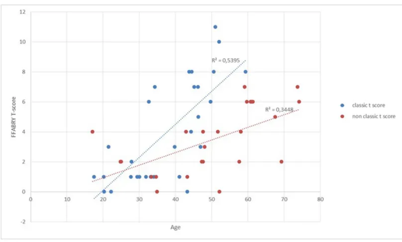
p = 0.008; HR (classical/ females) = 16.7; p < 10−4;Fig 4), and there was a trend toward a higher risk of severe neurological events (N-score = 2) in males with the classical compared to the nonclassical phenotype (Cox analysis; HR = 7.7; p = 0.08,Fig 4). There was no significant difference among females (HR = 2.5; p = 0.2). T-score evolution illustrated that the overall severity increased with time in the classical group compared to the nonclassical group (Fig 5).
LysoGb3 plasma levels, anti-agalsidase antibodies and FFABRY
LysoGb3 plasma levels were higher in males with the classical phenotype compared to those with the nonclassical phenotype or females in both treatment-naïve (respective medians 101.8, 5.8 and 3.2; Kruskal Wallis p < 0.04) and in treated (respective medians 18.9, 6.7 and 4.5; p < 10−4) patients. As expected, there was no correlation between lysoGb3 plasma levels and FFABRY scores after stratification by phenotype among treated patients. Two female and 18 male patients were positive for anti-agalsidase antibodies. If considering only males who had been exposed to agalsidase, the presence of antibodies was significantly associated with the classical phenotype (56% vs. 20%, p < 0.02). Their characteristics have already been reported [20].
Genotype-phenotype correlation
Among the 30 different genetic variants observed in males, nonsense variants were associated with the classical phenotype (9/13 patients with nonsense variants). The four patients classified in the nonclassical group had no cornea verticillata: a c.802-3_802-2del carrier with a K1H3N0 score at 17.1 years, two c.847C>T carriers with K0H1N0 and K0H1N1 scores at 33.4 and 47.3 years, and a c.522T>A carrier with a K2H0N0 score at 47.7 years. Five variants were observed in both the classical and nonclassical groups: c.334C>T, c.847C>T, c.802-3_802-2del, c. 902G>A and c.1010T>C. Their characteristics are described inTable 4.
Discussion
The nonclassical phenotype of FD has become the most prevalent [7,12]. However, most clini-cal and fundamental research studies addressing with FD have not stratified patients based on phenotype. Here, we propose a simple algorithm to distinguish patients. We demonstrate that CV assessed by slit-lamp and acral pain, but not angiokeratoma, are statistically sufficient to
Fig 4. Renal, cardiac and neurological severe event-free survival curves (black: Classical group males, green: Nonclassical group males, red: Females).
distinguish males with the classical phenotype from those with the nonclassical phenotype, with significant differences in terms of severity. As expected, males with the classical pheno-type have more severe renal disease than do males with the nonclassical phenopheno-type, but the
Table 3. FFABRY and MSSI scores (comparison with Mann-Whitney test p�: Classical versus nonclassical males; p��: Males versus females).
Males Females Classical Nonclassical p� p�� n 29 25 50 Age total 39.84 [29.49, 46.29] 51.76 [42.96, 60.76] 0.001 51.00 [37.99, 58.51] 0.001 < 35 y n (median [IQR]) 14 (28.69 [21.69, 32.51]) 6 (29.20 [24.90, 34.35]) 0.509 10 (27.16 [25.09, 32.51]) 0.766 35–50 y n (median [IQR]) 11 (44.50 [43.52, 46.30]) 6 (47.46 [44.28, 47.66]) 0.228 13 (43.14 [38.39, 46.56]) 0.233 > 50 y n (median [IQR]) 4 (51.58 [50.93, 53.93]) 13 (60.76 [58.10, 67.58]) 0.024 27 (58.22 [52.95, 65.26]) 0.074
FFABRY Heart score 1.00 [0.00, 2.00] 2.00 [1.00, 3.00] 0.085 1.00 [0.00, 2.00] 0.025
< 35 y (median [IQR]) 0.00 [0.00, 1.00] 1.00 [0.25, 1.75] 0.130 0.00 [0.00, 0.00] 0.213 35–50 y (median [IQR]) 1.00 [1.00, 2.50] 1.50 [0.25, 2.75] 0.716 0.00 [0.00, 2.00] 0.113
> 50 y (median [IQR]) 3.50 [3.00, 4.00] 3.00 [2.00, 3.00] 0.040 2.00 [1.00, 2.00] 0.001
FFABRY Kidney score 1.00 [0.00, 5.00] 0.50 [0.00, 2.00] 0.214 0.00 [0.00, 2.00] 0.228
< 35 y (median [IQR]) 0.00 [0.00, 0.75] 0.00 [0.00, 0.75] 1.000 0.00 [0.00, 0.00] 0.902 35–50 y (median [IQR]) 2.00 [1.00, 5.00] 0.00 [0.00, 0.00] 0.024 1.00 [0.00, 2.00] 0.026
> 50 y (median [IQR]) 4.50 [4.00, 5.00] 2.00 [1.50, 3.00] 0.004 2.00 [0.00, 2.00] 0.004
FFABRY Neurological score 1.00 [0.00, 1.00] 1.00 [0.00, 1.00] 0.340 1.00 [0.00, 1.00] 0.669
< 35 y (median [IQR]) 0.50 [0.00, 1.00] 0.00 [0.00, 0.00] 0.165 1.00 [0.00, 1.00] 0.231 35–50 y (median [IQR]) 1.00 [0.25, 1.00] 1.00 [0.25, 1.00] 0.859 0.00 [0.00, 1.00] 0.743
> 50 y (median [IQR]) 1.00 [1.00, 1.25] 1.00 [0.00, 1.00] 0.076 1.00 [0.00, 1.00] 0.311
FFABRY Total score 3.00 [1.00, 7.00] 4.00 [2.00, 6.00] 0.882 2.50 [1.00, 4.75] 0.312
< 35 y (median [IQR]) 1.00 [1.00, 1.75] 1.50 [1.00, 2.00] 0.724 1.00 [0.00, 1.00] 0.487 35–50 y (median [IQR]) 5.50 [3.25, 7.00] 2.50 [2.00, 3.75] 0.043 2.00 [1.00, 4.25] 0.030 > 50 y (median [IQR]) 9.00 [8.00, 10.25] 6.00 [4.00, 6.00] 0.003 3.00 [2.00, 5.00] 0.002 MSSI cardiac 2.00 [0.00, 9.00] 10.00 [3.00, 14.00] 0.013 2.00 [0.00, 10.75] 0.025 < 35 y (median [IQR]) 0.50 [0.00, 2.75] 4.50 [0.00, 9.00] 0.505 0.00 [0.00, 1.50] 0.348 35–50 y (median [IQR]) 6.00 [2.00, 10.50] 6.50 [1.50, 9.25] 0.724 0.00 [0.00, 2.00] 0.047 > 50 y (median [IQR]) 1.50 [0.00, 6.75] 14.00 [13.00, 16.00] 0.190 9.00 [1.50, 14.00] 0.067 MSSI general 5.00 [3.00, 8.00] 2.00 [1.00, 4.00] 0.001 2.00 [1.00, 4.00] <0.001 < 35 y (median [IQR]) 4.50 [2.00, 6.00] 2.00 [1.25, 2.00] 0.054 1.50 [1.00, 4.75] 0.167 35–50 y (median [IQR]) 5.00 [4.00, 8.50] 5.50 [3.25, 7.00] 0.577 1.00 [1.00, 3.00] 0.005 > 50 y (median [IQR]) 6.50 [5.00, 9.25] 1.00 [1.00, 2.00] 0.006 2.00 [1.50, 4.00] 0.006 MSSI renal 4.00 [0.00, 18.00] 0.00 [0.00, 8.00] 0.052 0.00 [0.00, 8.00] 0.111 < 35 y (median [IQR]) 0.00 [0.00, 0.00] 0.00 [0.00, 0.00] 0.342 0.00 [0.00, 0.00] 0.558 35–50 y (median [IQR]) 8.00 [6.00, 18.00] 0.00 [0.00, 0.00] 0.015 4.00 [0.00, 4.00] 0.010 > 50 y (median [IQR]) 13.00 [8.00, 18.00] 8.00 [0.00, 8.00] 0.017 0.00 [0.00, 8.00] 0.030 MSSI neurological 7.00 [3.00, 10.00] 2.00 [0.00, 5.00] 0.001 4.50 [1.25, 6.00] 0.002 < 35 y (median [IQR]) 6.00 [2.25, 8.00] 4.00 [2.25, 5.75] 0.534 5.50 [5.00, 7.50] 0.640 35–50 y (median [IQR]) 6.00 [3.50, 12.00] 4.00 [0.75, 7.25] 0.362 5.00 [1.00, 6.00] 0.348 > 50 y (median [IQR]) 9.00 [7.75, 10.75] 1.00 [0.00, 2.00] 0.004 3.00 [0.00, 5.50] 0.004 MSSI total 24.00 [14.00, 33.00] 20.00 [12.00, 24.00] 0.080 14.50 [7.00, 21.75] 0.017 < 35 y (median [IQR]) 14.00 [8.75, 18.75] 10.50 [4.75, 18.50] 0.535 11.00 [5.50, 18.50] 0.704 35–50 y (median [IQR]) 29.00 [24.50, 32.50] 16.50 [12.00, 24.75] 0.039 13.00 [7.00, 15.00] 0.002 > 50 y (median [IQR]) 35.50 [33.75, 37.25] 22.00 [18.00, 24.00] 0.005 19.00 [9.00, 26.00] 0.014 https://doi.org/10.1371/journal.pone.0233460.t003
former also experience cardiomyopathy earlier, and some of them also have stroke early, inde-pendent of arrhythmia. Females cannot be separated into classical and nonclassical pheno-types, likely due to the X-inactivation status in the different organs.
Hemangiomas, which are frequent, can easily be misdiagnosed as angiokeratoma, which may explain the reduced specificity of angiokeratoma for the classical phenotype. In contrast, CV is a pivotal criterion in our classification. Although CV can be related to exposure to amio-darone, its association with the severity of FD has already been described, as observed in 94% of classical Fabry cases [28]. A recent paper reported that CV is often underdiagnosed in FD
Fig 5. T-score evolution in the classical group compared to the nonclassical group (with associated linear regression curve).
https://doi.org/10.1371/journal.pone.0233460.g005
Table 4. Characteristics of patients with genetic variants observed in both classical and nonclassical groups (eGFR: Estimated glomerular filtration by CKD-EPI equation in ml/min/1.73m2; RT: Renal transplant).
Variant Classical patient Nonclassical patient
age Treatment duration (y) FFABRY score eGFR age Treatment duration (y) FFABRY score eGFR c.334C>T 34.4 6.2 K5 H1 N1 T7 RT 48.1 13.4 K0 H3 N0 T3 105 45.2 3.4 K3 H3 N1 T7 52 59.4 5.6 K4 H3 N1 T8 23 c.802-3_802-2del 50.6 1.3 K4 H3 N1 T8 25 17.1 0 K1 H3 N0 T4 136 c.847C>T 49.8 11.1 K5 H1 N0 T6 RT 33.4 13.3 K0 H1 N0 T1 116 47.3 13 K0 H1 N1 T2 102 c.902G>A 17.5 0.4 K1 H0 N0 T1 136 24.9 0 K0 H2 N0 T2 121 c.1010T>C 46.3 4.3 K2 H3 N0 T5 81 60.9 4.8 K3 H2 N1 T6 57 https://doi.org/10.1371/journal.pone.0233460.t004
using the slit-lamp approach [29]. The present work concerned 4 males whose phenotype was not mentioned and 10 females. However, whether a more sensitive approach using in vivo cor-neal confocal microscopy (IVCM) may reveal minimal deposits in nonclassical patients and females is not the question. Hence, we suggest that an unquestionable CV, with obvious depos-its assessed by slit-lamp, is associated with a classical FD phenotype. Similarly, our second piv-otal criterion is the existence past or actual of acral pain and not the proof of small fiber neuropathy assessed by paraclinical exams.
Our study was based on data for 104 adult patients; a small number of patients is inherent with the rarity of FD. However, FFABRY is a multicenter database including patients from dif-ferent pedigrees, difdif-ferent medical specialties (nephrology, cardiology, internal medicine, and genetics) and different locations in France, which allows for clinical and genetic heterogeneity (42 different variants) that benefits analyses. We used a linear regression model to assess the slope of eGFR evolution in the cohort, which is limited by the heterogeneity of patients, espe-cially females. Analysis of X-inactivation may allow for a better classification of the female phe-notype and help in stratifying the risk of severe events; unfortunately, this analysis remains unavailable in routine practice [30]. As experts managing lysosomal diseases in a dedicated ter-tiary center, we assume that treatment with ERT or chaperone therapy has not modified the overall natural history of the disease or the prognosis of patients. Nonetheless, to the best of our knowledge, regression but no disappearance of CV has ever been reported [31].
In the era of evidence-based medicine, severity scores have become mandatory for evaluat-ing therapeutics. With FFABRY, we propose a clinically and statistically objective severity scor-ing system for FD. The FFABRY score was developed based on the natural history of FD and the severe clinically relevant events we observed in our center of expertise for lysosomal dis-eases. Our N-score highlights the importance of cochlear disorders and headaches, which are associated with the risk of cerebral stroke in males, whereas white matter lesions (WMLs) were not related to any specific symptom.
FFABRY allowed us to establish a portrait of current Fabry patients, distinguishing males with classical and nonclassical phenotypes and females with their proper clinical specificity. Interestingly, we observed no systematic genotype-phenotype correlation. Some patients shar-ing the same genetic variant were classified into two different groups. The FFABRY scores as well as individual clinical criteria demonstrated that our classification performed better than genotype for describing disease severity. Other scoring systems have already been developed for FD. The MSSI, which has been the most commonly used scoring system, was developed on the basis of data from 24 males and 15 females registered in the Fabry Outcome Survey (FOS) database (Shire-Takeda)[19]. The MSSI includes 26 variables, empirically weighted, among which hemorrhoids, facial appearance and subjective fitness assessment are notable; however, the MSSI does not include renal transplantation. The Disease Severity Scoring System (DS-3), which was elaborated by experts from the Fabry Registry (Sanofi-Genzyme), also includes nonobjective items such as the “patient reported domain” or sweating capacity. WMLs are included, though they are asymptomatic and not associated with poorer outcomes. Finally, the different items and their weightings have been empirically and not statistically established in these two scoring systems. Moreover, the number of items makes them challenging to use and time consuming. The Fabry International Prognostic Index (FIPI) appears to be much more robust, as it has been developed on the basis of multivariate analyses of data from 1483 patients from the FOS registry [32]. Although FIPI appears to be an effective prediction tool, it does not allow assessment of the actual clinical severity. Indeed, five of the six variables included in the cardiac item refer to extracardiac symptoms (eGFR, proteinuria, deafness, vertigo, and angiokeratoma). The online Fabry Stabilization index (FASTEX) was recently validated [33], and the authors established an attractive tool for personal follow-up of individuals.
Nevertheless, clinical phenotypes are not distinguished in this system, which could be mislead-ing for interindividual comparisons, makmislead-ing the scormislead-ing system useless in group studies. After stratification according to phenotype, FFABRY scores allowed a rapid and clinically relevant evaluation of disease severity. Analyses of the K score highlight the suboptimal management of FD females in our cohort. Our study also emphasizes windows of opportunity for the diagnosis and introduction of specific treatments, as follows: before 30 years old in males with the classi-cal phenotype, 45 years old in males with the nonclassiclassi-cal classiclassi-cal phenotype and 50 years old in females. Regarding the obviously different prognoses observed with the FFABRY score, we believe that the classical and nonclassical phenotypes should be considered as two different subtypes of FD in males, with different management strategies. As for Niemann-Pick disease A and B or Gaucher types 1, 2 and 3, it would be useful to rename the phenotypes for male patients, with classical as type 1 and nonclassical as type 2, to fully take into account such obvi-ous clinical differences and possible pathophysiological differences.
Identifying acral pain and cornea verticillata is a rapid and simple approach that statistically discriminates Fabry phenotypes.
Supporting information
S1 Data. Clinical and biological data of included patients. (XLSX)
Acknowledgments
We gratefully thank the Socie´te´ Nationale Franc¸aise de Me´decine Interne and Vaincre les mal-adies lysosomales patient association for their support as well as Isabelle Citerne and Epicon-cept’s team for their help.
Author Contributions
Conceptualization: Wladimir Mauhin, Olivier Lidove.
Data curation: Wladimir Mauhin, Olivier Benveniste, Damien Amelin, Cle´mence Montagner, Foudil Lamari, Catherine Caillaud, Claire Douillard, Bertrand Dussol, Vanessa Leguy-Seguin, Pauline D’Halluin, Esther Noel, Thierry Zenone, Marie Matignon, Franc¸ois Maillot, Kim-Heang Ly, Ge´rard Besson, Marjolaine Willems, Fabien Labombarda, Agathe Masseau, Christian Lavigne, Didier Lacombe, He´lène Maillard, Olivier Lidove.
Formal analysis: Wladimir Mauhin.
Investigation: Wladimir Mauhin, Cle´mence Montagner, Foudil Lamari, Catherine Caillaud, Claire Douillard, Bertrand Dussol, Vanessa Leguy-Seguin, Pauline D’Halluin, Esther Noel, Thierry Zenone, Marie Matignon, Franc¸ois Maillot, Kim-Heang Ly, Ge´rard Besson, Marjo-laine Willems, Fabien Labombarda, Agathe Masseau, Christian Lavigne, Didier Lacombe, He´lène Maillard, Olivier Lidove.
Methodology: Wladimir Mauhin, Olivier Benveniste, Damien Amelin, Olivier Lidove. Project administration: Wladimir Mauhin, Olivier Benveniste.
Supervision: Wladimir Mauhin.
Validation: Wladimir Mauhin, Olivier Benveniste.
Writing – original draft: Wladimir Mauhin, Olivier Benveniste, Damien Amelin, Foudil Lamari, Catherine Caillaud, Olivier Lidove.
Writing – review & editing: Wladimir Mauhin, Olivier Benveniste, Damien Amelin, Cle´m-ence Montagner, Foudil Lamari, Catherine Caillaud, Claire Douillard, Bertrand Dussol, Vanessa Leguy-Seguin, Pauline D’Halluin, Esther Noel, Thierry Zenone, Marie Matignon, Franc¸ois Maillot, Kim-Heang Ly, Ge´rard Besson, Marjolaine Willems, Fabien Labombarda, Agathe Masseau, Christian Lavigne, Didier Lacombe, He´lène Maillard, Olivier Lidove.
References
1. Brady RO, Gal AE, Bradley RM, Martensson E, Warshaw AL, Laster L. Enzymatic defect in Fabry’s dis-ease. Ceramidetrihexosidase deficiency. N Engl J Med. 1967; 276: 1163–1167.https://doi.org/10.1056/ NEJM196705252762101PMID:6023233
2. Smid BE, van der Tol L, Biegstraaten M, Linthorst GE, Hollak CEM, Poorthuis BJHM. Plasma globo-triaosylsphingosine in relation to phenotypes of Fabry disease. J Med Genet. 2015; 52: 262–268. https://doi.org/10.1136/jmedgenet-2014-102872PMID:25596309
3. Young-Gqamana B, Brignol N, Chang H-H, Khanna R, Soska R, Fuller M, et al. Migalastat HCl reduces globotriaosylsphingosine (lyso-Gb3) in Fabry transgenic mice and in the plasma of Fabry patients. PloS One. 2013; 8: e57631.https://doi.org/10.1371/journal.pone.0057631PMID:23472096
4. Schiffmann R, Warnock DG, Banikazemi M, Bultas J, Linthorst GE, Packman S, et al. Fabry disease: progression of nephropathy, and prevalence of cardiac and cerebrovascular events before enzyme replacement therapy. Nephrol Dial Transplant Off Publ Eur Dial Transpl Assoc—Eur Ren Assoc. 2009; 24: 2102–2111.https://doi.org/10.1093/ndt/gfp031PMID:19218538
5. Meikle PJ, Hopwood JJ, Clague AE, Carey WF. Prevalence of lysosomal storage disorders. JAMA. 1999; 281: 249–254.https://doi.org/10.1001/jama.281.3.249PMID:9918480
6. Poorthuis BJ, Wevers RA, Kleijer WJ, Groener JE, de Jong JG, van Weely S, et al. The frequency of lysosomal storage diseases in The Netherlands. Hum Genet. 1999; 105: 151–156.https://doi.org/10. 1007/s004399900075PMID:10480370
7. Spada M, Pagliardini S, Yasuda M, Tukel T, Thiagarajan G, Sakuraba H, et al. High incidence of later-onset fabry disease revealed by newborn screening. Am J Hum Genet. 2006; 79: 31–40.https://doi.org/ 10.1086/504601PMID:16773563
8. Burton BK, Charrow J, Hoganson GE, Waggoner D, Tinkle B, Braddock SR, et al. Newborn Screening for Lysosomal Storage Disorders in Illinois: The Initial 15-Month Experience. J Pediatr. 2017; 190: 130– 135.https://doi.org/10.1016/j.jpeds.2017.06.048PMID:28728811
9. Hopkins PV, Klug T, Vermette L, Raburn-Miller J, Kiesling J, Rogers S. Incidence of 4 Lysosomal Stor-age Disorders From 4 Years of Newborn Screening. JAMA Pediatr. 2018; 172: 696–697.https://doi.org/ 10.1001/jamapediatrics.2018.0263PMID:29813145
10. Wittmann J, Karg E, Turi S, Legnini E, Wittmann G, Giese A-K, et al. Newborn screening for lysosomal storage disorders in hungary. JIMD Rep. 2012; 6: 117–125.https://doi.org/10.1007/8904_2012_130 PMID:23430949
11. von Scheidt W, Eng CM, Fitzmaurice TF, Erdmann E, Hu¨bner G, Olsen EG, et al. An atypical variant of Fabry’s disease with manifestations confined to the myocardium. N Engl J Med. 1991; 324: 395–399. https://doi.org/10.1056/NEJM199102073240607PMID:1846223
12. Arends M, Wanner C, Hughes D, Mehta A, Oder D, Watkinson OT, et al. Characterization of Classical and Nonclassical Fabry Disease: A Multicenter Study. J Am Soc Nephrol. 2017; 28: 1631–1641.https:// doi.org/10.1681/ASN.2016090964PMID:27979989
13. van der Tol L, Smid BE, Poorthuis BJHM, Biegstraaten M, Deprez RHL, Linthorst GE, et al. A system-atic review on screening for Fabry disease: prevalence of individuals with genetic variants of unknown significance. J Med Genet. 2014; 51: 1–9.https://doi.org/10.1136/jmedgenet-2013-101857PMID: 23922385
14. Ortiz A, Germain DP, Desnick RJ, Politei J, Mauer M, Burlina A, et al. Fabry disease revisited: Manage-ment and treatManage-ment recommendations for adult patients. Mol Genet Metab. 2018; 123: 416–427. https://doi.org/10.1016/j.ymgme.2018.02.014PMID:29530533
15. Citro V, Cammisa M, Liguori L, Cimmaruta C, Lukas J, Cubellis MV, et al. The Large Phenotypic Spec-trum of Fabry Disease Requires Graduated Diagnosis and Personalized Therapy: A Meta-Analysis Can Help to Differentiate Missense Mutations. Int J Mol Sci. 2016; 17.https://doi.org/10.3390/ijms17122010 PMID:27916943
16. Oder D, Nordbeck P, Wanner C. Long Term Treatment with Enzyme Replacement Therapy in Patients with Fabry Disease. Nephron. 2016; 134: 30–36.https://doi.org/10.1159/000448968PMID:27576727
17. Chen F, Du M, Blumberg JB, Chui KKH, Ruan M, Rogers G, et al. Association Between Dietary Supple-ment Use, Nutrient Intake, and Mortality Among US Adults: A Cohort Study. Ann Intern Med. 2019; 170: 604–613.https://doi.org/10.7326/M18-2478PMID:30959527
18. Tonneijck L, Muskiet MHA, Smits MM, van Bommel EJ, Heerspink HJL, van Raalte DH, et al. Glomeru-lar Hyperfiltration in Diabetes: Mechanisms, Clinical Significance, and Treatment. J Am Soc Nephrol JASN. 2017; 28: 1023–1039.https://doi.org/10.1681/ASN.2016060666PMID:28143897
19. Whybra C, Kampmann C, Krummenauer F, Ries M, Mengel E, Miebach E, et al. The Mainz Severity Score Index: a new instrument for quantifying the Anderson-Fabry disease phenotype, and the response of patients to enzyme replacement therapy. Clin Genet. 2004; 65: 299–307.https://doi.org/ 10.1111/j.1399-0004.2004.00219.xPMID:15025723
20. Mauhin W, Lidove O, Amelin D, Lamari F, Caillaud C, Mingozzi F, et al. Deep characterization of the anti-drug antibodies developed in Fabry disease patients, a prospective analysis from the French multi-center cohort FFABRY. Orphanet J Rare Dis. 2018; 13: 127. https://doi.org/10.1186/s13023-018-0877-4PMID:30064518
21. Kanda Y. Investigation of the freely available easy-to-use software “EZR” for medical statistics. Bone Marrow Transplant. 2013; 48: 452–458.https://doi.org/10.1038/bmt.2012.244PMID:23208313 22. Tøndel C, Kanai T, Larsen KK, Ito S, Politei JM, Warnock DG, et al. Foot process effacement is an early
marker of nephropathy in young classic Fabry patients without albuminuria. Nephron. 2015; 129: 16– 21.https://doi.org/10.1159/000369309PMID:25531941
23. Riccio E, Sabbatini M, Bruzzese D, Annicchiarico Petruzzelli L, Pellegrino A, Spinelli L, et al. Glomerular Hyperfiltration: An Early Marker of Nephropathy in Fabry Disease. Nephron. 2019; 141: 10–17.https:// doi.org/10.1159/000493469PMID:30466100
24. Marian AJ, Braunwald E. Hypertrophic Cardiomyopathy: Genetics, Pathogenesis, Clinical Manifesta-tions, Diagnosis, and Therapy. Circ Res. 2017; 121: 749–770.https://doi.org/10.1161/CIRCRESAHA. 117.311059PMID:28912181
25. Baig S, Edward NC, Kotecha D, Liu B, Nordin S, Kozor R, et al. Ventricular arrhythmia and sudden car-diac death in Fabry disease: a systematic review of risk factors in clinical practice. Eur Eur Pacing Arrhythm Card Electrophysiol J Work Groups Card Pacing Arrhythm Card Cell Electrophysiol Eur Soc Cardiol. 2018; 20: f153–f161.https://doi.org/10.1093/europace/eux261PMID:29045633
26. Mehta A, Clarke JTR, Giugliani R, Elliott P, Linhart A, Beck M, et al. Natural course of Fabry disease: changing pattern of causes of death in FOS—Fabry Outcome Survey. J Med Genet. 2009; 46: 548– 552.https://doi.org/10.1136/jmg.2008.065904PMID:19473999
27. Authors/Task Force members, Elliott PM, Anastasakis A, Borger MA, Borggrefe M, Cecchi F, et al. 2014 ESC Guidelines on diagnosis and management of hypertrophic cardiomyopathy: the Task Force for the Diagnosis and Management of Hypertrophic Cardiomyopathy of the European Society of Cardi-ology (ESC). Eur Heart J. 2014; 35: 2733–2779.https://doi.org/10.1093/eurheartj/ehu284PMID: 25173338
28. van der Tol L, Sminia ML, Hollak CEM, Biegstraaten M. Cornea verticillata supports a diagnosis of Fabry disease in non-classical phenotypes: results from the Dutch cohort and a systematic review. Br J Ophthalmol. 2016; 100: 3–8.https://doi.org/10.1136/bjophthalmol-2014-306433PMID:25677671 29. Leonardi A, Carraro G, Modugno RL, Rossomando V, Scalora T, Lazzarini D, et al. Cornea verticillata
in Fabry disease: a comparative study between slit-lamp examination and in vivo corneal confocal microscopy. Br J Ophthalmol. 2019.https://doi.org/10.1136/bjophthalmol-2019-314249PMID: 31401555
30. Echevarria L, Benistan K, Toussaint A, Dubourg O, Hagege AA, Eladari D, et al. X-chromosome inacti-vation in female patients with Fabry disease. Clin Genet. 2016; 89: 44–54.https://doi.org/10.1111/cge. 12613PMID:25974833
31. Prominent regression of corneal deposits in Fabry disease 16 years after initiation of enzyme replace-ment therapy.—PubMed—NCBI. [cited 27 Nov 2019]. Available:https://www.ncbi.nlm.nih.gov/pubmed/ ?term=Prominent+regression+ofcorneal+deposits+in+Fabrydisease+16+years+afterinitiation+of +enzymereplacement+therapy
32. Hughes DA, Malmena¨s M, Deegan PB, Elliott PM, Ginsberg L, Hajioff D, et al. Fabry International Prog-nostic Index: a predictive severity score for Anderson-Fabry disease. J Med Genet. 2012; 49: 212–220. https://doi.org/10.1136/jmedgenet-2011-100407PMID:22315436
33. Cairns T, Wanner C. Will the FAbry STabilization indEX make its way to everyday clinical practice? Clin Kidney J. 2019; 12: 61–64.https://doi.org/10.1093/ckj/sfy126PMID:30747153
