HAL Id: dumas-02438505
https://dumas.ccsd.cnrs.fr/dumas-02438505
Submitted on 14 Jan 2020HAL is a multi-disciplinary open access archive for the deposit and dissemination of sci-entific research documents, whether they are pub-lished or not. The documents may come from teaching and research institutions in France or abroad, or from public or private research centers.
L’archive ouverte pluridisciplinaire HAL, est destinée au dépôt et à la diffusion de documents scientifiques de niveau recherche, publiés ou non, émanant des établissements d’enseignement et de recherche français ou étrangers, des laboratoires publics ou privés.
Valeur pronostique de l’électrocardiogramme de repos
12 dérivations chez les patients porteurs d’une
cardiomyopathie hypertrophique sarcomérique
Coralie Marie
To cite this version:
Coralie Marie. Valeur pronostique de l’électrocardiogramme de repos 12 dérivations chez les patients porteurs d’une cardiomyopathie hypertrophique sarcomérique. Sciences du Vivant [q-bio]. 2019. �dumas-02438505�
1
N° de thèse : 3090 Année 2019
UNIVERSITE DE BORDEAUX
U.F.R. DES SCIENCES MEDICALES
Thèse en vue de l’obtention du
DIPLOME D’ETAT DE DOCTEUR EN MEDECINE
PROGNOSTIC VALUE OF 12-LEAD RESTING
ELECTROCARDIOGRAM IN PATIENTS WITH
SARCOMERIC HYPERTROPHIC CARDIOMYOPATHY
VALEUR PRONOSTIQUE DE L’ELECTROCARDIOGRAMME DE REPOS 12
DERIVATIONS CHEZ LES PATIENTS PORTEURS D’UNE CARDIOMYOPATHIE
HYPERTROPHIQUE SARCOMERIQUE
Présentée et soutenue publiquement
Le 15 Octobre 2019
Par MARIE Coralie
Née le 26 Juillet 1990 à Aunay sur Odon
JURY
Pr DOS SANTOS Pierre
PU-PH - CHU Bordeaux / Président
Pr REANT Patricia
PU-PH - CHU Bordeaux / Directrice
Pr LAFITTE Stéphane
PU-PH - CHU Bordeaux / Juge
Pr ROORYCK-THAMBO Caroline
PU-PH - CHU Bordeaux / Juge
2
Serment d’Hippocrate
Au moment d’être admise à exercer la médecine, je promets et je jure d’être fidèle aux lois de l’honneur et de la probité.
Mon premier souci sera de rétablir, de préserver ou de promouvoir la santé dans tous ses éléments, physiques et mentaux, individuels et sociaux.
Je respecterai toutes les personnes, leur autonomie et leur volonté, sans aucune discrimination selon leur état ou leurs convictions. J’interviendrai pour les protéger si elles sont affaiblies, vulnérables ou menacées dans leur intégrité ou leur dignité. Même sous la contrainte, je ne ferai pas usage de mes connaissances contre les lois de l’humanité.
J’informerai les patients des décisions envisagées, de leurs raisons et de leurs conséquences. Je ne tromperai jamais leur confiance et n’exploiterai pas le pouvoir hérité des circonstances pour forcer les consciences.
Je donnerai mes soins à l’indigent et à quiconque me les demandera. Je ne me laisserai pas influencer par la soif du gain ou la recherche de la gloire.
Admise dans l’intimité des personnes, je tairai les secrets qui me seront confiés. Reçue à l’intérieur des maisons, je respecterai les secrets des foyers et ma conduite ne servira pas à corrompre les mœurs.
Je ferai tout pour soulager les souffrances. Je ne prolongerai pas abusivement les agonies. Je ne provoquerai jamais la mort délibérément.
Je préserverai l’indépendance nécessaire à l’accomplissement de ma mission. Je n’entreprendrai rien qui dépasse mes compétences. Je les entretiendrai et les perfectionnerai pour assurer au mieux les services qui me seront demandés.
J’apporterai mon aide à mes confrères ainsi qu’à leurs familles dans l’adversité.
Que les hommes et mes confrères m’accordent leur estime si je suis fidèle à mes promesses ; que je sois déshonorée et méprisée si j’y manque..
3
Remerciements
Au Professeur Pierre DOS SANTOS, Je vous remercie de me faire l’honneur de présider mon jury. Je vous suis également reconnaissante pour ces précieux enseignements de physiologie myocardique et d’insuffisance cardiaque délivrés durant mon internat.
Au Professeur Patricia REANT, Cela a été un honneur d’avoir pu travailler avec vous sur ce projet. Merci pour votre soutien et votre disponibilité.
Au Professeur Stéphane LAFITTE, C’est un honneur pour moi que vous ayez accepté de prendre part à ce jury. Un grand merci également pour ces nombreux enseignements d’échocardiographie cardiaque.
Au Professeur Caroline ROORYCK-THAMBO, Je suis honorée que vous ayez accepté de juger mon travail. Je vous prie de recevoir mes remerciements les plus sincères.
Au Docteur Josselin DUCHATEAU, C’est un honneur pour moi que vous ayez accepté de faire partie de ce jury. Je vous remercie d’en avoir été le rapporteur. Un grand merci également pour ces précieux enseignements de rythmologie délivrés.
Aux équipes de Cardiologie de Bordeaux et de la Réunion Saint-Denis, aux Chefs de clinique et Assistants qui m’ont appris tellement durant ces quatre années.
A mes nombreux co-internes avec qui cela a été un plaisir de travailler, et en particulier Océane et Samuel, les internes cocotiers.
A ma Sœur et mes Parents, je ne vous remercierai jamais assez pour tout ce que vous avez pu faire pour moi. Vous avez toujours été d’un soutien sans faille, merci de m’avoir donné envie de croquer la vie !
A mes amis du lycée et de la faculté de médecine de Caen, une vingtaine de folie passée avec vous !
4
Table of contents
ABBREVIATIONS LIST ... 5 PREAMBULE ... 6 Définition ... 6 Diagnostic ... 7 Physiopathologie ... 9 Pronostic ... 10 Problématique ... 15 ABSTRACT ... 16 RESUME ... 17 INTRODUCTION... 19MATERIAL AND METHODS ... 20
RESULTS ... 24
DISCUSSION ... 31
CONCLUSION ... 33
5
Abbreviations list
ACTC1: cardiac alpha-actin AF: atrial fibrillation ATP: antitachycardia pacing
CMH: cardiomyopathie hypertrophique CMR : cardiac magnetic resonance
DAI: défibrillateur automatique implantable ECG: électrocardiogramme/electrocardiogram ETT: échocardiographie transthoracique FA: fibrillation atriale
FEVG: fraction d’éjection ventriculaire gauche HCM: hypertrophic cardiomyopathy
HF: heart failure
HVG: hypertrophie ventriculaire gauche IC: insuffisance cardiaque
ICD: implantable cardiac defibrillator IRM: imagerie par résonance magnétique LBBB: left bundle branch block
LV: left ventricular
MYBPC3: cardiac myosin binding protein C MYH7: B-myosin heavy chain
MYL2: myosin regulatory light chain MYL3: myosin essential light chain NYHA: New York Heart Association RBBB: right bundle branch block TNNT2: cardiac troponin T TNNI3: cardiac troponin I TPM1: alpha-tropomyosin SCD: sudden cardiac death
TVNS: tachycardie ventriculaire non soutenue VG: ventricule gauche
6
Préambule
Les cardiomyopathies hypertrophiques sarcomériques (CMH) sont des atteintes primitives
complexes du muscle cardiaque de transmission génétique avec un pronostic variable difficile
à établir. Après une stabilité clinique sur plusieurs années, la mort subite par fibrillation
ventriculaire, l’évolution vers l’insuffisance cardiaque et la survenue de fibrillation atriale en
sont les principales complications.
L’estimation du risque évolutif s’appuie en pratique sur l’histoire clinique personnelle
et familiale, des paramètres échocardiographiques tels que l’hypertrophie pariétale maximale
ou le gradient maximal d’obstruction intraventriculaire gauche et des paramètres de Holter- électrocardiogramme (ECG). Mais qu’en est-il de l’ECG de repos 12 dérivations, outil
quelque peu oublié, dans l‘estimation du pronostic des CMH ?
Définition.
L’hypertrophie ventriculaire gauche (HVG) est définie comme une épaisseur pariétale
supérieure ou égale à 11mm mise en évidence en échocardiographie transthoracique (ETT)
ou en Imagerie par Résonance Magnétique (IRM). Celle-ci peut être observée dans
différentes cardiopathies : cardiopathies à post-charge élevée (hypertrophie adaptative d’un
rétrécissement aortique ou d’une hypertension artérielle) où l’hypertrophie est généralement
non majeure (entre 11 et 13mm) et les CMH primitives parmi lesquelles on distingue les
CMH sarcomériques, les pathologies infiltratives telles que l’amylose cardiaque, la maladie
de Fabry (déficit congénital en alphagalactosidase) et autres atteintes métaboliques, les
mitochondriopathies et enfin les différents syndromes rapportés tels que le syndrome de
7
Nous nous intéresserons ici aux CMH sarcomériques, entité décrite pour la première
fois en 1958 par Donald Teare. La prévalence de celles-ci est estimée à 1/500 adultes. C’est
la plus fréquente des cardiopathies héréditaires [2][3].
Diagnostic.
Le diagnostic de CMH repose sur la mise en évidence, par imagerie cardiaque (ETT, IRM
myocardique ou Scanner cardiaque) d’une HVG non expliquée par des conditions de charge
anormales, à savoir une épaisseur pariétale (d’un ou plusieurs segments ventriculaires
gauches) supérieure ou égale à 13mm dans les formes familiales ou supérieure ou égale à 15
mm dans les formes sporadiques [1].
Dans un premier temps, l’existence d’une hypertrophie myocardique peut être
suspectée grâce à l’ECG de repos. C'est en effet, le premier examen de dépistage facilement
accessible et réalisable chez tout patient. Dans les cohortes, environ 80% des patients atteints
de CMH possèdent des troubles de la repolarisation, à type de sus-décalage du segment ST,
d’ondes T positives géantes ou encore de sous-décalage du segment ST, le plus fréquemment
dans les dérivations précordiales antérieures [4][5]. Différents critères d’HVG ont également
été décrits, non spécifiques des CMH, plus ou moins sensibles et spécifiques d’une HVG. Les
plus utilisés sont l’indice de Sokolow-Lyon et l’indice de Cornell avec, respectivement, une
sensibilité de 33% et 41% et une spécificité de 94% et 90% [6]. L’indice de Sokolow-Lyon
est globalement plus corrélé à la masse ventriculaire gauche tandis que l’indice de Cornell, à
l’épaisseur maximale ventriculaire gauche [7]. Un ECG strictement normal est très rare chez
les porteurs de CMH avec une prévalence estimée entre 4 et 6% dans diverses cohortes
[4][8].
Différents paramètres échocardiographiques permettent d’orienter le diagnostic
8
droit ou du septum inter-atrial, un épanchement péricardique et un aspect en cocarde du strain
longitudinal orientent vers une maladie de surcharge comme une amylose cardiaque [1]. La
classification Maron est fortement utilisée pour localiser cette hypertrophie dans les CMH,
elle comporte quatre types : Type I (10 %) où l’hypertrophie concerne la partie antéro-basale
du septum, Type II (20 %) avec une hypertrophie de l’ensemble du septum, Type III (52 %)
avec une hypertrophie du septum et de la paroi libre du ventricule gauche (VG) et le Type IV
(18 %) retrouvant une hypertrophie localisée à d'autres régions du VG (apex ou segment de
paroi libre) [9]. L’ETT de repos, possiblement complétée par une ETT d’effort, s’attache à
rechercher l’existence d’une obstruction au niveau de la chambre de chasse du VG avec la
mesure de son gradient maximal spontané et provoqué, l’existence d’anomalies de la valve
mitrale (notamment un mouvement systolique antérieur de la grande valve mitrale) ou de
l’appareil sous-valvulaire (hypertrophie ou apicalisation des piliers, par exemple), ou encore
une élévation des pressions ventriculaires gauches et un massif atrial dilaté [1].
L’IRM myocardique a également un rôle important, aidant à confirmer l’existence
d’une hypertrophie pathologique et à rechercher différents arguments en faveur d’une
étiologie [1].
Le bilan génétique est un autre élément clé du diagnostic permettant de rechercher
une mutation d’une protéine du sarcomère, une mutation du gène de la transthyrétine
orientant vers une amylose cardiaque ou encore le dosage de l’alphagalactosidase, enzyme en
cause dans la maladie de Fabry (gène GLA) [1].
Enfin, les principaux symptômes rapportés par les patients porteurs d’une CMH sont
une dyspnée, des douleurs thoraciques, des palpitations ou des syncopes, principalement
d’effort. Mais de nombreux patients sont aussi asymptomatiques et la découverte sera réalisée
9
Physiopathologie.
Cette pathologie repose sur une mutation d’un gène codant pour une protéine du sarcomère
des cardiomyocytes, cette mutation se transmet principalement de façon autosomique
dominante et la pénétrance est incomplète. Celle-ci doit être systématiquement recherchée, et
notamment au niveau des principaux gènes rapportés mis en cause : MYH7 (B-myosin heavy
chain), MYBPC3 (cardiac myosin binding protein C), TNNI3 (cardiac troponin I), MYL2
(myosin regulatory light chain), MYL3 (myosin essential light chain), TNNT2 (cardiac
troponin T), TPM1 (alpha-tropomyosin) et ACTC1 (cardiac alpha-actin). Une mutation est
habituellement identifiée chez 30 à 60% des patients atteints de CMH et concerne dans plus
de 75% des cas les gênes MYBPC3 et MYH7 avec pour ces gènes, de nombreux locus
pouvant être atteints. Les mécanismes physiopathologiques de ces mutations ne sont pas
encore totalement élucidés.
A l’échelle cellulaire, c’est l’atteinte du sarcomère qui est mise en cause. Le
sarcomère des cardiomyocytes est constitué de deux types de filaments, les filaments épais de
myosine parcourus par la connectine ou la titine localisés dans la partie centrale du sarcomère
et les filaments fins d’actine rattachés aux stries Z, localisés aux extrémités du sarcomère. Le
chevauchement des ces filaments et ainsi le raccourcissement des sarcomères va entraîner la
contraction des cellules musculaires. Le chevauchement est permis par l’augmentation de la
concentration calcique activant la troponine C inhibant la troponine I qui empêchait le contact
actine-myosine. Ainsi, il est aisé de comprendre qu’une mutation d’un gène codant pour une
protéine du sarcomère ira altérer le fonctionnement de celui-ci et donc le processus même de
contraction-relaxation du cardiomyocyte. Deux modèles physiopathologiques s’opposent à ce
niveau : une hypercontraction avec une augmentation de l’activité ATPase, une vitesse de
10
force de contraction plus grande, et, à l’inverse, une hypertrophie qui serait adaptative
secondaire à une force de contraction diminuée [11].
A l’échelle microscopique, il existe une désorganisation myocytaire et myofibrillaire
avec des myocytes difformes et larges. Ces anomalies seront principalement représentées
dans les zones où l’HVG est plus importante. Des zones de fibrose sont également retrouvées
associées à une dysplasie de l’intima et de la média des artères coronaires distales et à un
certain degré d’ischémie myocardique [12][13].
Tous ces mécanismes concourent à la survenue d’un remodelage ventriculaire à type
d’HVG souvent asymétrique avec une cavité VG de petite taille de fonction systolique
globale préservée et d’altération diastolique.
Néanmoins, cette présentation phénotypique sera variable d’un patient à l’autre en
termes de degré et de localisation de l’hypertrophie, de degré de fibrose ou encore de la
présence ou non d’une obstruction intra-ventriculaire. Cette variabilité provient en partie d’un
grand nombre de locus pouvant être atteints et explique que le pronostic global d’un patient à
l’autre soit autant variable [13].
Pronostic.
L’histoire d’un patient porteur d’une CMH est complexe et incertaine mais globalement
marquée par divers évènements cardiovasculaires : mort subite, évènements rythmiques
ventriculaires majeurs, syncope d’étiologie multiple, troubles du rythme supraventriculaires,
décompensations cardiaques ou encore évènements cardio-emboliques.
Le pronostic de cette pathologie s’est amélioré au fil des années avec un taux annuel
de mortalité toutes causes estimé à plus de 5% dans les années 1960, actuellement estimé à
11
actuellement [14]. Ce pronostic sera meilleur chez les patients traités dans des centres experts
avec un taux de mortalité globale de l’ordre du 1% par an [26].
La mort subite reste cependant la cause de mortalité la plus fréquente chez les
patients. De nombreuses études au fil des années se sont attachées à rechercher les facteurs
prédictifs d’évènements rythmiques dont la survenue d’une mort subite.
Le degré d’HVG est notamment un paramètre extrêmement important car il est
directement lié au risque de mort subite et est un prédicteur indépendant de la survenue
d’évènements. Dans l’étude de Maron et Spirito publiée en 2000 portant sur 480 patients
suivis durant 6.5 ans, le risque de mort subite augmentait de façon linéaire avec la taille de
l’hypertrophie ventriculaire. Pour les patients porteurs d’une hypertrophie atteignant au
moins 30mm (10% des patients), le risque cumulé de mort subite était respectivement de 20%
et 40% à 10 et 20 ans de suivi. D’un autre côté, pour les patients porteurs d’une hypertrophie
modérée avec une épaisseur maximale inférieure à 19mm, le taux était bien moindre, proche
de zéro à 10 ans de suivi et moins de 3% à 20 ans [15].
La présence d’une obstruction intra-ventriculaire est également de mauvais pronostic
comme démontré par Maron en 2003 dans une étude portant sur 1101 patients suivis durant
6.3 ± 6.2 ans. 25 pourcents de ces patients présentaient une obstruction de repos avec un
gradient d’au moins 30mmHg. En analyse multivariée, la mortalité due à la CMH (mort
subite ou décès par insuffisance cardiaque ou évènement embolique) était significativement
plus élevée dans le groupe obstructif (RR 1.6, p=0.02) et l’évolution vers l’insuffisance
cardiaque symptomatique NYHA 3 ou 4 ou la mortalité par insuffisance cardiaque était
également plus élevée (RR 2.7, p < 0.001). Le risque n’augmentait pas avec la présence d’un
gradient plus élevé [16]. De plus, la présence d’une obstruction est un facteur indépendant de
12
obstruction serait présente chez 70% des patients porteurs d’une CMH selon Maron, 37% dès
le repos et 33% mise en évidence à l’effort [18].
Un des autres marqueurs longtemps étudiés est la mise en évidence d’épisodes de
tachycardie ventriculaire non soutenue. Plusieurs études s’accordent à associer ces épisodes
rythmiques à un sur-risque de mort subite principalement chez les jeunes patients de moins de
30 ans [19].
Enfin, l’antécédent personnel de syncope inexpliquée est un facteur prédictif de mort
subite. Dans l’étude de 2009 de Spirito et Maron portant sur 1511 patients, ce risque était
bien précisé. En effet, il existait une association en analyse multivariée entre la survenue
d‘une syncope inexpliquée dans les 6 mois précédents l’évaluation initiale et celle d’une mort
subite lors du suivi. Tandis, que ce lien n’était pas retrouvé avec l’antécédent de syncope
ancienne (> 5 ans) chez les patients âgés de plus de 40 ans [20].
Ainsi, un des points clés de la prise en charge d’un patient porteur d’une CMH est la
gestion de ce risque d’évènements rythmiques. Les dernières recommandations européennes
de 2014 ont d’ailleurs marqué un tournant dans la prise en charge de ce risque avec la
publication d’un score de risque de mort subite à 5 ans permettant de statuer sur l’indication
ou non d’un défibrillateur automatique implantable (DAI) en prévention primaire. Ce score
repose sur sept critères : l’âge du patient, l’épaisseur maximale ventriculaire gauche, le
gradient intraventriculaire maximal au repos ou au Valsalva, le diamètre antéro-postérieur de
l’oreillette gauche mesuré sur la coupe parasternale grand axe, la mise en évidence d’épisodes
de tachycardie ventriculaire non soutenue (TVNS), l’antécédent de syncope d’origine
indéterminée et l’antécédent familial de mort subite (mort subite survenue avant l’âge de 40
13
avérée quel que soit l’âge). Un risque supérieur ou égal à 6% fait porter l’indication du DAI
prophylactique [21][1].
Parallèlement à la survenue d’évènements rythmiques, la CMH est une cause
importante d’insuffisance cardiaque. Dans une cohorte datant de 2010 portant sur 293
patients, 17% ont évolué sur un mode d’insuffisance cardiaque symptomatique sévère NYHA
3 ou 4. Trois profils d’insuffisance cardiaque étaient alors décrits [22]. Le premier,
l’insuffisance cardiaque à FEVG altérée concernait 30% des patients de la cohorte. Cette
évolution se faisait en général de façon lentement progressive avec un amincissement des
parois et une dilatation ventriculaire gauche. La prévalence de cette progression sous forme
de dysfonction systolique est variable dans les différentes études, entre 2.4 et 15.7%. Le
second survenait par obstruction intraventriculaire gauche symptomatique. Le troisième,
l’insuffisance cardiaque à fraction d’éjection ventriculaire gauche (FEVG) préservée et sans
obstruction, qui avait le plus mauvais pronostic et la progression la plus rapide. Parmi tous
ces patients insuffisants cardiaques, 64% avaient également présenté une fibrillation atriale.
Cette évolution sur le mode de l’insuffisance cardiaque est globalement due à un remodelage
ventriculaire avec zones de fibrose et d’ischémie localisées ou extensives. Dans une autre
cohorte portant sur 1844 patients CMH, le décès par insuffisance cardiaque était la première
cause de mortalité juste avant le décès par mort subite [23]. En termes de transplantation
cardiaque, les données épidémiologiques sont peu nombreuses ; dans une étude de Maron
publiée en 2010, la prévalence des CMH dans la population de transplantés cardiaques aux
Etats-Unis était de 1% par an [24].
Également, la fibrillation atriale est fréquente chez les patients porteurs d’une CMH
avec une prévalence de 20-30% voire même plus de 40% chez les patients de plus de 70 ans.
C’est un facteur avéré de mauvais pronostic entrainant une augmentation des taux
14
globale. Une stratégie agressive et précoce de maintien en rythme sinusal est primordiale
[25].
Comme nous le disions précédemment, la prise en charge des patients atteints de
CMH dans des centres experts a permis une amélioration du pronostic global. Cela a été
démontré dans une étude publiée en 2014 comparant les résultats du traitement médical et des
stratégies invasives chez 1065 patients dont 67% étaient obstructifs. Chez les patients
porteurs d’une obstruction, 20% étaient traités médicalement, 45% avaient subi une
alcoolisation septale et 33% une myomectomie septale. Les stratégies invasives permettaient
une amélioration de la survie globale chez les patients porteurs d’une obstruction sévère
symptomatique avec une survie équivalente à celle des non-obstructifs et ceci
majoritairement dans les centres experts. Chez les patients obstructifs peu symptomatiques, la
stratégie médicamenteuse donnait également de très bons résultats. En ce qui concerne la
mort subite, le taux était plus bas chez les patients obstructifs ayant subi une myomectomie
septale que dans le groupe alcoolisation septale ou traitement médical [26].
Pour finir, le pronostic des patients pourrait être variable selon la mutation en cause. Il
semblerait que les mutations du gène MYBPC3 aient un pronostic plus favorable que celles
portant sur MYH7 [27]. L’atteinte du gène Troponine T serait quant à lui potentiellement
plus défavorable avec un taux de mort subite élevé avant l’âge de 30 ans [30]. Ce concept de
mutation “maligne” ou “bénigne” est actuellement controversé car c’est surtout la localisation
du locus en cause qui sera source d’un pronostic variable [27][29]. L’existence de mutations
multiples serait également défavorable [28]. De plus, dans une étude de 2003, une mutation
était identifiée chez seulement 63% des patients testés, ce qui montre que nous ne
15
Problématique
Face à cette pathologie complexe aux multiples facettes, qu’en est-il de l’ECG de repos 12
dérivations, outil peu coûteux, facilement accessible et fiable, dans l‘estimation du pronostic
16
Abstract
Introduction: We aimed to determine 12-lead electrocardiogram (ECG) predictive value of major cardiovascular events in patients with sarcomeric hypertrophic cardiomyopathy (HCM)
in a monocentric observational retrospective study.
Material and Methods: A 12-lead resting ECG was carried out during the initial consultation and a qualitative and quantitative analysis was performed. The study endpoints
were major rhythmic events, including aborted or not sudden cardiac deaths, appropriate
implantable cardiac defibrillator therapies and poorly tolerated sustained ventricular
tachycardias; heart failure (HF) events including deaths and hospitalizations for acute HF and
heart transplantations, and a first access of atrial fibrillation (AF).
Results: 427 patients were included. Only 1.6% of patients had a strictly normal ECG. The most prevalent anomalies were inverted T waves (69%), pseudo-necrosis Q waves (28%),
and left atrial enlargement (28%). During a median follow-up of 3.1±2.1 years, 37 patients
(8.7%) presented HF events or surrogates, 14 patients (3.3%) experienced a major rhythmic
event and 19.4% had a first access of AF. At univariate analysis, a statistically significant
association was shown between HF events and surrogates and QRS duration ≥ 120ms (HR
3.97, 95% CI 2.04 to 7.72, p<0.001), complete left bundle branch block (LBBB) (HR 3.2,
95% CI 1.3 to 7.7, p=0.009), prolonged corrected QT interval (HR 1.02, 95% CI 1.01 to 1.02,
p<0.01), lateral inverted T waves (HR 2.02, 95% CI 1.06 to 3.9, p=0.03) and anteroseptal J
waves (HR 7.5, 95% CI 1.02 to 54.9, p=0.048). A statistically significant association was
shown between major rhythmic events and QRS duration ≥ 120ms (HR 4.4, 95% CI 1.5 to
12.7, p=0.006), complete LBBB (HR 3.9, 95% CI 1.1 to 14.3, p=0.03) and apical inverted T
waves (HR 3.3, 95% CI 1.2 to 9.6, p=0.03). Prolonged QRS (HR 1.02, 95% CI 1.004 to 1.03,
17
corrected QT interval (HR 1.01, 95% CI 1.005 to 1.02, p=0.001) and first-degree
atrioventricular block (HR 2.3, 95% CI 1.4 to 3.9, p=0.001) were associated with first access
of AF. In multivariate analysis, none of those parameters was a predictor of de novo AF.
Conclusion: A 12-lead resting electrocardiogram remains a simple, low-cost, reproducible and available exam which enables to guide HCM patient’s diagnosis and prognosis.
Résumé
Introduction : L’objectif était de rechercher, à partir d’une analyse
d’électrocardiogrammes (ECG) de repos 12 dérivations, des facteurs pronostiques
d’évènements cardiovasculaires majeurs chez les patients porteurs d’une myocardiopathie
hypertrophique sarcomérique (CMH), par une étude rétrospective observationnelle
monocentrique.
Matériels et Méthode : Un ECG de repos 12 dérivations était recueilli lors de la consultation initiale et une analyse qualitative et quantitative était réalisée. Les critères de jugement
principaux étaient les évènements rythmiques majeurs, incluant mort subite récupérée ou non,
thérapies appropriées du défibrillateur automatique implantable et les tachycardies
ventriculaires mal tolérées, les évènements d’insuffisance cardiaque (IC) incluant les décès et
les hospitalisations pour IC aigue et les transplantations cardiaques et enfin la survenue d’un
18
Résultats : 427 patients ont été inclus. Seuls 1.6% des patients avaient un ECG strictement normal. Les anomalies les plus fréquemment retrouvées étaient les ondes T inversées (69%),
les ondes Q de pseudo-nécrose (28%) et l’hypertrophie atriale gauche (28%). Durant une
médiane de suivi de 3.1±2.1 ans, 37 patients (8.7%) ont présenté des évènements liés à une
IC, 14 patients (3.3%) des évènements rythmiques majeurs et 19.4% ont présenté un premier
accès de FA. En analyse univariée, une association statistiquement significative était
retrouvée entre les évènements d’IC et la durée des QRS ≥ 120ms (HR 3.97, IC 95% 2.04 à
7.72, p<0.001), la présence d’un bloc de branche gauche complet (HR 3.2, IC 95% 1.3 à 7.7,
p=0.009), d’un QT corrigé allongé (HR 1.02, IC 95% 1.01 à 1.02, p<0.01), d’ondes T
inversées en latéral (HR 2.02, IC 95% 1.06 à 3.9, p=0.03) et d’ondes J en antéro-septal (HR
7.5, IC 95% 1.02 à 54.9, p=0.048). Une association statistiquement significative était
retrouvée entre les évènements rythmiques majeurs et la durée des QRS ≥ 120ms (HR 4.4, IC
95% 1.5 à 12.7, p=0.006), la présence d’un bloc de branche gauche complet (HR 3.9, IC 95%
1.1 à 14.3, p=0.03) et d'ondes T inversées apicales (HR 3.3, IC 95% 1.2 à 9.6, p=0.03). La
présence de QRS (HR 1.02, IC 95% 1.004 à 1.03, p=0.006) et d’ondes P (HR 1.02, IC 95%
1.001 à 1.03, p=0.03) élargis, d’un QT corrigé allongé (HR 1.01, IC 95% 1.005 à 1.02,
p=0.001) et d’un bloc atrioventriculaire du premier degré (HR 2.3, IC 95% 1.4 à 3.9,
p=0.001) étaient associés à la survenue d’un premier accès de FA. En multivarié, aucun de
ces paramètres n’était un prédicteur de FA.
Conclusion : L’ECG de repos 12 dérivations reste un examen simple, peu coûteux, reproductible et facilement accessible qui permet de guider le diagnostic et le pronostic d’un
19
Introduction
Hypertrophic cardiomyopathy (HCM) is the most common genetic cardiac disease with a
variable prognosis difficult to establish. Sudden cardiac death (SCD) due to ventricular
arrhythmia, heart failure (HF) and atrial fibrillation (AF) are the main complications. The
prognosis has improved over the years. Currently, all-cause mortality annual rate is less than
3% and annual SCD rate is around 1%. However, SCD is the most common cause of death
[14].
Many studies have focused on finding the predictive factors of worse events. Degree
of left ventricular (LV) hypertrophy [15], intra-LV obstruction [16], non-sustained ventricular
tachycardia [19] and unexplained syncope [20] were the main described predictors of SCD.
Sudden cardiac death can be avoided by using an implantable cardioverter defibrillator (ICD)
when the risk is high. This risk is estimated by calculating a score according to the 2014
Europeans guidelines [1].
However, the electrocardiographic parameters have not been much studied and are not
integrated into risk score despite a simple, inexpensive, available and reproducible
acquisition. A normal ECG is often associated with a better prognosis and a favorable
phenotype.
In the current study, we aimed to determine electrocardiographic predictors of SCD,
20
Material and methods
This is a single-center, observational, retrospective cohort study performed in a tertiary
referral center (Bordeaux university cardiologic hospital, France). Included patients were
monitored at the cardiological center from 2009 until April 2018 and were admitted in the
national clinical research observatory of hypertrophic cardiomyopathies “REMY”. Their
agreement to participate was necessary for their inclusion in the REMY file. Information
concerning the study and data collections were provided to all patients.
The inclusion criteria were: patient older than 15 years old, with diagnosis of HCM
based on LV hypertrophy on echocardiography (≥ 15 mm in sporadic forms or ≥ 13 mm in
hereditary forms) unexplained by abnormal loading conditions [1].
The exclusion criteria were: age < 15 years old, a significant valvular stenosis,
prosthetic aortic valve, other proven or strongly suspected disease that may produce
ventricular hypertrophy (cardiac amyloidosis or Fabry disease in particular), a ventricular
pacing not allowing the analysis of repolarization or an ECG of poor quality.
Various clinical parameters were collected such as extracardiac personal history,
cardiovascular risk factors, age of diagnosis, family history of HCM or SCD, personal history
of syncope < 5 years, ventricular and supraventricular arrhythmia, NYHA dyspnea, drug
therapies (ongoing treatment at baseline), and history of invasive therapeutics, such as an
ICD in primary or secondary prevention or a pacemaker, alcohol septal ablation,
myomectomy, mitral valve surgery or AF and His bundle ablations.
A 12-lead resting electrocardiogram (standard calibration 10mm/1mV, a paper speed
of 25mm/s) was performed during the initial consultation. All ECGs were analyzed by one
21
recommendations for the Standardization and Interpretation of the Electrocardiogram
[31][32][33] Cardiac rhythm was analyzed: sinus or not, heart rate, supraventricular
arrhythmia (AF, common flutter, atrial tachycardia), ventricular ectopic beats (their origin,
morphology, number) and atrial or ventricular pacing. P waves were studied: duration, right
atrial hypertrophy (amplitude of the P wave in D2-D3 ≥ 2.5mm) and left atrial hypertrophy
(duration of the P wave ≥ 120ms). Conduction disorders were assessed: duration of the PR
interval, atrioventricular block, QRS duration, cardiac axis, left or right bundle branch block
(LBBB or RBBB) and unspecified intraventricular conduction disturbance. Criteria of LV
hypertrophy were used: Sokolow-Lyon score (SV1 + RV5 or RV6 ≥ 35mm) and Cornell
voltage score (SV3 + RVL ≥ 28mm in men and ≥ 20mm in women), massive LV
hypertrophy defined by a Sokolow-Lyon score ≥ 50mm. Other ECG parameters were
analyzed such as right ventricular hypertrophy, fragmented QRS, pre-excitation defined by a
PR measuring ≤ 120ms with delta wave, abnormal J waves defined by an elevation ≥ 0.2mV
in a systematized territory and QT interval and corrected QT interval (according to the Bazett
formula). Repolarization abnormalities were studied : inverted T waves with an amplitude
between -0.1 and -0.5mV, negative T waves between -0.5mV and -1.0mV, giant negative T
waves with an amplitude ≤-1.0mV, giant positive T waves with an amplitude ≥1.0mV, ST
segment elevation (≥ 0.1mV in at least two leads or ≥ 0.2mV in precordial V1, V2 and V3) or
significant ST segment depression (≥ 0.1mV). The presence of pseudo-necrosis was
appreciated: pathological Q waves, defined by depth ≥ 1/3 of the associated R-wave and/or a
duration ≥ 40ms in at least two systematized leads except on aVR lead or the lack of
progressive R-wave voltage increase in the precordial leads.
A standard transthoracic bidimensional echocardiography at rest, associated with
exercise echocardiography, was performed at baseline. Different parameters were collected
end-22
diastolic LV volume and LV ejection fraction using Simpson’s biplane method indexed to the
body surface area [34]. The presence of an intra-LV obstruction was assessed at rest,
spontaneous or provoked by the Valsalva maneuver (maximal peak gradient ≥ 30mmHg) and
during exercise (maximal gradient ≥ 50mmHg). Dimensions of left atrium were accessed by
measuring the anteroposterior diameter on the parasternal long axis section and the left
atrium volume in biplane on the apical four and two-chamber views. Presence of mitral
insufficiency was quantified in 4 grades (mild, moderate, moderate to severe, severe) and
anterior systolic motion of the mitral valve was analyzed. Estimation of LV filling pressures
measuring E/Ea ratio at the lateral mitral and septal annular, right ventricular failure (defined
by an S’VD <10cm/s) and finally, the longitudinal LV strain at rest were analyzed.
The various parameters collected allowed SCD risk score calculation according to
current recommendations [1]. Results of the genetic evaluation performed were collected.
Patients were monitored every six months in our center or by their usual cardiologist.
The different events that had occurred were collected during consultation or contacting their
cardiologist. The first study endpoint was major rhythmic events: poorly tolerated sustained
ventricular tachycardia longer than 30 seconds, ventricular arrhythmia reduced by
antitachycardia pacing (ATP) or appropriate ICD shock, aborted cardiac arrest and SCD
defined as unexpected and sudden collapse with no previous complaints. The second study
endpoint was HF events or surrogates: death by HF, hospitalization for HF and heart
transplant. The last study endpoint was the occurrence of first episode of AF in patients
without history of supraventricular arrhythmia.
Continuous variables were analyzed by calculating the mean and standard deviation
and categorical variables were expressed as absolute numbers and percentages. Univariate
23
regression model to access hazard ratios and 95% confidence intervals. A p < 0.05 was
considered statistically significant. Because of its feasibility, a multivariate analysis was
performed (variables with p < 0.05 in univariate analysis). This statistical analysis was
24
Results
Four hundred and twenty-seven patients over 581 were finally included. The reasons for
exclusion of other patients are detailed in a flow chart (Fig.1). Characteristics of the cohort at
inclusion are presented in Table 1. There were 302 men (70%) and 125 women (30%). The
average age at baseline was 55.0±14.3 years old. Fifty-four (12.6%) patients were highly
symptomatic (NYHA ≥ III-IV), 287 (67.2%) patients were on beta-blockers. The mean of
maximum LV wall thickness was 19.2±4.4 mm, and 158 (37%) patients had an intra-LV
obstruction. Only three patients had LV systolic dysfunction. The median follow-up duration
was 3.1±2.1 years.
25 Table 1: Characteristics of HCM cohort patients at baseline
Characteristics n = 427 patients
Male gender (n, %) 302 (70)
Age (years) mean ± SD 55 ± 14.3
NYHA ≥ III-IV (n, %) 54 (12.6)
Unexplained syncope (n, %) 31 (7.2)
Family history of SCD (n, %) 105 (24.6)
Genetic investigations (n, %) 408 (95.5)
Identified mutation on MYBPC3 (n, %) 94 (22)
Identified mutation on MYH7 (n, %) 81 (18.9)
Several identified mutations (n, %) 17 (4)
ICD in primary prevention (n, %) 31 (7.3)
ICD in secondary prevention (n, %) 6 (1.4)
Beta-blocker (n, %) 287 (67.2)
Maximal LV wall thickness (mm) mean ± SD 19.2±4.4
Intra-LV obstruction (rest or during exercise) (n, %) 158 (37) Anteroposterior diameter of the left atrium (mm) mean ± SD 41.4±8.3
LV end diastolic dimension (mm) mean ± SD 43.3±7.1
LV ejection fraction ≤ 40% (n, %) 3 (0.7)
Anterior systolic motion of the mitral valve (n, %) 129 (30.2)
Pulmonary hypertension (n, %) 19 (4.4)
Right ventricular failure (n, %) 22 (5.1)
LV global longitudinal strain (%) mean ± SD -15.6 ± 4.5
ICD: implantable cardiac defibrillator, LV: left ventricular, MYBPC3: cardiac myosin binding protein C, MYH7: B-myosin heavy chain, NYHA: New York Heart Association, SCD: sudden cardiac death.
Electrocardiographic parameters are listed in Table 2. Only 1.6% of patients had a
strictly normal ECG. The most prevalent abnormality was inverted T waves (69%). One
hundred and twenty-two patients (28.6%) had pseudo-necrosis Q waves, mainly in
anteroseptal (62 patients - 50.8%). Ninety-five (22.2%) had a ST segment elevation, mainly
in aVR and 85 (19.9%) had a ST segment depression, mostly in lateral. 23.1% (99 patients)
26 Fig. 2: Example of ECG anomalies. A: LV hypertrophy and ST depression in lateral leads. B: pseudo-necrosis Q waves and pseudo-STEMI in inferior leads. C: inverted T waves in lateral leads.
Table 2: Electrocardiographic characteristics
Characteristics n = 427 patients
Normal ECG (n, %) 7 (1.6)
Heart rate (beat/min) mean ± SD 65,6±13.1
Ventricular ectopic beats (n, %) 12 (2.8)
Left atrial enlargement (n, %) 119 (27.8)
QRS duration ≥ 120ms (n, %) 64 (15)
Fragmented QRS (n, %) 40 (9.4)
LV hypertrophy according to Sokolow-Lyon score (n, %) 89 (20.8) LV hypertrophy according to Cornell voltage score (n, %) 148 (34.6)
Massive LV hypertrophy (n, %) 32 (7.5)
Prolonged corrected QT interval ≥ 440ms (n, %) 99 (23.1)
Prolonged corrected QT interval ≥ 480ms (n, %) 27 (6.3)
Short corrected QT interval < 380ms (n, %) 54 (12.6)
Pseudo-necrosis Q waves (n, %) 122 (28.6)
Left anterior fascicular block (n, %) 29 (6.8)
Complete RBBB (n, %) 28 (6.5)
Complete LBBB (n, %) 26 (6.1)
Unspecified intraventricular block (n, %) 37 (8.6)
ST segment elevation (n, %) 95 (22.2)
ST segment depression (n, %) 85 (19.9)
Inverted T waves (n, %) 296 (69.3)
Negative T waves (n, %) 76 (17.8)
Giant negative T waves (n, %) 16 (3.7)
Giant positive T waves (n, %) 56 (13.1)
J waves (n, %) 63 (14.7)
ECG: electrocardiogram, LBBB: left bundle branch block, LV: left ventricular, RBBB: right bundle branch block.
27
After a median follow-up of 3.1±2.1 years, 22 patients (5.1%) died. Thirty-seven
patients (8.7%) presented HF events or surrogates including three heart transplants and five
HF deaths. Fourteen patients (3.3%) experienced a major rhythmic event including six (1.4%)
SCD. Among patients without history of supraventricular arrhythmia (361 patients), 70
(19.4%) had a first access of AF.
At univariate analysis (Tables 3 and 4), a statistically significant association was
shown between HF events and surrogates and QRS duration ≥ 120ms, complete LBBB,
prolonged corrected QT interval, lateral inverted T waves and anteroseptal J waves. There
were also factors associated such as impaired LV fraction ejection, left atrium enlargement,
elevated LV filling pressures, pulmonary hypertension, right ventricular failure, NYHA ≥
III-IV and identified mutation on MYBPC3. At univariate analysis, a statistically significant
association was shown between major rhythmic events and QRS duration ≥ 120ms, complete
LBBB and apical inverted T waves. Degree of maximal LV wall thickness, unexplained
syncope and SCD risk score were also associated. Multivariable analysis was not authorized
28 Table 3: Univariate analysis to identify ECG predictors of heart failure and major rhythmic
events Heart failure events and surrogates
(n = 37 events for 427 patients)
Major rhythmic events and surrogates (n = 14 events for 427 patients)
Characteristics HR (95% CI) P value HR (95% CI) P value
Ventricular ectopic beats 3.7 (1.1-12) 0.03 3.3 (0.4-2.4) 0.30
Normal ECG 0.05(0-1.1.10^4) 0.62 0.05(0.0-2.8.10^7) 0.81 QRS duration ≥ 120ms 3.9 (2.04-7.7) <0.01 4.4 (1.5-12.8) 0.006 Fragmented QRS 1.3 (0.5-3.5) 0.53 2.4 (0.7-8.8) 0.17 QTc interval 1.02 (1.01-1.02) <0.01 1.0 (0.99-1.02) 0.47 QTc interval ≥ 480ms 7.7 (3.6-16.4) <0.01 3.4 (0.8-15.4) 0.11 Massive LV hypertrophy 1.7 (0.6-4.9) 0.30 1.1 (0.1-8.6) 0.92 Pseudo-necrosis Q waves 1.2 (0.62.4) 0.69 0.9 (0.3-3.1) 0.97 Complete LBBB 3.2 (1.3-7.8) 0.009 3.9 (1.1-14.3) 0.03 Complete RBBB 2.9 (1.2-7.1) 0.02 2.9 (0.6-12.9) 0.17 ST segment elevation 1.2 (0.5-2.6) 0.62 1.01 (0.3-3.6) 0.99 ST segment depression 1.3 (0.6-2.8) 0.46 2.4 (0.8-7.1) 0.12 Inverted T waves 1.7 (0.7-3.9) 0.19 2.5 (0.6-11.1) 0.23
Apical inverted T waves 1.1 (0.5-2.5) 0.71 3.3 (1.2-9.6) 0.03 Lateral inverted T waves 2.02 (1.6-3.9) 0.03 1.3 (0.4-3.9) 0.60
Negative T waves 1.1 (0.5-2.5) 0.83 1.3 (0.4-4.6) 0.71
Giant positive T waves 1.2 (0.5-2.9) 0.67 2.07 (0.3-15.8) 0.48
J waves 0.3 (0.08-1.3) 0.12 0.04 (0.00-26.3) 0.33
Anteroseptal J waves 7.5 (1.02-54.9) 0.048
ECG: electrocardiogram, LBBB: left bundle branch block, LV: left ventricular, QTc: corrected QT, RBBB: right bundle branch block.
29 Table 4: Univariate analysis to identify other predictors of heart failure and major rhythmic
events Heart failure events and surrogates
(n = 37 events for 427 patients)
Major rhythmic events and surrogates (n = 14 events for 427 patients)
Characteristics HR (IC à 95%) P value HR (IC à 95%) P value
Male 1.02 (0.5-2.1) 0.95 1.6 (0.4-5.8) 0.47 Age 1.0 (0.9-1.02) 0.83 0.9 (0.9-1.0) 0.03 NYHA class 2.5 (1.6-3.9) <0.001 1.1 (0.5-2.4) 0.70 Betablockers 3.9 (1.4-10.9) 0.01 1.1 (0.3-3.5) 0.88 Family history of SCD 1.3 (0.7-2.7) 0.39 2.3 (0.8-6.7) 0.12 NSVT 24 hour Holter-ECG 0.5(0.07-3.6) 0.49 1.5 (0.2-11.5) 0.70 Unexplained syncope 0.7 (0.2-3.1) 0.68 3.6 (1.02-13.1) 0.047 Maximal LV wall thickness 1.02 (0.9-1.1) 0.53 1.1 (1.1-1.2) 0.002 Intraventricular obstruction 1.9 (0.7-2.9) 0.25 0.6 (0.1-2.3) 0.51 AP diameter of left atrium 1.1 (1.06-1.1) <0.01 1.1 (1.0-1.1) 0.05 Left atrium volume in biplane 1.03 (1.02-1.04) <0.01 1.03 (1.01-1.1) < 0.001 LV end diastolic dimension 1.04 (0.9-1.1) 0.10 1.02 (0.9-1.1) 0.64 LV ejection fraction 0.94 (0.9-0.98) <0.001 1.03 (0.96-1.1) 0.36 Global longitudinal strain 0.9 (0.9-1.1) 0.83 0.9 (0.9-1.02) 0.16 LV filling pressures 1.1 (1.06-1.1) <0.001 0.9 (0.9-1.1) 0.63 Pulmonary hypertension 6.9 (3.1-15.5) <0.001 11.2 (3.3-37.6) <0.001
S’VD (cm/s) 0.8 (0.7-0.9) 0.02 0.9 (0.7-1.1) 0.35
HCM risk-SCD (with maximal gradient determined at rest or Vasalva)
1.01 (0.8-1.2) 0.94 1.1 (1.1-1.3) 0.02
HCM risk-SCD (with maximal gradient determined at rest, Valsalva or during exercise)
1.01 (0.8-1.2) 0.86 1.1 (1.01-1.3) 0.02
AP: anteroposterior, ECG: electrocardiogram, LV: left ventricular, NYHA: New York Heart Association, NSVT: non-sustained ventricular tachycardia, SCD: sudden cardiac death.
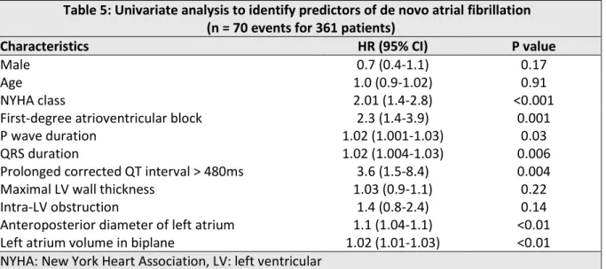
At univariate analysis, prolonged P waves and QRS duration, prolonged corrected QT
interval and first-degree atrioventricular block were associated with a first access of AF
(Table 5). From multivariate analysis, none of the ECG parameters were statistically
30 Table 5: Univariate analysis to identify predictors of de novo atrial fibrillation
(n = 70 events for 361 patients)
Characteristics HR (95% CI) P value
Male 0.7 (0.4-1.1) 0.17
Age 1.0 (0.9-1.02) 0.91
NYHA class 2.01 (1.4-2.8) <0.001
First-degree atrioventricular block 2.3 (1.4-3.9) 0.001
P wave duration 1.02 (1.001-1.03) 0.03
QRS duration 1.02 (1.004-1.03) 0.006
Prolonged corrected QT interval > 480ms 3.6 (1.5-8.4) 0.004
Maximal LV wall thickness 1.03 (0.9-1.1) 0.22
Intra-LV obstruction 1.4 (0.8-2.4) 0.14
Anteroposterior diameter of left atrium 1.1 (1.04-1.1) <0.01
Left atrium volume in biplane 1.02 (1.01-1.03) <0.01
NYHA: New York Heart Association, LV: left ventricular
Table 6: Multivariate analysis to identify predictors of de novo atrial fibrillation (n = 70 events for 361 patients)
Characteristics HR (95% CI) P value
Male 0.7 (0.4-1.3) 0.30
Age 1.03 (1.01-1.1) 0.003
First-degree atrioventricular block 1.2 (0.6-2.2) 0.61
P wave duration 1.01 (0.9-1.03) 0.08
QRS duration 1.01 (1.0-1.02) 0.47
Prolonged corrected QT interval > 480ms 1.1 (0.2-4.6) 0.94
31
Discussion
Hypertrophic cardiomyopathy is a disease with variable clinical history and prognosis
difficult to establish. Many studies have previous focused on clinical and echocardiographic
prognostic factors research and especially SCD risk factors.
This study reflects the abundance of electrocardiographic anomalies found in HCM.
These anomalies are used to establish a diagnosis and can help to stratify risk of rhythmic
events but also that of HF events and AF.
ECG anomalies.
A normal electrocardiogram is very rare in HCM. Therefore, a normal ECG has a high
negative predictive value from the absence of HCM. Contrary to what has been described, in
our study, it is not associated with better prognosis; may be due to a lower prevalence than
reported from other groups (1.6% versus 4-6%) [8].
In our study, some predictors were found such as QRS duration and repolarization
anomalies to predict HF and major rhythmic events and prolonged corrected QT interval to
predict HF events. This had already been suggested in a limited number of studies about
HCM. QRS duration would be an independent predictor of major cardiovascular events
[4][35]. Fragmented QRS [36][37][38], pseudo-STEMI [4] and prolonged correct QT interval
[39][40] would predict a high rhythmic risk, pseudo-necrosis Q waves would also be
associated with high cardiovascular mortality rate [23] and in patients with history of aborted
SCD, J waves would be more frequent [41-45]. Finally, for AF, the suggested predictors in
our univariate analysis were first-degree atrioventricular block, QRS and P wave duration and
prolonged corrected QT interval. The last two factors had already been mentioned in other
32
Pathophysiology.
Precise signification of these anomalies remains unclear. Relation between
electrocardiographic anomalies and phenotype expression on ETT had already been
suggested in a few studies [9][23][39]. An article published in 2011 showed that the number
and severity of ECG abnormalities were related to phenotype expression by Cardiac
Magnetic Resonance (CMR) in terms of LV hypertrophy and presence and extent of late
gadolinium enhancement. Therefore, patients with normal ECG had a more favorable
presentation on CMR [48].
Most of suggested electrocardiographic predictors were depolarization or
repolarization anomalies, testifying of myocardial cell action potential aberration.
Experimental studies highlighted implication of early after depolarizations and L-type Ca2+
current, which was involved in plateau of maintained depolarization, in emergence of
ventricular arrhythmias in HCM [49][50].
These electrocardiographic aberrations were also testifying of myocyte disarray. In
HCM, there is ventricular remodeling with fibrosis areas, increased interstitial tissue and
ischemic zones with medial hypertrophy of small coronary arteries. These lead to a decrease
of ventricular compliance, an increase of LV filing pressures and a decrease of LV ejection
fraction [12][13]. These findings explain HF events and AF occurrence.
Implications in clinical practice.
Having improved prevention of HCM-related SCD, the prognosis in older patients is related
to the prevention of HF and cardioembolic disease [51]. Identifying patients at risk is
33
depolarization anomalies, signs of electrical and mechanical myocyte disarray, non-specific
of HCM, could be predictors.
To predict SCD, HF and AF risks of HCM, all complementary tools must be used in
clinical practice such ECG abnormalities, clinical and ETT data including global longitudinal
strain analysis [52] but also late gadolinium enhancement in CMR imaging [48]. Patients
with significant ECG aberrations will need to have closer follow-up.
Perspectives.
As already suggested, an ECG anomalies score could be created and integrated in the
SCD-risk score [48][53]. Also, it might be interesting to evaluate three-dimensional 252-lead
electrocardiography in order to understand the mechanisms of ventricular and atrial
arrhythmias in HCM [54].
The interest of monitoring the evolution of ECGs over time should also be
determined.
Study limitations.
The main limitation of our study is observational retrospective design. Other limitations are
the small sample size, the short follow-up time of about three years and the low rate of study
endpoints which decrease the statistical power. In addition, as a referral center, this could be a
selection bias.
Conclusion
A 12-lead resting ECG remains a simple, low-cost, reproducible and available exam which
34
References
[1] 2014 ESC Guidelines on diagnosis and management of hypertrophic cardiomyopathy: The Task Force for the Diagnosis and Management of Hypertrophic Cardiomyopathy of the European Society of Cardiology (ESC). Eur Heart J 2014;35:2733.
[2] Maron BJ, Gardin JM, Flack JM, Gidding SS, Kurosaki TT, Bild DE. Prevalence of Hypertrophic Cardiomyopathy in a General Population of Young Adults: Echocardiographic Analysis of 4111 Subjects in the CARDIA Study. Circulation 1995;92:785.
[3] Semsarian C, Ingles J, Maron MS, Maron BJ. New perspectives on the prevalence of hypertrophic cardiomyopathy. J Am Coll Cardiol. 2015 Mar 31;65(12):1249-1254.
[4] Biagini E, Pazzi C, Olivotto I, Musumeci B, Limongelli G, Boriani G, et al. Usefulness of Electrocardiographic Patterns at Presentation to Predict Long-term Risk of Cardiac Death in Patients With Hypertrophic Cardiomyopathy. Am J Cardiol.2016;118:432.
[5] Savage DD, Seides SF, Clark CE, Henry WL, Maron BJ, Robinson FC, Epstein SE. Electrocardiographic findings in patients with obstructive and nonobstructive hypertrophic cardiomyopathy. Circulation. 1978 Sep;58(3 Pt 1):402-8.
[6] Casale PN, Devereux RB, Kligfield P, Eisenberg RR, Miller DH, Chaudhary BS, Phillips MC. Electrocardiographic detection of left ventricular hypertrophy: development and prospective validation of improved criteria. J Am Coll Cardiol. 1985 Sep;6(3):572-80.
[7] Konno T, Nagata Y, Teramoto R, Fujino N, Nomura A, Tada H et al. Usefulness of Electrocardiographic Voltage to Determine Myocardial Fibrosis in Hypertrophic Cardiomyopathy. Am J Cardiol. 2016 Feb 1;117(3):443-9.
[8] McLeod CJ, Ackerman MJ, Nishimura RA, Tajik AJ, Gersh BJ, Ommen SR. Outcome of Patients with Hypertrophic Cardiomyopathy and a Normal Electrocardiogram. J Am Coll Cardiol.2009;54:229. [9] Maron BJ, Wolfson JK, Ciró E, Spirito P. Relation of electrocardiographic abnormalities and patterns of left ventricular hypertrophy identified by 2-dimensional echocardiography in patients with hypertrophic cardiomyopathy. Am J Cardiol.1983;51:189.
[10] Sabater-Molina M, Pérez-Sánchez I, Hernández Del Rincón JP, Gimeno JR. Genetics of hypertrophic cardiomyopathy: A review of current state. Clin Genet. 2018 Jan;93(1):3-14.
[11] Marian AJ. Pathogenesis of diverse clinical and pathological phenotypes in hypertrophic cardiomyopathy. Lancet. 2000 Jan 1;355(9197):58-60.
[12] Davies MJ, McKenna WJ. Hypertrophic cardiomyopathy pathology and pathogenesis. Histopathology. 1995 Jun;26(6):493-500.
[13] Burke MA, Cook SA, Seidman JG, Seidman CE. Clinical and Mechanistic Insights Into the Genetics of Cardiomyopathy. J Am Coll Cardiol. 2016 Dec 27;68(25):2871-2886.
[14] P M Elliott, J R Gimeno, R Thaman, J Shah, D Ward, S Dickie et al. Historical trends in reported survival rates in patients with hypertrophic cardiomyopathy. Heart. 2006 Jun; 92(6): 785–791. [15] Spirito P, Bellone P, Harris KM, Bernabo P, Bruzzi P, Maron BJ. Magnitude of left ventricular hypertrophy and risk of sudden death in hypertrophic cardiomyopathy. N Engl J Med. 2000 Jun 15;342(24):1778-85.
35 [16] Maron MS, Olivotto I, Betocchi S, Casey SA, Lesser JR, Losi MA, Cecchi F, Maron BJ. Effect of left ventricular outflow tract obstruction on clinical outcome in hypertrophic cardiomyopathy. N Engl J Med. 2003 Jan 23;348(4):295-303.
[17] Elliott PM, Gimeno JR, Tomé MT, Shah J, Ward D, Thaman R et al. Left ventricular outflow tract obstruction and sudden death risk in patients with hypertrophic cardiomyopathy. Eur Heart J. 2006 Aug;27(16):1933-41. Epub 2006 Jun 5.
[18] Maron M.S., Olivotto I., Zenovich A.G., Link M.S., Pandian N.G., Kuvin J.T. Et al. Hypertrophic cardiomyopathy is predominantly a disease of left ventricular outflow tract obstruction. Circulation. 2006;114:2232–2239.
[19] Monserrat L, Elliott PM, Gimeno JR, Sharma S, Penas-Lado M, McKenna WJ. Non sustained ventricular tachycardia in hypertrophic cardiomyopathy: an independent marker of sudden death risk in young patients. J Am Coll Cardiol. 2003 Sep 3;42(5):873-9.
[20] Spirito P, Autore C, Rapezzi C, Bernabò P, Badagliacca R, Maron MS et al. Syncope and risk of sudden death in hypertrophic cardiomyopathy. Circulation. 2009 Apr 7;119(13):1703-10.
[21] O’Mahony C, Jichi F, Pavlou M, Monserrat L, Anastasakis A, Rapezzi C, et al. A novel clinical risk prediction model for sudden cardiac death in hypertrophic cardiomyopathy (HCM Risk-SCD). Eur Heart J 2014;35:2010.
[22] Paola Melacini, Cristina Basso, Annalisa Angelini, Chiara Calore, Fabiana Bobbo, Barbara Tokajuk et al. Clinicopathological profiles of progressive heart failure in hypertrophic cardiomyopathy. Eur Heart J. 2010 Sep; 31(17): 2111–2123.
[23] Xiao Y, Yang KQ, Yang YK, Liu YX, Tian T, Song L et al. Clinical Characteristics and Prognosis of End-stage Hypertrophic Cardiomyopathy. Chin Med J (Engl). 2015 Jun 5;128(11):1483-9.
[24] Maron MS, Kalsmith BM, Udelson JE, Li W, DeNofrio D. Survival after cardiac transplantation in patients with hypertrophic cardiomyopathy. Circ Heart Fail. 2010 Sep;3(5):574-9.
[25] Vaidya K, Semsarian C, Chan KH. Atrial Fibrillation in Hypertrophic Cardiomyopathy. Heart Lung Circ. 2017 Sep;26(9):975-982.
[26] Vriesendorp PA, Liebregts M, Steggerda RC, Schinkel AF, Willems R, Ten Cate FJ et al. Long-term outcomes after medical and invasive treatment in patients with hypertrophic cardiomyopathy. JACC Heart Fail. 2014 Dec;2(6):630-6.
[27] Landstrom AP, Ackerman MJ. Mutation type is not clinically useful in predicting prognosis in hypertrophic cardiomyopathy. Circulation. 2010 Dec 7;122(23):2441-9; discussion 2450.
[28] Kelly M, Semsarian C. Multiple mutations in genetic cardiovascular disease: a marker of disease severity? Circ Cardiovasc Genet. 2009 Apr;2(2):182-90.
[29] Ackerman MJ, VanDriest SL, Ommen SR, Will ML, Nishimura RA, Tajik AJ, Gersh BJ. Prevalence and age-dependence of malignant mutations in the betamyosin heavy chain and troponin T genes in hypertrophic cardiomyopathy: a comprehensive outpatient perspective. J Am Coll Cardiol. 2002 Jun 19;39(12):2042-8.
[30] Moolman JC1, Corfield VA, Posen B, Ngumbela K, Seidman C, Brink PA, Watkins H. Sudden death due to troponin T mutations. J Am Coll Cardiol. 1997 Mar 1;29(3):549-55.
[31] Hancock EW, Deal BJ, Mirvis DM, Okin P, Kligfield P, Gettes LS. AHA/ACCF/HRS Recommendations for the Standardization and Interpretation of the Electrocardiogram: Part V: Electrocardiogram Changes Associated With Cardiac Chamber Hypertrophy: A Scientific Statement
36 From the American Heart Association Electrocardiography and Arrhythmias Committee, Council on Clinical Cardiology; the American College of Cardiology Foundation; and the Heart Rhythm Society: Endorsed by the International Society for Computerized Electrocardiology. Circulation 2009;119:e25131.
[32] Rautaharju PM, Surawicz B, Gettes LS. AHA/ACCF/HRS Recommendations for the Standardization and Interpretation of the Electrocardiogram: Part IV. J Am Coll Cardiol 2009;53:982. [33] Surawicz B, Childers R, Deal BJ, Gettes LS. AHA/ACCF/HRS Recommendations for the Standardization and Interpretation of the Electrocardiogram: Part III. J Am Coll Cardiol 2009;53:976. [34] Julius M.Gardin, David B.Adams, Pamela S.Douglas and all. Recommendations for a standardized report for adult transthoracic echocardiography: A report from the American Society of Echocardiography's Nomenclature and Standards Committee and Task Force for a Standardized Echocardiography Report. Journal of the American Society of Echocardiography Volume 15, Issue 3, March 2002, Pages 275-290.
[35] Bongioanni S, Bianchi F, Migliardi A, Gnavi R, Pron PG, Casetta M, et al. Relation of QRS Duration to Mortality in a Community-Based Cohort with Hypertrophic Cardiomyopathy. Am J Cardiol. 2007;100:503.
[36] Das MK1, Zipes DP. Fragmented QRS: a predictor of mortality and sudden cardiac death. Heart Rhythm. 2009 Mar;6(3 Suppl):S8-14.
[37] Kang KW, Janardhan AH, Jung KT, Lee HS, Lee MH, Hwang HJ. Fragmented QRS as a candidate marker for high-risk assessment in hypertrophic cardiomyopathy. Heart Rhythm. 2014 Aug;11(8):1433-40.
[38] Konno T, Hayashi K, Fujino N, Oka R, Nomura A, Nagata Y et al. Electrocardiographic QRS Fragmentation as a Marker for Myocardial Fibrosis in Hypertrophic Cardiomyopathy. J Cardiovasc Electrophysiol. 2015 Oct;26(10):1081-7.
[39] Johnson JN, Grifoni C, Bos JM, Saber-Ayad M, Ommen SR, Nistri S et al. Prevalence and clinical correlates of QT prolongation in patients with hypertrophic cardiomyopathy. Eur Heart J. 2011 May;32(9):1114-20.
[40] Gray B, Ingles J, Medi C, Semsarian C. Prolongation of the QTc interval predicts appropriate implantable cardioverter-defibrillator therapies in hypertrophic cardiomyopathy. JACC Heart Fail. 2013 Apr;1(2):149-55.
[41] Li Y, et al. J Int Med Res. 2013. J wave is associated with increased risk of sudden cardiac arrest in patients with hypertrophic cardiomyopathy.
[42] Charles Antzelevitch, Gan-Xin Yan and Sami Viskin, Rationale for the Use of the Terms J Wave Syndromes and Early Repolarization. J Am Coll Cardiol. 2011 Apr 12; 57(15): 1587–1590.
[43] Haïssaguerre M, Derval N, Sacher F, Jesel L, Deisenhofer I, de Roy L et al. Sudden cardiac arrest associated with early repolarization. N Engl J Med. 2008 May 8;358(19):2016-23.
[44] Yan GX, Antzelevitch C. Cellular basis for the electrocardiographic J wave. Circulation. 1996 Jan 15;93(2):372-9.
[45] Tsuda T, Hayashi K, Konno T, Sakata K, Fujita T, Hodatsu A et al. J Waves for Predicting Cardiac Events in Hypertrophic Cardiomyopathy. JACC Clin Electrophysiol. 2017 Oct;3(10):1136-1142.
37 [46] Tuluce K, Yakar Tuluce S, Kahya Eren N, Kocabas U, Akyildiz Akcay F, Gunduz R et al. Predictors of Future Atrial Fibrillation Development in Patients with Hypertrophic Cardiomyopathy: A Prospective Follow-Up Study. Echocardiography. 2016 Mar;33(3):379-85.
[47] Nielsen JB, Graff C, Pietersen A, Lind B, Struijk JJ, Olesen MS, Haunsø S, Gerds TA, Svendsen JH, Køber L, Holst AG. J-shaped association between QTc interval duration and the risk of atrial fibrillation: results from the Copenhagen ECG study. J Am Coll Cardiol. 2013 Jun 25;61(25):2557-64. [48] Delcrè SD, Di Donna P, Leuzzi S, Miceli S, Bisi M, Scaglione M et al. Relationship of ECG findings to phenotypic expression in patients with hypertrophic cardiomyopathy: a cardiac magnetic resonance study. Int J Cardiol. 2013 Aug 10;167(3):1038-45.
[49] Elisa Passini, Ana Mincholé, Raffaele Coppini, Elisabetta Cerbai, Blanca Rodriguez, Stefano Severi and Alfonso Bueno-Orovioa. Mechanisms of pro-arrhythmic abnormalities in ventricular repolarisation and anti-arrhythmic therapies in human hypertrophic cardiomyopathy. J Mol Cell Cardiol. 2016 Jul; 96: 72–81.
[50] Tomaselli GF, Beuckelmann DJ, Calkins HG, Berger RD, Kessler PD, Lawrence JH et al. Sudden cardiac death in heart failure. The role of abnormal repolarization. Circulation. 1994 Nov;90(5):2534-9.
[51] Maron BJ, Olivotto I, Spirito P, Casey SA, Bellone P, Gohman TE, et al. Epidemiology of hypertrophic cardiomyopathy-related death: revisited in a large non-referralbased patient population. Circulation 2000;102:858.
[52] Vergé MP, Cochet H, Reynaud A, Morlon L, Peyrou J, Vincent C et al. Characterization of hypertrophic cardiomyopathy according to global, regional, and multi-layer longitudinal strain analysis, and prediction of sudden cardiac death. Int J Cardiovasc Imaging. 2018 Jul;34(7):1091-1098. [53] Olivotto I, Cecchi F, Poggesi C, Yacoub MH. Patterns of disease progression in hypertrophic cardiomyopathy: an individualized approach to clinical staging. Circ Heart Fail. 2012 Jul 1;5(4):535-46.
[54] Ashok J Shah, Meleze Hocini, Patrizio Pascale, Laurent Roten, Yuki Komatsu, Matthew Daly et al. Body Surface Electrocardiographic Mapping for Non-invasive Identification of Arrhythmic Sources. Arrhythm Electrophysiol Rev. 2013 Apr; 2(1): 16–22.