The Future of Biomedicine–
Delivered
POLY(AMINO ACID) BLOCK COPOLYMERS
FOR DRUG DELIVERY AND OTHER BIOMEDICAL APPLICATIONSSTIMULI-RESPONSIVE MATERIALS
AS INTELLIGENT DRUG DELIVERY SYSTEMS
ELECTROSPUN NANOFIBERS
FOR DRUG DELIVERY SYSTEMSCHITOSAN: A VERSATILE PLATFORM
FOR PHARMACEUTICAL APPLICATIONSPolymers in
Therapeutics and
Nanomedicine
Aldrich Materials Science Sigma-Aldrich Co. LLC 6000 N. Teutonia Ave. Milwaukee, WI 53209, USA To Place Orders Telephone 800-325-3010 (USA) FAX 800-325-5052 (USA)
International customers, contact your local Sigma-Aldrich office (sigma-aldrich.com/worldwide-offices).
Customer & Technical Services
Customer Inquiries 800-325-3010
Technical Service 800-325-5832
SAFC® 800-244-1173
Custom Synthesis 800-244-1173
Flavors & Fragrances 800-227-4563
International 314-771-5765
24-Hour Emergency 314-776-6555
Safety Information sigma-aldrich.com/safetycenter
Website sigma-aldrich.com
Email [email protected]
Subscriptions
Request your FREE subscription to Material Matters:
Phone 800-325-3010 (USA)
Mail Attn: Marketing Communications
Aldrich Chemical Co., Inc Sigma-Aldrich Co. LLC P.O. Box 2060 Milwaukee, WI 53201-2060 Website aldrich.com/mm Email [email protected] Online Versions
Explore previous editions
of Material Matters aldrich.com/materialmatters
Now available for your iPad®
aldrich.com/mm
Material Matters (ISSN 1933–9631) is a publication of Aldrich
Chemical Co., Inc. Aldrich is a member of the Sigma-Aldrich Group.
Vol. 9, No. 3
Yong Zhang, Ph.D. Aldrich Materials Science
©2014 Sigma-Aldrich Co. LLC. All rights reserved. SIGMA, SAFC, SIGMA-ALDRICH, and ALDRICH, are trademarks of Sigma-Aldrich Co. LLC, registered in the US and other countries. Material Matters is a trademark of Sigma-Aldrich Co. LLC. iPad is a registered trademark of Apple Inc. Pluronic is a registered trademark of BASF Corporation. RESOMER is a registered trademark of Evonik Roehm GMBH LLC. Sigma brand products are sold through Sigma-Aldrich, Inc. Purchaser must
The third issue of Material Matters™ in 2014 is focused on Polymers in Therapeutics and Nanomedicine. This issue covers a variety of topics including synthetic and natural polymers that deliver a therapeutic payload using stimuli-response, self-assembly, or electrospun fibers.
The first article, by Carmen Scholz (USA), reviews poly(amino acid) block copolymers. These biocompatible polymers are inspired by the activity and capabilities of enzymes. By synthesizing PEGylated polymers using one or two amino acids, the resulting self-assembled polymeric micelles or membranes allow for drug delivery in a variety of configurations. They can also be used to enable attachment of a drug delivery system to a surface.
In our second article, Amit Singh and Mansoor M. Amiji (USA) review stimuli-responsive materials as intelligent drug delivery systems. There are a variety of smart materials that are able to be tuned to deliver a drug as a result of several different types of stimuli. Highlights include pH-, redox-, enzyme-, thermo-, light-, ultrasound-, magnetically, and electrically responsive polymers.
Sang Jin Lee, James J. Yoo, and Anthony Atala (USA), review electrospun nanoscale fibers used in drug or protein delivery systems in our third article. They highlight several different apparatus for electrospinning, as well as methods to control the fiber properties. The resulting scaffolds were shown to have the ability to deliver a variety of drugs and proteins in a controlled manner.
The fourth article, by Raphaël Riva and Christine Jérôme (Belgium), reviews chitosan as a platform for pharmaceutical applications. Chitosan is a natural polymer which can be used to encapsulate drugs and has been shown to improve the therapeutic efficiency and bioavailability, as well as enable targeted delivery. Specifically, chitosan has been used with difficult-to-deliver genes and hydrophobic drugs.
Each article in this issue is accompanied by a list of polymers available from Aldrich® Materials Science. Contact us at [email protected] if you need any material that you cannot find in our catalog, or would like a custom grade for your development work. We welcome your new product requests and suggestions as we continue to grow our polymer offering.
About Our Cover
Polymers in therapeutics and nanomedicine allow for improved solubility, decreased toxicity, controlled release, and can help enable site-specific delivery. Polymers are used to generate numerous biomolecule–polymer conjugates, small molecule drug–polymer conjugates, supramolecular drug-delivery systems, and can be used as a platform for controlled release. This issue’s cover art artistically illustrates a variety of drug-delivery systems, including functionalized multivalent polymer micelles, well-defined nanostructures, and polyplexes flowing through a blood vessel.
We welcome fresh product ideas. Do you have a material or compound you wish to see featured in the Aldrich® Materials Science line? If it is needed to accelerate your research, it matters. Send your suggestion to [email protected] for consideration.
Bryce P. Nelson, Ph.D.
Aldrich Materials Science Initiative Lead
Articles
Poly(Amino Acid) Block Copolymers for Drug Delivery and
Other Biomedical Applications
73
Stimuli-Responsive Materials as Intelligent Drug Delivery Systems
82
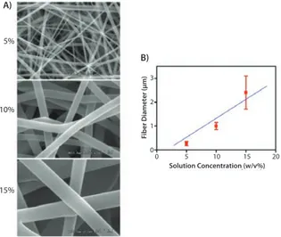
Electrospun Nanofibers for Drug Delivery Systems
89
Chitosan: A Versatile Platform for Pharmaceutical Applications
95
Featured Products
Triblock Copolymers
A list of BAB triblock copolymers
77
Poly(ethylene glycol)s
A selection of PEG PCL diblock and PEG macroCTA polymers
77
Synthetic Poly(amino acid)s
A list of synthetic homopolymers and copolymers
78
PolyNIPAMs
A selection of polyNIPAMs and functionalized polyNIPAMs
86
Well-defined Biodegradable Polymers
A selection of polylactides, end-functionalized poly(l-lactide)s, and block
copolymers
92
Chitosans
A list of chitosans from white mushroom and animal origins
98
Professor Richard Hoogenboom at Ghent University (Belgium) has recommended the addition of functionalized poly(2-alkyl-2-oxazoline), or poly(2-oxazoline) polymers to our product portfolio for biomedical applications. This includes alkyne-terminated poly(2-ethyl-2-oxazoline) (Aldrich Prod. No. 778338). Poly(2-oxazoline) polymers demonstrate a lower critical solution temperature (LCST) behavior in aqueous solutions and can be used as thermo-responsive materials for biomedical
applications because their transition temperatures are close to that of the human body.1,2,3 Poly(2-oxazoline) has also been used in various other
bio-inspired methods and for polymer therapeutics.4,5 In particular, our
alkyne-terminated clickable poly(2-oxazoline) products can be used as versatile building blocks for the construction of a large variety of complex, well-defined polymer architectures (including cyclodextrin core star-polymers) as well as virus and peptide polymer conjugates.1,6,7
References
(1) Chapman, R.; Bouten, P.J.M.; Hoogenboom, K.A.J.; Perrier, S. Chem. Comm. 2013, 49, 6522. (2) Hoogenboom, R. Angew. Chem. Int. Ed. 2009, 48, 7978–7994.
(3) Hoogenboom, R.; Thijs, H.M.L.; Jochems, M.J.H.C.; van Lankvelt, B.M.; Fijten, M.W.M.; Schubert, U.S. Chem. Commun., 2008, 5758–5760.
(4) Manzenrieder, F.; Luxenhofer, R.; Retzlaff, M.; Jordan, R.; Finn, M.G. Angew. Chem. Int. Ed. 2011,
50, 2601–2605.
(5) Hoogenboom, R.; Schlaad, H. Polymers 2011, 3, 467–488.
(6) Luxenhofer, R.; Han, Y.; Schulz, A.; Tong, J.; He, Z.; Kabanov, A.V.; Jordan, R. Macromol. Rapid
Commun. 2012, 33, 1613–1631.
(7) Fijten, M.W.M.; Haensch, C.; van Lankvelt, B.M.; Hoogenboom, R.; Schubert, U.S. Macromol.
Chem. Phys. 2008, 209, 1887–1895.
Poly(2-ethyl-2-oxazol ine), alkyne terminated Poly ethyl oxazol ine[1171957-24-6] C3H3(C5H9NO)nOH
N HO O H3C CH n
acetyl ene poly ethyl oxazol ine 5kDa
average Mn 5,000, ≤1.2
powder
Store at: 2-8°C
CONTROLLED RADICAL
POLYMERIZATION GUIDE
Enabling Well-defined Copolymers
Request your FREE CRP guide online at
aldrich.com/crpmm
y
y
Procedures written by CRP experts for different polymerizations.
Authors include:
- Edmondson
- Haddleton
- Matyjaszewski
- Moad
- Wooley
y
y
Mini-reviews of the technology help determine:
- The right techniques
- The right reagents
- The right conditions
y
y
Controlled Radical Polymerization Tools:
- New initiators
- Reversible Addition/Fragmentation Chain Transfer (RAFT) agents
- Atom Transfer Radical Polymerization (ATRP) ligands
POLY(AMINO ACID) BLOCK COPOLYMERS
FOR DRUG DELIVERY AND OTHER BIOMEDICAL APPLICATIONS
Carmen Scholz
Department of Chemistry, University of Alabama in Huntsville Huntsville, AL 35899
Email: [email protected]
Introduction to Polyamino Acids:
The Next Best Thing to Proteins
Humankind has utilized protein materials throughout its existence, starting with the use of materials such as wool and silk for warmth and protection from the elements and continuing with the use of recombinant DNA techniques to synthesize proteins with unique and useful properties. Proteins consist of amino acids linked in a pre-determined and genetically-prescribed sequence. There are twenty amino acids found in naturally occurring proteins. Commonly known as the standard proteinogenic amino acids, the sequence of these building blocks form the basis of protein function. The specific function of the protein impacts the sequence and abundance of particular amino acids. For example, the protection of the Bombyx mori pupa within the silk cocoon is a primary natural function of silk protein. As can be expected, the very hydrophobic and chemically non-reactive glycine and alanine comprise 70% of the amino acids in silk protein. Enzymes, however, are proteins that perform significantly different functions and, as such, are comprised of a different set of amino acids. Enzymes fulfill biochemical catalytic functions and their amino acid sequence enables catalytic activity through the composition and molecular architecture of the catalytic pocket. The stability of the enzyme is maintained via its unique tertiary structure and the solubility of the enzyme in its (aqueous) medium of activity is guaranteed by functionalized amino acids that form the outside of the three dimensional structure.
This capability to enable unique and highly-targeted biological function makes proteins ideal candidates for drug delivery systems because proteins have the ability to:
y
y Form an ideal “cradle” for a variety of therapeutic drugs, including delicate hydrophilic proteins or hydrophobic small molecules. y
y Interact with, fold, and transport DNA. y
y Deliver a therapeutic to a targeted location using site-specific interactions, exploit transmembrane transport mechanisms, and deliver a therapeutic payload to a specific site of action, free of side-effects.
However, our ability to engineer protein structure as well as understanding the infinite resulting interaction possibilities is still in its infancy. Despite of our current understanding of the intricacies of proteomics, the synthesis of fault-free (i.e., sequence-correct) proteins in appreciable amounts is still unresolved.
The limitations in our ability to synthesize proteins tailored for delivery of specific drugs has led researchers to concentrate on “the next best thing”: poly(amino acid)s (PAAs). While this category of polymers is also sometimes referred to as “polypeptides”, this author suggests the use of the term is slightly misleading because “peptide” refers to a polymeric structure with a specific amino acid sequence. However, in the macromolecules considered here, typically only two or three different amino acids are used for synthesis and no specific amino acid sequence is achieved.
PAAs are similar to proteins (and peptides) in that they consist of amino acid building blocks linked by means of amide bonds. This backbone architecture and the use of naturally occurring l-amino acids for their
synthesis ensure their biocompatibility. Subsequently, their potential degradation products are l-amino acids, which will be readily absorbed
by the body. Most PAAs consist of no more than two different amino acids that are linked to yield either random or block copolymers.
When l-amino acids are used in the synthesis of PAAs, the polymeric
products retain the stereoregularity of the monomers. This results in the creation of isotactic PAA polymers. This is structurally very important as the stereoregularity is responsible for the formation of distinct secondary structures. Typically, helices are expected as secondary structures for isotactic polymers. The resulting PAA copolymers also exhibit the chemical characteristics of the l-amino acid building blocks,
such as polarity, electrostatic, and solubility behavior. These physical characteristics are exploited when amphiphilic block copolymers are synthesized. Amphiphilic block copolymers readily undergo self-assembly into gels, micelles, membranes, vesicles, nanoparticles, nanogels, and other three-dimensional structures. The synthesis and utilization of PAA block copolymers is discussed in this article.
Synthesis of Poly(amino acid)s
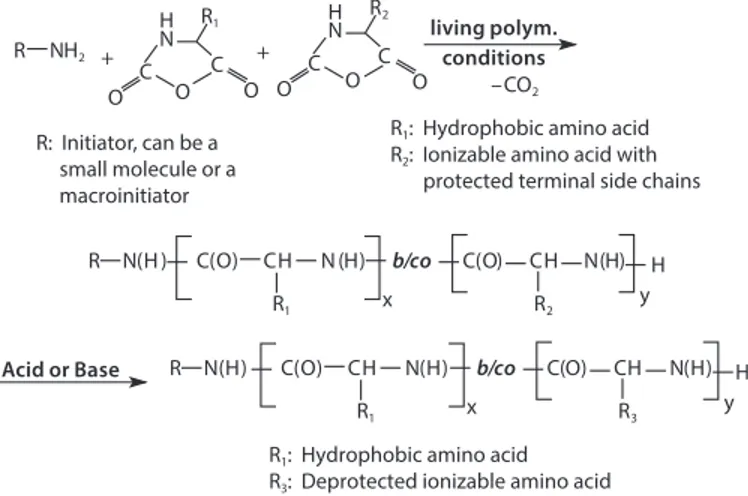
Poly(amino acid)s and their syntheses were discussed in detail by Kricheldorf.1 PAAs are typically synthesized by the ring-opening
polymerization of amino acid N-carboxyanhydrides (NCAs) initiated by an amino-terminated reagent, as shown in Figure 1. Results indicate the polymerization follows a living, ring-opening polymerization. This polymerization technique allows for the synthesis of PAA block copolymers as well as random copolymers, making it highly versatile. If the amino acid NCAs are added successively to the initiator (the addition of the NCA of amino acid A followed by the addition of the NCA of amino acid B), AB block copolymers are formed. If the NCAs of the two amino acids are added to the initiator as a mixture, random copolymers are formed. Additional polymer architectures of varying complexity can be achieved due to the living nature of the polymerization.
–CO2
R1 x R2
R N(H ) C(O) CH N (H) b/co C(O) CH N(H) y H + N R1 C C H O O O + N R2 C O C H O O R NH2 R1 x R3 R N(H) C(O) CH N(H) C(O) CH N(H) y H living polym. conditions
R1: Hydrophobic amino acid
R2: Ionizable amino acid with
protected terminal side chains R: Initiator, can be a
small molecule or a macroinitiator
R1: Hydrophobic amino acid
R3: Deprotected ionizable amino acid
Acid or Base b/co
Figure 1. Schematic of NCA polymerization leading to the synthesis of PAA block copolymers and PAA random copolymers.
When synthesizing PAAs for drug delivery and biomedical applications, it is crucial to tightly control the molecular weight within a narrow range. This can be accomplished by employing living polymerization techniques to produce block copolymers. To do this, Hadjichristidis et al. employed high vacuum techniques to purify the monomers, solvents, and initiator. The polymerization was then performed in a specialized vessel that guaranteed absolutely anhydrous conditions.2 Deming et al. developed
syntheses that use transition metal catalysts to gain control over the molecular weight and polydispersity of the synthesized PAAs.3 Our own
studies have shown that the first few addition reactions (i.e., the addition of the first few amino acid building blocks to the initiator) are most crucial to controlling the reaction and producing PAAs of the desired molecular weight and polydispersity.4 In the beginning of the polymerization, the
nascent oligomers are not yet long enough to form a helical structure, but tend to form β-sheets due to strong hydrogen bond interactions. The rigidity of these structures suppresses the ability of the growing chain to undergo continued nucleophilic attacks on NCA monomers and these small β-sheet structures have a tendency to precipitate out of the polymerization solution, effectively stopping the polymerization.5
If the β-sheet formation is not kept under control, the resulting final product consists of a few long chains that exceed the intended chain length as determined by the Monomer:Initiator ratio and low molecular weight oligomers.
It was found that urea effectively suppresses the hydrogen bond formation between nascent PAA chains. As a result, the premature precipitation of product is suppressed. This allows for the polymerization to follow a living mechanism.4 Because urea suppresses the formation
of β-sheets, there is sufficient time for the amino acid chains to reach a length of about 10 repeat units, enabling formation of α-helices. Once the β-sheet stage is overcome, polymerization proceeds in a controlled manner, where chain ends remain reactive (living) and block copolymers can readily form (Figure 1).
Poly(amino acid) Block Copolymers:
Concepts for Drug Delivery Systems
Amino acids have diverse chemical properties and can be subdivided according to the nature of their side chains, including those that are aliphatic, aromatic, sulfur-containing, and those that contain ionizable groups. When designing a PAA for use in drug delivery, amino acids are selected based on chemical properties that meet the needs of both the drug and the solvent environment. Typically, amphiphilic block copolymers are formed from a hydrophilic (ionizable) and a hydrophobic amino acid (Figure 1). This amphiphilicity will induce self-assembly of the block copolymer when exposed to a selective solvent.6 In the case of
drug delivery, where polymers are exposed to an aqueous environment, the hydrophilic segment of the block copolymer forms the outer layer or corona of the self-assembled structure, while the hydrophobic block self-assembles into a tightly packed structure that functions as a “cargo compartment” for hydrophobic drugs (Figure 2A).
Deming et al. studied the self-assembly and packing of PAA diblock and triblock copolymers and provided an excellent model to illustrate the polymer interactions and their subsequent self-assembly (Figure 2).7
Specifically, they studied copolymers consisting of poly(l-Leu), a
hydrophobic amino acid and poly(l-Lys), an ionizable amino acid.
Polymers of the following general structures were prepared: diblock copolymers: p(l-Lys)-block-p(l-Leu) and triblock copolymers: p(l
-Lys)-block-p(l-Leu)-block-p(l-Lys), and individual blocks varied in length. The resulting
block copolymers have a positively charged p(l-Lys) block that contributes
to its solubility in water and where it forms a distorted (stretched) random coil. The hydrophobic p(l-Leu) block forms an α-helix as a secondary
structure that is maintained when exposed to an aqueous environment. In the case of the triblock copolymers, there are two p(l-Lys) blocks that
are adjacent to the p(l-Leu) block.
When used as drug delivery vehicles, amphiphilic diblock copolymers generally self-assemble into classic spherical polymeric micelles upon exposure to a selective solvent such as water (Figure 2A). In most cases, the hydrophobic segment packs tightly to avoid contact with the water and the hydrophilic segment readily solubilizes to form a shell or corona around the hydrophobic core. However, in the case p(l-Lys)-block-p(l-Leu)
diblock copolymers the very stable p(l-Leu) helical structure, assembles
into a stiff rod shape, which impedes the formation of a spherical packing structure and a flat lamellar packing of the rods (Figure 2B) predominates. The rods line up in parallel and are held in place through hydrophobic intermolecular interactions. However, the hydrophilic blocks with increasing chain lengths assume a stretched random coil configuration and fill increasing molar volumes. In addition, electrostatic repulsion between the positively charged p(l-Lys) chains renders the situation even
more energetically unfavorable. As the chain length of the hydrophilic blocks increases, curved membranes are formed. These are the result of asymmetric packing of the hydrophilic blocks (Figure 2C). The resulting
increase in corona volume is energetically favorable. Structures with curvature in two directions can give rise to the formation of polymeric giant unilamellar vesicles. The repulsive effect between the p(l-Lys)
blocks can also be resolved by twisting the plane of the p(l-Leu) blocks
into fibrils, (Figure 2D). While the helices of the p(l-Leu) blocks maintain
parallel packing, the hydrophilic blocks gain volume by expanding around the forming fibrils which extend in one direction only.
A) B)
D) C)
Figure 2. Self-assembly arrangement of PAA di- and triblock copolymers into (A) spherical micelles; (B) flat membranes; (C) curved membranes, and (D) fibrils, with the cross section extending out of the plane of the paper and the insert depicting how the helices assemble into twisted fibers. Reprinted with permission from Ref. 7, Copyright 2004, American Chemical Society.
In the case of p(l-Lys)-block-p(l-Leu)-block-p(l-Lys) triblock copolymers,
where the two hydrophilic blocks are covalently attached to the hydrophobic core curved structures (Figure 2C) will not form. Instead, asymmetric packing of the p(l-Lys) blocks is impossible and fibrils readily
form. Both the curved and the fibrilar structure models can be used to explain the gelation behavior observed for the p(l-Lys)-block-p(l-Leu)
and p(l-Lys)-block-p(l-Leu)-block-p(l-Lys) block copolymers.8 Due to their
amphiphilic nature, these copolymer systems can be readily loaded with hydrophobic (in the core) as well as hydrophilic (in the corona) drugs. One can further expand on the above concept of PAA-based drug delivery systems, by using a macroinitiator to start the ring opening polymerization of amino acid NCAs. Poly(ethylene glycol) (PEG), is one of the most thoroughly investigated biocompatible polymers. With its large excluded volume, PEG conveys a “stealth character” to the structures to which it is attached. This essentially makes the structures “invisible” to adjacent protein molecules. The effectiveness of PEG to reduce protein interaction has been explained in physico-chemical terms by its low interfacial free energy in water and by its unique coordination with water molecules, as each PEG repeat unit coordinates with two to three water molecules. PEGylated PAA structures are used as drug delivery systems, employing polymeric micelles or nanoparticles with PEG to form the corona. They are also used to biocompatibilize surfaces of implantable devices intended to be in contact with protein-containing (bodily) fluids. In the case of drug delivery systems, PEGylation prevents uptake by the reticuloendothelial system and PEG-coated surfaces are efficient in suppressing biofouling.
The synthesis of PEGylated PAAs follows the scheme presented in Figure 1, with R representing NH2-terminated PEG. For biomedical
applications, PEGs with molecular weights of 2,000 to 10,000 Da are typically used to ensure renal clearance of degradation products. It is also possible to use bifunctional, α,ω-diamino PEGs as macroinitiators. The resulting block copolymers will have the PEG block in the middle, allowing the formation of interesting self-assembled molecular architectures, such as flower-like micelles.
A) B)
Figure 3. Surface modification with PEGylated PAA. A) The multi-point attachment of the PAA chain to the surface (e.g., by the formation of covalent bonds between l-Cys (yellow) and a gold surface) with the PEG-block (blue) extending away from the surface, thereby conveying a stealth character. B) Attachment of amino acid oligomers by single-point attachment and their subsequent modification with PAA-based polymeric micelles. The X indicates reactive terminal side groups, e.g., amino groups in l-Lys. A covalent bond is established between the PAA oligomer and the PAA polymeric micelle.
PEGylated PAAs can be attached directly to a surface and this surface attachment can be utilized for drug delivery applications.9 The attachment
of PEGylated PAAs containing (l-Cys) repeat units can be achieved using
gold-covered surfaces.10 To do this, l-Cys NCA is typically copolymerized
with another amino acid (shown in green in Figure 3A), such as l-Lys NCA
or l-Glu NCA, to achieve the optimal spacing (dilution) of the reactive l-Cys residues. The l-Cys repeat units react instantaneously with gold
surfaces. The repeating l-Cys units in each block copolymer chain result in
multiple anchor points to the surface (Figure 3A).
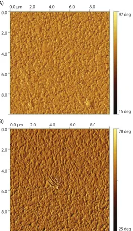
Because the PAA block functions as a multivalent anchor, the PEG block is free to extend away from the surface upon exposure to an aqueous environment. This architecture accomplishes two objectives: 1) PEG is strongly attached to the surface through multiple covalent thiol-gold linkages, and 2) the PAA block remains closely bound to the surface. These two factors contribute to the overall biocompatibility of the surface by masking “holes” that may form between individual PEG chains. It is well known the formation of dense polymer brushes is inherently difficult on surfaces using a “grafting to” method. This is because the concentration gradient built up by the already attached chains provides a strong kinetic hindrance against additional grafting. Furthermore, the large excluded volume that PEG exerts in an aqueous environment, while beneficial for preventing protein interactions, hinders the formation of dense PEG brushes in “grafting to” procedures even further. Figure 4shows complete coatings are achieved on gold surfaces with both, PEG-block-p(l-Glu)110
-block-p(l-Cys)10 and PEG-block-poly((l-Glu)110-co-(l-Cys)10). The polymer
architecture, PAA random copolymer vs. block copolymer, has little to no influence on the polymer morphology on the surface. The coatings are homogenous and pinhole-free.
97 deg 15 deg 0.0 µm 2.0 4.0 6.0 8.0 0.0 2.0 4.0 6.0 8.0 A) 78 deg 25 deg 0.0 µm 2.0 4.0 6.0 8.0 0.0 2.0 4.0 6.0 8.0 B)
Figure 4. 10×10 µm AFM phase images of gold surfaces coated with PEG-b-p(l-Glu)110-block-
p(L-Cys)10 (A) and PEG-block-p((l-Glu)110-co-(l-Cys)10)(B).
Another concept combines surface coating with the option of decorating the surface with PAA-based polymeric micelles that could be drug-loaded; the concept is schematically depicted in Figure 3B. In this procedure, oligo-(l-Lys) was attached covalently to the gold surface by a terminal
thiol group, which is the result of using cysteamine as the initiator in the ring-opening polymerization of l-Lys NCA. Subsequently, the surface
was decorated with micelles prepared from PEGylated PAAs (HOOC-CH2
-PEG114-block-poly(Bz-l-Glu)13) that carry carboxyl groups in their corona
and were, therefore, reactive toward p(l-Lys).11 Terminal carboxyl groups
in the poly(Bz-l-Glu) side chains are protected as a benzyl ester to achieve
the amphiphilicity that drives micellization. The resulting micelles are maintained due to the strong hydrophobic interaction of the benzyl groups in the micelle core. The micelles were covalently attached to the poly(l-Lys) using an amide linkage that formed between the terminal
carboxyl groups of the polymeric micelles and the amino functions of poly(l-Lys), shown in Figure 5.
97 deg 20 deg 0.0 µm 2.0 4.0 6.0 8.0 0.0 2.0 4.0 6.0 8.0
Figure 5. 10×10 µm AFM phase image of a gold surface coated with thiol-terminated p(l-Lys)15
and decorated with polymeric micelles formed by HOOC-CH2-PEG114-block-p(Bz-l-Glu)13.
Summary
The synthesis and application of PAA block copolymers in drug delivery and surface modification of biomedical products was discussed. Various self-assembly structures, including spherical polymeric micelles and membranes which enable gel formation, were examined. It was shown amino-terminated PEG can be used as a macroinitiator in the synthesis of PAAs, yielding block copolymers. PEGylated PAAs also self-assemble into polymeric micelles. Furthermore, if the PAA segment contains an amino acid that is reactive toward a surface (e.g., l-Cys reacts instantaneously
with gold), the PAA block can be employed as a multi-point anchor to attach PEG covalently in a dense packing by a “grafting to” method to a surface. In a different surface modification approach, PAA oligomers attached covalently to a surface can be further decorated with PAA polymeric micelles, enabling attachment of a drug delivery system to a surface.
References
(1) Kricheldorf, H.R. “Polypeptides” in: Models of Biopolymers by Ring Opening Polymerization (ed.: S. Penczek) CRC Press, Boca Raton, FL, 1990, pp 3–132.
(2) Hadjichristidis, N.; Iatrou, H.; Pispas, S.; Pitsikalis, M. J. Polym. Sci., Part A: Polym. Chem. 2000,
38(18), 3211–3234.
(3) Deming, T.J., JACS 1997, 119 (11), 2759–2760.
(4) Ulkoski, D.; Armstrong, T.; Scholz, C. “Investigating the Secondary Structure of Poly(amino acids)” in: Tailored Polymer Architectures for Pharmaceutical and Biomedical Applications (ed. C. Scholz, J. Kressler) ACS Symposium Series 1135 2013, pp 69–85.
(5) Komoto, T.; Kim, K.Y.; Oya, M., Kawai, T. Makromolek. Chemie 1974, 175(1), 283–299. (6) Kwon, G.S.; Kataoka, K., Adv. Drug Delivery Rev. 1995, 16, 295–309.
(7) Breedveld, V.; Nowak, A.P.; Sato, J.; Deming, T.J.; Pine, D.J. Macromolecules 2004, 37, 3943–3953. (8) Deming, T.J.; Soft Matter. 2005, 1, 28–35.
(9) Guenther, M.; Gerlach, G.; Wallmersperger, T.; Avula, M.N.; Cho, S.H.; Xie, X.; Devener, B.V.; Solzbacher, F.; Tathireddy, P.; Magda, J.J.; Scholz, C.; Obeid, R.; Armstrong, T. Adv. Sci. Technol. 2013, 85, 47–52.
(10) Obeid, R.; Armstrong, T.; Peng, X.; Busse, K.; Kressler, J.; Scholz, C. Journal of Polymer Science,
Part A, Polymer Science 2014, 52(2), 248–257.
Triblock Copolymers
For more information on this product line, visit aldrich.com/block.
BAB Triblock Copolymers
Name Structure Molecular Weight PDI Degradation Time Prod. No.
Polylactide-block-poly(ethylene glycol)-block-polylactide HO O O O H CH3 O O CH3 x y z PEG average Mn900 PLA average Mn1,500 average Mn (1,500-900-1,500) < 1.2 PDI <12 months 659630-1G PEG average Mn10,000 PLA average Mn1,000 average Mn (1,000-10,000-1,000) < 1.2 PDI <12 months 659649-1G Poly(lactide-co-glycolide)-block-poly(ethylene glycol)-block-poly(lactide-co-glycolide) O O H O O x y CH3 m H O O O O CH3 O O n x y m PEG average Mn1,000 PLGA average Mn2,200 average Mn (1,100-1,000-1,100) < 2.0 PDI 2-3 weeks 764817-1G Poly(lactide-co-caprolactone)-block-poly(ethylene glycol)-block-poly(lactide-co-caprolactone) H O O O O O O H3C y x m n H O O O O CH3 y x m
PEG average Mn5,000 (DCM, PEO)
PLCL average Mn5,700 average Mn (1,000-10,000-1,000) < 1.3 PDI 1-2 months 764833-1G Poly(lactide-co-glycolide)-block-poly(ethylene glycol)-block-poly(lactide-co-glycolide) O O H O O x y CH3 m H O O O O CH3 O O n x y m PEG average Mn1,000 PLGA average Mn2000 average Mn (1,000-1,000-1,000) < 1.2 PDI 1-2 weeks 764787-1G
Poly(ethylene glycol)s
For more information on this product line, visit aldrich.com/peg.
PEG PCL Diblocks
Name Structure Molecular Weight PDI Degradation Time Prod. No.
Poly(ethylene
glycol)-block-poly(ε−capro-lactone) methyl ether O O
H3C m OH O n PCL average Mn ~5,000 PEG average Mn ~5,000 average Mn ~10,000 (total) < 1.4 PDI >12 months 570303-250MG 570303-1G Poly(ethylene oxide)-block-polycapro-lactone, 4-arm R = * O O m O O n OH RO OR OR RO PCL average Mn ~2,500 PEG average Mn ~2,500 average Mn ~5,000 (total) < 1.2 PDI >12 months 570346-1G Poly(ethylene
glycol)-block-poly(ε−capro-lactone) methyl ether H O O
3C m OH O n PCL average Mn ~13,000 PEG average Mn ~5,000 average Mn ~18,000 (total) ≤ 1.4 PDI >12 months 570311-250MG 570311-1G PCL average Mn ~32,000 PEG average Mn ~5,000 average Mn ~37,000 (total) < 1.4 PDI >12 months 570338-250MG 570338-1G
PEG macroCTAs
Name Structure Molecular Weight PDI Prod. No.
Polystyrene, DDMAT terminated
HO S SC12H25
O
H3C CH3Phn S
average Mn 10,000 ≤ 1.1 PDI 772569-1G
Poly(ethylene glycol) methyl ether
4-cyano-4-[(dodecylsulfanylthiocarbonyl)sulfanyl]pentanoate SCH2(CH2)10CH3 S S CN H3C O O n O H3C average Mn 10,000 ≤ 1.1 PDI 753033-1G
Poly(ethylene glycol) methyl ether (4-cyano-4-pentanoate
dodecyl trithiocarbonate) S SCH2(CH2)10CH3 S H3C O O CN O H3C n average Mn 5,400 ≤ 1.1 PDI 751626-1G 751626-5G
Poly(ethylene glycol) methyl ether
2-(dodecylthiocarbonothioylthio)-2-methylpropionate H3CO O S SC12H25 O CH3CH3S n average Mn 5,000 ≤ 1.1 PDI 736325-1G
Poly(ethylene glycol) methyl ether (2-methyl-2-propionic acid
dodecyl trithiocarbonate) SCH2(CH2)10CH3 S S CH3 H3C O O n O H3C average Mn 10,000 ≤ 1.1 PDI 752495-1G
Name Structure Molecular Weight PDI Prod. No.
Poly(ethylene glycol) 4-cyano-4-(phenylcarbonothioylthio)
pentanoate S S H3C O O CN O H3C n average Mn 10,000 ≤ 1.1 PDI 764930-1G
Poly(ethylene glycol) methyl ether (4-cyano-4-pentanoate
dodecyl trithiocarbonate) S SCH2(CH2)10CH3 S H3C O O CN O H3C n average Mn 2,400 ≤ 1.1 PDI 751634-1G 751634-5G
Poly(ethylene glycol) methyl ether
2-(dodecylthiocarbonothioylthio)-2-methylpropionate H3CO O S SC 12H25 O CH3CH3S n average Mn 1,100 ≤ 1.1 PDI 740705-1G
Poly(ethylene glycol) 4-cyano-4-(phenylcarbonothioylthio)
pentanoate S S H3C O O CN O H3C n average Mn 2,000 ≤ 1.1 PDI 764914-1G
Poly(ethylene glycol) methyl ether (4-cyano-4-pentanoate
dodecyl trithiocarbonate) S SCH2(CH2)10CH3 S H3C O O CN O H3C n average Mn 1,400 ≤ 1.1 PDI 752487-1G 752487-5G
Synthetic Poly(amino acid)s
For more information on this product line, visit aldrich.com/polyaminoacid.
Homopolymers
Name Molecular Weight Prod. No.
Poly-l-histidine hydrochloride ≥5000 P2534-10MG
P2534-25MG P2534-100MG P2534-500MG Poly(γ-ethyl-l-glutamate) >100,000 P8035-1G Poly-l-proline 1,000-10,000 P2254-50MG P2254-100MG P2254-500MG P2254-1G
Poly-dl-lysine hydrobromide 1,000-4,000 P0171-100MG
Poly-d-lysine hydrobromide 1,000-5,000 P0296-10MG
P0296-50MG P0296-100MG P0296-500MG P0296-1G Poly-dl-alanine 1,000-5,000 P9003-25MG P9003-100MG P9003-1G Poly-l-tryptophan 1,000-5,000 P4647-100MG Poly-l-tyrosine 10,000-40,000 P1800-100MG
Poly-d-lysine hydrobromide ≥300,000 P1024-10MG
P1024-50MG P1024-100MG P1024-500MG P1024-1G
Poly-l-lysine hydrochloride 15,000-30,000 P2658-25MG
P2658-100MG P2658-500MG P2658-1G
Poly-l-ornithine hydrochloride 15,000-30,000 P2533-10MG
P2533-50MG P2533-100MG P2533-500MG
Poly-l-lysine–FITC Labeled precursor poly-l-lysine • HBr15,000-30,000 15,000-30,000
P3543-10MG P3543-25MG
Poly-(α,β)-dl-aspartic acid sodium salt 2,000-11,000 P3418-100MG
P3418-1G
Poly-d-glutamic acid sodium salt 2,000-15,000 P9917-100MG
Poly-dl-ornithine hydrobromide 3,000-15,000 P8638-25MG
P8638-100MG P8638-250MG P8638-500MG Poly-γ-benzyl-l-glutamate 30,000-70,000 P5011-500MG P5011-1G Poly-dl-tryptophan 5,000-15,000 P8514-500MG
Name Molecular Weight Prod. No.
Poly-l-arginine hydrochloride 5,000-15,000 P4663-10MG
P4663-50MG P4663-100MG Poly-l-asparagine 5,000-15,000 P8137-25MG P8137-100MG Poly-l-threonine 5,000-15,000 P8077-25MG P8077-100MG P8077-250MG
Poly (α,β-[N-(3-hydroxypropyl)-dl-aspartamide]) 5,000-20,000 P0937-100MG
Poly-l-histidine 5,000-25,000 P9386-10MG P9386-50MG P9386-100MG Poly-ε-Cbz-l-lysine 500-4,000 P4510-1G Polyglycine 500-5,000 P8791-100MG P8791-500MG
Poly-l-glutamic acid sodium salt 750-5,000 P1943-100MG
Poly(l-lactide) 85,000-160,000 P1566-5G
Poly-l-lysine hydrochloride >30,000 P9404-25MG
P9404-100MG P9404-500MG P9404-1G
Poly-l-lysine, succinylated >50,000 P3513-100MG
P3513-1G
Poly-l-glutamic acid sodium salt 1,500-5,500 P1818-25MG
P1818-100MG
Poly-dl-ornithine hydrobromide 15,000-30,000 P0421-100MG
P0421-250MG
Poly-l-arginine hydrochloride 15,000-70,000 P7762-10MG
P7762-50MG P7762-100MG
Poly-dl-lysine hydrobromide 25,000-40,000 P9011-25MG
P9011-100MG
Poly-l-lysine–FITC Labeled precursor poly-l-lysine • HBr30,000-70,000 30,000-70,000
P3069-10MG P3069-50MG
Poly-d-lysine hydrobromide 4,000-15,000 P6403-10MG
P6403-50MG P6403-100MG
Poly-l-ornithine hydrobromide 5,000-15,000 P4538-10MG
P4538-50MG P4538-500MG
Poly-l-lysine hydrobromide 500-2000 P8954-25MG
P8954-100MG P8954-500MG Poly-γ-benzyl-l-glutamate 70,000-150,000 P5386-100MG P5386-1G Poly(d,l-lactide) 75,000-120,000 P1691-1G P1691-5G
Poly-dl-ornithine hydrobromide >30,000 P0671-25MG
P0671-100MG P0671-500MG
Poly-l-proline >30,000 P3886-500MG
P3886-1G
Poly-dl-lysine hydrobromide >40,000 P4158-25MG
P4158-100MG P4158-500MG
Poly-l-arginine hydrochloride >70,000 P3892-10MG
P3892-50MG P3892-100MG
Poly-l-lysine hydrobromide 1,000-5,000 P0879-25MG
P0879-100MG P0879-500MG P0879-1G
Poly-d-glutamic acid sodium salt 15,000-50,000 P4033-10MG
P4033-100MG P4033-1G
Poly-l-tryptophan 15,000-50,000 P4772-100MG
Poly-γ-benzyl-l-glutamate 150,000-350,000 P5136-1G
Poly-l-glutamic acid sodium salt 3,000-15,000 P4636-25MG
P4636-100MG P4636-500MG P4636-1G
Name Molecular Weight Prod. No.
Poly-l-ornithine hydrobromide 30,000-70,000 P3655-10MG
P3655-50MG P3655-100MG P3655-500MG P3655-1G >100,000 P4638-10MG P4638-50MG P4638-100MG P4638-500MG P4638-1G
Poly-l-glutamic acid sodium salt 15,000-50,000 P4761-25MG
P4761-100MG P4761-500MG P4761-1G
Poly-d-lysine hydrobromide 30,000-70,000 P7886-10MG
P7886-50MG P7886-100MG P7886-500MG P7886-1G
Poly-l-lysine hydrobromide 15,000-30,000 P7890-25MG
P7890-100MG P7890-500MG P7890-1G
Poly-l-glutamic acid sodium salt 50,000-100,000 P4886-25MG
P4886-100MG P4886-500MG P4886-1G >50,000 G0421-25MG G0421-100MG G0421-1G
Poly-l-lysine hydrobromide 30,000-70,000 P2636-25MG
P2636-100MG P2636-500MG P2636-1G
Poly-d-lysine hydrobromide 70,000-150,000 P0899-10MG
P0899-50MG P0899-100MG P0899-500MG P0899-1G
Poly-d-lysine hydrobromide 150,000-300,000 P1149-10MG
P1149-100MG P1149-500MG
Poly-l-lysine hydrobromide 40,000-60,000 P3995-500MG
P3995-1G
70,000-150,000 P1274-25MG
P1274-100MG P1274-500MG P1274-1G
Poly-l-glutamic acid sodium salt 50,000-100,000 81328-100MG
Poly-l-lysine hydrobromide 150,000-300,000 P1399-25MG
P1399-100MG P1399-500MG P1399-1G
Poly-d-lysine hydrobromide 30,000-70,000 81358-500MG
Poly-l-lysine hydrobromide ≥300,000 P1524-25MG
P1524-100MG P1524-500MG P1524-1G 4,000-15,000 81331-50MG 81331-250MG 20,000-30,000 81333-50MG 81333-250MG 30,000-70,000 81338-250MG 70,000-150,000 81339-25MG 81339-100MG ≥300,000 81356-100MG 81356-500MG
Copolymers
Name Molecular Weight Feed Ratio Prod. No.
Poly(Arg, Pro, Thr) hydrochloride 10,000-30,000 Arg:Pro:Thr (6:3:1) P9431-25MG
Poly(Ala, Glu, Lys, Tyr) hydrobromide 20,000-30,000 Ala:Glu:Lys:Tyr (6:2:5:1) P1152-10MG P1152-25MG
Poly(d-Glu, d-Lys) hydrobromide 20,000-50,000 D-Glu:D-Lys (6:4) P7658-25MG
P7658-100MG
Poly(Glu, Ala) sodium salt 20,000-50,000 Glu:Ala (6:4) P1650-100MG
Poly(Glu, Ala, Tyr) sodium salt 20,000-50,000 Glu:Ala:Tyr (6:3:1) P3899-25MG
P3899-100MG
Poly(Glu, Lys, Tyr) sodium salt 20,000-50,000 Glu:Lys:Tyr (6:3:1) P4409-10MG
P4409-25MG P4409-500MG
Poly(Lys, Phe) 1:1 hydrobromide 20,000-50,000 - P3150-10MG
P3150-25MG P3150-100MG P3150-500MG
Poly(Lys, Tyr) hydrobromide 20,000-50,000 Lys:Tyr (4:1) P4659-10MG
P4659-25MG P4659-250MG
Poly(Glu, Tyr) sodium salt 5,000-20,000 Glu:Tyr (4:1) P7244-25MG
P7244-250MG
Poly(Glu, Lys) hydrobromide 75,000-125,000 Glu:Lys (1:4) P8619-100MG
Poly(Glu, Tyr)−Agarose - Glu:Tyr (4:1) P6835-5ML
Poly(Glu, Lys) hydrobromide 150,000-300,000 Glu:Lys (1:4) P0650-100MG
Poly(Glu, Ala, Tyr) sodium salt 20,000-50,000 Glu:Ala:Tyr (1:1:1) P4149-10MG
P4149-25MG
Poly(Glu, Tyr) sodium salt 20,000-50,000 Glu:Tyr (4:1) P0275-10MG
P0275-25MG P0275-100MG P0275-250MG P0275-500MG
Poly(Arg, Pro, Thr) hydrochloride 5,000-20,000 Arg:Pro:Thr (1:1:1) P9306-25MG
Poly(Lys, Tyr) hydrobromide 50,000-150,000 Lys:Tyr (1:1) P4274-100MG
Poly(Glu, Glu-OEt) 70,000-150,000 Glu:Glu-OEt (1:1) P4785-250MG
Poly(Glu, Tyr) sodium salt 20,000-50,000 Glu:Tyr (1:1) P0151-25MG
20,000-50,000 - 81357-50MG
Poly(d,l-lactide-co-glycolide) 66,000-107,000 lactide:glycolide (75:25) P1941-1G
STIMULI-RESPONSIVE MATERIALS
AS INTELLIGENT DRUG DELIVERY SYSTEMS
Amit Singh and Mansoor M. Amiji*
Department of Pharmaceutical Sciences, School of Pharmacy Northeastern University, Boston, MA 02115 USA
*Email: [email protected]; Tel: 617-373-3137
Introduction
Materials science has revolutionized the central paradigm of drug delivery, especially for cancer therapeutics, such that the physicochemical properties of the delivery system can be customized to develop so-called
smart or intelligent systems that can deliver the therapeutic molecule
on-demand. Much of the advances in the design of materials for drug delivery has been inspired by a growing understanding of tumor microenvironments and the exploitation of the subtle differences in the tumor bio-milieu. Cancer is a complex disease involving many different cell types, extracellular matrices, immune factors, signaling molecules, and physiological phenomena. The significant diversity of the cell types involved in cancer poses a significant challenge for targeting a tumor with a therapeutic drug. Acquired multidrug resistance (MDR) to existing chemotherapies further compounds the problem, inevitably leading to poor clinical outcomes. The tumor microenvironment is remarkably diverse; it is characterized by a variety of characteristics, including abnormal tumor vasculature, absence of lymphatic drainage, hypoxia, lower pH gradient, redox environment, high interstitial pressure, and high protease activity.1 At the cellular level, the tumor is comprised not
only of cancer cells but also a diverse population that includes stromal cells, endothelial cells, components of immune cells, and cancer stem cells (CSCs).2 These heterogeneities within the tumor impart survival
advantages and promote tumor growth, and also help to progress and disseminate the disease to distant sites. Alternatively, subtle differences in tumor physiology can be used to stimulate a response using a smart polymer system, enabling the design of therapeutic strategies with tumor specificity.
Internally Regulated Systems
Internally regulated vectors (also known as self-regulated or closed-loop systems) respond to a stimulus from within the body such as pH, redox, presence of proteases, or other factors to regulate drug release. Change in the bio-milieu at the diseased site triggers a chemical or physical change in the delivery system, which leads to the release of the payload. The release profile is entirely dependent on the physiological status of the site of the disease and cannot be modulated externally.
pH-Responsive Systems
pH-responsive systems take advantage of the significant variations in pH to stimulate localized drug delivery to different regions of the body such as the gastrointestinal tract, tumor microenvironment, or to the endosomal/lysosomal compartments of the cell (Figure 1). Cancer cells prefer aerobic glycolysis as their primary source of energy irrespective of the oxygen concentration. This leads to accumulation of lactate in the tumor microenvironment which lowers the pH in the extracellular matrix. This phenomenon is often referred to as the “Warburg effect”. This not only serves the incessant demand for energy of a rapidly dividing cancer cell but also supplies the essential precursors for other macromolecule biosynthesis.4 pH-responsive vectors are typically designed using
polymers that contain ionizable weak acids or weak bases to exploit the acidic microenvironment for controlled drug delivery into the tumors. These materials depend on protonation and deprotonation for the selective solubility in aqueous media. Acrylic acid (AAc) (Aldrich Prod. No. 147230), methacrylic acid (MAAc) (Aldrich Prod. No. 155721), maleic anhydride (MA) (Aldrich Prod. No. M188), N,N-dimethylaminoethyl methacrylate (DMAEMA) and 2-(methacryloyloxy)ethyl dihydrogen phosphate are some examples of weak acids and their derivatives that have been explored while poly(amidoamine) (PAA or PAMAM) is a common example of a polymeric weak base that has been extensively used for the design of pH-responsive delivery vectors. Poly(β-aminoester) (PbAE) is another polymer that possesses strong pH dependent solubility and PbAE has also been used in the design of pH-responsive delivery systems. A comprehensive in vitro and in vivo study using pH-responsive poly(ethylene oxide)-PbAE (PEO-PbAE) copolymer system demonstrated higher apoptosis in MDA-MB-231 breast cancer cells and effectively accumulated into the SKOV3 human ovarian cancer xenograft model.5–7
Several pH-responsive vectors have been suitable for delivery of biologics such as gene, siRNA, miRNA, peptides and proteins as well, which demonstrated the versatility of the delivery system.8,9
A) B) C) duodenum pH ~ 5 stomach pH = 1 ~ 3 jejunum pH ~ 6 ileum pH = 6.6 ~ 7.5 tumor cells blood vessel healthy
tissue nucleus cell membrane
endo-lysosome Figure 1. Schematic representation of differential pH environment at the organ (A), tissue (B), and cellular (C) level that could be exploited by pH-responsive drug delivery systems. Reprinted with permission from Reference 8, American Chemical Society 2010.
Redox-responsive Systems
Tumors exhibit characteristic oxidizing extracellular and reducing intracellular environments generating a redox potential that has become the driving force for the development of redox-responsive delivery vectors. Redox-responsive systems tend to lose their structural integrity in response to the significantly higher cytosolic and nucleus concentration of glutathione tripeptide (2–10 mM) compared to the extracellular matrix (2–20 µM). Due to this response, disulfide bonds (S–S) are the most studied redox-sensitive linkage used to develop polymer-, lipid- or protein-based delivery systems. Shell shedding copolymers such as PEG-S-S-poly(ε-caprolactone) (PEG-S-S-PCL), PCL-S-S-poly(ethyl ethylene phosphate) (PCL-S-S-PEEP), S-S-PAA-g-PEG, or dextran-S-S-PCL have been successfully employed as redox-responsive delivery systems with faster drug delivery kinetics. This improvement is evident by the in vitro activity of the payload and excellent in vivo tumor growth regression.10
Our group has developed a thiolated gelatin-based protein nanoparticle system that demonstrated tremendous capability in delivering both small molecules and genes for targeting pancreatic cancer cells in pancreatic human adenocarcinoma bearing tumor xenografts.11 In a separate
study, these nanoparticles were loaded with plasmid encoding vascular endothelial growth factor (VEGF-1) in an orthotopic MDA-MB-231 human breast adenocarcinoma model which resulted in reduced tumor growth as well as angiogenesis.12 Adopting a layer-by-layer (LbL) assembly
approach, poly(vinylpyrrolidone) (PVP) (Aldrich Prod. No. 81430) coated on silica nanoparticles was used as a sacrificial template to form disulfide crosslinked poly(methacrylic acid) (PMA) capsules for use in the delivery of proteins and peptides for use as vaccines and as small-molecule anticancer drugs.10
Enzyme-responsive Systems
Proteases are an integral part of tumor physiology. Cancer-associated proteases (CAPs) such as matrix metalloproteinases (MMPs), cathepsin, and urokinase plasminogen activators (uPAs) play a crucial role in tumor tissue remodeling and in disease progression, invasion, and dissemination. MMPs have been shown to be overexpressed in a majority of cancers and are generally accepted to be important contributors to cancer progression and invasiveness.13 As a result, enzyme-responsive vectors
have been designed with an enzyme-specific peptide in order to trigger delivery when the substrate is degraded by the enzymatic activity within the tumors. One example of these specially designed enzyme-responsive vectors was the local delivery of chemotherapeutic drugs by application of a protease sensitive matrix. Cisplatin conjugated to a protease cleavable peptide CGLDD was further bound to a PEG-diacrylate hydrogel wafer. This approach resulted in prompt drug release in response to the presence of MMP-2 or MMP-9. Dextran-PVGLIG-methotrexate conjugate showed a similar response from the MMPs to release the drug and demonstrated tumor inhibitory effect in vivo.14
1,2-dioleoyl-sn-glycero-3-phosphoethanolamine (DOPE) (Sigma Prod. No. 76548) with acetylated dipeptide (N-Ac-AA) along with dioleoyl trimethylammonium propane (DOTAP) and phosphatidylethanolamine (PE) has been used to make non-fusogenic liposomes that turn non-fusogenic when activated by elastase or proteinase K, thereby improving the intracellular uptake.14
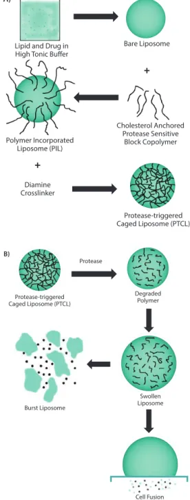
In a similar approach, DOPE functionalized with PEG using a protease responsive linker (e.g., GPLGIAGQ) was blended with disteraoyl phosphatidylcholine (DSPC), cholesteryl chloroformate, and cholesten-5-yloxy-N-(4-((1-imino-2-,β-D-thiogalactosyl ethyl)amino)butyl) formamide (Gal-C4-Chol) to make liposomes with galactose on the surface for targeting hepatic cells. These galactose moieties are shielded by bulky PEG groups, which limit their uptake. However, in the presence of MMP-2, the peptide that links PEG to the liposomal surface is cleaved to expose the targeting ligand, facilitating their rapid uptake.14 In a more recent effort, a cholesterol-anchored graft
1,2-dioleoyl-sn-glycero-3-phosphocholine (DOPC, Sigma Prod. No. P6354), 1,2-dipalmitoyl-sn-glycero-3-phosphocholine (DPPC, Sigma Prod. No. P0763), and cholesterol (47.5:5:47.5 respectively) (Figure 2A). These liposomes with crosslinked polymers showed improved stability with resistance to osmotic swelling or leaking. In the presence of the enzyme, the crosslinking rapidly degraded; this caused drug release through swelling of the liposome (Figure 2B).15 Though the clinical
application of these nanovectors is yet to be established, preclinical studies indicate that these systems hold tremendous promise in augmenting therapeutic efficacy of drugs that otherwise show poor bioavailability.
A)
Lipid and Drug in High Tonic Buffer
Bare Liposome Polymer Incorporated Liposome (PIL) Cholesterol Anchored Protease Sensitive Block Copolymer Diamine Crosslinker Protease-triggered Caged Liposome (PTCL)
+
+
Protease-triggered Caged Liposome (PTCL) Degraded Polymer Protease Burst Liposome Swollen Liposome Cell Fusion B)Figure 2. A) Scheme showing the multiple steps involved in the synthesis of uPA-sensitive, polymer-caged liposomes. B) Proposed mechanism of action of the polymer-caged liposomes
Externally Regulated Systems
Externally regulated systems, also referred to as open-loop systems, are vectors whose drug delivery capability can be governed by a stimulus from outside the body. Since the duration and strength of the external stimulus can be precisely controlled, the drug release profile of these nanovectors can be temporally and spatially controlled to achieve an on-demand supply of the drug at a desired dose. Heat, light, sound, magnetic, and electrical stimuli have all been explored and are discussed below.
Thermo-responsive Systems
Thermo-responsive materials undergo a phase change below or above a particular temperature. These changes are referred to as lower or upper critical solution temperature (LCST or UCST), respectively. Such materials are insoluble above or below the critical temperature but transform to a completely soluble form upon crossing the transition temperature. Poly(N-isopropyl acrylamide) (PNIPAAm) is one such polymeric material that is not ideal for drug delivery applications. PNIPAAm becomes hydrophobic at 32 °C in water and at temperatures that are similar to physiological conditions. However, altering the side chains, the molecular weight of the polymer, the polymeric architecture, or copolymerizing with other hydrophilic or hydrophobic polymers enables customization of the transition temperature to suit biomedical applications. PNIPAAm derivative, therefore, have been explored extensively as a material for thermo-sensitive drug delivery vectors and have been incorporated into micelles, liposomes, hydrogels and nanogels, polymersomes, interpenetrating networks, films, and the surface of inorganic nanoparticles.16 Copolymers of PNIPAAm with
poly(N,N-diethylacrylamide) (PDEAAm), poly(N-vinylcaprolactone) (PVCL), PLGA, poly[2-(dimethylamino) ethyl methacrylate] (PDMAEMA), PEG, gelatin, and chitosan have been used for the delivery of chemotherapeutic drugs and biologics. Beside drug delivery, considerable efforts have been made to develop thermo-sensitive materials for use in other applications including surfaces and scaffolds for tissue growth and engineering, imaging, and diagnostics.17
Light-responsive Systems
Light is a popular choice as an external stimulus since its intensity and penetration depth can be precisely controlled. As a result, light-sensitive materials have become increasingly popular as drug delivery systems. Azobenzene, o-nitrobenzene, coumarin, and pyrene derivatives are routinely used for devising light-responsive drug delivery vectors. Jiang et al. developed a UV-responsive micelle system with poly(ethylene oxide) (PEO) and poly(methacrylate) which included pyrene sidechains as diblock copolymers. The authors used Nile red dye (Sigma Prod. No. 19123) as a surrogate to demonstrate the efficient release of the payload as a function of irradiation time and illumination power.18
UV-irradiation, however, is not conducive for biological applications, especially for prolonged periods. For this reason, alternative materials responsive to visible or NIR wavelengths are being explored. Inorganic nanomaterials, especially anisotropic noble metal nanoparticles, show excellent NIR absorption characteristics and have been used as light-absorbing materials to facilitate drug delivery. Kang et al. used silica-coated gold nanorods with strong absorption maxima around 850 nm as a template to polymerize crosslinked acrylamide on the surface of the nanoparticle. Doxorubicin (DOX) loaded particles showed enhanced cell cytotoxicity upon irradiation with NIR light (808 nm) but the nanoparticles that had not been irradiated or irradiation in the absence of nanoparticles
showed little effect. These results confirmed light-mediated payload release.18 In a similar approach, Hribar, et al. developed a polymer-gold
nanorod composite that allowed a controlled and precise release of small molecules (<800 Da) upon irradiation with NIR-light. The PbAE macromers (A6), tert-butyl acrylate (tBA) monomers, and 2-hydroxyethyl acrylate (10:20:70% w/w, respectively) were polymerized to form the polymer blend. A high concentration of gold nanorods and light-responsive material could then be loaded in the core of the polymer microparticles (Figure 3A–4C). In vitro release of loaded DOX demonstrated a strong dependence on the light irradiation (Laser ON) (Figure 3D) and generated increased toxicity in T6–17 cells due to light-induced drug release (Figure 3E).19 A) B) C) E) +NR +DOX +NR –DOX Brightfield Fluorescence Time (hours) Ce ll Ac tivity (% ) 0 50 100 150 200 250 300 3.5 3 2.5 2 1.5 1 0.5 0 120 100 80 60 40 20 0 +DOX –DOX MS–LASER MS+LASER (1 cycle) MS+LASER (3 cycles) ON ON ON ON ON ON
OFF OFF OFF OFF OFF OFF
LASER
D)
Cu
mulativ
e Release (µg)
Figure 3. A) Environmental scanning electron micrograph of polymeric microspheres (MS) made of A6:HEA:tBA (10:20:70). The scale bar corresponds to 50 µm. B) Backscattered micrograph of the microsphere showing the embedded gold nanorods (bright spots, scale bar = 20 µm) and a magnified image of the highlighted area showing the nanorods within the microsphere (scale bar = 200 nm). C) Bright field and florescence images of the microsphere showing the microspheres. The DOX loaded microspheres show intense fluorescence (top panel, right) while those without DOX do not show any background fluorescence (bottom panel, right). Scale bar in all images corresponds to 100 µm. D) Cumulative drug release from the microspheres as a function of laser pulse (Wavelength = 808 nm) applied at physiological temperature (37 °C). E) Histogram plot of T6–17 cells activity after exposure to MS alone with laser and DOX-loaded MS with a laser pulse of 1 and 3 cycles (Laser power = 1.1 W, 5 min/cycle). Reprinted with permission from Reference 19, American Chemical Society 2011.
Ultrasound-responsive Systems
The intensity of ultrasound energy such as light can be tailored for its intensity and focused over a small area in the body. This maximizes the drug release efficiency and, therefore, is often referred as high intensity focused ultrasound (HIFU). Due to its wide application in clinical imaging and diagnosis, ultrasound-based on-demand drug releasing vectors are seen as a natural material for the development of “theranostic” delivery systems. Ultrasound energy mediates drug release from a delivery vector by three main mechanisms: (1) heat generation, (2) acoustic cavitation, and (3) acoustic radiation forces. Some of the important parameters which can influence the performance of ultrasound-based techniques include the time and nature of application. The rationale behind ultrasound-based delivery is largely regulated by heat generation; therefore, the nanoparticle delivery systems designed for ultrasound-based delivery are similar in composition to thermo-responsive systems. Microbubble technology, initially developed for contrast enhancement in ultrasound imaging, has since been exploited for acoustic cavitation to alter cell permeability and has also been used as a delivery vehicle.20
Ibsen, et al. prepared nested liposomes synthesized by a method in which a microbubble was created within the liposome to impart ultrasound-responsiveness and demonstrated that it could be used for delivery of both small and large molecules. Similarly, mRNA-lipoplex loaded microbubbles were employed as a vaccine to successfully transfect and express the reporter gene in dendritic cells. This led to a slight shift in maturation and, in turn, induced T-cell response.21 As a result,
ultrasonic-assisted delivery is an attractive approach with significant potential.
Magnetically Regulated Systems
Like thermo- and ultrasound-responsive vectors, delivery systems responsive to magnetic fields rely on the induction of hyperthermia to release the payload. The ability to direct and concentrate these vectors in a specific area of the body through the application of an external magnetic field gives an added advantage to such delivery systems. Traditionally, magnetic nanoparticles such as superparamagnetic iron oxide nanoparticles (SPIONs) are incorporated into polymeric, lipidic, or protein delivery systems to impart them with magnetic properties. SPIONs have also been extensively used as magnetic resonance imaging (MRI) contrast agents, and their presence in a delivery vector offers an imaging modality that possesses stimuli-responsiveness. SPIONs coated with the thermo-responsive polymer PNIPAAm and loaded with DOX showed a rapid drug release above the lower critical solution temperature (LCST) due to magnetic field induced hyperthermia but a slow sustained release below the LCST. In vivo studies in buffalo rats implanted with hepatocellular carcinoma revealed magnetically guided increased accumulation and drug release in tumors. This resulted in an improved contrast-based MR imaging and efficient therapeutic potential.22
Majewski, et al. demonstrated the utility of the magnetically active vectors as a gene delivery system. Most importantly, the study demonstrated that internalized magnetic nanoparticles can be used for selective isolation of the transfected cells from the population.23 Due to the added versatility
of the magnetically regulated delivery system, they are now often used to design dual and multiple stimuli-responsive vectors to harvest the benefits of individual stimuli-responsiveness.24
Electro-responsive Systems
Certain materials (organic and inorganic) exhibit conductive properties and can be used to design delivery systems that are responsive to an external electric field. Common examples of electro-sensitive materials for drug delivery include polypyrrole (PPy, Aldrich Prod. No. 577030), ferrocene, and carbon nanotubes. Weak electric pulses (~1 V) are generally used for such applications and these electric fields are preferred over other externally applied stimuli due to several advantages: an electric pulse is (1) easy to control and apply, (2) does not need sophisticated and elaborate instrumentation, and (3) can be easily integrated to design chip-based devices. Ge, et al. prepared dodecyltrimethylammonium bromide (DTAB) micelles with decyl alcohol as a cosurfactant, and PPy was polymerized in the hydrophobic core. These nanoparticles were then loaded with a thermo-sensitive PLGA-PEG-PLGA block polymer that showed temperature-dependent sol-gel transformation. The polymer exists as a solution at 4 °C but rapidly forms a gel at a physiological temperature of 37 °C. Daunorubicin and fluorescein were loaded into the nanoparticles that were embedded in the polymer matrix. The resulting material maintained its solid hydrogel form at body temperature and demonstrated an electric-pulse dependent drug release. The subcutaneously injected, soluble form rapidly forms a gel in FVB mice and successfully released the payload in vivo upon application of the electric
pulse. In the control group, the absence of an external stimulus resulted in insignificant release of the cargo.25 Adopting a novel approach, Zhu,
et al. grafted 4-(3-cyanophenyl) butylene (CPB) as an electric field-active
“nanoimpeller” onto the wall of mesoporous silica. Due to their large inherent dipole moment, the grafted CPBs reorient under the influence of an applied electric field. The CPBs then rapidly release the guest molecules (ibuprofen) from their pores.26
Future Perspectives
The plethora of available literature on stimuli-responsive delivery systems demonstrates the growing importance for these systems. However, a majority of these systems have not made it past the pre-clinical stage and only a handful of examples currently have entered clinical trials.27 The
need for a precise control over the “response” to the applied “stimulus” makes their clinical translation challenging. The complex synthetic steps and formulation of multiple components further compounds the issue. The majority of stimuli-responsive delivery systems are still in the early stages of development and the optimization of the synthesis procedures is needed before they can transition into the clinical world. Accuracy and precision over the stimulus will also need improvement from preclinical to the clinical level. Externally applied “physical” stimuli are easy to control and manipulate but internal “biological” triggers are not as easily controlled. Tumors show considerable variation in their physiological status between patients, organs, or even the same tumors in different species. External stimuli, on the other hand, need improvement to achieve better tissue penetration without causing any damage, which would require the optimization of several contributing parameters. Other factors can also have a negative impact on delivery systems for the stimuli-responsive vectors. The EPR effect that leads to accumulation of the delivery vectors into the tumor is a well-accepted phenomenon in preclinical studies but has not yet been confirmed in clinical settings. Additionally, a majority of diseases show a complex microenvironment consisting of diseased cells, tissue interstitium, immune cells and other structural cells of the tissue that limits the ability of the delivery system to access the desired target cells. These physical and physiological barriers further impede the optimal performance of these delivery systems. In the case of cancer, heterogeneity at the cellular and physiological levels significantly limits the ability of these vectors to access their targets. A majority of the stimuli-responsive systems discussed have been tested
in vitro but few have in vivo applications, an aspect that needs immediate
focus. The goal is to design simplified systems with positive stimuli-responsive characteristics. This achievement will drastically improve the chances for clinical applications. Though several major hurdles are yet to be overcome, stimuli-responsive systems have ultimately shown tremendous promise as alternatives to the existing delivery approaches.
References
(1) Iyer, A.K.; Singh, A.; Ganta, S.; Amiji, M.M. Role of integrated cancer nanomedicine in over-coming drug resistance. Advanced drug delivery reviews 2013, 65, (13–14), 1784–802. (2) Meacham, C.E.; Morrison, S.J. Tumour heterogeneity and cancer cell plasticity. Nature 2013,
501, (7467), 328–37.
(3) Ganta, S.; Devalapally, H.; Shahiwala, A.; Amiji, M. A review of stimuli-responsive nanocarriers for drug and gene delivery. Journal of controlled release 2008, 126, (3), 187–204.
(4) Talekar, M.; Boreddy, S.R.; Singh, A.; Amiji, M. Tumor aerobic glycolysis: new insights into therapeutic strategies with targeted delivery. Expert opinion on biological therapy 2014. (5) Devalapally, H.; Shenoy, D.; Little, S.; Langer, R.; Amiji, M. Poly(ethylene oxide)-modified
poly(beta-amino ester) nanoparticles as a pH-sensitive system for tumor-targeted delivery of hydrophobic drugs: part 3. Therapeutic efficacy and safety studies in ovarian cancer xenograft model. Cancer chemotherapy and pharmacology 2007, 59, (4), 477–84.