Sputum biomarkers in IPF: Evidence for raised
gene expression and protein level of IGFBP-2,
IL-8 and MMP-7
J. Guiot*, M. Henket, J. L. Corhay, C. Moermans, R. Louis
Pneumology Department, CHU Liège, Liège, Belgium, CHU Liège, Pneumology Department, Domaine universitaire du Sart-Tilman, Liège, Belgium
*J.Guiot@chu.ulg.ac.be
Abstract
Background
Idiopathic pulmonary fibrosis (IPF) is a rare lung disease of unknown origin leading rapidly to death. This paper addresses the issue of whether sputum induction is a suitable tool to study respiratory tract inflammation and potential biomarkers in IPF compared to COPD, a fibrosing airway wall disease.
Methods
In a cross-sectional analysis, 15 IPF patients, 32 COPD and 30 healthy subjects underwent sputum induction. Total sputum cell counts and the amount of TGF-β, IGF-1, IGF-2, IGFBP-1, IGFBP-2, IGFBP-3, IL-8, IL-13, MMP-7, MMP-9, YKL-40, TNF-αand KL-6 in spu-tum supernatant were analysed. We also profiled gene expression of cells in the induced sputum for TGF-β, MMP-7, YKL-40, IGFBP-2, IL-6, IL-8 and TNF-α.
Results
IPF patients, like COPD, had increased sputum absolute number of neutrophils, eosino-phils, macrophages and epithelial cells compared to HS. IPF sputum supernatants had increased concentrations of IGFBP-2, IL-8, TGF-β, MMP-7, MMP-9 and KL-6 (p<0.05, p<0.0001, p<0.05, p<0.05, p<0.0001, p<0.05 respectively) when compared to healthy sub-jects where COPD had higher IL-6 and TNF-αlevels than IPF (p<0.05 and p<0.05 respec-tively) and HS (p<0.0001 and p<0.001 respectively) and higher IL-8 and MMP-9 than HS (p<0.0001 and p<0.001 respectively). Conversely to IL-6 and TNF-α, MMP-7 was increased in IPF compared to COPD (p<0.05). The KL-6 and MMP-7 protein levels in sputum were inversely correlated with total lung capacity (TLC, % of predicted) in IPF patients (r = -0.73 and r = -0.53 respectively). Sputum gene expression analysis identified a significant in-crease for IGFBP-2, IL-6, IL-8 and MMP-7 in IPF compared to HS (p<0.05, p<0.01, p<0.05 and p<0.0001 respectively) and for IGFBP-2, YKL-40, IL-6, IL-8 and MMP-7 compared to COPD (p<0.01, p<0.01, p<0.05, p<0.01 and p<0.0001 respectively). Furthermore, gene expression of TGF-βwas increased in IPF compared to COPD (p<0.001) but not to HS.
a1111111111 a1111111111 a1111111111 a1111111111 a1111111111 OPEN ACCESS
Citation: Guiot J, Henket M, Corhay JL, Moermans
C, Louis R (2017) Sputum biomarkers in IPF: Evidence for raised gene expression and protein level of IGFBP-2, IL-8 and MMP-7. PLoS ONE 12 (2): e0171344. doi:10.1371/journal.pone.0171344
Editor: Rory Edward Morty, University of Giessen
Lung Center, GERMANY
Received: October 29, 2016 Accepted: January 19, 2017 Published: February 8, 2017
Copyright:© 2017 Guiot et al. This is an open access article distributed under the terms of the
Creative Commons Attribution License, which permits unrestricted use, distribution, and reproduction in any medium, provided the original author and source are credited.
Data availability statement: Data contains
information that could be used to identify study participants and is available upon request from the corresponding author at (J.Guiot@chu.ulg.ac.be).
Funding: The authors received no specific funding
for this work.
Competing interests: The authors have declared
that no competing interests exist.
Abbreviations: ATS, American thoracic society;
BAL, Bronchoalveolar lavage; COPD, Chronic obstructive pulmonary disease; DLCO, Diffusion
Conclusion
Our data show clear increase in expression and production of IGFBP-2, IL-8 and MMP-7 in sputum from patients with IPF that may contribute to the disease.
Introduction
Idiopathic pulmonary fibrosis (IPF) is a rare lung disease of unknown origin leading rapidly to death [1]. The diagnostic approach is complex and requires a multidisciplinary discussion [1]. Although bronchoalveolar lavage (BAL) is an important tool for evaluating interstitial lung diseases (ILDs), induced sputum, which has demonstrated clinical interest in airway diseases [2], has been proposed as a less invasive alternative [2–4]. The research with sputum in ILDs has mainly focused on the cellularity so far and there has been few studies that have investi-gated the molecular inflammatory pathways in IPF using sputum analysis [5].
Many biomarkers have already been studied both in serum and BAL from ILDs and in par-ticular in IPF where inflammation seems to take part of the disease [6] in addition to the remod-elling process [7]. Among those we can identify surfactant protein A or SP-A, surfactant protein D or SP-D which has recently be identified to be correlated with pulmonary function and mor-tality in IPF [8], the Krebs von den Lungen 6 or KL-6, type A immunoglobulin or IgA, periostin [9–13], insulin-like growth factor binding protein or IGFBP-2 [14] and a chitinase-3-like-1 human cartilage glycoprotein or YKL-40 [15–16] which were found to be increased in serum from IPF patients. As for BAL, matrix metalloproteinase-7 (MMP-7) [17] and YKL-40 [15] were the main markers found to be raised in IPF compared to healthy subjects.
The aim of our study was to evaluate the potential of measuring biomarkers of the fibrosing process in sputum from patients with IPF. As comparators we have recruited a group of COPD, recognized to be an airway disease featuring fibrosis of the airway wall at the periphery of the lung [18].
Materials and methods
We analysed the induced sputum of patients with IPF (n = 15) in comparison to healthy sub-jects (n = 30) and COPD (n = 32). The diagnosis of IPF was made according to the interna-tional recommendations of the ATS [1] using the respiratory function, HRCT scan, BAL (when available), as well as the clinical history of the patient. We excluded all other causes of ILD (such as asbestosis, hypersensitivity pneumonitis, pneumopathy associated with connec-tive tissue diseases or toxic pneumonitis). All cases were discussed in our multidisciplinary dis-cussion team about interstitial lung diseases composed of: a pulmonologist, a specialist in pulmonary rehabilitation, a rheumatologist, a radiologist, a pathologist, a specialist in occupa-tional medicine. COPD were diagnosed according to the GOLD [19] and healthy subjects were recruited by advertisement in ambulatory care waiting room of the hospital. They all denied any chronic respiratory disease and had FEV1 and FVC values above 80% predicted.
The protocol was approved by the ethics committee of CHU Liège, and all subjects gave written consent before their enrolment (Belgian number: B707201422832; ref: 2014/302).
Sputum induction and processing
After premedication with 400 micrograms inhaled salbutamol, sputum was induced by inhala-tion of hypertonic (NaCl 5%) or isotonic (NaCl 0.9%) saline according to the FEV1 value (> or < than 65% predicted). Saline was combined with additional salbutamol delivered by an
capacity of CO in the lung; FEV1, Forced expiratory volume in one second; FVC, Forced vital capacity; HRCT, High resolution computed tomography; IgA, Type A immunoglobulin; IGF, Insulin-like growth factor; IGFBP, Insulin-like growth factor binding protein; IL-6, Interleukine-6; IL-8, Interleukine-8; ILD, Interstitial lung disease; IPF, Idiopathic pulmonary fibrosis; KL-6, Krebs von den Lungen-6; MMP, Matrix metalloproteinase; SD, Standard deviation; SP-A, Surfactant protein-A; SP-D, Surfactant protein-D; TGF-β, Transforming growth factor beta; TLC, Total lung capacity; YKL-40, Chitinase-3-like-1, human cartilage glycoprotein-39.
ultrasonic nebulizer (Ultra-Neb 2000; Devilbiss, Somerset, PA, USA) with an output set at 0.9 ml/min. Each subject inhaled the aerosol for three consecutive periods of 5 min for a total of 15 min. For safety reasons, FEV1 was monitored throughout the induction and it was stopped if FEV1 fell by more than 20% from baseline. The whole sputum was collected in a plastic con-tainer, weighed, and homogenized by adding three volumes of phosphate-buffered saline (PBS), vortexed for 30 s, and centrifuged at 800g for 10 min at 4˚C. Supernatant was separated from cell pellet and stored at− 80˚C. The cells were resuspended in a solution containing 5mM dithiothreitol without Ca++and Mg++and gently rocked for 20 min at room tempera-ture. The cell suspension was then centrifuged again at 400g at 4˚C for 10 min. Squamous cells, total cell counts and cell viability checked by trypan blue exclusion were performed with a manual hemocytometer [20]. The differential leukocyte count was obtained using a cytospin stained with Rapi Diff II (Atom Scientific, Manchester UK) on 500 non squamous cells.
Biomarkers measurements in the induced sputum
We analysed several biomarkers assumed to be critical growth factors or chemokines in the induced sputum. The concentration of IL-6, IL-13, TNF-α, MMP-7, YKL-40 were assessed by ELISA multiplex using Fluorokine1 Multianalyte Profiling Kits (R and D Systems, Minneap-olis, MN, USA) according to the manufacturer’s instructions. The detection limit for this assays were 7-25-7-39-61 pg/ml respectively The concentration of the other proteins were measured separately by ELISA: TGF-β, MMP-9, IL-8, IGF-1, IGFBP-1, IGFBP-2 and IGFBP-3 (DuoSet kit, R and D systems); IGF-2 (Mediagnost, Reutlingen, Germany); KL-6 (Lumipulse G KL-6 Fujirebio Europe). The detection limits for these kits were 7-25-25-32-30-125-130-450pg/ml and 100 U/ml respectively.
Gene expression in the induced sputum
PCR was used to profile gene expression of cells in the induced sputum for TGF–β, MMP-7, YKL-40, IGFBP-2, IL-6, IL-8 and TNF-α in a subgroup of patients (HS n = 14; COPD n = 15, IPF n = 12). mRNA expression was measured in total sputum cells by Taqman reverse tran-scription quantitative polymerase chain reaction (RT-qPCR). RNA extraction, and RT-qPCR were performed according to description of da Silva et al [21]. The house keeping gene rRNA 18S was used as the reference gene and the fold change was calculated from the median of healthy subject group. Sequences of primers and probes used are listed inTable 1. All Probes were labeled with reporter and double-quencher dyes 506-carboxyfluorescein/ZEN/30Iowa Black FQ (506-FAM/ZEN/30IBFQ). Probes and primers were synthesized by IDT (Integrated
DNA Technologies, Inc., Coralville, IA).
Primers and probes for 18S rRNA, IL-6 and IL-8 were designed by Gielen and al. [22]. The sequences are listed inTable 1. PrimeTime1qPCR Assays provide by IDT (Integrated DNA Technologies, Inc., Coralville, IA) were used for IGFBP-2, MMP-7, TNF-α, TGF-β1 and YKL-40. We only analysed samples with more than 50% of viability with less than 30% squamous cells. We chose a cut-off of 20 CT for the 18S and excluded samples in which more than 20 CT was needed to pick up 18S.
Pulmonary function tests
Lung function tests were performed in our routine respiratory laboratory at CHU Liège. All spirometric tests performed for this study were measured using the pneumotachograph JaegerMasterlab system (Erich Jaeger GmbH, Wuzburg, Germany). The expiratory volume in one second (FEV) and forced vital capacity (FVC) were measured in accordance with the rec-ommendations of the European Respiratory Society (ERS)[23]. The results are expressed in ml
and as percentage of predicted normal values. The Tiffeneau index or FEV / FVC is expressed as percentage. The diffusion capacity of CO (DLCO) and the ratio DLCO / VA were measured by the single breath testing technique (Sensor Medics 2400 He / CO Analyzer System, Biltho-ven, Netherlands).
Statistical analysis
Demographic and functional data were expressed as mean± standard deviation (SD). Induced sputum cellularity was expressed as median (minimum-maximum) because of a not normal distribution. When the data showed normal distribution, they were compared with a one-way ANOVA, followed by Tukey-Kramer’s post-hoc testing. When the data did not show a normal distribution, they were compared by Kruskall-Wallis followed by Mann-Whitney testing. Cor-relations between variables were assessed using Spearman’s rank correlation test. A p<0.05 was considered as significant.
Results
Subject demographic and functional characteristics
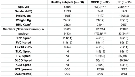
The demographic, functional and treatment characteristics of the subjects are given inTable 2. Both disease groups were slightly older than healthy subjects. As expected both IPF and COPD
Table 1. Sequence of primers and probes.
Gene name Accession number sequence
IGFBP-2 NM 000597.2 Forward TTC ACA CAC CAG CAC TCC
reverse ACC TCT ACT CCC TGC ACA T
Probe AGC ATG GCC TGT ACA ACC TCA AAC A
TGF-β NM 000660.6 Forward GTT CAG GTA CCG CTT CTC G
reverse CCG ACT ACT ACG CCA AGG A
Probe ACC CGC GTG CTA ATG GTG GAA
MMP-7 NM 002423.4 Forward GAA TGT CCC ATA CCC AAA GAA TG
reverse GAT GAG GAT GAA CGC TGG A
Probe CAT ACA GGA AGT TAA TCC CTA GAC TGC TAC CA
IL-6 NM 000600.4 Forward CCA GGA GCC CAG CTA TGA AC
reverse CCC AGG GAG AAG GCA ACT G
Probe CCT TCT CCA CAA GCG CCT TCG GT
IL-8 NM 000584.3 Forward CTG GCC GTG GCT CTC TTG
reverse CCT TGG CAA AAC TGC ACC TT
Probe CAG CCT TCC TGA TTT CTG CAG CTC TGT GT
YKL-40 NM 001276.2 Forward TCT GGG TGT TGG AGG CTA T
reverse GCT CAA CAC ACT CAA GAA CAG
Probe TGT CTG TCG GAG GAT GGA ACT TTG G
TNF-α NM 000594.3 Forward TCA GCT TGA GGG TTT GCT AC
reverse TGC ACT TTG GAG TGA TCG G
Probe AGA TGA TCT GAC TGC CTG GGC C
18s (rRNA) x03205.1 Forward CGC CGC TAG AGG TGA AAT TCT
reverse CAT TCT TGG CAA ATG CTT TCG
Probe ACC GGC GCA AGA CGG ACC AGA
Primers and probes for 18S rRNA, IL-6 and IL-8 were designed by Gielen and al (22).
PrimeTime®qPCR Assays provided by IDT (Integrated DNA Technologies, Inc., Coralville, IA) were used for IGFBP-2, MMP-7, TNF-α, TGF-βand YKL-40. doi:10.1371/journal.pone.0171344.t001
had reduced FEV1 and FVC compared to healthy subjects but only COPD had a reduced FE1/ FVC ratio. Total lung volume (TLC), residual lung volume (VR), lung diffusing capacity (DLCO) and transfert coefficient (KCO) were sharply reduced in the IPF group.
Sputum cellularity
Induced sputum (IS) from IPF patients showed an increased cellular concentration in compar-ison to healthy subjects (Table 3). There was a reduction in the proportion of macrophage and lymphocyte in IPF in comparison to healthy subjects. When expressed in absolute values, neu-trophils, macrophages, eosinophils and epithelial cells were all significantly increased in IPF compared to healthy subjects (Table 3). There was no significant difference between IPF and COPD.
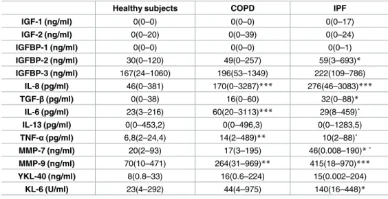
Sputum supernatant biomarkers
Several biomarkers were measured in sputum supernatant (Table 4). There was a significant increase of IGFBP-2, IL-8, TGF-β, MMP-7, MMP-9 and KL-6 (p<0.05, p<0.0001, p<0.05, p<0.05, p<0.0001, p<0.05 respectively) in IPF patients compared to HS. COPD had higher IL-6 and TNFα levels than IPF (p<0.05 and p<0.05 respectively) and HS (p<0.0001 and p<0.001 respectively) and higher IL-8 and MMP-9 than HS (p<0.0001 and p<0.001 respectively). Con-versely to IL-6 and TNF-α, MMP-7 was increased in IPF compared to COPD (p<0.05).
Table 2. Subject characteristics.
Healthy subjects (n = 30) COPD (n = 32) IPF (n = 15)
Age, yrs 55(9) 63(9)*** 72(9)***˚ Gender (M/F) 11/19 24/8 12/3 Height, cm 169(8) 171(9) 170(12) Weight, Kg 72(12) 71(17) 76(13) BMI, Kg/m2 25(3) 24(4) 26(3) Smokers (Never/ex/Current), n 13/8/9 1/17/14 2/10/3 pack-yr 9(13) 47(32)*** 33(24)** FEV1%pred 111(11) 49(16) 69(15) FVC %pred 114(15) 81(14) 69(16) FEV1/FVC % 80(4) 48(10) 76(11) TLC, %pred nd 115(19) 68(14) RV, %pred nd 192(58) 65(49)˚˚˚ DLCO %pred nd 56(14) 36(16)˚ KCO %pred nd 76(20) 59(16) ICS (yes/no) 0/30 12/20 3/12 OCS (yes/no) 0/30 2/30 2/13
Data are expressed as mean (SD).
*p<0.05 compared to healthy subjects.
**p<0.001 compared to healthy subjects.
***p<0.0001 compared to healthy subjects. ˚ p<0.05 compared to COPD.
˚˚ p<0.001 compared to COPD. ˚˚˚ p<0.0001 compared to COPD.
FEV1: forced expiratory volume in one second; FVC: forced vital capacity; TLC: total lung capacity; RV: residual volume; DLCO: diffusion capacity of CO in the lung; ICS: inhaled corticosteroid; OCS: oral corticosteroid.
Table 3. Sputum cell counts.
Healthy subjects COPD IPF
Sputum weight g 3,7(0,8–22,4) 2,2(0,4–11,5)* 2,5(0,8–4,4)* cell x106/g 0,8(0,2–7,6) 2,9(0,4–55,9)*** 3,5(0,4–19,5)** Squamous cells % 18(1–31) 10(0–38)* 7(0–29)* Viability % 78(45,3–94,5) 70(24–100) 73(31–89) Macrophage % 25(4,4–80,5) 9(0–77,5)** 16(0–41,2)* x103/g 203(0–6085,8) 333(0–2275)** 716(0–5891,6)** Lymphocyte % 1,6(0–6,2) 1,5(0–6) 0,6(0–13,2)* x103/g 10 (0–388,6) 39(0–506) 14 (0–1887,6) Neutrophil % 64(6,5–94,6) 66(11–100) 74(44,4–100) x103/g 423(0–5266,2) 1429(129–55900)** 2775(201,6–13912,5)*** Eosinophil % 0,2(0–5,8) 2(0–81)*** 0,5(0–21,4) x103/g 0,4(0–207) 58(0–8455)*** 17(0–4181,6)* Epithelial cell % 2,9(0,5–25,3) 3.4(0–30.4) 1,8(0–16,4) x103/g 23(0–831,6) 94(0–594)* 86(0–469)*
Results are expressed as median (min-max).
*p<0.05 compared to healthy subjects.
**p<0.001 compared to healthy subjects.
***p<0.0001 compared to healthy subjects.
doi:10.1371/journal.pone.0171344.t003
Table 4. Sputum supernatant biomarkers.
Healthy subjects COPD IPF
IGF-1 (ng/ml) 0(0–0) 0(0–0) 0(0–17) IGF-2 (ng/ml) 0(0–20) 0(0–39) 0(0–24) IGFBP-1 (ng/ml) 0(0–0) 0(0–0) 0(0–1) IGFBP-2 (ng/ml) 30(0–120) 49(0–257) 59(3–693)* IGFBP-3 (ng/ml) 167(24–1060) 196(53–1349) 222(109–786) IL-8 (pg/ml) 46(0–381) 170(0–3287)*** 276(46–3083)*** TGF-β(pg/ml) 0(0–38) 16(0–60) 32(0–88)* IL-6 (pg/ml) 23(3–216) 60(20–3113)*** 29(8–459)˚ IL-13 (pg/ml) 0(0–453,2) 0(0–496,3) 0(0–1283,5) TNF-α(pg/ml) 6,8(2–24,4) 14(2–489)** 10(2–88)˚ MMP-7 (ng/ml) 20(2–93) 17(3–195) 46(0.008–190)*˚ MMP-9 (ng/ml) 70(10–471) 264(31–969)** 415(18–970)*** YKL-40 (ng/ml) 8(0.8–33) 16(0.6–224) 15(0.002–204) KL-6 (U/ml) 23(4–292) 44(4–975) 140(16–448)*
Results are expressed as median (min-max).
*p<0.05 compared to healthy subjects.
**p<0.001 compared to healthy subjects.
***p<0.0001 compared to healthy subjects. ˚ p<0.05 compared to COPD.
˚˚ p<0.001 compared to COPD. ˚˚˚ p<0.0001 compared to COPD.
Sputum cell gene expression
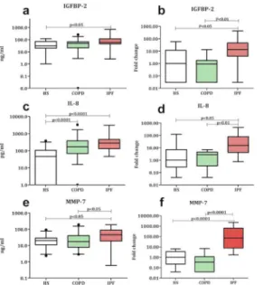
Gene expression analysis identified a significant increase for IGFBP-2, IL-6, IL-8 and MMP-7 in IPF compared to HS (p<0.05, p<0.01, p<0.05 and p<0.0001 respectively) and for IGFBP-2, YKL-40, IL-6, IL-8 and MMP-7 compared to COPD (p<0.01, p<0.01, p<0.05, p<0.01 and p<0.0001 respectively). (Fig 1)(Table 5). Furthermore, gene expression of TGF-β was also
increased in IPF compared to COPD (p<0.001).
Biomarkers and impaired lung function
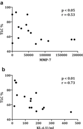
In IPF, there was an inverse correlation between sputum supernatant protein levels of MMP-7 and KL-6 and % predicted TLC (r = -0.53, r = -0.73 respectively) (Fig 2). Diffusion lung capac-ity was not significantly related to any of the biomarkers.
Fig 1. Protein and gene levels of IGFBP-2, IL-8 and MMP-7 in sputum of IPF patients. a) IGFBP-2 level in
supernatant showing a significant increase in IPF compared to HS. b) Sputum cell gene expression of IGFBP-2 showing a significant increase in IPF patients compared to COPD and HS. c) IL-8 level in supernatant showing a significant increase in COPD and IPF compared to HS. d) Sputum cell gene expression of IL-8 showing a significant increase in IPF compared to COPD and HS. e) MMP-7 level in supernatant showing a significant increase in COPD and IPF compared to HS. f) Sputum cell gene expression of MMP-7 showing a significant increase in IPF compared to COPD and HS.
doi:10.1371/journal.pone.0171344.g001
Table 5. Sputum cell gene expression.
COPD IPF HS vs IPF HS vs COPD COPD vs IPF
fold change fold change p value p value p value
IGFBP-2 0.91 13.70 0,0385 ns 0,0011 IL-6 4.67 24.88 0,0042 ns 0,0478 IL-8 2.78 15.53 0,0193 ns 0,0037 MMP-7 0.35 70.43 <0,0001 ns <0,0001 TGF-β 0.62 5.02 ns ns 0,0005 TNF-α 1.15 2.53 ns ns ns YKL-40 0.5 5.65 ns ns 0,0011
Data are presented as fold change relative expression to healthy subjects group. COPD: chronic obstructive pulmonary disease (n = 15); IPF: idiopathic pulmonary fibrosis (n = 12), HS: healthy subjects (n = 14).
Relationship between age, gender and biomarkers
Our groups were not fully matched with respect to age, sex and gender and smoking status. Therefore we performed a specific analysis in the HS group in which we didn’t find any corre-lation between biomarkers and age, gender and smoking status (data not shown).
Discussion
Here we report for the first time concomitant measurement of gene expression and protein levels of growth factors and matrix metalloproteinases in sputum from patients with IPF. We confirmed that TGF-β, IL-8, KL-6 and MMP-7, known to be biomarkers in IPF based on serum and BAL analysis, are also significantly higher in induced sputum from IPF patients. Furthermore IGFBP-2, recently identified as a new protein involved in IPF [14], was also sig-nificantly increased in sputum from IPF both at the gene and the protein level.
Sputum cell count of patients with IPF revealed an intense inflammatory pattern with raised total cell counts including granulocytes, macrophages and epithelial cells confirming previous finding of Beeh et al. [24]. This increased number of cells had already been described in BAL of IPF patients [25] but with a different proportion of cells, the granulocytic component being more prominent in sputum than in BAL at the expense of the lymphocyte and the macrophage fraction [2,26]. Interestingly, in our study, IPF patients did not differ from COPD with respect to sputum cell counts. This would indicate that it is difficult to identify the main localisation of the pathological process (lung versus airways) based on the sole sputum cell count.
Fig 2. Correlation between MMP-7 and KL-6 and TLC (% pred) in IPF. a) Correlation between level of
MMP-7 (ELISA) in supernatant of IPF patients and total lung capacity (TLC)(%predicted). b) Correlation between level of KL-6 (ELISA) in supernatant of IPF and TLC (%predicted).
TGF-β, widely known as a key actor of fibrosis and known to be elevated in BAL of IPF patients [27], was increased in sputum supernatant of IPF compared to healthy subjects. More-over gene expression of TGF-β was significantly raised in IPF compared to COPD, another disease featuring remodelling, though in the latter it mainly affects the airway compartment.
Our study also confirmed the importance of MMP-7, a matrix metalloproteinase specifi-cally found to be raised both at gene and protein level in sputum from IPF compared to COPD and HS. While MMP-9 was increased in both IPF and COPD, it is remarkable that the raised expression and protein amount of MMP-7 was limited to patients with IPF compared to HS. Our results are in keeping with those of Zuo et al. who reported a greater MMP-7 gene expres-sion in lung tissues of IPF patients [28] and with those of Huh et al. who reported an increased level of MMP-7 in BAL of IPF [29]. Supporting the role of MMP-7 in disease progression [17,
30], we interestingly found a negative correlation between MMP-7 levels and TLC (%pred), a physiological marker of pulmonary restriction. As our study is cross sectional it does not dem-onstrate that sputum MMP-7 would associate with a loss of lung volume over time but this would require a longitudinal study to confirm.
We found a significant and specific increase in KL-6 in sputum supernatant from IPF. As with MMP-7 there was also a negative correlation between TLC (%pred) and sputum protein level of KL-6. KL-6 is well known as a biomarker in pulmonary fibrosis [9,31] and is present on the surface membrane of alveolar epithelial cells (AEC-II) and bronchiolar epithelial cells. Serum KL-6 has been previously described as predictive of acute exacerbation of IPF [9] but is not recognized as a marker of disease severity by itself. The fact that sputum levels of KL-6 relates to the loss of lung volume in our study suggests that sputum may be a more appropriate compartment than serum to evaluate the epithelial and alveolar damage occurring in IPF.
We have recently shown that serum IGFBP-2 was raised in IPF [14]. Here we extend our finding by showing that this protein was also specifically raised in sputum. The augmentation was seen both at the gene and the protein level making it very consistent. This is an original finding as the protein had not been measured in the airway/lung compartment of patients suf-fering from IPF before. Of interest is that gene expression of IGFBP-2 in IPF was not only increased compared to healthy subjects but also compared to COPD. There is however a previ-ous report of increased IGFBP-2 in BAL of children with diverse interstitial lung diseases [32]. Therefore we cannot claim that IGFBP-2, though being a useful biomarker and relevant to pathophysiology [14], is specific of IPF.
Gene expression of IL-6 in IPF was increased compared to both healthy subjects and COPD though the protein level remained comparable to that measured in healthy subjects. In IPF IL-6 is pro-mitogenic for fibroblasts due to the sustained activation of MAP Kinases in contrast to what is seen normal fibroblast [33]. In order to explain that, as opposed to gene expression, protein level was not increased, we cannot rule out the possibility of a local consumption of IL-6 that participates to the fibrosing process. Conversely the high protein level of IL-IL-6 in sputum supernatant from COPD contrasting with poor gene expression may be seen as a consequence of plasma exudation into the airways and is in keeping with our previous observation that spu-tum cells from COPD produced less IL-6 than those of HS [34]. Confirming previous studies [26], IL-8 was significantly increased in IPF compared to healthy subjects both at gene and at protein level while, as for IL-6, the increase in COPD was only seen at the protein level. IL-8 is known to be a strong chemotactic agent for neutrophils [35,36] and could explain the greater number of neutrophils infiltrating the airways in IPF and COPD.
As aforementioned, one of the potential limitation of the study is the cross sectional design that precludes any interpretation of the predictive values of these biomarkers in the follow-up of the patients. A further limitation is that we were unable to assess the expression and the lev-els of the growth factors in patients receiving anti-fibrotic therapy. Finally the lack of perfect
matching between our groups regarding age, gender and tobacco may be seen as a potential confounding factor. However we have to say that it is extremely difficult to recruit a large number of healthy subjects above the age of 65 yrs. To address this issue of potential confound-ing factors we have assessed the relationship between age, gender and tobacco smokconfound-ing and protein and gene expression of biomarkers in the group of healthy subjects and we did not find any significant correlation.
Conclusion
Our data show clear increase in expression and production of several growth factors and matrix metalloproteinases and chemokine in sputum from patients with IPF. Whether sputum analysis may become a suitable and less invasive tool than BAL to predict and monitor evolu-tion of the disease and the response to treatment should be investigated in prospective longitu-dinal trials.
Acknowledgments
Thank you to all co-authors for their contribution
Author contributions
Conceptualization: JG RL JLC CM. Data curation: JG MH. Formal analysis: JG. Investigation: JG. Methodology: JG JLC RL. Project administration: MH. Supervision: JG. Validation: JG RL. Visualization: JG RL. Writing – original draft: JG.Writing – review & editing: JG RL JLC CM.
References
1. Ganesh Raghu HRC, Egan Jim J., Martinez Fernando J., Behr Juergen, Brown Kevin K., Colby Thomas V., et al. An Official ATS/ERS/JRS/ALAT Statement: Idiopathic Pulmonary Fibrosis: Evidence-based Guidelines for Diagnosis and Management. American Journal of Respiratory and Critical Care Medi-cine. 2011; 183(6):788–824. doi:10.1164/rccm.2009-040GLPMID:21471066
2. Antoniou KM, Alexandrakis M, Tzanakis N, Tsiligianni I, Tzortzaki EG, Siafakas NM, et al. Induced spu-tum versus bronchoalveolar lavage fluid in the evaluation of patients with idiopathic pulmonary fibrosis. Respiration; international review of thoracic diseases. 2005; 72(1):32–8. doi:10.1159/000083398
PMID:15753632
3. Fireman E, Topilsky I, Greif J, Lerman Y, Schwarz Y, Man A, et al. Induced sputum compared to bronchoalveolar lavage for evaluating patients with sarcoidosis and non-granulomatous interstitial lung disease. Respiratory medicine. 1999; 93(11):827–34. PMID:10603633
4. Fireman E, Lerman Y. Induced sputum in interstitial lung diseases. Current opinion in pulmonary medi-cine. 2006; 12(5):318–22. doi:10.1097/01.mcp.0000239547.62949.82PMID:16926645
5. Antoniou KM, Tzortzaki EG, Alexandrakis MG, Zervou M, Tzanakis N, Sfiridaki K, et al. Investigation of IL-18 and IL-12 in induced sputum of patients with IPF before and after treatment with interferon gamma-1b. Sarcoidosis, vasculitis, and diffuse lung diseases: official journal of WASOG / World Associ-ation of Sarcoidosis and Other Granulomatous Disorders. 2005; 22(3):204–9.
6. Balestro E, Calabrese F, Turato G, Lunardi F, Bazzan E, Marulli G, et al. Immune Inflammation and Dis-ease Progression in Idiopathic Pulmonary Fibrosis. PloS one. 2016; 11(5):e0154516. doi:10.1371/ journal.pone.0154516PMID:27159038
7. Vuga LJ, Milosevic J, Pandit K, Ben-Yehudah A, Chu Y, Richards T, et al. Cartilage oligomeric matrix protein in idiopathic pulmonary fibrosis. PloS one. 2013; 8(12):e83120. doi:10.1371/journal.pone. 0083120PMID:24376648
8. Doubkova M, Karpisek M, Mazoch J, Skrickova J, Doubek M. Prognostic significance of surfactant pro-tein A, surfactant propro-tein D, Clara cell propro-tein 16, S100 propro-tein, trefoil factor 3, and prostatic secretory protein 94 in idiopathic pulmonary fibrosis, sarcoidosis, and chronic pulmonary obstructive disease. Sar-coidosis, vasculitis, and diffuse lung diseases: official journal of WASOG / World Association of Sarcoid-osis and Other Granulomatous Disorders. 2016; 33(3):224–34.
9. Ohshimo S, Ishikawa N, Horimasu Y, Hattori N, Hirohashi N, Tanigawa K, et al. Baseline KL-6 predicts increased risk for acute exacerbation of idiopathic pulmonary fibrosis. Respiratory medicine. 2014; 108 (7):1031–9. doi:10.1016/j.rmed.2014.04.009PMID:24835074
10. Samukawa T, Hamada T, Uto H, Yanagi M, Tsukuya G, Nosaki T, et al. The elevation of serum napsin A in idiopathic pulmonary fibrosis, compared with KL-6, surfactant protein-A and surfactant protein-D. BMC pulmonary medicine. 2012; 12:55. doi:10.1186/1471-2466-12-55PMID:22963039
11. Ten Klooster L, van Moorsel CH, Kwakkel-van Erp JM, van Velzen-Blad H, Grutters JC. IgA in serum: An old acquaintance as a new prognostic biomarker in Idiopathic Pulmonary Fibrosis. Clinical and experimental immunology. 2015.
12. Tajiri M, Okamoto M, Fujimoto K, Johkoh T, Ono J, Tominaga M, et al. Serum level of periostin can pre-dict long-term outcome of idiopathic pulmonary fibrosis. Respiratory investigation. 2015; 53(2):73–81. doi:10.1016/j.resinv.2014.12.003PMID:25745852
13. Ley B, Brown KK, Collard HR. Molecular biomarkers in idiopathic pulmonary fibrosis. American journal of physiology Lung cellular and molecular physiology. 2014; 307(9):L681–91. doi:10.1152/ajplung. 00014.2014PMID:25260757
14. Guiot J, Bondue B, Henket M, Corhay JL, Louis R. Raised serum levels of IGFBP-1 and IGFBP-2 in idio-pathic pulmonary fibrosis. BMC pulmonary medicine. 2016; 16(1):86. doi:10.1186/s12890-016-0249-6
PMID:27215343
15. Korthagen NM, van Moorsel CH, Barlo NP, Ruven HJ, Kruit A, Heron M, et al. Serum and BALF YKL-40 levels are predictors of survival in idiopathic pulmonary fibrosis. Respiratory medicine. 2011; 105 (1):106–13. doi:10.1016/j.rmed.2010.09.012PMID:20888745
16. Korthagen NM, van Moorsel CH, Zanen P, Ruven HJ, Grutters JC. Evaluation of circulating YKL-40 lev-els in idiopathic interstitial pneumonias. Lung. 2014; 192(6):975–80. doi:10.1007/s00408-014-9647-9
PMID:25274153
17. Rosas IO, Richards TJ, Konishi K, Zhang Y, Gibson K, Lokshin AE, et al. MMP1 and MMP7 as potential peripheral blood biomarkers in idiopathic pulmonary fibrosis. PLoS medicine. 2008; 5(4):e93. doi:10. 1371/journal.pmed.0050093PMID:18447576
18. Jeffery PK. Remodeling in asthma and chronic obstructive lung disease. Am J Respir Crit Care Med. 2001; 164(10 Pt 2):S28–38.
19. Vestbo J, Hurd SS, Agusti AG, Jones PW, Vogelmeier C, Anzueto A, et al. Global strategy for the diag-nosis, management, and prevention of chronic obstructive pulmonary disease: GOLD executive sum-mary. Am J Respir Crit Care Med. 2013; 187(4):347–65. doi:10.1164/rccm.201204-0596PPPMID:
22878278
20. Delvaux M, Henket M, Lau L, Kange P, Bartsch P, Djukanovic R, et al. Nebulised salbutamol adminis-tered during sputum induction improves bronchoprotection in patients with asthma. Thorax. 2004; 59 (2):111–5. doi:10.1136/thorax.2003.011130PMID:14760148
21. da Silva J, Hilzendeger C, Moermans C, Schleich F, Henket M, Kebadze T, et al. Raised interferon beta, type 3 interferon and interferon stimulated genes—evidence of innate immune activation in neu-trophilic asthma. Clinical and experimental allergy: journal of the British Society for Allergy and Clinical Immunology. 2016.
22. Gielen V, Johnston SL, Edwards MR. Azithromycin induces anti-viral responses in bronchial epithelial cells. The European respiratory journal. 2010; 36(3):646–54. doi:10.1183/09031936.00095809PMID:
23. Miller MR, Crapo R, Hankinson J, Brusasco V, Burgos F, Casaburi R, et al. General considerations for lung function testing. The European respiratory journal. 2005; 26(1):153–61. doi:10.1183/09031936. 05.00034505PMID:15994402
24. Beeh KM, Beier J, Kornmann O, Buhl R. Sputum matrix metalloproteinase-9, tissue inhibitor of metallo-protinease-1, and their molar ratio in patients with chronic obstructive pulmonary disease, idiopathic pul-monary fibrosis and healthy subjects. Respiratory medicine. 2003; 97(6):634–9. PMID:12814147
25. Domagala-Kulawik J, Skirecki T, Maskey-Warzechowska M, Grubek-Jaworska H, Chazan R. Bronch-oalveolar lavage total cell count in interstitial lung diseases—does it matter? Inflammation. 2012; 35 (3):803–9. doi:10.1007/s10753-011-9378-5PMID:21882075
26. Beeh KM, Beier J, Kornmann O, Buhl R. Neutrophilic inflammation in induced sputum of patients with idiopathic pulmonary fibrosis. Sarcoidosis, vasculitis, and diffuse lung diseases: official journal of WASOG / World Association of Sarcoidosis and Other Granulomatous Disorders. 2003; 20(2):138–43.
27. Meloni F, Caporali R, Marone Bianco A, Paschetto E, Morosini M, Fietta AM, et al. BAL cytokine profile in different interstitial lung diseases: a focus on systemic sclerosis. Sarcoidosis, vasculitis, and diffuse lung diseases: official journal of WASOG / World Association of Sarcoidosis and Other Granulomatous Disorders. 2004; 21(2):111–8.
28. Zuo F, Kaminski N, Eugui E, Allard J, Yakhini Z, Ben-Dor A, et al. Gene expression analysis reveals matrilysin as a key regulator of pulmonary fibrosis in mice and humans. Proceedings of the National Academy of Sciences of the United States of America. 2002; 99(9):6292–7. doi:10.1073/pnas. 092134099PMID:11983918
29. Huh JW, Kim DS, Oh YM, Shim TS, Lim CM, Lee SD, et al. Is metalloproteinase-7 specific for idiopathic pulmonary fibrosis? Chest. 2008; 133(5):1101–6. doi:10.1378/chest.07-2116PMID:18071010
30. Pardo A, Cabrera S, Maldonado M, Selman M. Role of matrix metalloproteinases in the pathogenesis of idiopathic pulmonary fibrosis. Respiratory research. 2016; 17:23. doi:10.1186/s12931-016-0343-6
PMID:26944412
31. Kobayashi J, Kitamura S. KL-6: a serum marker for interstitial pneumonia. Chest. 1995; 108(2):311–5. PMID:7634858
32. Chadelat K, Boule M, Corroyer S, Fauroux B, Delaisi B, Tournier G, et al. Expression of insulin-like growth factors and their binding proteins by bronchoalveolar cells from children with and without intersti-tial lung disease. The European respiratory journal. 1998; 11(6):1329–36. PMID:9657575
33. Moodley YP, Scaffidi AK, Misso NL, Keerthisingam C, McAnulty RJ, Laurent GJ, et al. Fibroblasts iso-lated from normal lungs and those with idiopathic pulmonary fibrosis differ in interleukin-6/gp130-medi-ated cell signaling and proliferation. The American journal of pathology. 2003; 163(1):345–54. doi:10. 1016/S0002-9440(10)63658-9PMID:12819039
34. Moermans C, Heinen V, Nguyen M, Henket M, Sele J, Manise M, et al. Local and systemic cellular inflammation and cytokine release in chronic obstructive pulmonary disease. Cytokine. 2011; 56 (2):298–304. doi:10.1016/j.cyto.2011.07.010PMID:21880505
35. Schnyder B, Bogdan JA Jr., Schnyder-Candrian S. Role of interleukin-8 phosphorylated kinases in stim-ulating neutrophil migration through fibrin gels. Laboratory investigation; a journal of technical methods and pathology. 1999; 79(11):1403–13. PMID:10576211
36. Ziegenhagen MW, Zabel P, Zissel G, Schlaak M, Muller-Quernheim J. Serum level of interleukin 8 is elevated in idiopathic pulmonary fibrosis and indicates disease activity. Am J Respir Crit Care Med. 1998; 157(3 Pt 1):762–8.