Université de Montréal
Modulation of Nociception and Pain by Attention and
Stress
par
Natalie Cardinal-Aucoin
Département de Physiologie Faculté de Médecine
Mémoire présenté à la Faculté Médecine en vue de l’obtention du grade de maitrise
en Sciences Neurologiques
Novembre, 2013
Résumé
Les facteurs psychologiques tels que l'hypnose, l'émotion, le stress et l’attention exercent un effet modulant puissant sur la nociception et la douleur. Toutefois, l’influence de l'attention sur la nociception et la douleur, ainsi que les mécanismes neuronaux sous-jacents, ne sont pas clairs. La littérature actuelle sur la modulation attentionnelle des réponses spinales nociceptives, telles que mesurées par le réflexe RIII, et de la perception de l’intensité de la douleur est discordante et souvent contradictoire. Ce mémoire fournit un nouveau cadre pour examiner la modulation du réflexe RIII et de la douleur par l’attention. Une tâche de discrimination sensorielle a été décomposée en trois composantes attentionnelles : la vigilance, l’orientation, et le contrôle exécutif. Auparavant, la nature multidimensionnelle de l’attention fut largement ignorée dans la littérature. Nous démontrons que les composantes attentionnelles ont des effets modulatoires distincts sur la nociception et la douleur et suggérons que ceci représente une partie de la confusion présente dans la littérature. En prenant compte du stress indépendamment, nous démontrons, pour la première fois, que le stress inhibe la modulation attentionnelle du réflexe RIII ce qui indique une interaction et dissociation de la modulation des réponses nociceptives par l’attention et le stress. Ces résultats importants clarifient, en grande partie, les contradictions dans la littérature, puisque les tâches cognitives produisent souvent des augmentations du stress ce qui confond l’interprétation des résultats. De plus, la tâche de discrimination inclut des stimuli visuels et somatosensoriels et révèle que l’influence de l'attention sur la douleur est spatialement spécifique tandis que la modulation attentionnelle de la nociception est spécifique à la modalité des stimuli, au moins en ce qui concerne les modalités examinées. A partir de ces résultats, un nouveau modèle de la modulation attentionnelle des processus de la douleur, basée sur les composantes attentionnelles, a été
proposé. Celui-ci est appuyé par la littérature et fournit une explication systématique et intégratrice des résultats antérieurement contradictoires. De plus, à partir de ce modèle, plusieurs mécanismes neuronaux ont été proposés pour sous-tendre la modulation attentionnelle de la nociception et de la douleur.
Mots clés : Douleur, nociception, réflexe RIII, attention, stress, modulation de la douleur, composants attentionnels.
Abstract
Psychological factors such as hypnosis, emotion, stress, and attention produce powerful modulatory effects on nociception and pain. However, the influence of attention on nociception and pain and the underlying neural mechanism responsible are unclear. The current literature on attentional modulation of spinal nociceptive responses, as measured by the RIII reflex, and pain perception (pain intensity) is inconsistent and often contradictory. The present thesis provides a new component-based framework for the examination of attentional modulation of the RIII reflex and pain. A delayed-discrimination task was decomposed into the three components of attention – namely alerting, orienting, and executive control (sensory working memory). Previously, the multidimensional nature of attention was largely ignored in the pain literature. We show that each component of attention exerts a distinct modulatory effect on nociception and pain and suggest that this accounts for some of the confusion in the literature. By considering stress separately, we demonstrate for the first time that stress blocks attentional modulation of the RIII reflex, indicating an interaction and dissociation of attention- and stress-mediated modulation of spinal nociceptive responses. This important finding clarifies much of the disagreement in the literature, since cognitive tasks often induce increases in stress that consequently confound interpretation. Additionally, both visual and somatosensory stimuli were included in the discrimination task, revealing that the influence of attention on pain intensity is spatially-specific whereas attentional modulation of nociception is modality-specific, at least for the modalities investigated. From these findings a component-based model for the attentional modulation of pain processes is proposed. This model is substantially supported by the literature and provides a meaningful and cohesive
explanation of the seemingly contradictory results across studies. Moreover, this model suggests potential neural mechanisms underlying the attentional modulation of pain.
Keywords : Pain, nociception, RIII reflex, attention, stress, pain modulation, attentional
Table des matières
Résumé... i
Abstract... iii
Table des matières... v
Liste des tableaux... iv
Liste des Figures... x
Liste des abréviations... xi
Remerciements... xiii
CHAPTER 1: GENERAL INTRODUCTION... 1
1.1 From Toe to Head: What is pain and how does it work?... 4
1.1.1 Neurobiology of Pain... 5
1.2 Toolbox for Studying Pain: The Visual Analogue Scale and Nociceptive Flexion Reflex... 8
1.2.1 Supraspinal... 8
1.2.2 Spinal... 10
1.2.3 Autonomic... 11
1.3 Look here!!! The interplay between pain and attention... 12
1.3.1 Attention: Theories and Networks... 12
1.3.2 Effect of attention on sensory processing and integration... 15
1.3.3 Attention and Pain... 16
1.3.4 Stress, Attention and Pain... 22
1.4 Objectives... 26
CHAPTER 2: ARTICLE - Effects of components of attention and stress on pain and the nociceptive flexion reflex... 28
Abstract... 29
Introduction... 30
Materials and Methods... 31
Participants... 31
Study Design... 32
Painful Electrical Stimulation and NFR measurement... 32
Attention Task... 33
Vibrotactile and Visual Stimuli... 36
Stress Manipulation... 36 High-Stress Group... 37 Relaxation/Low-Stress Group... 37 Control Group... 37 Procedure... 38 Preliminary Session... 38 Experimental Session... 39 Data Analysis... 40 Task Performance... 40
RIII Reflex, Pain Ratings and SCR... 40
Manipulation checks... 41
Effects of Stress on Attentional Modulation of Pain-related Responses...42
Engagement in a Task... 43
Components of Attention: Alerting, Orienting & Working-memory... 46
Direction of Attention and Attention Process... 48
Discussion... 52
The interaction and dissociation of attention- and stress- mediated modulation of nociception... 52
Effects of different components of attention on supraspinal modulation of nociception and pain... 54
Alerting... 54
Orienting... 55
Executive Processing... 55
The dissociation of nociception and pain... 56
Attention-related modulation of pain perception: influenced by the location, but not by the modality, of the attended stimulus... 56
Attention-related modulation of nociception: influenced by the modality of the attentional stimuli and inhibited by stress... 57
Clarifying the Literature: A New Model of Attentional Modulation of Pain-Related Responses... 58
Conclusion... 60
References... 62
CHAPTER 3: GENERAL DISCUSSION...71
3.1 Attentional Modulation of the RIII and Pain: New Insights...72
3.2 A new model of attentional modulation of nociception and pain... 74
3.3 Reviewing the literature on attentional modulation of the NFR and pain: a re-evaluation of the literature... 74
3.3.1 Stress: a confound in the literature... 74
3.3.2. Components of attention... 78
3.3.3 Spatially-specific nature of pain... 79
3.3.4 Modality-specific nature of the RIII... 81
3.4 Neural mechanisms of attentional modulation of pain... 84
Liste des tableaux
Table 1 - Summary of ANOVAs of RIII reflex, pain ratings, and SCR on transformed data for all experimental conditions... 44
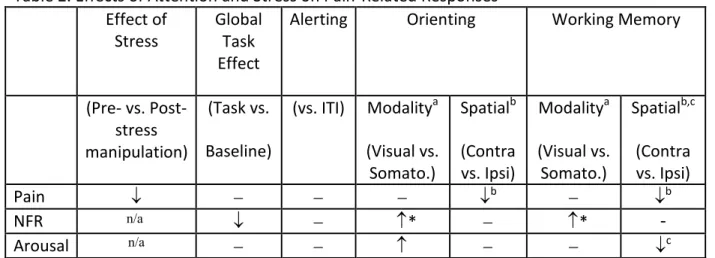
Table 2 - Effects of Attention and Stress on Pain-Related Responses... 53
Table S1 - Table S1. Mean Reaction Times (ms) and SEM of Attention Task Performance.. 66
Table S2 - Mean Accuracy (Hits and False Alarm Rates) and SEM of Attention Task Performance... 66
Liste des figures
Figure 1 - Experimental Paradigm... 35
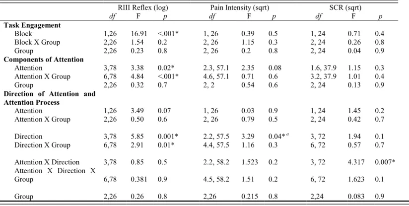
Figure 2 - Engaging in the task produces a significant decrease in NFR (A) in all three stress groups... 45
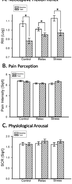
Figure 3 - Orienting and working-memory yielded a significant decrease in NFR (A) in the relax group only... 47
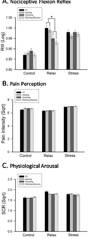
Figure 4 - Direction of attention yielded a modality effect for NFR in the relax group only (A) and a spatial effect for pain perception (B)... 49
Figure 5 - Direction of attention yielded a modality effect during orienting and a spatial effect during working-memory... 51
Figure 6 - Effects of attention and stress on pain-related responses... 59
Figure S1 - Pre-treatment of data... 65
Figure S2 - Pain intensity ratings are reduced by experimental stress manipulation... 67
Figure S3 - Means and standard errors of pain intensity ratings for all experimental conditions by group...68
Figure S4 - Means and standard errors of SCR for all experimental conditions by group...69
Figure S5 - Means and standard errors of RIII reflex amplitude for all experimental conditions by group... 70
Liste des abréviations
ACC – anterior cingulate cortex ANT – attention network testBOLD – blood oxygenation level dependent BP – blood pressure
CIP - congenital insensitivity to pain ECG – electrocardiogram
EEG – electroencephalogram
fMRI – functional magnetic resonance imaging GSR – galvonic skin response
HR – heart rate INS – insula
LC – locus coeruleus NE – norepinephrine
NFR - Nociceptive Flexion Reflex NRS – numerical rating scale PAG- periaqueductal grey matter
PASAT – paced auditory serial addition task PET – positron emission tomography
PFC – prefrontal cortex
PMR – progressive muscle relaxation RR – respiration rate
RT – reaction time
S1 – primary somatosensory cortex S2 – secondary somatosensory cortex SCR – skin conductance response SIA – stress-induced analgesia THAL – Thalamus
TSST – Trier social stress test VAS – visual analogue scale
VLM – ventrolateral reticular formation VRS – verbal rating scale
Remerciements
I would first like to thank my research supervisors, Dr. Gary Duncan and Dr. Pierre Rainville, for their guidance and support throughout the entire process. Thank you to all members of the lab for their participation and assistance in the experiments that make up this thesis. To all of the participants who were subjected to painful shocks, this would not have been possible without you. Thank you to Stephane Caron for your all of your help, motivation, and friendship. Special thanks to my brother, Michael Cardinal-Aucoin, for all of your help and support through the good, the bad, and the ugly. Finally, thank you to my family and friends who have encouraged, supported, and motivated me every step of the way.
Chapter 1: General Introduction
“The greatest evil is physical pain.” (Saint Augustine (354-430).
Pain is the number one reason that North Americans seek medical attention (Statistics Canada, 2010). Although our current understanding of pain has long surpassed our predecessors’ theories that these sensations emanate from evil, the appeal for a more thorough understanding of the peripheral and central neural mechanisms underlying pain and the factors that influence these mechanisms is ever present and remains highly relevant.
Pain is a complex sensory and affective experience that acts as the body’s alarm to present and potential physical harm. Nociception is the processing of information by the peripheral and central nervous system elicited by the activation of nociceptors, whereas pain is a multidimensional experience involving higher order processing that underlies the subjective sensation. Nociception and pain are of great physiological importance and are thought to have evolved to provide organisms with a system that alerts them to potential physical threat and produce protective withdrawal and aversion responses (Perl, 2011). The multidimensional nature of pain (with its auto-defensive qualities) distinguishes it from the other senses, such as vision, touch, smell, sound, and taste, and imparts it with a unique and distinctive nature. Although a deficit in one of the other sensory systems can lead to numerous obstacles and difficulties, individuals lacking the ability to perceive pain, such as those suffering from congenital insensitivity to pain (CIP), experience significantly higher incidence of injury and untreated illness and lower life expectancy (Nagasako et al., 2003), highlighting the biological importance of such a system. Despite its protective role, under certain circumstances pain can
become maladaptive as a result of changes in normal pain processing, and can lead to chronic pain conditions (Perl, 2011). Developing better treatment options for acute and chronic pain remains a prominent goal in clinical research and fuels the need for further advances in fundamental research on pain.
Pain is a composite of multiple components and is influenced by a wide range of elements – physiological, pharmacological, and psychological/cognitive factors affect pain processing and perception (Price et al., 2004). It is this complex multi-faceted nature of the pain experience that simultaneously incites further examination and challenges our ability to do so. To date, much interest has been focused on pharmacological modulation of pain in an attempt to address the clinical need for pain relief. As polypharma is becoming an ever-growing problem, some attention has been redirected to psychological and cognitive factors that influence pain processing. Previous research has demonstrated that pain perception is influenced by higher order processes such as emotion, expectation, and attention. The ability to modulate pain by cognitive factors such as attention is an area of current interest and remains to be thoroughly explored and understood.
The main focus of this work is to examine the influence of higher order processes, specifically attention and stress, on nociception and pain. In order to undertake this feat, the current state of the field is first assessed by analyzing what is presently known about these processes, what remains to be determined, and the tools available for investigation in this area of study. To this end, an overview of pain transmission from receptor to cortex will be presented, followed by an overview of factors currently recognized to modulate pain, and finally a description of the Nociceptive Flexion Reflex (NFR), also known as the RIII reflex, as an objective measure of spinal nociceptive transmission. With this foundation in place, the
current literature on the modulation of the NFR and pain by attention will be described, highlighting the gaps in our current understanding. Hypotheses were developed based on the current literature and form the basis that direct the research presented in this thesis.
1.1 From Toe to Head: What is pain and how does it work?
Humankind has long sought to understand the origins and nature of pain. Prior to our familiarization with the human nervous system, some of the earliest beliefs about pain emphasized spiritual and theological sources. Pain was believed to result from bodily intrusions of evil spirits, which were thought to be remedied by spiritual incantations and prayer, or it was assumed to represent an imbalance of “vital fluids” remedied by treatments such as bloodletting. With the practice of dissecting human cadavers and the exploration of human anatomy came a shift towards an empirical approach to the investigation of pain. The publication of Rene Descartes’ philosophy of the body as a machine, with its famous drawing of a pain pathway depicting a nerve fiber travelling through the body from the site of disturbance to the brain, marked a change from a mystical to a more scientific theory of pain. Aristotle and Plato regarded pain as an emotion; Descartes described it as a mechanical sensation.
Today, the International Association for the Study of Pain defines pain as "an unpleasant sensory and emotional experience associated with actual or potential tissue damage, or described in terms of such damage". Thus, pain must be described as both a “mechanical sensory” and “emotional” experience. The sensory-discriminative component can be paralleled with neural processing present in other sensory systems and is made up of spatial and temporal aspects, as well as the quality and intensity of the sensation. The
motivational-affective dimension of pain can be described as the emotional response that accompanies the sensory experience. A third dimension of pain, the cognitive-evaluative aspect, pertains to how pain perception is modulated by the cognitive appraisal of the sensory and affective experience. Together, these three dimensions of pain perception act in concert to produce the pain experience.
1.1.1 Neurobiology of Pain:
The pain experience typically begins with stimulation of nociception-specific receptors (nociceptors), which initiates a signal transduction cascade that ultimately leads to activation of supraspinal structures (Kandel et al., 2012). In contrast to the composition of other receptors of the somatosensory system, nociceptors are the free nerve endings of Aδ and C fibers, which are characterized by the nature of the stimuli that preferentially triggers a response: mechanical nociceptors are triggered by intense pressure; thermal nociceptors respond to temperatures above 45oC and below 5oC; polymodal nociceptors are stimulated by the former as well as by chemical agents; and finally, silent nociceptors are visceral afferents activated only following sensitization, such as in the presence of inflammation or specific chemical agents. The nociceptive signal initiated by stimulation of nociceptors is transmitted via Aδ and C fibers, which synapse in specific laminae of the dorsal horn of the spinal cord. Projection neurons in the spinal cord relay this information, directly or indirectly, via six main ascending pathways (spinothalamic tract, spinoreticular tract, spinomecenphalic tract, cervicothalamic tract, spinohypothalamic tract, and, spinocervical tract) to supraspinal structures involved in pain processing and perception, including thalamus (THAL), primary (S1) and secondary somatosensory cortices (S2), insula (INS), anterior cingulate cortex
(ACC), and prefrontal cortex (PFC). This nociceptive information, derived from the activation of nociceptors and the subsequent processing of this activity by the peripheral and central nervous system, is the foundation of the multidimensional pain experience. Nociceptive signals transmitted via these ascending pathways relay information that is integrated, along with other pertinent information, at the supraspinal level to then produce the multifaceted experience of pain.
The conceptual model of pain as a multidimensional construct is not purely philosophical; the sensory-discriminative, affective-motivational, and cognitive-evaluative components of pain are represented at the neural level. Mapping and neuroimaging techniques have given rise to the identification of brain areas involved in pain processing and perception. Brain regions that have been reported most often as involved in the pain experience, as evidenced by increased blood-oxygenation-level-dependent (BOLD) response, include S1, S2, THAL, ACC, INS and dorsolateral PFC (Apkarian et al., 2005). These same areas have also been identified by mapping studies in non-human primates as termination sites of nociceptive tracts (Dum et al., 2009). These regions, which are typically activated during the application of a painful stimulus, are not exclusively involved in pain processing. The non-pain-related functions of these structures provide insight and support to their hypothesized roles in pain processing. As a well established principle of neural functioning, S1, S2, and THAL have been shown to be directly involved in the perception of the spatial, temporal, and qualitative nature of innocuous stimulus intensity as a fundamental function in the somatosensory system. Not surprisingly, S1, S2, and THAL are generally considered to be primarily implicated in the sensory-discriminative aspect of pain. Functional magnetic-resonance-imaging (fMRI) studies have revealed that increased activation in these brain structures positively correlates with pain
intensity ratings of participants during application of painful stimuli, demonstrating their involvement in the sensory aspect of pain processing (Hofbauer et al., 2001; Coghill et al., 2013). Similar parallels can be found between the functions supported by other brain structures involved in pain processing and their role in the context of pain processing. Attention, motivation, error detection, and emotion are widely accepted as functional roles of the ACC as evidenced by numerous fMRI studies(Davis et al., 1997; Kandel et al., 2012). Functions of the INS include but are not limited to emotion, interoception, and autonomic regulation. In line with this, positive correlations have been found between pain unpleasantness ratings and the BOLD response to noxious stimuli in the ACC and INS, supporting their involvement in the affective-motivational dimension of pain (Rainville et al., 1999). Finally, the dorsolateral PFC is commonly associated with higher order functions including learning, memory, attention, and decision-making and is considered to be responsible for the cognitive-evaluative dimension of pain processing.
This general construct of a pain processing system that is composed of brain structures involved primarily in a particular dimension of pain is further supported by recent fMRI studies. Studies on hypnosis have found that selectively modulating either pain unpleasantness or pain intensity result in positively correlated changes in BOLD response in the dorsal ACC and S1, respectively, demonstrating a functional dissociation of these components of pain processing(Rainville, 1997; Rainville et al., 1999). The neural processing of pain can therefore be seen as a complex multidimensional system that involves a matrix of brain structures and peripheral pathways that integrates sensory-discriminative, motivational-affective and cognitive-evaluative components to produce the experience we call pain.
1.2 Toolbox for Studying Pain: The Visual Analogue Scale and Nociceptive Flexion Reflex
Our understanding of the neural mechanisms underlying the pain experience has been greatly advanced by the advent of methods that allow for the study of pain processing at various levels of the nervous system. The sensation of pain is most commonly associated with in changes in autonomic, spinal, and supraspinal activity, and therefore techniques to specifically measure each of these are of fundamental importance in order to advance our understanding of pain as a whole. Of the presently available methods, those most often employed in current research on pain include, but are not restricted to, galvanic skin response (GSR), heart rate (HR), electrocardiogram (ECG), respiratory rate, blood pressure (BP), NFR, subjective pain ratings, electroencephalogram (EEG), positron emission tomography (PET) and fMRI. Despite the inherent limitations of each method, together these tools provide critical information on how pain affects the nervous system and provides vital insight into what is happening at the autonomic, spinal, and supraspinal levels.
1.2.1 Supraspinal
In order to study what is going on at the supraspinal level we cannot simply open a person’s head and examine the inner workings of the brain. To circumvent this obstacle, well established methods are available that provide measures of neural activity and subjective experience. The most widely used technique in current neuroscience research is BOLD fMRI, which takes advantage of predictable changes in blood oxygenation levels related to neural activity. From this relationship, a functional map of brain activation can be constructed providing information about brain regions involved in a particular function. The imaging
approach to the study of pain has led to the elucidation of a network of brain regions involved in the supraspinal response to pain, often referred to as the “pain matrix”, and has provided the ability to associate the functions of these structures to particular aspects of pain.
Subjective pain ratings are also a primary tool that can offer insights into the processing of nociceptive information at the supraspinal level. Several pain rating scales and questionnaires have been created to measure the subjective experience of pain for use in both the clinical and experimental settings. Three of the most commonly used scales are the verbal rating scale (VRS), numerical rating scale (NRS), and the visual analogue scale (VAS) (Lara-Muñoz et al., 2004; Leon et al., 2004; Williamson & Hoggart, 2005; Ferreira-Valente et al., 2011; Hjermstad et al., 2011). The VRS is comprised of a list of descriptive terms that represent various levels of pain intensity, for example “no pain” or “moderate pain”. Despite its reliability, validity, and ease of use, this scale is ordinal and has no interval relationship between descriptors. However, the NRS, which allows subjects to choose a number relative to verbal anchors, does exhibit interval properties and has also been validated as a reliable measure of subjective pain response. Finally, the VAS consists of descriptive verbal anchors, such as “no pain” and “most intense imaginable pain,” at each extremity of a line that graphically represents the intensity of pain experienced. Pain rating is accomplished by marking the area on the line that corresponds to the subjective feeling. Although this method is slightly more cumbersome, in that it requires the use of a paper or computer, it exhibits ratio properties, is linear, and is strongly correlated with stimulus intensity. Studies have confirmed that, despite individual strengths and weaknesses, each of these three scales is a valid and reliable subjective measure of pain with significant positive correlations with stimulus intensity.
The scales described above provide a useful tool with which to examine the subjective nature of pain; however it is important to keep in mind that pain is a complex multidimensional experience. Studies have demonstrated a dissociation between the sensory and affective components of pain and have established the importance and necessity of using both pain intensity (sensory) and pain unpleasantness (affective) rating scales. Pharmacological and psychophysical research has revealed that while some manipulations primarily modulate the sensory component, as with distraction or administration of fentanyl, others act predominantly on the affective aspect, as seen with placebo or administration of diazepam (Fernandez & Turk, 1992; Auvray et al., 2010). Therefore, the implementation of both pain intensity and unpleasantness ratings is essential. Pain is, however, composed of not 2 but 3 components: sensory-discriminative, affective-motivational, and cognitive-evaluative. Experimental manipulations of cognitive factors, such as attention in the present research, allow for an investigation of the third component of pain and help to illuminate the neural mechanisms at play and produce a comprehensive understanding of the “pain” associated with the nociceptive event.
1.2.2 Spinal
The NFR is a widely used objective neurophysiological tool that has been demonstrated to correlate with subjective pain reports (Skljarevski & Ramadan, 2002; Sandrini et al., 2005). It was originally observed in animals as a withdrawal reflex of the ipsilateral limb following noxious electrical stimulation (Sherrington, 1910). Human studies of the NFR eventually identified two excitatory components of the NFR – RII and RIII, separated by a silent period. The first excitatory period, RII, reflects the tactile information conducted
via A-Beta fibers, whereas the RIII component is dependent on A-delta fibers, with a contribution from C fibers, and reflects transmission of nociceptive information. The RIII is a polysynaptic reflex that can be measured in humans by recording the EMG activity of the biceps femoris in response to nociceptive electrical stimulation of the sural nerve, in a temporal window of 90-180 ms following the shock (Sandrini et al., 2005). Because of the strong correlation between pain intensity and reflex size, the RIII has been used as an objective measure of pain in both clinical and experimental settings. The use of this method as a measure of spinal nociceptive transmission has proved invaluable to the study of the neural mechanisms underlying pain and the effect of various conditions on the modulation of pain processing and perception. However, despite numerous reports of a significant correlation between the objective measure of the NFR and subjective pain ratings, recent findings suggest that this correlation is not present under all conditions. Several studies have shown a dissociation between the RIII and pain ratings, putting into question its validity in the clinical setting. Despite this, it remains an important tool in experimental neurophysiology and provides an important method for the investigation of nociceptive transmission.
1.2.3 Autonomic
The autonomic system is highly interconnected with the nociceptive system, sharing many of the same brain regions involved in both the perception and modulation of pain, such as ACC, INS, periaqueductal grey matter (PAG), and ventrolateral reticular formation (VLM) (Benarroch, 2006; Leone et al., 2006). As such, measures of autonomic activity provide an important window into the influence of pain on the nervous system. The methods currently used in pain research that tap into autonomic functioning are GSR, HR, ECG, respiration rate
(RR), and BP. Studies have demonstrated a predictable increase in GSR and HR in response to painful stimuli. Additionally, the cardiac cycle, RR, and BP have been linked to subjective and objective measures of pain. Together, acquiring data on the supraspinal, spinal, and autonomic response to pain allows for a more comprehensive view of the neuroscience of pain.
1.3 Look here!!! The interplay between pain and attention
“Pain insists upon being attended to. God whispers to us in our pleasures, speaks in our consciences, but shouts in our pains. It is his megaphone to rouse a deaf world.” (C.S. Lewis)Pain solicits attention; it directs focus to present or potential physical harm, thus affecting the relative salience of environmental stimuli. In turn, attention exerts a modulatory effect on pain. Attention plays a direct and important role in many cognitive and neurological functions including learning, memory, emotion, spatial processing, and sensory perception. It coordinates where and to what degree our focus is directed, thereby dictating how we sample and experience our environment. The interaction between attention and pain can therefore be described as a complex tug of war between competing stimuli and the constant reassessment of where to allocate biological and neurological resources.
1.3.1 Attention: Theories and Networks
The study of attention is historically important in the evolution of cognitive neuroscience and the emergence of experimental psychology. Furthermore, some of the first experiments in psychophysiology explored attention. The sustained interest in this domain is not surprising given the importance it plays in how we perceive the world around us. Attention is a complex dynamic equilibrium of lenses and filters through which we experience our
environment, both external and internal. It dictates what stimuli are highlighted and brought to the forefront of our experience, which percepts are ignored and how much weight is allotted to every attribute of our perception of reality.
The neurological underpinnings of the attention system are composed of three main anatomically and functionally distinct attentional networks that work together and influence processing systems. Three major components of attention– namely alerting, orienting and executive control – were initially identified and established as functionally discrete elements of attentional processing (Petersen & Posner, 2012). Alerting is defined as establishing (phasic alerting) and sustaining (tonic alerting) a state of increased vigilance; orienting is the prioritizing of the sensory input of a location or modality; executive control consists of control processes including error detection, conflict resolution, and decision-making.
More recent studies have revealed that these individual components of attention, while working together, are in fact dissociable. The Attention Network Test (ANT), a task created to isolate the components of attention, provided much support from human and animal studies for the idea that these systems function independently of one another. Fan and colleagues (2002) developed the ANT by combining a cued reaction time (RT) task with a flanker task(Fan et al., 2002). The objective of the task is to correctly identify the direction, left or right, of a central arrow located between four flankers. On some trials, cues are provided informing participants about the time and location of stimulus presentation. In this task, the influence of alerting is measured by the effect of providing a temporal cue on participant RTs, whereas orienting is reflected by changes in performance due to the presence of spatial cues. The involvement of executive functioning is established by differences in RT between trials with congruent and incongruent flankers.
The use of the ANT and variations of this task has led to the realization that individual differences in the strength of one component of attention are distinct from abilities of another and that improvements in functional capacity of one component are isolated from capabilities of another (Callejas et al., 2005). Moreover, pharmacological and imaging studies have demonstrated that these components of attention are not only functionally dissociable but are supported by anatomically distinct networks of brain structures and neurotransmitter systems (Petersen & Posner, 2012).
The alerting network has been revealed to rely on the norepinephrine (NE) system with projection from the locus coeruleus (LC) to frontal and parietal regions and lateralized to the right hemisphere. Pharmacological studies using drugs that influence the release of NE show that increased NE release improves alerting and decreased NE release inhibits the warning-signal effect (Morrocco and Davidson, 1998).
The orienting network on the other hand has been shown to rely on the cholinergic system. Studies with drugs that influence acetylcholine have an effect on orienting but not alerting (Davidson & Marrocco, 2000), further demonstrating a dissociation between these attention networks. Recent research suggests that orienting relies on two systems, a top-down dorsal system made up of the frontal eye fields and intraparietal sulcus/superior parietal lobe and a bottom-up ventral system involving the temporoparietal junction and ventral frontal cortex (Corbetta and Shulman, 2002).
The work of Dosenbach (2007, 2008) provides evidence for a dual network theory for the executive control system consisting of a frontoparietal system involved in moment to moment processing of a task, such as task switching/initiation and real-time adjustments, and a cingulo-operculum system, responsible for task set maintenance, acting as a stable background
for overall performance. Both systems act to produce top-down control. Together, alerting, orienting, and executive control networks support state of readiness, focal awareness, and complex mental operations, thereby influencing perception and cognition.
1.3.2 Effect of attention on sensory processing and integration
Our knowledge of the components of attention and the networks that support them is fundamental to our understanding of how we relate to external and internal environments. It is therefore important to consider the influence of attention on sensory processing and integration. Each component of attention influences sensory processing in a specific and unique way. Alerting facilitates the perception of stimuli by increasing overall arousal and/or acting as a warning signal in preparation for response to a target. Presentation of alerting cues has been shown to support improved ability to obtain information from a stimulus (Fernandez-Duque & Posner, 1997; Wang & Fan, 2007; Weinbach & Henik, 2012). Additional facilitation of information acquisition and processing is provided by the orienting component of attention. Orienting can be either a top-down or bottom-up selection of what information is most salient at any given time. The orienting network is responsible for both spatial and modality-specific allocation of attentional resources, acting as a spotlight on pertinent stimuli. Results from studies using a divided-attention task provide much support for this, showing enhanced ability to process information when orienting towards a relevant target and degraded performance when orienting away from the relevant target or towards another distractor target (Bashinski & Bachrach, 1980; Eriksen & Hoffman, 1973; Jonides, 1981; Mountcastle, 1978; Posner & Davidson, 1980; Nissen et al., 1978; Geffen & Wale, 1979; Sexton & Geffon, 1979; Mozolic et al., 2008). This is true of the influence of orienting for both location and modality of the
relevant stimuli. Finally, executive functioning influences sensory processing by highlighting and maintaining a stable background of task-relevant information and integrating this information in a meaningful way (reviewed by Jurado & Rosselli, 2007). Thus, attention and its various components play a critical role in how the sensory environment is sampled, processed, and integrated.
1.3.3 Attention and Pain
Pain is a sensory experience, and attention is well known to influence sensory processing and integration. Therefore, it is not surprising that attention has been shown to exert a powerful modulatory effect on pain. However, the neurological mechanisms underlying this modulation of pain by attention remain unclear. Several theories have been put forth to explain how attention influences pain, but a complete and comprehensive model of this interaction is still conspicuously unavailable.
Consider the processing of painful stimuli as the inner workings of a factory where several steps are required for the production of a final product, in this case pain. Modifications at any stage of the process will influence the outcome in specific ways. If, for example, the factory employees ignore arriving raw materials to be used in product manufacturing – directing attention away from painful stimuli - the result is a reduction in machine operation and less output of the final product - decreased activity in brain structures involved in sensory processing of pain and reduction of perceived pain intensity and unpleasantness. Here, attentional modulation of pain is the result of altered efficacy of processing of primary nociceptive inputs. One prominent theory of the attention-pain interaction suggests that, at least in part, the nociceptive system is influenced by attention in an equivalent manner to other
sensory modalities. If this is true, our understanding of the influence of attention on sensory processing would suggest that orienting towards the target stimulus, in this case pain, would enhance perception, whereas attention directed away from the target stimulus would decrease perception.
Bushnell et al. (1985) manipulated spatial attention (humans) and the modality (somatosensory or visual) to which attention was directed (monkeys) during sensory discrimination of innocuous and noxious thermal stimuli. In humans, subjects were provided with a visual cue (valid or invalid) indicating where to attend - left arm, right arm, or no cue - and instructed to detect the occurrence of a change in stimulus intensity. Ability to discriminate between stimuli - as measured by response latencies, percent undetected change, and percent early responses - was significantly better when subjects received cues as compared to without cues. Stimulus discrimination was also improved when cues validly indicated the location where the stimulus change occurred compared to invalidly cued trials. In monkeys, similar results were obtained when attention was manipulated between the visual and somatosensory modality. When attention was directed to the somatosensory as compared to the visual modality, monkeys’ ability to discriminate between stimulus intensities (same performance measures as humans) was significantly improved. These attention-dependant performance differences in humans and monkeys were present for both innocuous warm (humans and one monkey) and noxious thermal trials. These data demonstrate that nociceptive and innocuous inputs are similarly modulated by direction of attention (location and modality), although they do not exclude the possibility of additional nociceptive-specific attentional processes.
In addition to altered capacity for the discrimination of noxious stimuli, perceptual differences resulting from attentional modulation would translate into a relative increase in perceived pain intensity when attending to a noxious stimulus and decreased perception of pain intensity when attending to other stimuli. In a study by Miron et al. (1989), participants performed a sensory discrimination task of visual and noxious thermal stimuli and provided pain intensity and unpleasantness ratings following each trial. Cues were provided on each trial indicating the modality in which the change would occur (directed attention) or that the change may occur in either modality (divided-attention). Participants’ ability to discriminate a change in nociceptive stimulus intensity was dependent on direction of attention as evidenced by higher percent detection and greater speed of detection on correctly versus neutral and incorrectly signaled trials i.e. greater discriminability when attention was directed toward noxious stimuli. Additionally, pain intensity and unpleasantness ratings were also dependent on the attentional condition. Pain intensity and unpleasantness ratings were lower when attention was directed to the visual (falsely signaled trials) compared to the somatosensory modality (correctly signaled trials). Thus, Miron et al. (1989) replicated the effects of attention on the discriminability of noxious stimuli shown by Bushnell et al. (1985) and further demonstrated that attentional modulation also influences subjective pain perception.
In a magnetoencephalography study, Nakamura et al. (2002) investigated the effect of the degree of attention toward painful infra-red heat stimuli on S2 activity. Three conditions of attention to pain were examined: 1) low attention (subjects were instructed to ignore pain), 2) mid-level attention (subjects rated pain following an auditory tone), and high attention to pain (subjects associated a high or low tone with one of two pain stimulus intensities and were rewarded for accuracy). Low attention to pain resulted in less S2 activity compared to higher
attention to pain conditions. These results further support the idea that attentional modulation of pain is, at least in part, related to altered pain processing similar to perceptual attention-related changes in other sensory modalities. Therefore, attention-attention-related changes in pain perception may result from direct modulation within areas related to the processing of noxious stimuli, or more indirectly as a result of the attentional modulation of sensory processing observed in other modalities – a redistribution of processing resources away from the nociceptive pathways.
A second theory on the attentional modulation of pain relates to higher order processing during execution of an attention task. In this case, to return to the factory analogy, if most factory workers are occupied with responsibilities other than product manufacturing, there will be a shift in which machines are operating, but there is a reduction in product output. In much the same way, resources being allocated to neural processing of an attention task will lead to increased activation of regions involved in task-related functions, decreased activity in pain processing structures, and decreases in pain intensity and unpleasantness ratings. In this case, pain perception is modulated as a result of resources being allocated differentially at the supraspinal level, resulting in a decrease in processing and integration of nociceptive information. Again, this prioritizing of neural processing of task-relevant information during an attention task mirrors the influence of attention on other sensory modalities. Dual-task interference studies demonstrate the limited capacity for neural processing when engaging in multiple tasks. These detriments in neural processing have been shown to occur at early sensory processing stages as well as later processing, integration and higher order functioning during performance of cognitive tasks (Kasper et al., 2008; Rissman
et al., 2009; Tombu et al., 2011). This effect of central processing bottlenecks is likely also involved in pain modulation during executive processing. Petrovic et al. (2000) examined the effects of engaging in a cognitive task on pain perception and brain activity using PET. Participants rated pain intensity induced by a cold-pressor test under two conditions: 1) pain alone, and 2) during performance of a computerized maze task. The pain-alone condition resulted in activation of characteristic pain-related regions including contralateral S1, bilateral S2, ACC, and INS. During the attention task, activity in somatosensory association areas and PAG/midbrain were significantly decreased and orbitofrontal regions increased. In conjunction with these changes in regional blood flow during performance of the attention task, participant’s pain ratings were reduced. These results suggest that the increased activity during attentional processing of the cognitive task, such as in the PFC, may be involved in the modulation of pain processing and reductions in subjective pain ratings.
Empirical support for these theories notwithstanding, there is evidence for a third pain-specific neural mechanism of attentional modulation. In this case, if the factory manager issues a memo to order a stop on incoming raw materials or to turn on/off some machines, there will be a shift in machine operation and a subsequent decrease in production of the product. Here, the performance of a distracting task may engage higher order structures initiating descending inhibition (or facilitation) of pain processing at the spinal level or altering activity at the supraspinal level which leads to increased activation of regions involved in attentional processing, decreased activity in pain processing structures, possible changes in activity of additional structures, and decreases in pain intensity and unpleasantness ratings.
Recent imaging studies on attention-mediated changes in brain response to pain provide insight into the mechanisms at play and suggest that, in addition to attentional modulation processes involved in other sensory modalities, pain-specific mechanisms – including descending inhibitory systems - may be recruited. Valet et al. (2004) used fMRI to examine the attentional modulation of pain by the Stroop task. In comparison with the pain-alone condition, performance of the Stroop task resulted in significantly increased activation of the orbitofrontal cortex, perigenual ACC, PAG, and posterior THAL, as well as decreased pain ratings. Additionally, distraction resulted in reductions in the activity evoked from pain alone, particularly in the medial THAL, the midcingulate, anterior-ventral INS and lateral PFC. Moreover, Valet et al. (2004) report a functional interaction between these structures during the distraction task suggesting that these regions mediate the observed changes in pain processing and related reductions in pain ratings.
Tracey et al. (2002) used high-resolution fMRI to investigate PAG activity during attention to pain compared to distraction from pain. During the attention to pain condition, participants were instructed to focus on the pain whereas in the distraction condition, participants were instructed to think of something other than the painful stimulus. During the distraction condition, PAG activity was significantly higher and correlated with reductions in pain ratings. In conjunction with other studies that report increased activity in frontal cortex regions and PAG and reductions in S1 and S2, with related decreases of subjective pain ratings when participants attend away from painful stimuli (Nakamura, Paur, Zimmermann, & Bromm, 2002; Peyron et al., 1999; Bantick et al., 2002), these results suggest that attentional modulation of pain involves a descending modulatory system.
In a recent study, high resolution fMRI of the spinal cord during a high- versus low-working memory load distraction task with concurrent application of thermal pain resulted in reductions of neuronal response to pain in the dorsal horn and paired decreased pain ratings, suggesting the involvement of a descending inhibitory system in attentional modulation of pain (Sprenger et al., 2012). In a second experiment, administration of naloxone, an opïoid antagonist, partially blocked this effect, demonstrating that the observed changes were partly due to an endogenous opïoid-mediated analgesia. Taken together, this may suggest the contribution of a descending opïoid-mediated system in the attentional modulation of pain. However, it remains possible that these reported changes in nociceptive processing are due to stress-related pain modulation, which has been associated with descending opïoid-mediated inhibition of nociceptive responses and is often not considered in studies on attentional modulation of pain. As such, in order to address this potential confound, the paradigm in the study presented in this thesis manipulates stress levels independently from the attention task, thereby dissociating the effects of stress from that of attention on pain processing.
1.3.4 Stress, Attention and Pain
Stress is an important factor to consider in the investigation of the attentional modulation of pain. Research over the past three decades has demonstrated that stress typically has a suppressive effect on pain (Butler & Finn, 2009). This psychological modulation of pain, commonly referred to as stress-induced analgesia (SIA), has received much interest. The anatomical, molecular, and neurochemical mechanisms (including the contribution of opïoid and non-opïoid mediated systems) underlying SIA have been largely investigated. However, despite the overwhelming evidence from numerous animal and human studies supporting an
analgesic effect of stress, a number of investigators have reported increases in pain during exposure to stress – stress-induced hyperalgesia – a phenomenon that is still not well understood (Imbe et al., 2006; Richebe et al., 2011).
Given the significant modulatory effect of stress on pain, it is important to control for the influence of this factor during the investigation of other types of psychological modulation of pain. Unfortunately, the effects of stress appear to have been overlooked in many studies on attentional modulation of pain, and this may provide an explanation for some seemingly contradictory findings in the literature. One major cause for this is the stressful nature of some of the currently used attention/distraction tasks. In fact, some of the same tasks used as a “distraction task” in the study of attention, including mental arithmetic, the Stroop, tracing tasks, and other cognitive tasks, have been employed in research on stress as a “stress manipulation.” Regrettably, there is no quick fix to this issue, as highly demanding and engaging tasks that require focused attention are, by the nature of the task, stressful to some degree. One possible solution to this caveat is to differentially modulate stress and attention levels in an attempt to dissociate the involvement of each factor in the modulation of pain. Varying the difficulty level of the attention task would have the effect of altering stress and attention in parallel. Alternatively, an experimental manipulation of stress that is separate and distinct from the attention task could potentially modulate these psychological factors differentially and may help dissociate their effects.
1.3.5 Attention, Pain, and the RIII
Previous research has demonstrated that many physiological, pharmacological and psychological factors modulate both spinal and supraspinal levels of pain processing (Sandrini
et al., 2005). Although there is evidence that attention has a modulatory effect on pain - increased pain while attention is directed towards and decreased pain while attention is directed away from a noxious stimulus - the neural mechanisms underlying these changes remain unclear. The NFR, or RIII reflex, provides a method with which to examine the effects of attention on spinal nociceptive transmission to gain insight into the neural underpinnings of the observed changes in pain processing. The RIII has been utilized in pain research as an objective measure of spinal nociceptive transmission and has been shown to correspond with subjective pain ratings, making it a useful tool in clinical and experimental settings. However, recent work has demonstrated that the relationship between these measures does not always correlate, suggesting a possible dissociation between RIII and subjective pain ratings (Roy et al., 2011).
The current literature on the effects of attention and distraction on pain ratings and the RIII is inconsistent. Bathien and colleagues (1969, 1971, and 1972) found that certain, but not all, tasks that demanded the attention of participants resulted in a change in RIII amplitude. Willer et al (1979) showed that both pain sensation and RIII were inhibited during a mental task, whereas Dowman (2001) found that attentional set reduced pain ratings but had no effect on the RIII. Some more recent studies have been unable to replicate a modulatory effect of attention on the NFR, finding no significant difference in RIII threshold during a distraction task (France 2002, Terkelsen 2004, Hennighasuen et al 2007). Further research on the effects of attention on the RIII reflex and pain have yet to resolve these incongruencies.
The conflicting findings in the literature on attentional modulation of the RIII reflex and pain are due to several factors, both methodological and theoretical. First, as previously mentioned, a likely confound in a number of studies is the effect of stress on pain mechanisms.
Several studies examining the neural mechanisms underlying the effect of attention on nociception and pain made use of stress-inducing tasks such as mental arithmetic (Bathien & Hugelin, 1969; Bathien, 1971; Bathien & Morin, 1972; Willer et al., 1979; France et al., 2002; Terkelson et al., 2004; Edwards et al., 2006; McIntyre et al., 2006). Because stress has a modulatory effect on pain at the spinal and supraspinal levels, failure to control for stress level induced by distraction tasks hinders the interpretation of findings from these experiments. Additional studies have introduced other confounds such as emotion and expectancy, processes that have been shown to modulate both the RIII and pain ratings (Willer et al., 1979; Ruscheweyh et al., 2011), thereby obscuring the evaluation of the findings from these experiments. Additional methodological issues, such as variations in the intensity of stimulation of the sural nerve between studies, further obfuscate analysis of the results. Several studies stimulate at threshold intensity of the reflex (Edwards et al., 2006), while others have selected an intensity of 1.5 times the threshold (Terkelsen et al., 2004) and still others report using an unspecified level between threshold and tolerance (Dowman, 2001). This is of considerable concern since the level of stimulus intensity may affect the susceptibility of the NFR to modulation by cognitive factors. Additionally and importantly, the current literature fails to consider the multifaceted nature of the attention system, which is composed of several functionally and anatomically dissociable networks: alerting, orienting, and executive control. These components of attention may produce distinct effects on nociception and pain and may involve different underlying modulatory neural mechanisms. Overall, the current literature on attentional modulation of nociception and pain is confusing, inconsistent, and consequently largely uninterpretable.
1.4 Objectives
The present work aims to explain the conflicting findings in the literature on the attentional modulation of pain perception and NFR in order to gain further insight into their causal neural mechanisms. An in-depth review of the existing literature on attentional modulation of the RIII and pain was undertaken to critically analyze the current findings and generate hypotheses that may help clarify the ostensible contradictions (see general discussion). The development of these hypotheses into a novel functional framework along with the conception and execution of a study to test them constitute the focus of the current work.
In the present study, a component-based approach to the investigation of attentional modulation is proposed that separately examines the effects of alerting, orienting, and executive control on nociception and pain and includes stress as an additional variable. We isolated the components of attention in a discrimination task involving both visual and somatosensory stimuli. Throughout the experiment we measured the RIII reflex, skin conductance response (SCR), and pain ratings in response to painful electrical stimuli delivered to the sural nerve in order to examine the effects of these components of attention on nociception and pain. Furthermore, we made use of two previously validated methods to manipulate basal stress levels (music-induced relaxation and the Trier Social Stress Test: TSST) to dissociate the influence of attention and stress on pain processing. Exposure to music reduces anxiety and stress and has been shown to reduce subjective and physiological indices of stress such as heart rate, blood pressure, and the cortisol response to exposure to an external stressor (Knight & Rickard, 2001; Khalfa et al., 2003; Salamon et al., 2003). The
TSST has been demonstrated as a reliable procedure to increase subjective stress reports and salivary cortisol measures via motivated performance with social-evaluative threat and uncontrollability (Kirschbaum et al., 1993; Dickerson & Kemeny, 2004).
Chapter 2: Article
Effects of components of attention and stress on pain and the nociceptive flexion
reflex
Authors: Natalie Cardinal-Aucoin13-5, Gary H. Duncan2,3, Pierre Rainville1-5
Affiliations: Department of (1) neuroscience and (2) stomatology, (3) Groupe de recherche sur le système nerveux central (GRSNC), (4) Centre de recherche de l’Institut universitaire de gériatrie de Montréal and (5) Centre de Recherche en Neuropsychologie et Cognition, Université de Montréal, Montreal Qc, Canada
Abstract
The literature on attentional modulation of the nociceptive flexion reflex (NFR) and pain is inconsistent, possibly because the complex nature of attention processes and the possible interactions with stress have been overlooked. Here, the NFR and pain ratings were measured before and during a visual and somatosensory delayed-discrimination task designed to separate components of attention-related processes (alerting, orienting, and sensory working-memory), in three groups of healthy individuals following relaxation, stress-induction, or no manipulation (control). Pain was significantly reduced following induction, consistent with stress-induced analgesia while effects of attention components were observed mainly or only in the relaxation group. Alerting reduced both pain and the RIII. Top-down orientation away from noxious stimuli resulted in hypoalgesia (pain ratings) independent from the stimulus modality. In contrast, the NFR was larger when attention was directed towards the visual compared to the somatosensory modality. Beyond these orientation effects, executive control (working memory) had no additional effect on pain but showed a tendency to decrease further the NFR relative to baseline. The modulation of nociception by attention was observed only in the low-stress group. These findings highlight the influence of each component of attention on pain and the masking effect of stress on some of these modulatory effects. The spatially- and modality-specific nature of attentional modulations of pain and the NFR, respectively, further demonstrate the complexity of attentional influences on perceptual and spinal processes and clearly points to the multiplicity of underlying mechanisms.
Introduction
Psychological processes have been demonstrated to have a powerful modulatory effect on pain and nociception. Cognitive modulation of pain has been shown in studies on hypnosis, placebo, emotion, and attention (Price et al., 2004). The current literature on the effects of attention on both pain and nociception, however, is inconsistent. Early studies on the attentional modulation of spinal nociceptive transmission, as measured by the nociceptive flexion reflex (NFR), found either a decrease or no change in the reflex amplitude during performance of an attention task and, in one case, a slight increase in reflex amplitude (Bathien, 1971; Bathien & Hugelin, 1969; Bathien & Morin, 1972). Research on the effects of an attention task on both pain and spinal nociceptive transmission has yielded similarly equivocal results. Where some findings indicate a decrease in both pain and NFR (Willer, Boureau, & Albe-Fessard, 1979), others have found no change in NFR threshold or amplitude with variable findings on pain ratings (Dowman, 2001; France, Froese, & Stewart, 2002; Terkelsen, Andersen, Molgaard, Hansen, & Jensen, 2004). Others still have reported a decrease in pain ratings and concurrent facilitation of spinal nociceptive transmission (Louisa Edwards & Richard Clarke, 2006; McIntyre, Edwards, Ring, Parvin, & Carroll, 2006; Roy, Piche, Chen, Peretz, & Rainville, 2009). A more refined analysis of attentional processes involved in the modulation of pain and the NFR may help clarify these contradictory results.
Attention is a complex process involving several components: alerting, orienting and executive control. Behavioural and imaging studies have revealed that these components of attention are functionally dissociable from one another and involve distinct neural mechanisms (Fan, McCandliss, Fossella, Flombaum, & Posner, 2005). To our knowledge, no study has systematically investigated the specific influence of each component of attention on pain and the
NFR. Moreover, the modality and location to which attention is being directed needs be considered (e.g. visual or somatosensory; close to, or away from, the painful site). Furthermore, another dimension of psychological processing rarely accounted for across these studies is the effect of stress (task-related or unrelated). This is an important factor to consider as cognitive tasks are sometimes used to generate psychological stress and stress may modulate pain-related brain responses (e.g. Vachon-Presseau, 2013). Therefore, the individual components of attention, the sensory modality of the distracter, and the influence of stress must be accounted for in order to disentangle the conflicting literature on the modulation of the NFR and pain by attention.
This study employs a delayed-discrimination task designed to dissociate components of attention involved in alerting, orienting, and executive processes (here sensory working-memory). We examined the effects of these components in the visual and somatosensory modalities while varying the levels of both task-induced and social stress across subjects. In so doing, this research provides a novel conceptual model of how attentional mechanisms affect the spinal transmission of nociceptive signals and the perception of pain. Moreover, by dissociating the components of attention across sensory modalities and stress levels, these results might explain some of the inconsistencies in the literature on the attentional modulation of spinal nociceptive transmission and pain.
Materials and Methods
Participants
Thirty-eight healthy volunteers between the ages of 18-35 were recruited from the University of Montreal using on-campus notices, from Concordia University via their website, and through general internet advertisement. Five participants were excluded due to an inability to
obtain a stable RIII reflex and pain ratings. Of the thirty three remaining subjects, three were excluded because they did not complete the entire experimental protocol (1 participant withdrawal and 2 technical failures). Additionally, one participant was excluded post hoc due to an elevated Becks Depression Inventory-II (BDI-II) score (BDI-II score = 31). The final sample included 29 healthy volunteers aged between 20 and 35 years (25.6 ± 4.5 years; 15 men and 14 women) with no history of chronic pain, diabetes, colour-blindness, neurological or psychiatric disease. The experimental protocol was approved by The Research Ethics Board of the “Centre de recherche de l’Institut de gériatrie de Montréal”; all participants completed a consent form and were compensated for their participation.
Study design
The study relied on a mixed experimental design involving the manipulation of stress across three groups (high stress, relaxation/low stress, and control) and the manipulation of attention within-subject. Pain and NFR responses were assessed before (baseline) and throughout the different phases of an attention task. Stress was induced by the Trier Social Stress test (TSST) administered before the attention task and by additional negative feedback on task performance. Low stress was induced by listening to relaxing music prior to the task and supportive feedback on task performance. This design allowed us to assess effects of stress on pain and NFR responses at baseline (no task), the effects of different functional components of attention (see task description, below), and the potential interaction between stress and attention.