A new method for the determination of Leuconostoc mesenteroides cell number
7
0
0
Texte intégral
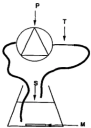
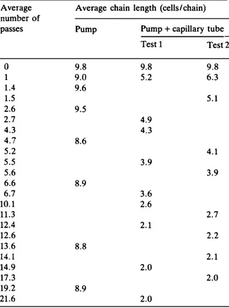
Figure
Documents relatifs