HAL Id: tel-01505677
https://pastel.archives-ouvertes.fr/tel-01505677
Submitted on 11 Apr 2017
HAL is a multi-disciplinary open access archive for the deposit and dissemination of sci-entific research documents, whether they are pub-lished or not. The documents may come from teaching and research institutions in France or abroad, or from public or private research centers.
L’archive ouverte pluridisciplinaire HAL, est destinée au dépôt et à la diffusion de documents scientifiques de niveau recherche, publiés ou non, émanant des établissements d’enseignement et de recherche français ou étrangers, des laboratoires publics ou privés.
between aptamers and pathogens
Timothy Aschl
To cite this version:
Timothy Aschl. Biochips based on silicon for detecting the interaction between aptamers and pathogens. Materials and structures in mechanics [physics.class-ph]. Université Paris Saclay (CO-mUE), 2016. English. �NNT : 2016SACLX103�. �tel-01505677�
NNT : 2016SACLX103
Thèse de doctorat
de
L’Université Paris-Saclay
préparée à
“L’Ecole Polytechnique”
Ecole Doctorale n° 573
Interfaces : approches interdisciplinaires, fondements, applications et innovation
Spécialité de doctorat : Chimie
ParM. Timothy Aschl
Biochips based on silicon for detecting the
interaction between aptamers and pathogens
Soutenance de thèse à Palaiseau, le 13 décembre 2016. Composition du Jury :
Mme. Hafsa Korri-Youssoufi Directeur de recherche, Université Paris-Sud Présidente M. Rabah Boukherroub Directeur de recherche, Université Lille 1 Rapporteur M. Jean-Louis Marty Professeur, Université de Perpignan Rapporteur M. François Ozanam Directeur de recherche, Ecole Polytechnique Examinateur Mme. Rosemarie Sauvage Responsable du domaine scientifique nanotechnologies, DGA Examinatrice Mme. Anne Chantal Gouget- Chargée de recherche, Ecole Polytechnique Directrice de thèse Laemmel
Acknowledgments
Ce travail de thèse a été réalisé au laboratoire de Physique de la Matière Condensée (PMC), à l’École Polytechnique cofinancé par la Direction générale de l’Armement (DGA).
Je veux remercier d’abord les membres du jury qui ont accepté d’évaluer ce travail de thèse. Tout d’abord, je souhaite exprimer toute ma gratitude à Hafsa Korri-Youssoufi présidente du jury. J’adresse ensuite mes remerciements aux rapporteurs, Jean-Louis Marty et Rabah Boukherroub qui ont lu l’ensemble de ce manuscrit avec beaucoup d’attention. Merci enfin à Rosemarie Sauvage de la DGA pour son soutien pendant ce travail.
Je souhaite exprimer toute ma gratitude à ma directrice de thèse, Anne-Chantal Gouget, qui a dirigé ce travail et a été présente tout au long de ces trois années de thèse. Je la remercie vivement pour sa disponibilité, pour le temps qu’elle a consacré à mes sollicitations, et pour les discussions « manips » quotidiennes. J’ai beaucoup apprécié l’autonomie qu’elle m’a laissé dans la réalisation de mes expériences. C’est grâce à elle que ce travail a pu aboutir.
J’adresse aussi mes remerciements à mon co-directeur de thèse, Philippe Allongue. Au-delà de sa rigueur et de sa disponibilité, je lui suis reconnaissant de m’avoir fait profiter de ses compétences scientifiques. Sur ce point, je lui suis particulièrement reconnaissant pour ses idées et conseils pendant la rédaction du manuscrit et la préparation de soutenance.
Il est tout particulièrement important pour moi de remercier Anne-Chantal et Philippe pour leurs nombreuses relectures du manuscrit, afin de le rendre à la fois bien structuré, clair et compréhensible
François Ozanam, grâce à ses conseils et sa disponibilité, a également contribué largement à l'obtention et l'interprétation des résutats présentés ici. Je le remercie de m'avoir transmis sa passion pour la science, ainsi que certaines de ses nombreuses connaissances.
Je remercie aussi Gilles Frison de LCM, qui a lancé des calculs DFT pour l’analyse des spectres infrarouges, en dernière minute. Ses calculs ont énormément apporté à l’analyse des résultats.
Lucio Martinelli et Fouad Maroun ont également contribué au succès de cette thèse. Leurs idées, propositions et coup de mains ont souvent aidé à résoudre efficacement les problèmes qui ont régulièrement ponctués cette recherche.
Pendant la thèse j’ai partagé le bureau et plein de fou-rires avec Denis, Khalid et Patrice. Je les remercie très sincèrement pour leur bonne humeur / humour pendant le temps que l’on a passé ensemble. Toujours quelques mots gentils ou des petites blagues… on s’est bien amusé dans ce bureau. J’espère que vous allez rester la bonne équipe que vous êtes et aussi que vous aurez quelqu’un de sympa à ma place.
Je tiens à remercier très chaleureusement Anne Moraillon, sans elle cette thèse n’aurait pas été possible. C’est par elle que j’ai appris tous les détails du travail quotidien de laboratoire, notamment en passant de longues heures à m’indiquer comment faire telles ou telles. Je lui adresse un grand merci pour tous les moments de bonne humeur et d’enthousiasme que nous avons partagés. Elle est devenue une vraie amie, je ne la remercierai jamais assez pour tous les bons moments passés ensemble. Je souhaite aussi de remercier tous les amis que j’ai eu la plaisir de rencontrer pendant ma thèse à l’X. C’est surtout grâce à leur soutien que j’ai pu réussir. Notamment je remercie pour notre amitié : Lucie Devys pour sa gentillesse ; Maxime Ardre pour son esprit ouvert et sa mentalité de « let’s do it » ; Samuel Thomas, Pierre Nocerino et Léa Mazé pour les diners, soirées de jeux et des conversations excellentes ; Alizée Dubois pour tous les heures sympas passées en faisant du sport ; Godefroy Lemenager pour son soutien infaillible ; Stefan Klaes pour son humour extraordinaire ; Hongye pour les discussions enrichissants ; Isabelle Maurin pour tous les discussions sympas, mais bien sûr aussi tous les autres que je ne peux tous nommer sans exploser le cadre. Mes remerciements doivent bien sûr aussi inclure mes amis d’Autriche qui m’ont soutenu pendant ma thèse. Notamment Thomas, Lisi, Berni, Judith, Gernot, Kerstin, Christian, Fabio, Chrisi, Barbara, Mike et Tom.
Je veux aussi exprimer toute ma gratitude à Maria Castellano Sanz qui m’a soutenu énormément. On a partagé de jours à jours nos hauts et nos bas de la vie de chercheurs : que notre amitié perdure, profonde et fidèle.
Il est tout particulièrement important pour moi d’exprimer ma gratitude à la famille Ink, qui m’a accueilli très chaleureusement parmi eux et qui m’ont appris à vivre en et apprécier la France. C’est surtout grâce à Marion que j’ai trouvé la force de faire ce travail. Je la remercie, avec toute ma gratitude, pour son soutien, sa patience et sa compréhension vis-à-vis du temps que j’ai passé à travailler. Nous avons passé de très beaux moments ensemble : elle et sa famille auront toujours une place dans mon cœur.
Mes remerciements vont enfin à ma famille. Je remercie ma sœur et mes frères pour leur soutien et leur amour, et je remercie surtout mes parents qui m’ont toujours soutenu dans n’importe quelle aventure, comme faire une thèse en France. Merci, de toujours croire en moi et d’être derrière moi.
C
ONTENTS
RESUME ... 1
GENERAL INTRODUCTION ... 7
1 STATE OF THE ART & STRATEGY ... 11
1.1 Introduction ... 12
1.2 Pathogens ... 12
1.2.1 Definition and types ... 12
1.2.2 Ochratoxin ... 13
1.2.3 Methods for detecting pathogens ... 15
1.3 Aptamer-based biosensors ... 18
1.3.1 Aptamers as probes in biosensors ... 18
1.3.2 Aptamers for the detection of OTA ... 21
1.3.3 Aptamer-based biosensors for the detection of OTA ... 22
1.3.4 Challenges for biosensors ... 24
1.4 Strategy proposed for detecting the interaction aptamer-OTA ... 25
1.4.1 Direct detection of the interaction aptamer-OTA by ATR-FTIR ... 26
1.4.2 Indirect detection of the interaction aptamer-OTA by fluorescence ... 26
2 SURFACE FUNCTIONALIZATION OF OXIDE FREE SILICON SURFACES ... 29
2.1 Introduction ... 30
2.2 State of the Art ... 31
2.2.1 Functionalization of silicon surfaces ... 31
2.2.2 Two-step amidation of acid terminated surfaces ... 35
2.3 Materials and Methods ... 39
2.3.1 Chemicals and substrates ... 39
2.3.2 Hydrogenated silicon surfaces ... 40
2.3.3 Photochemical hydrosilylation of undecylenic acid ... 40
2.3.4 Activation with EDC/NHS of acid-terminated surfaces ... 41
2.3.5 Aminolysis of PEG 750 on activated silicon surfaces ... 41
2.3.6 Adsorption test of E. coli Katushka K12 on PEG-surfaces ... 42
2.4.1 Hydrogenation of silicon surfaces ... 42
2.4.2 Grafting of undecylenic acid on silicon surfaces by hydrosilylation ... 44
2.4.3 Activation with EDC/NHS of acid-terminated surfaces ... 46
2.4.4 Aminolysis of an activated silicon surface with PEG 750 ... 48
2.4.5 Adsorption test of E. coli Katushka K12 on PEG-surfaces ... 50
2.5 Conclusion ... 53
3 CHARACTERIZATION OF OCHRATOXIN A IN PHYSIOLOGICAL BUFFER SOLUTIONS . 55 3.1 Introduction ... 56
3.2 Materials and methods ... 58
3.2.1 Chemicals and substrates ... 58
3.2.2 Preparation of OTA-solutions for in-situ IR spectroscopy ... 58
3.2.3 Preparation of OTA-solutions for UV-vis spectroscopy ... 59
3.3 Results and discussion ... 59
3.3.1 The relation of OTA-structure with IR response and UV-vis absorption ... 59
3.3.2 Calibration of OTA IR-intensity and UV-vis absorption to OTA-concentration in solution... 67
3.4 Conclusion ... 72
4 INTERACTION APTAMER – OTA ... 73
4.1 Introduction ... 74
4.2 Materials and methods ... 75
4.2.1 Chemicals, substrates and devices ... 75
4.2.2 Aminolysis with aptamers ... 75
4.2.3 Interaction of OTA and warfarin with aptamers... 76
4.2.4 Regeneration of OTA from its aptamers ... 76
4.3 Results and discussion ... 77
4.3.1 Aminolysis of activated silicon surfaces with aptamers ... 77
4.3.2 Interaction of OTA with its aptamer by IR on crystalline silicon surfaces ... 90
4.3.3 Regeneration of aptamers ... 94
4.3.4 Non-specific interaction of aptamers with warfarin ... 98
4.4 Conclusion ... 101
5 FLUORESCENT BIOCHIPS FOR OTA DETECTION ... 103
5.2 Materials and Methods ... 106
5.2.1 Chemicals and substrates ... 106
5.2.2 Transfer of protocols from crystalline silicon ... 107
5.2.3 Preparation of solutions for spotting ... 108
5.2.4 Blocking of remaining active surface sites ... 108
5.2.5 Hybridization with complementary oligonucleotides ... 108
5.2.6 Association with OTA ... 109
5.3 Results and discussion ... 110
5.3.1 PECVD-calibration ... 110
5.3.2 Fluorescent assays ... 118
5.4 Conclusion ... 135
6 GENERAL CONCLUSION AND OUTLOOK ... 137
LIST OF ABBREVIATIONS AND ACRONYMS ... 141
ANNEX ... 142
A.I. ATR-FTIR spectroscopy... 143
A.II. Scanning electron microscopy ... 148
A.III. Thermal vapor deposition ... 149
A.IV. Plasma enhanced chemical vapor deposition ... 149
A.V. Robotic spotter ... 150
A.VI. Fluorescence imaging ... 151
R
ESUME
La détection et l'identification des pathogènes est un enjeu crucial pour la biosécurité
des pays, en particulier dans la lutte contre le bioterrorisme.1 Les biocapteurs sont une
méthode de détection optimale, car ils permettent une détection facile et sensible des
cibles.2 Ils ont un potentiel fort car ils peuvent être conçus pour être compacts et
portables, tout en permettant la détection en temps réel d'agents pathogènes ou de produits chimiques dangereux.
Il y a plus de cinquante ans3, les premiers biocapteur pour la surveillance en continue
de la composition du sang ont été testés lors d’interventions chirurgicales. Depuis, les biocapteurs ont été au centre des intérêts de la recherche et ont aussi démontré une utilité dans une grande gamme d'applications (médicale, pharmaceutique,
biosécurité…).1,4,5 Généralement les biocapteurs sont constitués d'un élément de
reconnaissance biologique (biorécepteur) et d'un transducteur qui convertit une
réaction biochimique à un signal mesurable.2 Les biopuces sont généralement
constituées d’une matrice de biosenseurs, ce qui permet une analyse simultanée (multiplexée) de différentes réactions biochimiques. Les biorécepteurs peuvent être des systèmes ou des espèces biologiques, ainsi que des matériaux biomimétiques. Ils sont formés à partir de molécules biologiques, telles que des séquences d'acide
nucléique,6 de protéines (anticorps, antigènes, enzymes, ...),7,8 de cellules entières ou
de fragments subcellulaires.9,10 En particulier les aptamères, qui sont des brins simples
d'acides nucléiques (ARN et ADN), sont particulièrement intéressant. Ils sont une excellentes sondes alternatives, car ils peuvent présenter une affinité et une sélectivité élevées vers une large gamme de cibles spécifiques (des petites molécules chimiques à
des cellules entières).11 Les aptamères présentent également l'avantage d'être
modifiables à façon, et sont comparativement plus faciles et moins à produire.
Une des questions les plus importantes dans la technologie des biocapteurs est la fiabilité et la reproductibilité, en particulier l'immobilisation des sondes peut poser des
sérieux pour une immobilisation fiable des sondes, mais aussi pour la passivation des surfaces et pour éviter l'adsorption non spécifique.
Dans le laboratoire PMC, le greffage bien contrôlé de 1-alcènes sur des surfaces de silicium exemptes d'oxygène a été étudié pour une immobilisation fiable par des
liaisons stables et covalentes Si-C.13–16 En particulier, l'hydrosilylation photochimique
de l'acide undécylénique entraîne une monocouche organique terminée d’acide
carboxylique sur des surfaces de silicium hydrogénées.17 Les groupes d’acides
permettent une modification chimique supplémentaire de la surface avec des biomolécules par une stratégie d'amidation en deux étapes dans des conditions douces et en utilisant des réactifs de couplage de peptides de carbodiimide bien
connus.18,19 Cette chimie a été utilisée pour développer des micro-arrays, où une
couche mince d'un alliage de carbone de silicium amorphe hydrogéné (a-Si1-XCX:H) est
déposée sur des couches métalliques ou des nanostructures métalliques. Cela permet
d’optimiser la fluorescence des fluorophores greffés sur ces couches.14,20–22 Les
propriétés optiques de la couche a-Si1-XCX:H déposée par dépôt chimique en phase
vapeur assisté par plasma (PECVD) ont été réglées et optimisées, ce qui a donné une
sensibilité extrêmement élevée de 5 fM pour l'interaction ADN-ADN.23
L'objectif de cette thèse est d'étudier l'interaction des agents pathogènes avec les aptamères sur une architecture de biopuces stable et reproductible basée sur un
alliage de carbone de silicium amorphe hydrogéné (a-Si1-XCX:H) déposé sur un
rétroréflecteur en aluminium pour une détection fiable et sensible d'agents pathogènes par fluorescence. La surface de silicium permet le greffage de monocouches organiques à terminaison acide avec des liaisons Si-C robustes. Les couches terminées par des groups acides sont des candidats excellents pour une immobilisation fiable de sondes à terminaison amine par des liaisons peptidiques covalentes. Le substrat de silicium est non seulement la base d'une immobilisation fiable mais fournit également une plateforme d'analyse et de quantification par spectroscopie infrarouge, ainsi qu'une analyse avec fluorescence pour une sensibilité augmentée. Sur cette architecture, nous introduisons l'interaction de la toxine ochratoxine A (OTA) avec son ADN-aptamère 36mer comme système modèle.
Le procédé de modification multi-étapes que nous effectuons sur le silicium sera expliqué plus en détail au chapitre 2. Des surfaces de silicium à terminaison d'hydrogène sont utilisées comme base pour le greffage d'une monocouche d’acide organique par hydrosilylation photochimique. Il en résulte une monocouche très stable liée par des liaisons Si-C covalentes. La couche stable terminée par un acide constitue alors la base de l'étape suivante, dans laquelle les aptamères à terminaison amine sont immobilisés sur la surface par un processus d'amidation en deux étapes. L'amidation en deux étapes consiste d'abord d’une réaction d'activation de la surface avec le carbodiimide EDC en présence de NHS. Ceci est suivi d'une réaction d'aminolyse, conduisant à des aptamères immobilisés sur la monocouche organique par des liaisons peptidiques stables. Cette stratégie d'immobilisation donne au substrat une haute stabilité chimique par rapport au vieillissement et à une longue exposition dans des milieux physiologiques. Une fois les aptamères immobilisés sur la surface, l'interaction aptamère-OTA est réalisée.
Le manuscrit est conçu comme suit :
Le premier chapitre présente le concept des agents pathogènes, leurs classifications et leurs méthodes de détection. Le concept d'aptamères est discuté, avant d'expliquer plus en détail la stratégie de détection de l'interaction entre la toxine alimentaire ochratoxine A (OTA) et son aptamère.
Le deuxième chapitre présente en détail la fonctionnalisation des surfaces de silicium
pour l'immobilisation des groupes fonctionnels à terminaison NH2. Les modifications
contrôlées sont étudiées quantitativement par spectroscopie infrarouge à transformée de Fourier en géométrie de réflexion totale atténuée (ATR-FTIR). En particulier, comme
exemple, le PEG-NH2 a été immobilisé pour créer une surface anti-bio-fouling. La
couche d'oxyde de substrats de silicium a été enlevée par gravure HF. Ceci a permis le greffage de l'acide undécylénique par hydrosilylation photochimique, conduisant à une monocouche organique à terminaison carboxyle, fixée par des liaisons covalentes Si-C
possédant une densité surfacique dense de 𝑁𝐶𝑂= 2.5 ∙ 1014𝑐𝑚−2 et ayant une
couverture de surface de 𝜃 = 32%. L'étape suivante consiste à activer la surface avec EDC / NHS, ce qui conduit à des groupes ester-NHS sur la surface, qui à leur tour subissent une réaction d'aminolyse et sont ainsi remplacés par des (bio) molécules à
terminaison amine, comme des PEGs ou aptamères. Les molécules de PEG sont immobilisées avec succès sur des monocouches à terminaison ester-NHS. Une analyse quantitative de l'acide restant sur la surface permet de calculer la densité surfacique
des chaînes de PEG sur la surface de NPEG = 1.7 ∙ 1014 cm2, avec un rendement de
réaction de 𝜂 = 64%. Les propriétés anti-biofouling vis à vis E. coli Katushka K12 a été montrée qualitativement par fluorescence et microscopie optique pour trois surfaces PEG différentes (PEG 750, PEG 2000 et PEG 5000). Les trois surfaces PEG ont montré un comportement prometteur anti-biofouling.
Le troisième chapitre décrit la caractérisation de l'OTA dans différents milieux physiologiques par spectroscopie in-situ ATR-IR et UV-vis. Ce chapitre présente en détail l'étude de la relation de la structure d’OTA avec sa réponse infrarouge et UV-vis dans différents milieux. Cette relation a été d'abord étudiée pour l'OTA dans différents
milieux (méthanol et H2O) et à différents pH par infrarouge in-situ. Les mesures in situ
de l'OTA dans l'eau ont été effectuées dans des conditions de travail similaire à ceux qui prévalent lors de l'interaction de l'OTA avec son aptamère. On a trouvé que dans le
méthanol, l'OTA est sous sa forme déprotonée, alors que dans H2O à pH 7 il est sous
une forme une fois déprotonée. A pH 8.5 et 11, la forme ouverte et la forme deux fois déprotonée coexiste probablement. Des mesures de spectroscopie UV-vis ont été effectuées et le comportement similaire pour les différentes valeurs de pH a été observé, comme par IR, on ne peut pas différencier entre les structures protonée est une fois déprotonée, ainsi qu'entre les formes deux fois déprotonée est ouverte. Une
mesure de calibration d’IR in-situ a été effectuée avec succès dans H2O, pH 8.5 afin de
pouvoir quantifier l'OTA sur les surfaces après interaction avec les aptamères. Ceci a été suivi par une mesure de calibration UV-vis qui permet la quantification de l'OTA dans un tampon de régénération.
Le quatrième chapitre est l'étude de l'interaction de l'OTA avec son aptamère immobilisé sur des surfaces de silicium cristallin. Nous présenterons au début la
stratégie d'immobilisation des aptamères à terminaison NH2 sur des surfaces de
silicium fonctionnalisées. Le rendement de la réaction d'aminolyse a été augmenté en optimisant la composition du tampon et le pH, ainsi que la concentration en aptamère. Une fois qu'un tampon idéal a été trouvé pour la réaction d'aminolyse, le rendement a
été encore augmenté en répétant les réactions d'activation et d'aminolyse jusqu'à ce qu’aucune autre immobilisation d'aptamères n'ait lieu sur les surfaces. Après cette
optimisation nous avons obtenu une densité surfacique d'aptamère de 𝑁𝑎𝑝𝑡𝑎= 1.6 ∙
1014 𝑐𝑚−2. L'interaction de l'OTA avec son aptamère sur les surfaces a été démontrée
avec succès et quantifiée pour la première fois par ATR-FTIR, donnant une densité
surfacique OTA de 𝑁𝑂𝑇𝐴 = 1.9 ∙ 1013 𝑐𝑚−2 sur les aptamères. La régénération des
aptamères (élimination de l'OTA) a été étudiée avec deux méthodes. La quantification
des surfaces par ATR-FTIR a montré que 63% (ou 1.2 ∙ 1013 𝑐𝑚−2) de l'OTA ont été
enlevé, tandis que la quantification de la solution tampon de régénération par spectroscopie UV-vis a donné un densité inférieure par un facteur 3. Finalement, la spécificité de l'aptamère vis-à-vis de l'OTA a été démontrée en utilisant une molécule chimiquement similaire (warfarine), vers laquelle l'aptamère n'a montré aucune affinité.
Après avoir vu l'interaction de l'OTA avec ses aptamères directement par spectroscopie infrarouge, nous avons observé l'interaction OTA-aptamère indirectement par fluorescence sur l'architecture des biopuces. Le dernier chapitre est donc consacré au transfert des protocoles du système du silicium cristallin à
l'architecture de biopuces a-Si1-XCX:H pour la détection indirecte de l'interaction
OTA-aptamère par spectroscopie de fluorescence. L’architecture des biopuces est basée sur
un film de 44 nm a-Si1-XCX:H déposé sur un rétroréflecteur en aluminium. Dans une
première étape, le dépôt PECVD de a-Si1-XCX:H a été étalonné pour assurer une bonne
augmentation de signal de fluorescence. Le taux de dépôt pour un Si0.85C0.15:H a été
trouvé à 1.10 μm / h Deux méthodes de détection par fluorescence (signal OFF et signal ON) ont été étudiées avec des brins complémentaires de longueur 13 mer (13 base nucléique). Les résultats obtenu étaient prometteurs, avec un changement relatif de signal de 75% et 64% par l'association pour "signal OFF" et "signal ON", respectivement. Néanmoins, une sévérité accrue conduit à des résultats faussement positifs, où aucune différence n'a été trouvée entre une association « blanc » et une association avec l'OTA. La forte diminution de l'intensité de fluorescence des spots témoins a conduit à l'étude de la qualité de l'immobilisation des oligonucléotides, qui se sont révélé être stables et bien contrôlées. Enfin, deux directions différentes d'immobilisation d'aptamère (par la côté 3' et 5') ont été comparées vis-à-vis de trois
brins complémentaires différents. Les trois brins complémentaires étaient deux 18 mer, un à chaque extrémité, et un 36mer. La meilleure combinaison trouvait, fut l’aptamère immobilisé sur le 5’ avec un brin complémentaire d’une longueur 18 mer marqué sur le 5’, et qui présentait une perte de signal relative de 40,9% avec OTA et 23,9% sans OTA.
G
ENERAL INTRODUCTION
The detection and identification of pathogenic targets is a critical issue for national
biosecurity, especially in the fight against bioterrorism.1 Biosensors are an optimal
method of detection as they allow easy and sensitive detection of targets.2 They have a
high potential for in-field use as they can be designed to be compact and portable, while enabling real time detection of pathogens or dangerous chemicals.
Over fifty years ago the first biosensor for the continuous monitoring of the chemical
composition of blood during surgery was reported.3 Ever since biosensors have been in
the centre of research interests and have shown rapid growth in a wide range of
applications such as medical, pharmaceutical or biosecurity.1,4,5 Biosensors generally
consist of a biological recognition element (bioreceptor) and a transducer which
converts a biochemical reaction into a measurable signal.2 Biochips are usually
composed of large arrays of biosensors, which allow simultaneous (multiplex) analysis of different biochemical reactions. The bioreceptors are either biological systems or species, or biomimetic materials, and are formed from biological molecules, such as
nucleic acid sequences,6 proteins (antibodies, antigens, enzymes,…),7,8 whole cells and
sub-cellular fragments.9,10 Especially aptamers, which are ligand-binding single strands
of nucleic acids (RNA and DNA), have moved into the centre of interest.5 They are a
great alternative as probes as they can be engineered to exhibit a high affinity and selectivity towards a wide range of specific targets (from small chemical molecules to
whole cells).11 Aptamers also display the advantage of being site specifically modifiable
as well as comparably easy and cheap to produce.
One of the most important issues in biosensor technology is the reliability and reproducibility, especially the immobilization of probes can impose serious
limitations.12 Many techniques display serious drawbacks for reliable probe
immobilization, but also towards surface passivation and avoidance of non-specific adsorption.
In the laboratory PMC the well-controlled grafting of 1-alkenes on oxygen-free silicon surfaces for a reliable immobilization by covalent Si-C bonds has been studied
extensively.13–16 Especially the photochemical hydrosilylation of undecylenic acid
results in a carboxylic acid-terminated organic monolayer grafted on hydrogenated
silicon surfaces.17 The acid groups allow further chemical modification of the surface
with biomolecules by a two-step amidation strategy in mild conditions, using
well-known carbodiimide peptide coupling reagents.18,19
This well mastered chemistry was used to develop microarrays, where a layer of an
hydrogenated amorphous silicon carbon alloy (a-Si1-XCX:H) is deposited on metal thin
films or metallic nanostructures for enhanced fluorescence.14,20–22 The optical
properties of the a-Si1-XCX:H-layer deposited by plasma enhanced chemical vapor
deposition (PECVD) were tuned and optimized, resulting in an extremely high
sensitivity of 5 fM for DNA-DNA interaction.23
The goal of this thesis is to develop a stable and reproducible chemistry for biochips, for a reliable and sensitive detection of pathogens by fluorescence. For this amino-terminated aptamers are immobilized by peptide bonds on carboxyl-amino-terminated
monolayers. These organic monolayers are grafted on an a-Si1-XCX:H-layer which is
deposited on an aluminium back reflector. The interaction of the food and feed toxin Ochratoxin A (OTA) with its 36-base (or 36mer) DNA-aptamer is introduced as model system on our biochip architecture.
The first chapter introduces the concept of pathogens, their classifications and methods of detection. The concept of aptamers is discussed, before explaining in more detail the strategy for detecting the interaction between the food and feed toxin Ochratoxin A (OTA) and its aptamer.
The second chapter is dedicated towards the surface functionalization of oxide free crystalline silicon surfaces. The state of the art is compared with the methods we applied for a stable multi-step functionalization by covalent bonds, for the immobilization of amino-terminated functional groups. The well-controlled modifications are studied quantitatively by Fourier-transform infrared spectroscopy in attenuated total reflection geometry (ATR-FTIR).
The third chapter describes the characterization of OTA in different physiological media by in-situ ATR-IR and UV-vis spectroscopy. Calibration measurements for the quantification of OTA on surfaces will be discussed.
The fourth chapter is the study of the interaction of OTA with its immobilized aptamer on crystalline silicon surfaces. Quantitative ATR-IR and UV-vis spectroscopy allow to determine the density of OTA on surfaces. The specificity of the aptamer towards OTA is demonstrated by studying its affinity towards a chemically similar molecule.
The last chapter is dedicated to the transfer of the system from crystalline silicon to
the a-Si1-XCX:H biochip architecture for indirect detection of the interaction
1.1 Introduction
Pathogens pose a crucial risk for a country’s biosecurity, in the fight against bioterrorism, but also for the protection against infectious diseases. They pose danger to living beings and include bacteria, viruses, fungi and toxins. Biochips offer a way of rapidly detecting a wide range of pathogens and can be designed to be compact and
be used by semi-skilled personnel in emergency situations.1
As the subject of pathogens is very wide, this chapter provides an introduction into the concept of pathogens, the ways they are classified, and an overview over methods for their detection, especially biosensors. Aptasensors are a subgroup of biosensors which use single strands of DNA (aptamers) as probes. They will be discussed towards the detection of ochratoxin, which we introduce as a model system for studying the interaction of pathogens with aptamers with our biochip architecture. Therefore both aptamers and ochratoxins will be addressed. Finally the strategy for detecting directly and indirectly the interaction between OTA and its aptamer will be discussed.
1.2 Pathogens
1.2.1 Definition and types
Pathogens, or more specifically bio-threat agents, generally include bacteria, viruses, fungi and toxins that harm or kill people, animals and plants, and which can
deliberately be used as a weapon.1 Lists of potential agents are created by the NIAID
(National Institute of Allergy and Infectious Diseases, USA) or the ECDC (European Centre for Disease Prevention and Control) and include a large number of agents, reflecting the fact that many common pathogens have the capacity of causing harm to
humans or economic damage.1 The classification of biological agents is either done
operationally (lethal vs. incapacitating), legally (limited possession and transportation rights) or regulatory. The regulatory classification by the NIAID differs between three
national security risk, as they have high morbidity and mortality rates and furthermore can disseminate and be transmitted easily. Most of these agents require biosafety level 4 for laboratories. The Category B agents disseminate with moderate ease but have a low mortality rate and can be contained relatively easily. Category C agents are emerging pathogens which might be engineered for mass dissemination. This can be because they are easily available, easy to produce and easy to spread, and/or because
they have potential for high morbidity and mortality rates.1,24,25 Examples of pathogens
in these categories are listed in Table 1.
Table 1: Examples of pathogens of the NIAID priority categories A-C.
Category Examples
A Anthracis, smallpox, bubonic plague
B Brucellosis, Typhus, Escherichia coli, Salmonella and ricin toxin
C Tuberculosis, rabies virus and prions
1.2.2 Ochratoxin
Ochratoxins are a group of mycotoxins which are produced by secondary metabolism
by several species of fungi. It was first found in Aspergillus ochraceus by Scott26 in 1965
and then isolated by the group of van der Merwe27,28 in the same year. Other
ochratoxin producing fungi are Penicillium,29 and other members of Aspergillus.30–32
Ochratoxins contaminate food and feed sources,33 but meat can also contain
ochratoxin through secondary contamination.34,35 The group of the ochratoxins
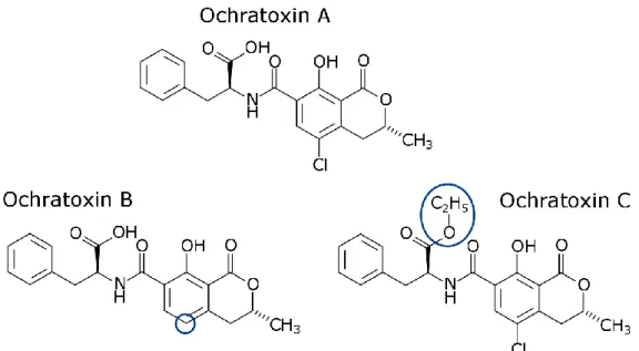
includes several related metabolites. While ochratoxin A is the most harmful, there are also other ones like the dechloro analog ochratoxin B, or its methyl ester ochratoxin C
Figure 1: Chemical structure of ochratoxin A, ochratoxin B and ochratoxin C. Blue circles highlight the differences.
OTA is severely toxic for humans and animals, displaying nephrotoxic37,38,
hepatotoxic,39,40 neurotoxic,41,42 teratogenic,43,44 immunotoxic,45,46 and potentially
carcinogenic47,48 effects on organisms.
The tolerable weekly intake of OTA has been set at 120 ng/kg by the European Food
Safety Authority (EFSA).49 More restrictive regulations have been established by other
agencies, for example the US Food and Drug Administration (FDA) considers the
tolerable daily intake to be 5 ng/kg.50
Since OTA has hazardous effects on the human health the testing of real food samples for it has been regulated by the European Union. While several possibilities for measuring the OTA content in food samples exist, the most commonly used is high-performance liquid-chromatography (HPLC) combined with fluorescence detection
systems (FD).51 HPLC-FD returns results quickly, and allows relatively precise
quantification with a limit of detection (LOD) of 0.55 ng/mL,52 but extensive sample
pretreatment is necessary, and parallel analysis for multiple targets is cumbersome. Other rather traditional methods of detection are:
Liquid chromatography – mass spectrometry (LC-MS/MS), LOD: 0.01 ng/mL 53
Enzyme-linked immunosorbent assays (ELISA), LOD: 0.3 ng/mL 54
1.2.3 Methods for detecting pathogens
The conventional methods for detecting pathogens can be very time-consuming and labor intensive. For example pathogenic bacteria are traditionally cultured on agar
plates with subsequent standard biochemical identification.56 More recently
developed methods are faster and can provide higher sensitivity and specificity. They include nucleic acid-based methods, immunology-based methods and biosensors.
Nucleic acid-based methods
The principle of the nucleic acid-based methods is based on the detection of
bacteria-specific DNA or RNA by hybridizing it with artificial complementary oligonucleotides.57
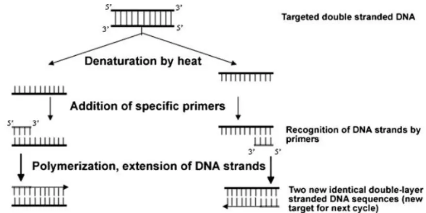
These methods include the probably most commonly used polymerase chain reaction (PCR), but also newer developments like quantitative PCR (qPCR), multiplex PCR (mPCR) and other strategies like microarray technology. PCR is a method for amplifying
nucleic acids, which was developed in the 80s by Mullis et al.58 The PCR process
generally consists of 20-50 cycles as displayed in Figure 2. In the first step (denaturation) the double-stranded DNA is heated to separate the strands. For the next step specific primers are added and anneal with the single strands of DNA. In the last step of the cycle the DNA-polymerase fills up the missing strands with free nucleotides, starting from the 3’-side of the primer and following the strand. The two new double strands can then be used for the next cycle, resulting in exponential
amplification.58–60
The main advantages of PCR lie in its high sensitivity and high specificity, but it can also be automated and yield reliable results. Nevertheless the results can be affected by PCR inhibitors and the DNA requires purification.
qPCR improves on normal PCR as in addition to PCRs advantages it allows rapid cycling, no post-amplification products are necessary and real-time monitoring is possible. At the same time it has to cope with the same disadvantages of PCR and being rather expensive and multiplex measurements are difficult.
Figure 2: Schema of one PCR cycle for the amplification of DNA.60
Immunology based methods
These methods rely on high specificity and affinity of antibodies towards their antigen. It is one of the few methods which has been successfully applied for the detection of
bacteria, viruses, spores and toxins.61 Some of the better known methods applied are
enzyme-linked immunosorbent assay (ELISA)62, magnetic immunoassay (MIA)63 and
lateral flow immunoassay (LFIA)64,65. ELISA is a very popular method, with the sandwich
method (see Figure 3) being its most effective form.57 For this method in the first step
(1) antibodies are immobilized before the targets are added (2). In the next step (3) an antigen detecting antibody binds to the target. Then an enzyme-labelled antibody binds to the detecting antibody (4). Finally (5), the enzyme’s substrate is added, which reacts to a detectable product.
Figure 3: Schema of sandwich ELISA process.
ELISA can be applied for a wide range of targets, is very specific and can be automated to reduce time and labor needs. Nevertheless it struggles with low sensitivity, the risk
of false high-positive results and especially the fact, that the antibody might interact
with similar antigens.66 Further challenges are posed by the controlled immobilization
of antibodies and the denaturation of the antibodies on surfaces.66
Biosensors
Biosensors are analytical devices with a biological component. They are used in many different applications, including environmental, electrochemical, clinical and
security.1,4,67,68 The analysis is based rather on function than on structure, and they can
be used to detect against a wide range of targets:
Toxins (food and feed stock contamination, security…)54,69
Carcinogens (food contamination, released by industrial processes…)70
Human vitals (blood glucose level, cancer…)71,72
Pesticides (food contamination, detection in environment, e.g. DDT)73,74
Drug detection (low level detection in environment or in pharmaceuticals)75,76
Microbial contamination (food, environmental hazards, security)77–79
Biosensors combine a biological recognition component (or bioreceptor) with a physico-chemical transducer and possibly an (electronic) amplifier, as displayed in Figure 4. The bioreceptor uses biological probes to interact with the analyte and directly converts its presence or concentration from a biological reaction into a physico-chemical effect, which is measurable by the transducer.
Figure 4: General structure of biosensors.
Transducer Sample Signal Target Probe Bioreceptor
The bioreceptors of biosensors can be separated into five different main types:
(i) Biological polymers including nucleic acids80,81, aptamers82–84 and peptides85
(ii) Proteins such as antibodies and antigens77,86
(iii) Enzymes67,87,88,abzymes (catalytic antibodies)89,90 and other proteins91,92
(iv) Whole cells and cellular fragments using plant or animal cells93,94,
microorganisms95, organelles96, cell receptors97,98, and tissue slices99
(v) Biomimetic materials100,101
There are various types of transducers, which convert different kinds of reactions into measurable signals and which depend on the intrinsic properties of the substrate. Electrochemical transducers detect an electrochemical reaction by monitoring the
change of an electrical signal at an electrode.102 Optical transducers detect the change
of refractive index,103 light interference,104 changes in local surface plasmon resonance
frequencies (LSPR)105 and other properties.106 Acoustic transducers are based on wave
propagation in solids. They exploit, for example, resonance-frequencies77 of
piezoelectric materials107,108 to detect surface interactions. There are also other
technologies which are used as transducers, among others gravimetric,109 thermal110
and magnetic.111
Biochips, in comparison to biosensors, allow the detection of multiple analytes in parallel. One well known and widely used example is the microarray, which allows the detection of a wide range of analytes on one slide. The two most common methods of detection with microarrays are fluorescence and SPR.
1.3 Aptamer-based biosensors
1.3.1 Aptamers as probes in biosensors
Aptamers are single stranded DNA or RNA molecules, which have been specifically selected to bind with high specificity and affinity to a certain target by a process called
becoming increasingly popular as alternative probes for biosensors, immunoassays and other analytical devices. Aptamers display several advantages over antibodies when used as probes:
(i) In-vitro selection and synthesis is faster, lowers the cost and can be conducted without animals, facilitating its use for toxic compounds and
increasing flexibility in selection conditions112–115
(ii) Synthesis can be conducted with high purity and reproducibility116
(iii) Smaller size enables higher surface densities117
(iv) Chemical modifications which don’t affect the affinity are relatively easy to conduct to enhance specificity, detection, stability and allow
immobilization11,118
(v) Higher thermal stability (reversible denaturation) and higher shelf live11,119
(vi) High versatility due to wider range of targets (cells, toxins, viruses, proteins,
even small molecules and metal ions)6,120–122
(vii) Low immunogenicity and low-toxicity123
(viii) Can show higher affinity and specificity towards target than antibodies115,120
Despite all these advantages, aptamers also have some limitations:
(i) Degradation by nucleases in biological media of RNA-aptamers limits the therapeutic applications. One of the propositions to avoid this degradation include the performance of SELEX with modified stability-enhanced
nucleotides124
(ii) Cross-reactivity with molecules of similar structure can be problematic, but binding can be avoided with negative SELEX, where aptamers that bind with
similar molecules are discarded125
(iii) Time and labor consuming generation, despite being easier and faster to produce than antibodies. Automated SELEX has been proposed by Cox et
al., which allows the generation of aptamers in several days126
The SELEX process which is used to find aptamers was developed in 1990 by the groups
process starts with a large library of randomized sequences (up to 1016) with lengths
usually around 30-50mer, but up to 100mer.115,121 In the first step (1), the sequences
are incubated with an immobilized target molecule. In the second step (2), the unbound oligonucleotides are removed from the target-bound ones. Then the oligonucleotides which were bound to the target are eluted (3) and amplified (4). The amplification is usually done with polymerase chain reaction (PCR) or real-time PCR
(RT-PCR).121 These amplified strands form the new library for the next round of this
cycle, where the stringency is increased (5) to raise the competition between the remaining oligonucleotides. The cycle is repeated 10-15 times, resulting in aptamers which recognize the target molecule. The stringency is set by the selection parameters (temperature, pH, buffer composition) and can be adapted to a small or broad range of working conditions for the aptamers.
Figure 5: Standard SELEX strategy. Starting with initial library of random sequences, the binding sequences (1) are isolated (2 -> 3) and amplified (4), before the cycle reiterates at more stringent conditions (5 -> 1).
Aptamers are mainly unstructured in solution, but form well-defined 3D-structures
when binding their ligands.127 The molecular architectures which are formed include
the ligand as an intrinsic part of its structure. Aptamers are also highly reusable, as they can preserve their structure when being subjected to repeated cycles of regeneration. The aptamers can be regenerated with heat, salt concentration, medium pH and chelating agents, while remaining undamaged if extreme limits are not
exceeded.11 The interaction of an aptamer with its target depends on the presence of
salts and the pH of the physiological media.128 The recognition of targets by an
aptamer is characterized by any combination of: hydrogen bonding (which can result in a pseudo-base pair involving the ligand), electrostatic interactions, intermolecular bonding, general acid-base interactions, shape complementarity, and folding of the
aptamer around the target, resulting in numerous intermolecular interactions.127
Aptamers as probes have shown a wide range of usability while being comparatively easy and cheap to produce. As the research on aptamer-based sensors advances they display more and more their competitiveness with alternative approaches.
1.3.2 Aptamers for the detection of OTA
The first aptamer towards OTA was published by Cruz-Aguado and Penner in 2008.128
The aptamer which they isolated contains 36 nucleotides with the sequence
GAT-CGG-GTG-TGG-GTG-GCG-TAA-AGG-GAG-CAT-CGG-ACA, and displays an affinity Kd of 49 nM
towards OTA. They also showed that the aptamer does not bind with chemically similar molecules like warfarin and N-acetyl-L-phenylalanine and has a strongly reduced affinity towards ochratoxin B. Upon binding with OTA this aptamer takes the
Figure 6: G-quadruplex structure of OTA-aptamer.
More recently, Barthelmebs et al. isolated two new 30mer aptamers against OTA with
dissociation constants of 96 nM and 130 nM.83 The specificity of the aptamers was also
tested towards ochratoxin B and phenylalanine. Despite this alternative, the more commonly used aptamer in literature is the first one found by Cruz-Aguado and Penner.
1.3.3 Aptamer-based biosensors for the detection of OTA
There is a large number of different aptamer based methods for the detection of OTA with electrochemical or optical detection methods. An overview of the methods and their sensitivities is given in Table 2. The different detection methods show a wide range of limit of detection (LOD). Depending on the underlying method the fluorescent and luminescent methods show high sensitivities with very low LODs (fM – pM range). Electrochemical methods also show relatively high sensitivities (pM – nM range), while the colorimetric methods are less sensitive (nM range). The response range of sensors is the range in which they show a linear response with the concentration of the target, and is therefore also an important characteristic of sensors.
Table 2: Examples of aptamer-based assays for OTA detection.
Detection method Method Response range LOD Ref.
Colorimetric Propagated chain reaction of hybridization events of
DNAzymes (catalytic DNA) in solution 0.01 - 0.32 nM 0.01 nM
129
Colorimetric Gold particles as indicator in solution 20 - 625 nM 20 nM 82
Fluorescent (FRET)
Au-nanoparticle (NP) coated with complementary, biotin-terminated strand (energy acceptor), Cy3- streptavidin (energy donor); in presence of OTA aptamer detaches and biotin and streptavidin interact resulting in signal decrease (in solution)
6.2 pM - 2.5 µM 3.7 pM 130
Fluorescent
Fluorophore labelled aptamer hybridized with quencher modified complementary strand; presence of OTA releases modified strand resulting in signal increase (in solution)
0.1 - 150 nM 5 fM 131
Luminescent
Aptamer immobilized on magnetic NP hybridizes with complementary strand on upconversion-NPs; OTA causes separation of strands and NPs, magnetic separation results in signal decrease (in solution)
0.25 fM - 2.5 nM 0.25 fM 132
LSPR Localised surface plasmon resonance (LSPR)-shift of
gold nanorods with immobilized aptamers (solution) 1 nM - 1 µM 1 nM
105
SERS Surface-enhanced Raman spectroscopy, aptamers
immobilized on Au-nanotriangles (on surface) 0.05 - 4 μM 0.05 µM
133
PCR-amplification signal
RT-PCR of DNA strands complementary to aptamer
after dehybridization in presence of OTA in solution 12.5 pM - 12.5 nM 2.5 fM
134
Electrochemical
Amplified electrochemical sandwich detection and methylene blue as redox indicator, aptamers immobilized on Au-NPs (in solution)
2.5 pM - 2.5 nM 0.75 pM 135
Electrochemical
Either OTA or Aptamers immobilized on
superparamagnetic NPs, reduced activity in presence of free OTA in solution
0.27 - 22.2 nM 0.27 nM 136
Electrochemical Voltammetry: Biotin labeled and free OTA compete to
bind with immobilized aptamers (in solution) 0.37 - 12.4 nM 0.17 nM
1.3.4 Challenges for biosensors
The performance of biosensors regarding sensitivity, selectivity, speed, stability, complexity, and processing depends strongly on the nature of the chosen bioreceptor and transducer. The substrate for the bioreceptor plays a major role, as its intrinsic properties define the possible measurement methods, which in turn also determine the biosensors speed, ease-of-use and possible miniaturization for portable devices. The need of high stability and reproducibility demands a reliable and well-controlled chemistry. This ensures dependable probe immobilization but also institutes the possibility of applying (antifouling) layers to avoid non-specific adsorption from targets such as proteins and bacteria. A reliable probe immobilization is also crucial for the dependability of the biosensor. The biosensors specificity and selectivity are mainly defined by the probes which are used. Another point to consider is that the probes are biological components and their production can be complex and their shelf life has to be considered before choosing a biosensor system.
1.4 Strategy proposed for detecting the interaction aptamer-OTA
The goal of this thesis is to study the interaction of pathogens with aptamers on a stable and reproducible biochip architecture based on an hydrogenated amorphous
silicon carbon alloy (a-Si1-XCX:H) deposited on an aluminium back-reflector for reliable
and sensitive detection of pathogens by fluorescence. The silicon surface enables the grafting of terminated organic monolayers with robust Si-C bounds. The acid-terminated layers are excellent candidates for a reliable immobilization of amine-terminated probes by covalent peptide bonds. The silicon substrate is not only the basis for a reliable immobilization but also provides a platform for analysis and quantification by infrared spectroscopy, as well as analysis with fluorescence for an increased sensitivity. On this architecture we introduce the interaction of the toxin ochratoxin A (OTA) with its 36mer DNA-aptamer as a model system.
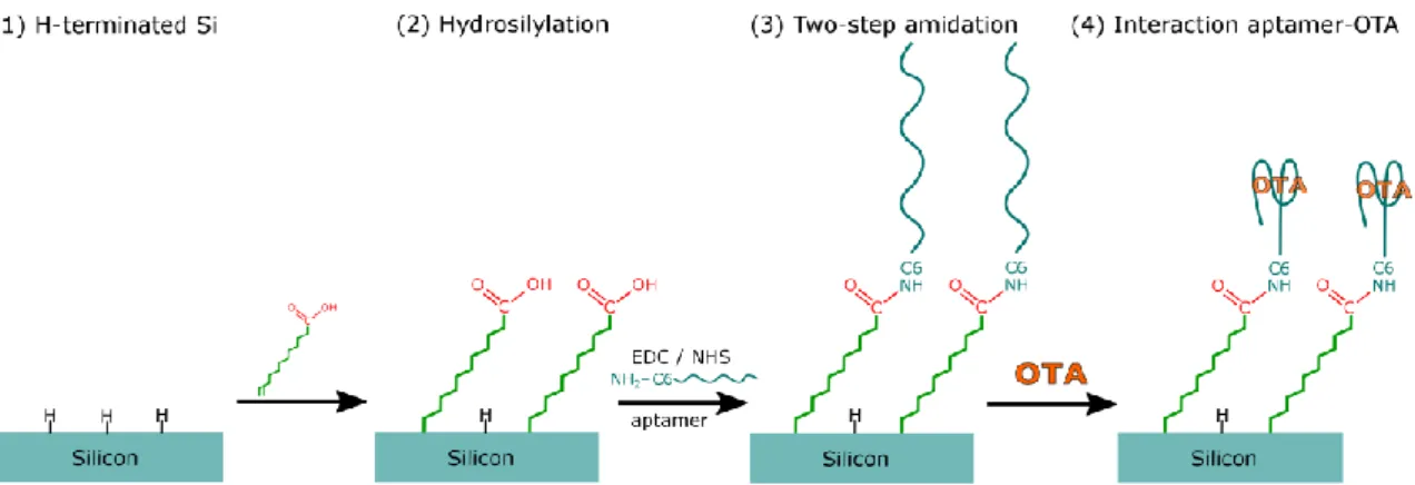
The well-controlled multi-step modification process which we carry out on silicon is displayed in Figure 7, and will be explained in more detail in chapter 2. Hydrogen terminated silicon surfaces (1) are used as a basis for the grafting of an acid-terminated organic monolayer by photochemical hydrosilylation (2). This results in a very stable monolayer attached by covalent Si-C bonds. The stable acid-terminated layer is then the basis for the next step (3), where the amino-terminated aptamers are immobilized on the surface by a two-step amidation process. The two-step amidation consists first of an activation reaction of the surface with the carbodiimide EDC in presence of NHS. This is followed by a subsequent aminolysis reaction, leading to aptamers immobilized on the organic monolayer by stable peptide bonds. This immobilization strategy provides the substrate with a high chemical stability with
respect to ageing and long exposure in physiological media. Once the aptamers are
Figure 7: Strategy for direct detection of the interaction aptamer-OTA on crystalline silicon surfaces.
Two detection methods were envisaged:
1.4.1 Direct detection of the interaction aptamer-OTA by ATR-FTIR
Carrying out the surface modifications on crystalline silicon (111) allows studying of all steps by Fourier-transform infrared spectroscopy in attenuated total reflexion geometry (ATR-FTIR). This enables the study and quantification of the surface functionalization, the probe immobilization and the interaction of OTA with its aptamer without any labelling.
1.4.2 Indirect detection of the interaction aptamer-OTA by fluorescence
The chemistry is transferred to the biochip architecture. While the chemistry remains largely the same (HF-etching, hydrosilylation, 2-step amidation), it is now conducted
on a thin layer of an amorphous silicon-carbon alloy (a-Si0.85C0.15:H) on an aluminium
back reflector. The role of the back reflector is to increase the fluorescence sensitivity
by constructive interference at the a-Si1-XCX:H layer.20
The local and defined deposition of different aptamers and oligonucleotides is carried out with a robotic spotter, giving it its biochip (multiplex) capabilities.
The biochips are based on an indirect detection of the interaction aptamer-OTA by fluorescence. Two different methods of detection on surfaces are proposed in
literature, “signal OFF” and “signal ON”. In the “signal OFF” detection method (Figure 8, left) short complementary oligonucleotides labelled with fluorophores are hybridized to the aptamers. Successful association with OTA removes the complementary strands, resulting in a decrease of fluorescence intensity. For the “signal ON” method (Figure 8 right), the attached aptamers have a fluorophore at their extremity, while the short complementary oligonucleotide has a quencher, which suppresses fluorescence of nearby fluorophores. Successful association of OTA will remove the quenchers and therefore increase the fluorescence signal.
Figure 8: Strategy for indirect detection of the interaction aptamer-OTA on crystalline silicon surfaces. Left side: "signal OFF", right side: "signal ON".
2 S
URFACE
F
UNCTIONALIZATION OF
2.1 Introduction
Silicon and its derivatives (e.g.: silicon dioxide, silicon carbide, silicon nitride)4,21,138,139 is
of interest as a substrate for biosensors because of the chemical and physical capacities (e.g.: surface reactivity, luminescence properties), and as it comes in various
forms (monocrystalline, polycrystalline, amorphous, porous, as nanoparticles)21,140–143.
This enables its use for a wide range of different detection methods, like optical,106,144
electronical,2,145 electrochemical,102,146 piezoelectrical,108,147 and gravimetrical.148,149
Silicon is to a certain degree biocompatible and can easily be miniaturized permitting
the implementation as in vivo devices.150 Furthermore, silicon surfaces can be readily
functionalized with a bio-recognition layer for probe immobilization and allows modular change of its physico-chemical properties for the development of patterned
or antifouling surfaces to avoid unspecific adsorption.151–153
The reliability, efficiency and sensitivity of biosensors strongly depends on the
robustness of the chemistry between the surface and the bio-recognition layer.106,154
Chemical stability and good reproducibility can be achieved by a well-controlled anchoring of the bio-receptive layer. Secondly, for the same reasons of reliability, efficiency and sensitivity, it also depends on the stability of further modifications of the bio-recognition layer which might be performed. These modifications are often the immobilization of probes and/or antifouling molecules. The applied chemistry must be dependable enough to guarantee stable binding, even when carrying out assay protocols in physiological media. Unreliable immobilization can impair the quality of the biosensors in terms of reproducibility and sensitivity, causing for instance reduced sensitivity due to probe loss. The immobilization of antifouling molecules is carried out to avoid unwanted non-specific adsorption on surfaces of for example proteins and bacteria. This is done to ensure that the detection is only dependent on the interaction of the probe with its target and is not affected by non-specific adsorption of the targets on the surface.
In this work we propose a multi-step process for well-controlled silicon surface modification for reliable biomolecular aptamer-target interaction. Our strategy (see Figure 9) consists of: Hydrogenation of silicon surfaces (1), to enable the grafting of carboxyl-terminated organic monolayers via stable Si-C bonds (2). This is followed by a
2-step amidation process (3 and 4), allowing the covalent immobilization of NH2
-terminated functional groups (as biological probes) in mild conditions. This chapter will start with the “state of the art” of surface functionalization, explaining in detail our method, while also pointing out some of the alternatives. In the final part the experimental results of the functionalization of oxide-free silicon surfaces will be discussed. The immobilization of the antifouling molecule poly(ethylene glycol) (PEG) was chosen as a proof of concept for the covalent binding of amine-terminated molecules on acid-terminated silicon surfaces. Its anti-biofouling properties towards bacteria are verified.
Figure 9: Strategy for silicon surface functionalization.
2.2 State of the Art
2.2.1 Functionalization of silicon surfaces
The functionalization of silicon surfaces generally follows two principle strategies,
either directly on the native oxidized silicon surfaces (SiOX), or on hydrogen-terminated
(a) Functionalization of native oxidized silicon surfaces
Two main methods are used for functionalizing the native silicon surface with organic
molecules. The first one developed was silanization, where alkoxysilane (Rm-Si-(OH)n)
or chlorosilane (Rm-Si-Cl) molecules are attached through covalent Si-O-Si bonds.155
Despite the fact that the silanization is relatively simple to carry out, it suffers under major disadvantages. Not only is the formation of reproducible and well-defined monolayers difficult, but also the Si-O bonds near the surface are easily hydrolyzed in
physiological buffers.156,157 The second method often used is the attachment of
phosphonate molecules where Si-O-P bonds are formed.158 Layers formed in this way
are easily reproducible, have higher surface coverages and don’t have the intrinsic risk
of multilayer formation.158,159 Despite resulting in organic layers which are more stable
than those obtained by silanization, it is still relatively easily hydrolyzed from the SiOX
surface.160 Another drawback is that the sample preparation requires heating and a
relatively long processing time (more than 48 h).161
(b) Functionalization of hydrogenated silicon surfaces
The removal of the native oxide layer from silicon surfaces by wet chemical etching opens up other pathways of stable and reliable surface modifications via stable covalent Si-C bonds which are not subject to hydrolysis. Hydrogenated surfaces are relatively easy to prepare and can be achieved by wet chemical etching in
fluoride-containing solutions at different pH.162,163 Despite the native oxide layer re-growing
relatively fast, these surfaces remain to some extent stable in air (at least one hour at
room temperature and 40% ambient humidity)164 and brief watering165 procedures.
Etching of Si (111) in basic NH4F results in atomically flat hydrogenated SiH surfaces163,
while etching in acidic HF yields atomically rough SiHX surfaces.165 Porous silicon can be
formed by different pathways, the first method found was stain etching, where the
silicon is etched with HF, nitric acid and water.166–168 Figure 10 shows the mechanism
which leads to the formation of hydrogenated silicon by HF etching on atomically rough surfaces. In the first step the removal of oxygen leads to Si-F at the interface. This causes the Si-Si back bond to be weakened by the highly electronegative fluoride.
The fluorinated silicon can then be removed by further HF attack in form of SiFX,
leaving as a consequence an H-terminated silicon surface.169,170
Figure 10: Mechanism of HF etching on silicon.171
In 1993 Linford and Chidsey reported the first formation of alkyl monolayers on non-oxidized silicon surfaces by covalent Si-C bonds, which were prepared by the pyrolysis
of diacyl peroxides ([CH3(CH2)nCO]2O2).172 Since then a wide range of different methods
have been developed to graft organic monolayers via stable Si-C bonds on different morphologies of hydrogenated silicon surfaces. The reaction can be conducted in two
ways: (i) electrochemically with Grignard reagents173,174, diazonium175 and other onium
salts (ammoniums and phosphoniums)176; or (ii) chemical hydrosilylation mediated
thermally172,177, photochemically17,151,178,179, or catalytically180,181.
The photochemical grafting of 1-alkenes is especially of interestbecause the reaction is
comparably fast and easy to handle and produces densely packed and stable monolayers with low surface oxidation. It also allows grafting of alkenes with different functional groups, which can then be used to link other molecular structures to silicon
surfaces.19,182,183 The choice of functional end groups R (see Figure 11) is limited by
their reactivity towards the hydrogenated silicon surface. This excludes the hydroxyl
(R-OH), amine (R-NH2) and aldehyde (R-CHO) groups, which would be of interest for
further functionalization. One way of grafting with functional end groups is by protecting them, then grafting the alkene chains, followed by the removal of the protection. In this context in the laboratory PMC the one-step and two-step grafting of
alkyl chains with the functional group COOH via stable Si-C bonds was studied in
(without or with protection, respectively).17 This method allows the grafting of
1-alkenes with the functional group carboxyl (R-COOH). In particular the direct photochemical grafting of undecylenic acid at 312 nm was studied, resulting in carboxylic acid (COOH) terminated monolayers. The obtained surfaces showed no formation of siloxane (Si-O-C=O), confirming that the carboxyl group does not react with the surface. The surfaces exhibited fairly dense organic monolayers (surface
densities up to 2.7∙1014 cm-2). The grafting also displayed no surface oxidation, yielding
excellent electronic properties of the interface.17
Figure 11: Scheme of the formation of organic monolayers through covalent Si-C bonds.
Linford et al. proposed a chain propagation reaction mechanism (see Figure 12) for the
UV-induced grafting.172 The UV light cleaves the Si-H and thereby creates a surface
radical. This surface radical will attack the terminally unsaturated alkene, with the binding of the alkene by its α-carbon to the Si atom. This results in a radical on the grafted molecule on the β-carbon, which then removes a hydrogen atom at a neighboring SiH site, resulting in a new surface radical for further alkene
attachment.172
Figure 12: Reaction mechanism of UV-induced grafting of 1-alkenes on hydrogen
The grafting of organic monolayers on silicon surfaces is also possible by visible light. Stewart and Buriak introduced an alternative route for grafting on porous hydrogen
terminated silicon under mild reaction conditions using white light.185 They proposed a
grafting mechanism, where the illumination with white light at moderate intensity
creates relatively long-lived surface-localised excitons (electron-hole pairs).186 The
electron-hole pair is separated and results in a positive localised surface charge. The unsaturated alkene can then undergo a nucleophilic attack by the hole, yielding a carbon centred radical nucleophilic. This radical can bond a hydride, and neutralize the
charge.186,187 Later on Hamers found a way of grafting on crystalline silicon surfaces by
temporarily terminating the silicon surface with iodine by exposure to 514 nm light.188
The group of Zuilhof then found a method of grafting alkyl monolayers directly on
crystalline silicon with visible light (447 nm) at room temperature.189,190
Wang et al. proposed that the reactions involved in grafting are based on the creation of a valence-band hole, which can be either created by an exciton mechanism or
photoemission. In the exciton mechanism the light creates an electron-hole pair.191,192
The valence-band hole can also be created by a photoemission reaction initiated by UV light, where an electron from the silicon surface is excited to an acceptor level of electron acceptors (which can include the reactant molecules), leaving a valence-band hole. The valence-band hole, created by either mechanism, can then induce a nucleophilic attack by the alkenes, resulting in a stable Si-C bond. The reaction mechanism is dependent on the wavelength of the light. At short wavelengths the photons excite the electrons to the electron-acceptor levels of the alkenes, resulting in
more efficient photoemission despite the fact that both are possible.192 At longer
wavelengths the valence-band hole creation is likely limited to the exciton mechanism. Therefore the grafting mechanism is defined by the wavelength and the electronic structure of the molecules.
2.2.2 Two-step amidation of acid terminated surfaces
The acid-terminated surfaces are good candidates for the attachment of biomolecules in mild conditions by amide bonds. They can be immobilized on acid terminated