REBUBLIQUE ALGERIENNE DEMOCRATIQUE ET POPULAIRE
MINISTERE DE L’ ENSEIGNEMENT SUPERIEUR ET DE LA RECHERCHE SCIENTIFIQUE UNIVERSITE FERHAT ABBAS SETIF
THESE
Présentée à la Faculté des Sciences Département de Biologie Pour l’obtention du diplôme de
DOCTORAT D’ ETAT
Option: Biologie AnimalePar
Mme. DAHAMNA Saliha
THEME
BIOCHEMICAL AND HISTOLOGICAL
INDICATORS OF ATRACTYLIS GUMMIFERA L.
TOXICITY
Soutenu le: 08 / 12 / 2007 Devant le Jury:
Président: Dr. Touabti A. Pr. Université de Sétif Directeur de thèse: Dr. Sekfali N. Pr Université de Sétif Co-Directeur de thèse: Dr. Abekane A. Pr. Université e Constantine Examinateur: Dr. Baghiani A. M.C. Université de Sétif
ACKNOWLEDGMENTS
I wish to express my gratitude to all who helped me during the course of this thesis. My sincere gratitude is due to my supervisors, Prof. Nadia Sekfali and Prof. Abdelhamid Aberkane for the encouragement, exceptional ideas, and for their invaluable scientific wisdom and knowledge that have kept me going and for sharing with me some of their experience.
I am especially grateful to Prof. Luciano Saso and Dr. Claudia Daniele (Department of Pharmacology of Natural Substances and General Physiology, University of Rome, Italy) for their fruitful co-operation during my stay in Italy.
My thanks are also due Prof. A. Taouabti and his colleagues and staff in the hospital central laboratory for their valuable contributions in blood analysis.
I wish also to express my sincere thanks to the chairman ( Prof. A. Touabti) and members of the jury ( Dr. A. Baghiani and Dr. S. Laroui) who accept to examine my work.
My warm thanks are due to all my friends , colleagues and the staff at the department of biology, faculty of science, university Ferhat Abbass, Sétif.
The special thanks go to my family: my husband Kamal and my children Safa, Wafa, Moussa, Sara and Salem . I want to thank my sisters Nora , Houria and my brother in law Daoud for happy moments and for all help throughout the entire period of this research undertakings.
I am also indebted to the following friends: Khennouf Seddik, Smain Amira, Fatima Amira, Hamama Bouriche and others for their loving and encouragement support in the course of my almost endless years of study.
SUMMARY
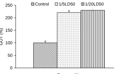
Herbal drugs are widely used and often contain high active pharmacological compounds. Hepatotoxicity of herbal remedies ranges from mild liver enzyme alterations to chronic liver disease and liver failure. Atractylis gummefera is one of the toxic plants in Algeria, it’s toxicity is due to its active substances mainly atractyloside and carboxyatractyloside. Hepatotoxic reactions have been observed often by ingestion of Atractylis gummefera.The intoxications, usually lead to collective death in rural places. Especially the children between 4-13 years. In this study animals were treated with different doses of rhizome extract according to their body weight. At one experiment, acute toxicity, one dose was used on rats which corresponds to 1/5 LD50 Atractylis gummifera and sacrified after the 1st day, 3rd 5th, 7th, 10th, and 14th, . Biochemical, haematological and
histological studies were carried on. The results showed an important alteration in liver tissue; necrosis in the hepatic parenchyma. Fluctuations in plasma glucose levels were observed in all treated groups. These generally followed by hypoglycaemia. Enzymes activities of plasma GOT and GPT were increased. These results indicate that Atractylis gummifera rhizome extract causes structural and functional changes of liver. The study of the toxicity of Atractylis gummifera rhizome extract on rabbits (Oryctolagus cunilicus & New Zealand White rabbits), treated with 1/5LD50 and 1/20 LD50 compared with K atractylate (6.5, 13, 32 umole/kg), The histopathological results showed lesions and morphological changes of the hepato-cellular and confirmed disturbances of the biochemical parameters , these changes were much underlines during the animal toxicity. The acute toxicity of this plant extract on rat male reproductive system showed a clear modification of sperm morphology, especially the flagella. Testicular and epididymal morphology was also impaired. It is concluded that Atractylis gummifera may cause morphological and functional alteration of the male reproductive tract. Another study, was focused on oxygen consumption and glucose absorption by rat intestine loops to get some knowledge about the effect of AG extract. Warburg technique was used and oxygen consumption was reduced by 30% . The inhibitory effect of rhizome extract on oxygen consumption, was clear in the presence of glucose, and reached approximately 56%.
Key words: Atractylis gummefera, Biochemical parameters, Toxicity, Histological alterations
Résumé
Les drogues végétales sont largement utilisées suite à leur contenu élevé en composés pharmacologiques actifs. La toxicité hépatique des médicaments d’origine végétale varie des altérations modérées de l’enzyme du foie à des maladies chroniques voire l’arrêt fonctionnel de cet organe. Atractylis gummifera est une des plantes toxiques algériennes, elle tire sa toxicité de ses substances actives notamment l’actractyloside et les carboxyatractyloside. Les réactions de toxicité hépatique ont été souvent observées suite à l’ingestion de l’Atractylis gummifera. Ces intoxications conduisent les plus souvent à la mort collective, en milieu rural, des enfants de 4 à 13 ans. Dans la présente étude les animaux ont été traités avec différentes doses l’extrait rhizomateux, en fonction du poids corporel. Dans une expérience, la dose utilisée sur des rats correspond au 1/5 de la DL50
d’Atractylis gummifera et ces sujets ont été sacrifiés après 1ier , le 3ieme , 5 ieme, 7 ieme, 10 ieme et 14 ieme jours. Des examens biochimiques, hématologiques et histologiques ont été opérés. Les résultats indiquent une altération importante des tissus du foie et des nécroses du parenchyme hépatique. Des fluctuations du niveau du plasma glucosique ( vs glucose plasmique) ont été notées chez tous les groupes traités, et sont généralement suivies par une hypoglycémie. Les activités enzymatiques du plasma GOT et GPT augmentent. Ces résultats indiquent que l’extrait rhizomateux d’Atractylis gummefera induit des changements structurels et fonctionnels du foie.
L’étude de la toxicité de l’extrait rhizomateux d’Atractylis gummifera sur les lapins
(Oryctogalus cunilicus et les lapins blancs de la Nouvelle Zélande), traités avec 1/5 DL50 et 1/20 DL50 ,
est comparée au K atractylate ( 6.5, 13 et 32 µmole/kg) Les résultats histo- pathologiques montrent des lésions et des changements morphologiques des cellules hépatiques et confirment les changements des paramètres biochimiques. Ces changements ont été mis en relief au cours de l’intoxication de l’animal. La toxicité de l’extrait de cette plante, sur le système reproducteur des rats mâles, montre une nette modification de la morphologie du sperme, notamment le flagelle. La morphologie des testicules et de l’épididyme a été aussi altérée. . Il a été conclu que l’Atractylis gummifera cause des altérations morphologiques et fonctionnelles de l’appareil reproducteur du rat mâle. L’autre expérience s’est focalisée sur la consommation de l’oxygène et l’absorption du glucose par l’intestin des rats pour avoir des informations sur l’effet de l’extrait AG. La technique Warburg a été utilisée et la consommation de l’oxygène a été réduite de 30%. L’effet inhibiteur de l’extrait rhizomateux sur la consommation de l’oxygène est net, en présence du glucose et atteint approximativement 56%.
ﺺﺨﻠﻣ
ﺎﻣ ﺎﺒﻟﺎﻏ ﻭ ﻝﺎﻤﻌﺘﺳﻻﺍ ﺔﻌﺳﺍﻭ ﺔﻴﺒﺸﻌﻟﺍ ﲑﻗﺎﻘﻌﻟﺍ ﺔﻴﻟﺎﻋ ﺔﻴﻟﺎﻌﻓ ﺕﺍﺫ ﺔﻴﻧﻻﺪﻴﺻ ﺕﺎﺒﻛﺮﻣ ﻰﻠﻋ ﻱﻮﺘﲢ . ﻰﻠﻋ ﻞﻤﻌﺗ ﺏﺎﺸﻋﻷﺎﺑ ﻱﻭﺍﺪﺘﻟﺍ ﺔﻄﺳﺍﻮﺑ ﺪﺒﻜﻟﺍ ﺔﻴﲰ ﺕﺎﺒﻧ ، ﻱﺪﺒﻛ ﻞﺸﻓ ﻭ ﺪﺒﻜﻟﺎﺑ ﺩﺎﺣ ﺽﺮﻣ ﱃﺇ ﺔﻳﺩﺆﻣ ﺪﺒﻜﻟﺍ ﺕﺎﳝﺰﻧﺇ ﰲ ﻞﻠﺧ ﺙﺍﺪﺣﺇ Atractylis gummefera ﺮﺋﺍﺰﳉﺍ ﰲ ﻑﻭﺮﻌﳌﺍ ﻭ ﺘﺣﺍ ﱃﺇ ﻪﺘﻴﲰ ﻊﺟﺮﺗ ﻭ ﺔﻣﺎﺴﻟﺍ ﺕﺎﺗﺎﺒﻨﻟﺍ ﺪﺣﺃ ﻮﻫ ﺩﺍﺩﻷﺎﺑ ﺔﺻﺎﺧ ﺔﻟﺎﻌﻓ ﺩﺍﻮﻣ ﻰﻠﻋ ﻪﺋﺍﻮ ) ATR ( atractyloside carboxyatractyloside ) CATR .( ﺪﻨﻋﻭ ﺔﻴﻔﻳﺮﻟﺍ ﻁﺎﺳﻭﻷﺍ ﰲ ﺔﺻﺎﺧ ﺔﺘﻴﳑ ﻥﻮﻜﺗ ﻭ ﺭﺮﻜﺘﺗ ﺎﻣ ﺎﺒﻟﺎﻏ ﺩﺍﺩﻷﺍ ﺕﺎﺒﻨﺑ ﻢﻤﺴﺘﻟﺍ ﺙﺩﺍﻮﺣ ﻥﺃ ﺎﻤﻛ ﲔﺑ ﺎﻣ ﻢﻫﺭﺎﻤﻋﺃ ﺡﻭﺍﺮﺘﺗ ﻦﻳﺬﻟﺍ ﻝﺎﻔﻃﻷﺍ 4 -13 ﻲﺟﻼﻋ ﺭﺪﺼﻤﻛ ﺕﺎﺒﻨﻟﺍ ﻝﺎﻤﻌﺘﺳﺍ ﱃﺇ ﺐﺒﺴﻟﺍ ﻊﺟﺮﻳﻭ ،ﺔﻨﺳ ﻮﻠﳊﺍ ﻩﺭﻭﺬﺟ ﻕﺍﺬﻣﻭ ﺽﺍﺮﻣﻷﺍ ﺾﻌﺒﻟ . ﺕﺎﻧﺍﻮﻴﳊﺍ ﺖﻠﻣﻮﻋ ) ﺐﻧﺍﺭﺃﻭ ﻥﺍﺮﺌﻓ ( ﻥﺍﻮﻴﳊﺍ ﻥﺯﻭ ﺐﺴﺣ ﻰﻠﻋ ﻭ ﺔﻋﺮﳉﺍ ﻰﻠﻋ ﺍﺩﺎﻤﺘﻋﺍ ﺍﺬﻫ ﻭ ﺩﺍﺩﻷﺍ ﺕﺎﺒﻧ ﺺﻠﺨﺘﺴﲟ . ﻝﺎﻤﻌﺘﺳﺍ ﺏﺭﺎﺠﺘﻟﺍ ﺪﺣﺃ ﻝﻼﺧ ﰎ ﺏ ﺓﺭﺪﻘﻣ ﺓﺪﺣﺍﻭ ﺔﻋﺮﲜ ﻥﺍﺮﺌﻔﻟﺍ ﺖﻠﻣﻮﻋ ﻦﻳﺃ ، ﺓﺩﺎﳊﺍ ﺔﻋﺮﳉﺍ 1 / 5 LD50 ﻥﺯﻭ ﺐﺴﺣ ﻰﻠﻋ ﺩﺍﺩﻷﺍ ﺕﺎﺒﻧ ﺺﻠﺨﺘﺴﻣ ﻦﻣ ﻥﺍﻮﻴﳊﺍ . ﺕﺎﻧﺍﻮﻴﳊﺍ ﺖﻠﺘﻗ ﻭ ﺪﺣﺍﻭ ﻡﻮﻳ ﺪﻌﺑ 3 ﻭ 5 ﻭ 7 ﻭ 10 ﻭ 14 ﺔﻠﻣﺎﻌﳌﺍ ﻦﻣ ﺎﻣﻮﻳ . ﺔﻴﺠﻴﺴﻨﻟﺍﻭ ﺔﻳﻮﻣﺪﻟﺍﻭ ﺔﻴﺋﺎﻴﻤﻴﻛﻮﻴﺒﻟﺍ ﺔﺳﺍﺭﺪﻟﺍ ﺖﲤ . ﻯﻮﺘﺴﻣ ﻰﻠﻋ ﺔﻣﺎﻫ ﺕﺍﲑﻐﺗ ﺞﺋﺎﺘﻨﻟﺍ ﺕﺮﻬﻇﺃ ﺪﺒﻜﻟﺍ ﺔﻴﻤﻴﺸﻧﺍﱪﺑ ﺓﺯﺮﻜﻧﻭ ﻱﺪﺒﻜﻟﺍ ﺞﻴﺴﻨﻟﺍ . ﺭﺎﻘﻣ ﺔﻠﻣﺎﻌﳌﺍ ﺕﺎﻧﺍﻮﻴﳊﺍ ﻞﻛ ﺪﻨﻋ ﺯﻮﻛﻮﻠﻐﻟﺍ ﺕﺎﻳﻮﺘﺴﻣ ﰲ ﺏﺬﺑﺬﺗ ﻚﻟﺬﻛﻭ ﺪﻫﺍﻮﺸﻟﺎﺑ ﺔﻧ . ﺔﻋﻮﺒﺘﻣ ﺖﻧﺎﻛ ﺪﻗﻭ ﺯﻮﻛﻮﻠﳉﺍ ﰲ ﻁﻮﺒ Hypoglycemie . ﺕﺎﳝﺰﻧﺍ ﺔﻴﻃﺎﺸﻧ ﰲ ﻉﺎﻔﺗﺭﺍ ﻚﻟﺬﻛﻭ TGO ﻭ TGP . ﺕﺎﺒﻧ ﺭﺬﺟ ﺺﻠﺨﺘﺴﳌﺍ ﻥﺄﺑ ﺞﺋﺎﺘﻨﻟﺍ ﲔﺒﺗ ﺪﺒﻜﻟﺎﺑ ﺔﻴﺠﻴﺴﻧﻭ ﺔﻴﺒﻴﻛﺮﺗ ﺕﺍﲑﻐﺗ ﺐﺒﺴﻳ ﺩﺍﺩﻷﺍ . ﻉﻮﻧ ﻦﻣ ﺐﻧﺍﺭﻷﺍ ﻰﻠﻋ ﺩﺍﺩﻷﺍ ﺕﺎﺒﻧ ﺭﺬﺟ ﺔﻴﻤﺴﻟ ﺔﺳﺍﺭﺩ ﰲ Oryctolagus cunilicus ﻭ
New Zealand White ﺔﻋﺮﲜ ﺔﻠﻣﺎﻌﳌﺍ 1 / 5 LD50 ﻭ 1 / LD50 20 ﺓﺩﺎﲟ ﺔﻧﺭﺎﻘﻣ Atractylate ﻝﺪﻌﲟﻭ ﻡﻮﻴﺳﺎﺗﻮﺒﻟﺍ ) 6.5 ﻭ 13 ﻭ 32 ﻝﻮﻣﻭﺮﻜﻴﻣ / ﻎﻠﻛ .( ﺙﻭﺪﺣ ﺔﻴﺋﺎﻴﻤﻛﻮﻴﺒﻟﺍ ﺕﺍﺮﺷﺆﳌﺍ ﺖﺒﺜﺗ ﺚﻴﺣ ﺔﻳﺪﺒﻜﻟﺍ ﺎﻳﻼﺨﻠﻟ ﺔﻴﺟﻮﻟﻮﻓﺭﻮﻣ ﺕﺍﲑﻐﺗﻭ ﺕﺎﻗﺰﲤ ﺙﻭﺪﺣ ﺔﻴﺠﻴﺴﻨﻟﺍ ﺞﺋﺎﺘﻨﻟﺍ ﺕﺮﻬﻇﺍ ﺪﺒﻜﻟﺎﺑ ﺕﻻﻼﺘﺧﺍ . ﺎﻣﻮﻤﻋ ﻥﺍﻮﻴﳊﺍ ﻢﻤﺴﺗ ﻝﻼﺧ ﺕﺍﲑﻐﺘﻟﺍ ﻩﺬﻫ ﻆﺣﻼﺗ . ﺯﺎﻬﳉﺍ ﻰﻠﻋ ﺩﺍﺩﻻﺍ ﺕﺎﺒﻧ ﺭﺬﺟ ﺺﻠﺨﺘﺴﲟ ﺓﺩﺎﳊﺍ ﺔﻴﻤﺴﻟﺍ ﺔﺳﺍﺭﺩ ﺕﺮﻬﻇﺃ ﻁﻮﺴﻟﺍ ﻯﻮﺘﺴﻣ ﻰﻠﻋﻭ ﻦﻣﺎﻴﺤﻠﻟ ﻲﺟﺭﺎﳋﺍ ﻞﻜﺸﻟﺍ ﰲ ﺕﺎﻫﻮﺸﺗﻭ ﺔﻠﻣﺎﻌﳌﺍ ﺪﻌﺑ ﺔﺤﺿﺍﻭ ﺕﺍﲑﻐﺗ ﻥﺍﺮﺌﻔﻟﺍ ﺭﻮﻛﺬﻟ ﻲﻠﺳﺎﻨﺘﻟﺍ . ﺺﻠﺨﺘﺴﻣ ﲑﺛﺄﺗ ﻥﺎﻛ ﻚﻟﺬﻛﻭ ﻰﻠﻋ ﺢﺿﺍﻭ ﻞﻜﺸﺑ ﻭ ، ﺩﺍﺩﻻﺍ ﺕﺎﺒﻧ ﺭﺬﺟ ﺦﺑﱪﻟﺍ ﺔﻴﺋﻼﻃﻭ ﺔﻴﺼﳋﺍ . ﺔﻴﺟﻮﻟﻮﻓﺭﻮﻣ ﺕﺍﲑﻐﺗ ﺐﺒﺴﻳ ﺩﺍﺩﻷﺍ ﺕﺎﺒﻧ ﻥﺄﺑ ﺔﺳﺍﺭﺪﻟﺍ ﻩﺬﻫ ﻦﻣ ﺺﻠﺨﺘﺴﻧﻭ ) ﺔﻴﻠﻜﺷ ( ﻱﺮﻛﺬﻟﺍ ﻱﺮﺛﺎﻜﺘﻟﺍ ﺯﺎﻬﳉﺍ ﻰﻠﻋ ﺔﻴﻔﻴﻇﻭﻭ . ﻰﻠﻋ ﺍﺩﺎﻤﺘﻋﺍ ﲔﺠﺴﻛﻭﻷﺍ ﻙﻼﻬﺘﺳﺍ ﺮﻳﺪﻘﺗ ﰎ ،ﺓﺮﻳﺎﻐﻣ ﺔﺳﺍﺭﺩ ﰲ ﺔﻴﻨﻘﺗ WARBURG ﺯﻮﻛﻮﻠﻐﻟﺍ ﺹﺎﺼﺘﻣﺍﻭ
ﺴﻣ ﲑﺛﺄﺗ ﺔﻓﺮﻌﳌ ﺭﺄﻔﻟﺍ ﻲﻌﻣ ﺕﺎﻘﻠﺣ ﻑﺮﻃ ﻦﻣ
ﺕﺎﺒﻧ ﺺﻠﺨﺘ
Atractylis gummifera L..
ﺽﺎﻔﳔﺍ ﺞﺋﺎﺘﻨﻟﺍ ﺕﺮﻬﻇﺃ
ﱄﺍﻮﲝ ﺔﻜﻠﻬﺘﺴﳌﺍ ﲔﺠﺴﻛﻭﻷﺍ ﺔﺒﺴﻧ
30
%ﺩﻮﺟﻭ ﰲ ﺎﺤﺿﺍﻭ ﻥﺎﻛ ﲔﺠﺴﻛﻭﻷﺍ ﻙﻼﻬﺘﺳﺍ ﻰﻠﻋ ﺕﺎﺒﻨﻟﺍ ﺭﺬﺟ ﺺﻠﺨﺘﺴﳌ ﻲﻄﻴﺒﺜﺘﻟﺍ ﲑﺛﺄﺘﻟﺍ ﻥﺃ ﺎﻤﻛ
ﺎﺒﻳﺮﻘﺗ ﻪﺘﺒﺴﻧ ﺖﻐﻠﺑ ﺚﻴﺣ ﺯﻮﻛﻮﻠﻐﻟﺍ
56
% .،ﺔﻴﺋﺎﻤﻴﻛﻮﻴﺒﻟﺍ ﺕﺍﺮﺷﺆﳌﺍ ،
، ﺔﻴﻤﺴﻟﺍ
ﻟﺍ
ﺔﻴﺠﻴﺴﻨﻟﺍ ﺕﺍﲑﻐﺘ
Atractylis gummifera
:
ﺔﻟﺍﺪﻟﺍ ﺕﺎﻤﻠﻜﻟﺍ
ABREVIATIONS
ADP : Adenosine Di Phosphate . AG: Atractylis Gummifera ATP: Adenosine Tri phosphate ATR: Atractyloside
GPT : Glutamate pyruvate Transaminase . GOT : Glutamate Oxaloacetate Transaminase . GSH: Reduced Glutathione
PAH: Phosphatase Alkaline . CSA : Cyclosporin .
LD 50: Lethal dose.
ANT : Adenine nucleotide Translocase. GSSG: Oxidized Glutathione . DTT: Dithiothreitol Transaminase . VRP: Verapamil NAC: N-Acetylcysteine SDH: Sorbitol dehydogenase STEV : Steviosid
GLDH : Glutamate Lactate Dehydrogenase . MDH: Malate Dehydrogenase
8-GT : Gamma Glutamyle Transferase. RBC : Red Blood Cells
WBC : White Blood Cells HB : Hemoglobine
LIST OF FIGURES
Figure 1: Atractilis gummifera L. Figure 2: Atractilis gummifera L.
Figure 3: Contents of atractyloside in rhizomes of Atratylis gummifera L. from Sardinia and Sicily in different time of the years
Figure 4: Chemical structure of atratylosides of Coffea arabica
Figure 5: A schematic mechanism of enzyme leakage under pathological conditions Figure 6: Atractylis gummifera L., Rhizome (a) aerial part (b)
Figure 7: Steps of the plant material extraction of Atractylis gummifera L.
Figure 8: Effects of Atractylis gummifera L. extract on plasma glucose (g/L) in rabbits after the treatment.
Figure 9: Effects of Atractylis gummifera L. extract on plasma GOT(IU/L) in rabbits after the treatment.
Figure 10: Effects of Atractylis gummifera L. extract on plasma GPT (IU/L) in rabbits after the treatment.
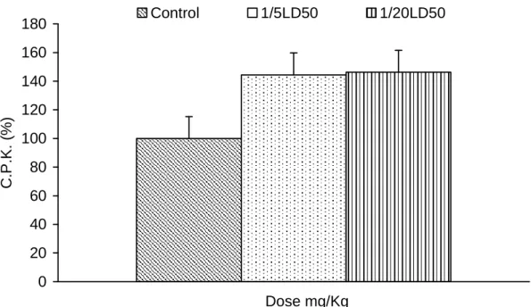
Figure 11: Effects of Atractylis gummifera L. extract on plasma CPK (IU/L) in rabbits after the treatment.
Figure 12: Effects of Atractylis gummifera L. extract on plasma Alkaline phosphatase (IU/L) in rabbits after the treatment.
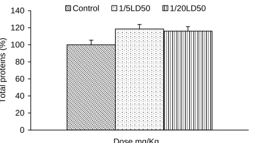
Figure 13: Effects of Atractylis gummifera L. extract on plasma proteins (g/L) in rabbits after the treatment.
Figure 14: Effects of Atractylis gummifera L. extract on red blood cells( RBC) in rabbits after the treatment
Figure 15: Effects of Atractylis gummifera L. extract on white blood cells (WBC) in rabbits after the treatment.
Figure 16: ffects of Atractylis gummifera L. extract on plasma Hb in rabbits after the treatment.
Figure 17: Effects of Atractylis gummifera L. extract on plasma hematocrite in rabbits after the treatment
Figure 18: Micrograph of rabbit blood stream treated with 76mg/kg of Atractylis gummifera extract, shows echinocytes transformation.
Figure 19: Micograph of a liver section from rabbit treated with 76 mg/kg Atractylis gummifera L. rhizome extract, showing centro-lobular hemorragy, necrosis and congetion of sinusoids.
Figure 20: Effects of Atractylis gummifera L. rhizome extract (76mg/Kg) on glucose in rats, after 1,3,5,7,10 and 14 days expressed as a percentage. Figure 21: : Enzymatic activities of plasma GOT and GPT in female rats treated
with Atractylis gummifera rhizome extract .
Figure 22: Micrograph of a section of liver from female rat treated with 76 mg/Kg Atractylis gummifera L. rhizome extract showing hepatocellular
necrosis,Peri-centro-lobular necrosis, sinusoids congestion and steatose.
Figure 23: Effect of Atractylis gummifera L. extract and K atractylate on plasma glucose
Figure 24: Effect of Atractylis gummifera L. extract and K atractylate on plasma GOT Figure 25: Effect of Atractylis gummifera L. extract and K atractylate on plasma GPT Figure 26: Effect of Atractylis gummifera L. extract and K atractylate on plasma
protein
Figure 27: Microgpraph of liver section from female rabbit treated with 76 mg/kg Atractylis gummifera rhizom extract showing hepato-cellular necrosis.
Figure 28: Microgpraph of liver section from female rabbit treated with 32 µmole/kg K atractylate showing hepato-cellular necrosis and hemorragy.
Figure 29: Microgpraph of kidney section from female rabbit treated with 76 mg/kg Atractylis gummifera rhizom extract showing congestion
Figure 30: Epididymal sperm velocity after acute Atractylis gummifera L. rhizome extract ingestion (120 mg/Kg).
Figure 31: Increased spermatozoal dysmorphology associated with acute Atractylis gummifera L. rhizome extract ingestion (120 mg/Kg). Figure 32: : Atractylis gummifera L.-induced sperm dismorphology: epididymal
spermatozoa were fixed, stained and examined for morphological anomalies.
Figure 33: Transverse sections of testes of normal and treated male rats with 120mg/Kg. of Atractylis gummifera L.rhizome extract.
Figure 35: QO
2consumption (100µl/100mg/wet fresh weight) from
intestinal rat loop in presence and in absence of DMSO
Figure 36: QO2 consumption (100µl/100mg/wet fresh weight) from intestinal rat loop
in presence of glucose and in presence of glucose + DMSO
Figure 37: QO2 consumption (100µl/100mg/wet fresh weight) from intestinal rat loop in presence and absence of glucose (12mM)
Figure 38: QO2 consumption (100µl/100mg/wet fresh weight) from intestinal rat loop in presence and in absence of DMSO.
Figure 39: Qo2 consumption (100µl/100mg/wet fresh weight) from intestinal rat loop in presence and absence of AG (15mg/ml) dissolved in DMSO
Figure 40: QO2 consumption (100µl/100mg/wet fresh weight) from intestinal rat loop in presence of glucose (12mM) ,in presence
LIST OF TABLES
Table. 1: Contents of atractyloside in rhizomes of Atratylis gummifera L. from Sardinia and Sicily in different time of the years
Table. 2: Case reports of Atratylis gummifera L. poisoning
GENERAL INTRODUCTION
In some regions of the world, plant poisoning continues to be an infrequent though important problem causing clinical morbidity and mortality. Among plants occasionally involved in human poisonings is Atractylis gummifera L., especially in countries where the plant grows spontaneously (Hamouda et al., 2000). Because Atractylis gummifera L. is easily confused with a wild artichoke most poisonings are unintentional. Many of the victims are children because Atractylis gummifera has sweet tasting juice and children enjoy chewing the root-like chewing gum (Stickel et al., 2000).
Atractylis gummifera L., is a toxic plant widely distributed through the worldwide, but especially in North Africa and the Mediterranean countries. This plant has been used in folk medicine or taken mistake (Caravaca et al., 1985). Atractyloside (ATR) is one of a group of diterpenoide glycosides. The primary mechanism of atractyloside poisoning is known to be inhibition of the mitochondrial ADP transporter and thus blocks oxidative phosphorylation, which prevents the synthesis of ATP and leads failure of gluconeogenesis and ultimately cell death (Obatomi and Bach., 1998).
The consumption of this plant, containing ATR causes acute fatal renal and hepatic necrosis in animals and humans (Obatomi and Bach, 1998).Reversible hypoglycaemia accompanied by depletion of hepatic glycogen was observed during in vivo investigations on the carbohydrate metabolism of rats and rabbits treated with the glycosidic fraction of the extract obtained from unroasted coffee-beans of coffee robusta which contains three glycosides closely related to ATR, which is a powerful competitive inhibitor of the ADP transport (Casio et al., 1994).
Clinical manifestations are related to the induced hypoglycemia and neurovegetative disorders or subsequent renal failure (Larrey, 1994). Epigastric pain, vomiting, respiratory depression and general anxiety have been reported. Patients poisoned demonstrated also an elevated serum alanine and aspartate aminotransferases (ALT and AST), indicating liver damage, which was associated with a 50% reduction of the prothrombin index, sharply demarcated and severe centrilobular hepatocellular necrosis (Stuart et al., 1981;Bhoola, 1983; Caravaca et al., 1985; Georgiou et al., 1988 and Hedili et al., 1989).
It has been previously demonstrated that ATR causes inhibition of energy processes in both liver and kidney tissues and exhibits cell-specific cytotoxicity in vitro (Obatomi et al., 1998a). It has been shown that tissue slices offer concrete scientific advantages for studying organ specific activity (Obatomi et al., 1998a, b).
Despite significant human and animal exposure and many reported deaths (Bhoola, 1983; Hutchings and Terblanche, 1989) there is no established mechanism of injury and thus there has been no rational approach to limit or prevent ATR toxicity. It is also not clear whether ATR metabolism is the same in man and in the laboratory animals although both species have been reported to exhibit common target toxicity in vivo and pig has been shown to be specifically sensitive to this compound (Georgiou et al., 1988; Stewart et al., 2000).
Although its toxicity, the antidote is not known so far. There are many studies associated with the toxicology and the effects of this plant, and its active substances (Lemaigre et al., 1975; Daniel et al., 1993). However the decrease of the toxicity or the anti-toxic response at the cellular level or body level has yet to be identified, but a better understanding of the mechanisms of toxicity may lead to the application of a number of compounds (natural or chemical compounds) that are effective in vitro
(Stewart and Steenkamp, 2000). These important developments emphasize the need to bring the current knowledge on ATR to the attention of the food, clinical and research toxicologists. More importantly, many aspects of ATR toxicity have not been investigated. There are only limited or no data available on pharmacokinetics, subchronic and chronic toxicity, reproductive toxicity, carcinogenicity and metabolism.
Although, the studies of the effects of some protective agents on renal and hepatic toxicity, in general, indicated that these protective agents exert cytoprotective effects in atractyloside-induced biochemical perturbation, which acted differently in liver and kidney (Obatomi et al., 2001).
The depletion of the tissue level of GSH was found to contribute significantly to the ultimate cytotoxicity of ATR. In the kidney tissue, PAH accumulation was also markedly reduced, while lipid peroxidation was more pronounced in the liver.
Flavonoides including flavones, isoflavones, and flavonones acted as antioxidant against peroxyl and hydroxyl radicals and several proxidants in the presence of cu+2 (Guohua et al., 1996).
It has been shown that cyclosporin A (CsA) and glutathione (GSH) can reduce ATR toxicity in vivo (Haouzi et al., 2002).
1. Literature review
1.1. Botany and ethnopharmacology of Atractylis gummifera L.
Atractylis gummifera is a thistle distributed worldwide and it is especially abundant in the Mediterranean regions: North Africa (Algeria, Morocco and Tunisia) and South Europe (Italy, Greece, Spain and Portugal). The plant has a long rhizome that can reach 30–40 cm length with a 7–8 cm diameter. The leaves are deeply divided into prickly lobes and grouped into rosettes. The flowers are pink and grouped into capitulum surrounded by bracts covered with spikes (Fig. 1). Yellowish white latex exudes from the base of the bracts, once the fruit is ripe (Bruneton, 1999). The rhizome contains the plant’s lactiferous system. The parenchymal cells of the rhizome abound in crystalline masses and the cortical parenchyma has numerous secreting lacunae.
Linnaeus named the species and classified it among the Asteraceae, in the Atractylis genus and as gummifera species. He also proposed that Atractylis gummifera could be the white chamaleon described by Theophrastus (1949). The species is also known as Acarna gummifera W. and Carlina gummifera Less. Two varieties of Carlina gummifera were proposed: α-typical and β-Fontanesii (Fiori and Paoletti, 1973), which differed in their plant morphology and geographical distribution:
α-typical grows in the south of Italy and Sicily whereas β-Fontanesii is found especially in Sardinia and Corsica. Atractylis gummifera was known as ‘masticogna’ (Sicilian), ‘musciurida’ (Sardinian), ‘chardon `a glu’ (French) and ‘el-addad’ (Arabian). Other common names are ‘birdlime’, ‘blue thistle’ and ‘chamaelon’, because its flowers continually change colour. The court-physician and botanist-pharmacologist of the khalif el-Hakim of Cahira, Mesuè the Younger (1000 a.d.), called it ‘mezereon’;
the same author, in his ‘De Simplicibus’ commented that the Persians named this deadly plant ‘rapiens vitam’ (Santi and Luciani, 1978).
The therapeutic as well as the toxic properties of Atractylis gummifera have long been recognized. In the first century a.d., the Greek pharmacologist and physician Pedanius Dioscorides of Anazarbos gave a thorough description of the plant and its properties. Theophrastus (300 BC) also noted the toxicological effects of the plant in animals; he described two kinds of ‘chamalaeon’, white and dark, differing in the colour of their root and properties. The root of the ‘chamalaeon’ type is white, thick and sweet and has a strong smell. Theophrastus wrote also that the white root had been used against worms, and as a poison for dogs and pigs when mixed with oil and wild cabbage. The dark ‘chamalaeon’ type resembles white chamelaeon, but has smaller and smoother leaves; the main root is thick and black and was known for its properties against leprosy (Theophrastus, 1949). Galen recommended the plant only for external applications (Bruneton, 1999). In the renaissance the plant was also well known, especially under the name of chameleone. Matthioli described, in his book, the 'chameleone bianco’ as a sweet, aromatic plant with a strong odour. He suggested using its root for various purposes, as a vermifuge boiled in wine, vinegar and wild marjoram; as a drink, mixed with wine against snake poison and mixed with ‘polenta’, sugar, water and oil as poison against dogs, pigs and mice (Matthioli, 1957).
In another renaissance book (Durante, 1585), the author accurately described both ‘chameleone bianco’ and ‘chameleone nero’. He recommended a root decoction of ‘chameleone bianco’ against urinary retention, and somnolence; and the root of ‘chameleone nero’ to decrease toothache, to refresh the breath and to remove skin stains such as freckles. In folk medicine, Atractylis gummifera has been used to treat several conditions including intestinal parasites, ulcers, snake-bite poisoning, hydropsy and drowsiness.
In traditional Arabic medicine it was used to cauterize abscesses. The plant was also known for its antipyretic, diuretic, purgative and emetic properties (Larrey and Pageaux, 1995). In the popular medicine of Northern African, it is still used to treat syphilitic ulcers, induce abortion and bleach the teeth (Capdevielle and Darraq, 1980; Georgiou et al., 1988). It is also used against parasites in folk veterinary medicine (Viegi et al., 2003). The dry rhizome is also usually burned in Arabic countries as incense to ward off bad fate (Hamouda et al., 2004).
1.2. Phytochemistry
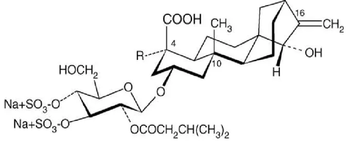
All the underground parts of this plant contain two toxic diterpenoid glucosides: atractyloside (ATR) and carboxyatractyloside (CATR) ( Fig. 2 and Fig. 3). ATR was isolated from the roots of Atractylis gummifera for the first time by Lefranc (1868). The corresponding aglycone (atractyligenine) is a non-volatile diterpene of the (−) kaurene family, with a perhydrophenanthrenic structure. The carbohydrate portion consists of a single D(+) glucose molecule, with only one free hydroxyl group (C-6); it is linked in C-2’ to a residue of isovaleric acid, in C-3’ and C-4’ to two residues of sulphuric acid and in C-1’, through a β-linkage, to the C-hydroxyl of atractyligenin (Piozzi, 1978). CATR was isolated for the first time in 1964 and called gummiferin (Stanisls and Vignais, 1964) and subsequently identified as 4-carboxyatractyloside (Danieli et al., 1971). CATR differs from ATR owing to the presence of a second carboxylic group in position C-4’ of the diterpene ring. CATR is present in fresh but
. 1: Chemical structure
Fig. 2: Chemical structure of toxic compounds from Atractylis gummifera (Danieli et
al., 1971)
R1 = SO3H; R2 = H; Atractyloside
R1 = SO3H; R2 = COOH ;Carboxyatractyloside
not in dried plants because it is decarboxylated to ATR during ageing or desiccation. CATR is also more toxic than ATR (Luciani and Carpenedo, 1978). Several factors, including the climate, the composition of the soil, the time of harvest and genetic factors, influence the content of diterpenoid glucosides in the rhizome of Atractylis gummifera. For example, differences reportedly exist between the rhizome content of ATR from Atractylis gummifera grown in Sardinia or in Sicily (Table. 1) (Fassina et al., 1962; Contessa and Fassina, 1978 ). A higher amount of ATR was found in rhizomes collected in autumn or winter, than in spring or summer (Table. 1), in agreement with the higher content of reserve substances and active compounds in the underground part of a plant during the time of quiescence. And last, a quantitative study showed that Atractylis gummifera of Sicilian origin had a higher ATR content than that of Sardinian origin, even though the two plants had been acclimatized in the same territory different from the original habitat (Toth, 1964).
ATR and CATR have also been also isolated from other plants from different genera including Callilepsis laureola, Xanthium strumarium, Iphiona aucheri, and Wedeila glauca (Obatomi and Bach, 1998). ATR analogues were also found in aqueous extracts of green and roasted beans of Coffea arabica. In particular, three glycosides were identified: 2-O-(2-O-isovaleryl-beta-d-glucopyranosyl)-atractyligenin,2-O-beta-D-glucopyranosyl-atractyligenin and 2-O-(3-O-betad-glucopyranosyl-2-O-isovaleriyl-beta-d- glucopyranosy)-atractyligenin (Fig. 4) (Obermann and Spiteller, 1976; Richer and Spiteller, 1978). Green beans of Coffea arabica contain 34.5– 62.4 mg/kg of ATR analogues whereas the roasted beans contain only 17.5–32.5 mg/kg (Obatomi and Bach, 1998).
Table 1: Contents of atractyloside in rhizomes of Atratylis gummifera L. from Sardinia , Sicily and Algeria in different time of the years ( Fassina et al., 1962 ; Contessa and Fassina, 1978)
Time of the harvest ATR content gram% dry weight June 0.12±0.013 October 0.19±0.036 Sardinia December 0.33±0.045 May 1.21±0.038 Sicily December 1.57±0.101 May 0.97 Algeria December 1.34
No published information is available on the ATR plant levels required to produce toxicity. Considering the quantity of ATR analogues present in coffee beans and the fact that some of them are non-toxic (Fontana et al., 1994), the risk related to coffee consumption is presumably negligible. Indeed, no published reports have described cases of coffee-induced ATR toxicity even in heavy coffee drinkers. ATR inhibits the oxidative phosphorylation of ADP. Because of its genin, atractyligenin, which is about 150-fold less toxic, has a similar but weaker activity, the glucose moiety, disulphoric and isovaleric acids probably increase the inhibitory effect of the atracyligenin moiety of ATR (Vignais et al., 1978).
Various atractyloside analogues were prepared and tested, to evaluate structure– activity relationships and to better understand the mechanism underlying the action of ATR. One of the most crucial groups for ATR toxicity is the carboxyl group in C-4’ of the diterpene ring: its reduction to alcohol produces a non-toxic compound, atractylitriol. ATR toxicity depends also on the C-16 methylene group: its reduction leaves a compound two- or three-fold less potent than ATR. Acetylation of the two free hydroxyl groups of ATR, C-15 of atractyligenin and the C-6 free hydroxyl group on the glucose molecule, decreases by 100-fold the inhibitory potency of ATR. The derivatives obtained by the removal of isovaleric acid or the sulphate groups are also less effective than ATR (Vignais et al., 1978). For example, Fontana et al. (1994) studied the effects of two atractylosides of Coffea arabica 2-O-(2-O-isovaleryl-beta-d-glucopyranosyl)-atractyligenin and 2-O-beta-d-glucopyranosyl-atractyligenin on carbohydrate metabolism in rats by measuring blood glucose levels, lactate and pyruvate, as well as hepatic, muscular and cardiac glycogen. In this study, the ATR analogues were used at a dose of about 150 mg/kg, corresponding to the ATR DL50 in rats, when administered by the S.C. route (Luciani and Carpenedo, 1978);
2-O-(2-O-isovaleryl-beta-dglucopyranosyl)-atractyligenin showed the same activity as atractyloside, conversely the other analogue glycoside without the isovaleric group, was nearly inactive.
1.3. Clinical aspects of Atractylis gummifera poisoning
About 100 cases of Atractylis gummifera poisoning have been described in the literature (Table 2) since the XIXth century (Hamouda et al., 2004). Larribaud (1954) reported the intoxication of two children aged 4 and 6 years, after ingestion of Atractylis gummifera rhizome in a region of Dellys (Algeria). The children arrived at the hospital with violent abdominal colic and abundant vomiting, followed by generalized contractions, cyanosis, agony and coma. The next morning both children died. The histopathologic findings showed congested intestine, peritoneal exudates and haemorrhages in stomach and kidney. In 1955, a report, describing the accidental poisoning of several Italian school children including three fatal cases, raised the interest of the scientific community in Atractylis gummifera(Santi and Cascio, 1955). Catanzano (1969) described the intoxication of two children who drank a decoction of Atractylis gummifera. The observed symptoms were vomiting, convulsions, muscular hypertonia and mydriasis; the arterial pressure values were 100/50mmHg in one child and 100/75mmHg in the other and the pulse rate was 100 bpm for both children. The laboratory findings showed increased leucocyte numbers (20,000/mm3) while the histopathologic findings showed hepatic necrosis. Georgiou et al., (1988) described the case of a 7-year-old boy, admitted to hospital 2 days after drinking an extract of Atractylis gummifera, taken to treat oxyuriasis. The symptoms were epigastric pain, vomiting and general anxiety. The patient’s condition progressively worsened and, despite treatment, the boy died 8 days after admission.
In Spain, five people were poisoned after taking an infusion of Atractylis gummifera, owing to a misidentification.
The patients suffered extensive liver damage and kidney failure. One of them died after gastrointestinal haemorrhage but the other four survived after dialysis (Caravaca-Magarinos et al., 1985). A report by Hamouda et al. (2000) stated that from 1983 to 1998 the Tunisian Poisoning Center collected 56 medical records of patients admitted to the toxicological intensive care unit for poisoning with 11 species of plants. The principal plants involved were Atractylis gummifera (18 cases; 32%) Datura stramonium L. (14 cases; 25%) and Ricinus communis L. (5 cases; 9%). Of these 56 cases 16 were lethal and all of them involved Atractylis gummifera.
These case reports provide useful information on the symptoms and laboratory findings that help to identify victims of Atractylis gummifera poisoning. The symptoms begin 6–36 h after the ingestion of the extract of Atractylis gummifera rhizome (Capdevielle and Darraq, 1980). The typical symptoms are gastrointestinal problems including nausea, vomiting, epigastric and abdominal pain, and diarrhoea (Capdevielle and Darraq, 1980; Georgiou et al., 1988). Some reports also describe general anxiety, headache, drowsiness, arrhythmia and convulsions (Hamouda et al., 2000). In several cases, these symptoms are followed by coma (Capdevielle and Darraq, 1980).
The laboratory findings (marked increased in SGOT, SGPT and bilirubin) may indicate severe hepatocellular damage and acute renal failure (Georgiou et al., 1988; Nogue et al., 1992). Post-mortem histopathologic examination discloses massive gastrointestinal haemorrhage, diffuse necrosis of the hepatic parenchyma with collapse of the interstitial connective tissue and accumulation of macrophages (Caravaca et al., 1985).
Table. 2: Case reports of Atratylis gummifera L. poisoning
Author Case reports
Madani 2006 Atractylis gummifera poisoning in a pregnant woman Skalli 2002 Atractylis gummifera L. poisoning: a case report
Nogue 1992 Acute kidney failure caused by Atractylis gummifera L. poisoning
Georgiou 1988 Hepatotoxicity due to Atractylis gummifera L.
Caravaca 1985 Renal and hepatic injuries in human intoxication with Atractylis gummifera L.
Capdevielle 1980 Poisoning by bird-line thistle (Atractylis gummifera L.) Lemaigre 1975 Fulminating hepatitis caused by glue thistle (Atractylis
gummifera L.), poisoning. Anatomo-pathological study of 4 cases
Catanzano 1969 2 cases of poisoning due to "gum thistle" (Atractylis
gummifera L.). Clinical development and anatomo-pathologic lesions
Thiodet 1961 Poisoning by the glue thistle (Atractylis gummifera L.). Clinical study apropos of 11 cases
Thiodet 1960 A fatal poisoning frequent in Algeria and little known:
poisoning by a gummiferous thistle (Atractylis gummifera L.) Santi 1955 Ricerche farmacologiche sul principio attivo dell’ Atractylis
gummifera L.
Larribaud 1954 Two cases of poisoning by gummy thistle, Atractylis gummifera L.
1.4. Toxicology and pharmacology
The toxic effect of Atractylis gummifera arises from ATR, a powerful inhibitor of oxidative phosphorylation in mitochondria. This action is exerted especially in cells rich in mitochondria such as hepatocytes and in proximal tubular epithelial cells, which contain carriers that allow ATR to cross the cell membrane. ATR interacts with the adenine nucleotide translocator (ANT) (Roux et al., 1996), a mitochondrial protein contained in the inner membrane. ANT has two major functions: it is responsible for the antiport of ATP and ADP, an important system for oxidative phosphorylation and it is part of the permeability transition pore complex, a non-specific pore involved in mitochondrial membrane permeabilization, an important event during apoptosis (Haouzi et al., 2002). ATR and ADP both interact in a similar way with ANT because they resemble one another in geometric and charge distribution (Stewart and Steenkamp, 2000): the polar character of the sulphate groups in ATR corresponds to the phosphate group in ADP; the glucose in the glycoside group of ATR corresponds to the ribose of ADP, and the condensed rings of ATR resemble the purine moiety of adenine.
The selective binding of ATR to ANT has two important consequences. First, ATR inhibits ADP transport and inhibits the access of extra mitochondrial ADP to a phosphorylation site located in the mitochondrial compartment, thus blocking oxidative phosphorylation and Krebs cycle oxidative reactions (Quintanilla et al., 1979; Kholodenko et al., 1988). Second, ATR induces opening of the mitochondrial permeability transition ATN-containing pores leading to membrane permeabilization and release of soluble intermembrane proteins, including cytochrome C. The translocation of cytochrome C from mitochondria to the cytosol is a crucial step in
Fasinduced apoptosis (Vancompernolle et al., 1998). The toxic effects due to the inhibition of mitochondrial phosphorylation are hepatic necrosis and renal failure in animals and humans.
The acute toxicity of ATR nevertheless differs according to the animal species and route of administration. Toxicity is higher in dogs than in mice and rats, suggesting an even higher toxicity in humans (Luciani and Carpenedo, 1978). Renal toxicity also differs among the various species: for example, rabbits and guinea pigs show no renal necrosis at ATR doses that are nephrotoxic for rats (Carpenedo et al., 1974).
Toxicity differs also in male rats between albino and Wistar strains: in albino rats ATR is not toxic at doses up to 200 mg/kg, whereas in Wistar strains 60 mg/kg can lead to death. Luciani and Carpenedo (1978) described the action of ATR in different species.
The major effects are on glycidic metabolism: ATR depletes glycogen in vivo by inhibiting glycogen synthesis, it increases blood lactic acid concentrations and decreases oxygen consumption. Animals poisoned with ATR usually die in hypoglycaemic convulsions.
An interesting feature of ATR poisoning is the long latency between the administration of ATR and the appearance of the first toxic symptoms, also when the compound is given intravenously. After an initial hyperglycaemic phase caused by depletion of skeletal muscle and hepatic glycogen a hypoglycaemic phase ensues that has important consequences on the whole organism, such as respiratory depression, hypoxaemia, acidosis due to increased plasma lactic acid, and finally convulsions. Because ATR toxicity remains after vagotomy and pancreatectomy
(Santi, 1958) it cannot depend either on an interaction with the autonomic nervous system or on glands implicated in glycaemic control.
In a study in rats, Hopps et al. (1997) described the effects of the ATR and some of its derivatives on renal function. They determined the urinary excretion of enzymes (beta-Nacetyl-d-glucosaminidase, alanine amino peptidase, gamma glutamyltransferase) and electrolytes (Na+, K+ and Cl−), and the blood and urinary concentrations of creatinine in rats treated with ATR, atractyligenin and two ATR derivatives (one lacking both the sulphate groups and the isovalerate group, the other lacking the sulphate groups alone). These experiments showed that ATR is highly toxic to the kidney, as shown by enzymuria and reduction of creatinine clearance, whereas atractyligenin leaves renal function almost unchanged. ATR toxicity is related to its chemical structure and increases when the hydroxyl groups in C-4’ is esterified with isovaleric acid or when the hydroxyl groups in C-3’ and C-4’ are esterified with sulphuric acid. Other important information on the toxic effects of ATR comes from in vitro studies using tissue slices (Obatomi et al., 1998; Obatomi and Bach, 1998).
This method allowed comparison of enzyme leakage, mitochondrial viability, changes in ATP levels, lipid peroxidation, oxidized glutathione (GSSG) and reduced glutathione (GSH) levels in kidney and liver tissues from different animals. ATR nephrotoxicity involved only the proximal tubule cells, whereas the glomerular cells appeared unaffected (Carpenedo et al., 1974). In the proximal tubule cells, ATR caused a significant concentration-dependent decrease in ATP content and a depletion of cellular GSH (Obatomi and Bach, 1998). The investigators proposed that ATR could act by interfering with cell transport; the presence of a sulphate moiety, suggested that ATR may undergo anion transport and then accumulate in the renal
cells. ATR also inhibited organic anion uptake in a dose- and time-dependent manner (Koechel and Krejci, 1992; Obatomi et al., 1998).
In the liver, the major effects are lipid peroxidation, GSH depletion and GSSG elevation. These changes suggest that ATR may induce its toxic effects through an oxidative process involving its methylene moiety thus producing a free radical. The reactive intermediate produced has not been identified, but reactive oxygen species such as superoxide anion, hydrogen peroxide or hydroxyl radicals may be involved (Obatomi et al., 1998).
1.5. Herbal hepatotoxicity
The liver is the central drug-metabolizing organ and is, therefore, a prime target of drug-related pathologies. Foreign compounds are predominantly bio transformed in the liver by the action of drug-metabolizing enzymes including microsomal cytochrome P450 enzymes, mixed-function mono-oxygenases, glutathione-S-transferases, sulfotransferases and UDP-glucuronosyltrans-ferases. Some of these can be induced through variable mechanisms which may lead to large inter individual variability in susceptibility for drug-related liver damage. Hepatic damage from conventional drugs is widely acknowledged and most physicians are well aware of them. Herbals as a cause of adverse hepatic reactions, however, have only recently been recognized as their use has become more widespread. Certain herbals have been identified as a cause of acute and chronic hepatitis, cholestasis, drug-induced autoimmunity, vascular lesions and even hepatic failure and cirrhosis. Risk factors for herbal toxicity have not been well identified, largely since hepatotoxic incidents have mostly been published as isolated case reports or small series
(Table. 3) However, a certain risk pattern has become evident, such as the observation that most affected individuals were females. This gender difference does not reflect a higher likelihood of women to use these preparations, but their higher susceptibility towards herb-induced liver damage (Flora et al., 1997), as is observed for the majority of adverse hepatic reactions induced by conventional drugs. As with chemically defined drugs, adverse hepatic reactions towards herbals cannot be predicted through diagnostic means, which makes the early recognition of liver damage important. Most individuals who take herbals do not admit their intake, even on repeated questioning, either because they do not consider herbals as 'drugs', or because they fear not to be taken seriously by their doctors for using herbals. Furthermore, doctors who recommend herbals or patients who take them advocate the long-standing use of herbals in traditional medicine as proof of safety, in particular, since most herbals are available without prescriptions and at low costs. Therefore, self-medication is frequent and, sometimes, patients even increase the dosage as liver disease worsens. Another problem is that herbals are usually mixtures of several ingredients or plants harvested during different seasons and extracted through variable procedures, which makes the identification of both the pharmacologically active and toxic compounds difficult. Also, contamination of herbals with microorganisms, fungal toxins such as aflatoxin, with pesticides, heavy metals, and synthetic drugs has been described (Yamamura et al., 1997).
Table 3: Selection of Herbal Preparations With Proven Hepatotoxicity (Larrey and Pageaux, 1995; Kaplowitz, 1997 )
Causative Plants Toxic Agents Symptoms Mechanism/Pathology
Crotalaria Senecio Heliotropium Symphytum officinale (Comfrey) Pyrrolizidine alkaloids Veno-occlusive disease Endothelial cell glutathione depletion, central vein necrosis, thrombosis, and fibrosis
Atractylis gummifera Atractylate, gummiferin
Hepatitis Inhibition of oxidative phosphorylation, hepatic necrosis
Callilepsis laureola Atractylate Hepatitis Hepatocyte necrosis
Chelidonum majus (greater celandine) Chelidonine, sanguinarine, berberine, coptisine? Hepatitis (cholestatic) Lymphocyte infiltration Larrea tridentata (chaparral) Guaiaretic acid derivatives
Hepatitis Not known Teucrium
chamaedrys (germander)
Furano-diterpenoids
Hepatitis Hepatocyte glutathione depletion and apoptosis Chinese herbal mixtures (artemisia, hare's ear, chrysanthemum, plantago seed, gardinia, red peony root, etc.)
Largely undefined
1.6. Atractylis gummifera and toxic hepatitis
Atractylis gummifera is a known cause of toxic hepatitis in the Mediterranean. It is used as an antipyretic, emetic and diuretic, and a bright fluid secreted from the plant is enjoyed by children as chewing-gum (Chauhan et al., 1991). The onset of hepatitis is usually acute and commences few hours after ingestion and following unspecific symptoms such as nausea, abdominal pain and headache. It is associated with a syndrome of neuro vegetative symptoms, hepato renal failure and pronounced hypo glycemia; the latter being caused by the inhibition of gluconeogenesis. Death due to fulminant hepatic failure is frequent. Consumption of Atractylis gummifera is particularly dangerous during spring time when toxins are concentrated in roots or when the plant is confused with wild artichoc (Abatroun. 1986). Toxicity has been ascribed to atractylosides and gummiferin which are inhibitors of the Krebs cycle and other mitochondrial functions and exert selective toxicity to hepatocytes and kidney epithelia in vitro via induction of oxidative stress, as mirrored by glutathione depletion and increased lipid peroxidation ( Obatomi et al., 2000) .
1.7. Treatment of Atractylis gummifera poisoning and studies on
possible therapeutic approaches
No specific pharmacological treatment is currently available to treat Atractylis gummifera intoxications. All therapeutic approaches, including fluid and electrolyte replacement, cardiovascular and respiratory support, seizure control and conventional therapeutic methods for severe hepatic and renal failure, are symptomatic (Stewart and Steenkamp, 2000). Some recommend that standard therapeutic practice should include induction of vomiting, bowel evacuation, gastric lavage and administration of activated charcoal (Ben Salah et al., 2001).
In an attempt to develop better pharmacological treatments for Atractylis gummifera intoxication, various compounds were evaluated. Catanzano et al. (1969)
showed the inefficacy of hydrocortisone (500 and 25 mg i.v.) administered to two poisoned children. In vivo experiments performed in dogs (Chardon et al., 1964) evaluated the efficacy of an intravenous administration of 100 ml of a solution containing 6–7% glucose, 0.1% dinitrophenol, 5mg of ATP and 11.25 mg of cytochrome C against poisoning induced by intravenous administration of an extract or dry root of Atractylis gummifera in saline solution. This treatment corrected the hypoglycaemia, hypotension and anuria and delayed death only when administered immediately after the extract. In an other study, Ishii and Bracht (1986) investigated the protective activity against ATR-induced liver toxicity of stevioside, a sweet glycoside isolated from the plant Stevia rebaudiana (Asteraceae) that can interfere with the transport of ATR across the cell membrane. In isolated perfused rat liver, stevioside decreased the effects of ATR on glycolysis, gluconeogenesis and oxygen uptake.
The cytoprotective effects of verapamil (VRP) and dithiothreitol (DTT) against ATR toxicity were investigated using precision-cut renal and liver slices assays (Obatomi et al., 2001a). The beneficial effect of VRP reflects its ability to reduce the ATR-induced increase in cytosolic Ca+2. VRP, a well-known calcium-channel blocker, completely blocked ATR-induced cell death, depletion of ATP, inhibition of gluconeogenesis in both kidney and liver slices, and provided protection against ATR-induced depletion of GSH in liver slices but not in kidney slices. Dithiothreitol (DTT), a sulphydryl-reducing agent and a metal chelator, reduced the reactive methylene double bond of ATR. DTT protected against ATR-induced enzyme linkage, inhibition of gluconeogenesis and depletion of GSH in kidney but exerted no protective effects against GSH and ATP depletion in the liver slices. Both verapamil and dithiothreitol nevertheless had protective effects only if used 30 or 60 min before
ATR exposure, but were ineffective after the exposure to ATR. Hence they cannot be used for therapeutic purposes.
In another study, Obatomi et al., (2001b) compared the role of ADP, calpain inhibitor I (CPI), stevioside and probenecid in protecting against ATR-induced toxicity in rat renal cortical slices. ADP, CPI and stevioside prevented the depletion of ATP and the reduction in gluconeogenesis, whereas probenecid gave no protection at all. Both in vivo and in vitro Haouzi et al., (2002) showed that ATR-permeability transition is counteracted by cyclosporin A (CsA) and GSH. CsA inhibited necrosis and apoptosis induced by ATR in human hepatic cells and pre-incubation with the GSH precursor N-acetylcysteine (NAC) reduced cytotoxic effect of ATR. In vivo ATR lethality was reduced by a regime, which enhances mitochondrial GSH levels; a diet enriched in sulphur amino acids or CsA administration significantly reduced both the lethal effect of ATR and cytoplasmic vacuolisation in hepatocytes and in proximal renal tubular cells.
1.8. Plasma Marker Enzymes
The history of the use of enzymes in diagnostic studies began with the introduction by Wolegemuth (1908) of the determination of amylase in plasma. Since that time further investigations have been carried out. It was reported that carbon tetrachloride can cause a decrease in cholinesterase (Brawer and Root, 1946). In recent decades many investigators have correlated clinical and serum tests which became routinely used and applied diagnostically in both medicine and toxicology. It was Ladenson et al., (1974); and Haymond and knight, (1975) who emphasised the superiority of plasma samples to serum samples for diagnostic purposes.
The classification of plasma enzymes is based on their function and tissue origin rather than upon their enzyme action (Bücher, 1958; Bücher et al , 1959) there are two major groups :
1. Plasma specific enzymes which are characterised by their secretion into the plasma, where they will express activity. These enzymes are found to be at higher activity level in plasma than in the tissue of origin. Examples of these are prothrombin, plasminogen, pseudocholinesterase. The level of activity of these enzymes decreases when there is no damage to the tissue from where they originate.
2. Non_specific plasma enzymes which can be divided into those of exocrine secretions and those concerned only with cellular metabolism; these enzymes are not normal functional constituents of blood and are only present because the blood was circulated through tissues of high enzyme activity.
The enzymes of cellular metabolites can be clearly associated with their source in certain pathological condition and include pancreatic α- amylase, prostatic phosphatase and pepsinogen. The cellular enzymes include those of tissue metabolism that are not active in blood because their substrates and co-factors are mainly absent. Enzymes of key metabolic pathways that may occur include glutamate ,lactate,malate and α - glycophosphate-dehydrogenase , 1.6-diphosphofructoaldolase glutamate oxaloacetate and glutamate pyruvate dehydrogenase , some of these are bound within the same two organelles such as mitochondria. In addition enzymes involved in the in cellular metabolism that are mainly associated with specific tissues and organ functions include the urea cycle enzymes such as arginase and ornithine carbamyl transferase, also alcohol and sorbitol dehydrogenase,
glucose-6-phosphatase and 1-phosphofructoaldolase all of those from liver; also glycerokinase from liver and kidney and alkaline phosphatase from bone.
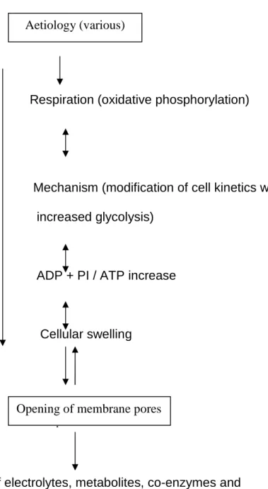
1.9. Mechanism of Enzyme Release from Cells
Plasma enzymes are good indicators of cell injury; they are commonly used in tissue function tests along with other tests designed to elucidate subtle response injury (Burke, 1978). Enzymes can be released from the tissue or the place of origin by different ways such as diffusion, active transport, permeability change or the shedding of structures such as the glycocalyx of plasma membranes (Fishman and Doellgast, 1975).
It has been shown that the appearance of cell necrosis (Schmidt et al., 1963; Schmidt and Schmidt, 1974; Wilkinson, 1978). However cell death may lead to the discharge of cell contents into the extra cellular fluid. The enzyme release may be due to an imbalance between energy production and cellular expenditure of energy. This can be shown in certain cases of histopathology such as hypoxia, when little hepatic necrosis occur with large increases in cell enzyme activities (Schmidt and Schmidt, 1974).
A schematic mechanism of enzyme leakage under pathological condition was made by Hess (1963) (Fig. 5), damage to the cellular respiration leading to pathological disturbance in energy metabolism. The consequent alteration in the water content of the cell, cellular swelling and permeability, sometimes possibly ends in cellular death.
This theory is supported by evidence provided by many investigators. It was found that in the incubated cells, the direct measurement of the intracellular ATP (Wilkinson & Robinson, 1974) showed little leakage occurred until it had fallen to at least
20% of the original level after which efflux was greatly increased. There are three types of enzymes that can be distinguished
Firstly, cytoplasmic enzymes, e.g. lactate dehydrogenase (SDH), secondly, mitochondrial enzymes such as glutamate dehydrogenase (GLDH), and finally from both cell compartments, for instance, glutamate oxaloacetate (GPT) and malate dehydrogenase (MDH). The first analysis of serum enzymes was made by (Schmidt and Schmidt, 1962) who suggested that the ratio of GOT: GPT may be useful in the diagnosis of chronic inflammatory lesions of the liver from necrotic processes.
Many studies have been carried out upon the distribution of the enzymes. It was reported (Schmidt and Schmidt, 1962) that the value of the ratio GOT + GPT/GLDH was useful for the differential diagnosis of obstructive jaundice. In addition patho-physiological conditions such as haemolytic syndromes (Gerlach et al., 1961) can be distinguished by enzyme ratios that include LDH: GOT, and (δ-GT): GOT ratios for hepatitis, toxic liver damage and alcoholic cirrhosis (Schmidt and Schmidt, 1976). Because (δ-GT) is located in the epithelium of intra hepatic biliary tracts, it serves as sensitive indicator of biliary obstruction, rising earlier and faster than either leucine or amino peptidase or alkaline phosphates. Ratios of GPT: (δ-GT), GOT: δ-GT and GPT. GLDH are also used as aids in differential diagnosis of the condition. The release of plasma enzymes from tissues was considered by Rees and Sinha (1960) and Zimmerman et al., 1965a) to reflect the sequence of cellular injury since a delayed plasma elevation from mitochondrial enzymes was obtained compared to cytoplasmic enzymes.
Respiration (oxidative phosphorylation)
Mechanism (modification of cell kinetics with
increased glycolysis)
ADP + PI / ATP increase
Cellular swelling
?
Leakage of electrolytes, metabolites, co-enzymes and soluble enzymes.
Death of cell
Fig. 5: A schematic mechanism of enzyme leakage under pathological conditions (Hess, 1963).
Aetiology (various)
2. General materials and methods
2.1. Plant material
Atractylis gummifera plant samples (rhizome and aerial part ) used in these studies were collected from different sites at Setif and Bordj- Bou- Arreridj regions. The plant samples were washed, cleaned and cut into small peaces then left to dry at room temperature in the shadow and then finely grinded to powder in a rotating knife grinder. The powder was sieved through a 1 mm mesh to remove large fragments. Each plant part powder was then used for the extraction procedure.
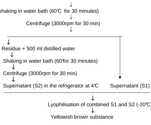
2.2. Extraction of plant materials
The extraction procedure was conducted according to the method used by Bey (1990). This method was used either to extract toxic compounds from rhizomes or aerial parts (Fig. 6 a and b; Fig. 7)
a
b
125 g of Atractylis gummifera (powder)+1000 ml of distilled water
↓
shaking in water bath (60°C for 30 minutes) ↓
Centrifuge (3000rpm for 30 min) ↓
↓↓↓↓
Residue + 500 ml distilled water ↓↓↓↓ Shaking in water bath (60°for 30 minutes) ↓↓↓↓ Centrifuge (3000rpm for 30 min) ↓↓↓↓ Supernatant (S2) in the refrigerator at 4°C Supernatant (S1)
↓↓↓↓
Lyophilisation of combined S1 and S2 (-20°C) ↓↓↓↓
Yellowish brown substance
Fig. 7: Steps of the plant material extraction of Atractylis gummifera L. ( Bey, 1990) after sleight modification.
2.3. Animal Tests
In the experiments performed two adult animal species were used. Albino Wistar rats and New Zealand rabbits. Rats were purchased from Pasteur Institute in Algiers and rabbits were provided by a local breeder in Setif region (Société
d’exploitation agricole, Mezloug, Setif). All animals were kept in the animal house with a natural lighting schedule for 1 to 3 weeks before experiment. They were fed with a standard pellet food and tap water.
2.4. Toxicological study experiments
The LD50 was determined using rats according to the method described by
Abatroun (1986). The symptoms of acute toxicity and post mortem finding were recorded.
2.5. Study design and dosage
The animals were divided into two groups: check and treated.
2.6. Assays
Plasma glucose, creatinine, urea , alkaline phosphatase , transaminases “AST and ALT”, CPK and total proteins were measured by Bio Merieux kits ( Anonymous, 1972). Hb, hematocrit , RBC and WBC were measured according to Dimianova, ( 1986) , Sultan et al., (1978), Dacie , (1982) respectivelly.
2.7. Histological techniques
2.7.1. Tissue preparation
Following the treatment with the Atractylis gummifera extract, animals were stunned and killed. This was done to coincide with the time when the level of certain Glutamate Oxaloacetate Transaminase (GOT) enzymes had increased in the plasma samples. The liver was then removed, washed with saline (0.9% NaCl) and cut into small cubes (approximately 5mm), and fixed in formalin (pH=7.0) or in Bouin’s solution, (Pantin.1959), for at least 24 hrs.
- Bouin ‘s solution
Picric acid (saturated aqueous solution) 75ml Formalin (40% formaldehyde) 25ml Glacial acetic acid 05ml
- Neutral 10%%%% formal saline, pH 7.0
Formalin (40% formaldehyde 250ml Sodium chloride 02ml Distilled water 250ml
2.7.2. Light microscopy
1. Fixation and dehydration of specimens
Specimens which had been fixed in Bouin’s solution or formalin washed in 70% alcohol for 24 hours (3-5 changes), dehydrated in series of alcohols 90%,100% and cleared in chloroform (3 changes) according to Mahoney (1973). After this, specimens were embedded in fresh wax and sectioned at thickness of 5-8 µm with microtome.
2. Staining method
Many staining techniques were tried. The useful chosen stain found was Hould method (1984) .
3. Hould’s staining method
Staining procedure
Treatment Time of treatment 1. Xylene 1 02 minutes 2. Xylene 2 02 minutes 3. Absolute alcohol 02 minutes 4. 90% alcohol 01 minutes 5. 70% alcohol 01 minutes 6. Distilled water 01 minutes 7. Haematoxylin 20 minutes 8. Tap water 03 minutes 9. Differentiation acid alcohol (1% hydrochloric acid in 70% alcohol) 10 seconds 10. Wash in tap water 02 minutes 11. 70% alcohol 02 minutes 12. 1% eosin in 70% alcohol 01 minutes 13. 90% alcohol 01 minutes 14. absolute alcohol 1 02 minutes 15. absolute alcohol 2 02 minutes 16. xylene 1 03 minutes
2.8. Chemicals
All drugs were reagent grade, and were purchased from Sigma chemical Corp., St Louis (MO, USA) or from Merck (Darmstadt, Germany) unless otherwise stated.
2.9. Statistical analysis
Data are shown as means ± standard error (SEM). The statistical analyses were performed with the analysis of variance. The results obtained at the end of each time phase were compared with those obtained from zero time for the same group using Student’s t-test for paired observations. At the same time, the results obtained at the end of each time phase were compared with those obtained for the control group at the same time phase. Biochemical data were analysed by Fischer and Dunnet tests.