En vue de l'obtention du
DOCTORAT DE L'UNIVERSITÉ DE TOULOUSE
Délivré par :
Institut National Polytechnique de Toulouse (Toulouse INP)
Discipline ou spécialité :
Dynamique des fluides
Présentée et soutenue par :
M. MAXIME BERGle mercredi 19 juin 2019
Titre :
Unité de recherche : Ecole doctorale :
Modélisation de l'écoulement sanguin et du transport de molécules dans la
microcirculation sanguine cérébrale: impact des occlusions capillaires dans
la maladie d'Alzheimer
Mécanique, Energétique, Génie civil, Procédés (MEGeP) Institut de Mécanique des Fluides de Toulouse ( IMFT)
Directeur(s) de Thèse :
MME SYLVIE LORTHOIS M. YOHAN DAVIT
Rapporteurs :
M. FRANCK NICOUD, UNIVERSITE DE MONTPELLIER M. PIERRE-YVES LAGREE, CNRS PARIS
Membre(s) du jury :
M. CHRISTIAN GEINDREAU, UNIVERSITE GRENOBLE ALPES, Président Mme NOZOMI NISHIMURA, CORNELL UNIVERSITY EU, Invité Mme REBECCA SHIPLEY, UNIVERSITY COLLEGE LONDRES, Membre
Mme SYLVIE LORTHOIS, CNRS TOULOUSE, Membre M. MICHEL QUINTARD, CNRS TOULOUSE, Membre
M. YOHAN DAVIT, CNRS TOULOUSE, Membre
abstract
The cerebral microvascular system is central in the remarkable machinery of the brain. It is responsible for the delivery of vital molecules (e.g. oxygen, glucose) and clearance of metabolic wastes (e.g. carbone dioxide, amyloid) to and from brain cells. Such a system is composed of small vessels (i.e. arterioles, venules and capillaries), embedded in brain tissue, which form a very large and intricate network spanning over the whole brain. Due to its critical role in brain homeostasis, the cerebral microvascular system is also involved in various pathologies, ranging from stroke to neurodegenerative diseases.
During the last decades, significant advances in imaging techniques have been made, such as multi-photon microscopy, that enabled the observation of the cerebral microvascular system with an unprecedented level of accuracy. However, these techniques generate large amounts of data that are difficult to interpret without a proper theoretical framework. In this thesis, we address this need by building computationally efficient models that accurately describe blood flow and molecular transport in the cerebral microvascular system, more specifically in large anatomical networks.
The biggest challenge in the resolution of blood flow and molecular transport problems dwells in the scale of such anatomical networks. Indeed, even though they represent only a fraction of the complete cerebral microvascular system, they are made of tens of thousands of vessels and exhibit highly complex geometries. Consequently, this prohibits the resolution of the blood flow and molecular transport problems by means of classic numerical methods, e.g. finite volume or finite elements. To get around this computational issue, we combine a pore network approach to upscaling methods (volume averaging and multiscale asymptotics) and Green’s functions to simplify the formulation of both blood flow and molecular transport problems while still capturing the underlying physics. In order to assess the relevance of such simplifications we systematically validate our models against in vitro and in vivo measurements, and against reference analytical solutions otherwise.
We then use our models to investigate the role of the cerebral microvascular system during the onset of Alzheimer’s disease. Indeed, it has been recently shown that a significant decrease in the cerebral blood flow is the earliest biophysical marker of the disease. Coincidently, our collaborators, Professors Schaffer and Nishimura from Cornell University, have observed in Alzheimer’s diseased mice that a small proportion (2% to 4%) of capillaries were abnormally occluded by white blood cells adhering to the inflammatory vessel walls. They subsequently injected antibodies inhibiting the white blood cells adhesion. This resulted in the unclogging of the capillary vessels, causing a substantial increase in cerebral blood flow and ultimately leading to a significant cognitive improvement of the diseased animals. Assuming that such antibody injections restored the cerebral blood flow to its baseline value leads to estimate that capillary occlusions were previously reducing the cerebral blood flow by 20% to 30%.
This raises the critical question: can 2% to 4% of capillary occlusions cause up to 30% reduction in cerebral blood flow? This question is challenging to answer only using experiments since it is tremendously difficult to isolate the contribution of such a biophysical process in vivo in mice or humans. Instead, we use our models to numerically investigate the impact of such capillary occlusions on cerebral blood flow. We find that 2% to 4% capillary occlusion cause up to 12% cerebral blood reduction making them a significant mechanism in the onset of Alzheimer’s disease. Finally, we go on to investigate the consequences of such occlusions on molecule exchanges.
résumé
Le système microvasculaire est un acteur essentiel du fonctionnement cérébral. Il est en effet responsable de l’approvisionnement des cellules en oxygène et glucose ainsi que de l’évacuation des déchets métaboliques comme le dioxyde de carbone. Ce système est composé d’une multitude de petit vaisseaux appelés artérioles, veinules et capillaires, qui sont entourés de tissu cérébral. Ces vaisseaux forment un immense réseau qui étend ses ramifications à travers tout le cerveau. A cause de son rôle prépondérant dans l’homéostasie cérébrale le système microvasculaire est impliqué dans de nombreuses pathologies, allant de l’accident vasculaire cérébral aux maladies neurodégénératives. Ces dernières décennies ont été marquées par des avancées significatives dans le domaine de l’imagerie du vivant (e.g. la microscopie multi-photonique) qui ont permis l’observation du système microvasculaire cérébral avec un niveau de précision sans précédent. Ces techniques génèrent cependant de grandes quantités de données qu’il est difficile d’analyser sans outils théoriques adaptés. C’est pourquoi, dans cette thèse, nous développons des modèles capables de décrire l’écoulement sanguin ainsi que le transport de molécules au sein de vastes réseaux microvasculaires anatomiques.
La principale difficulté dans la résolution de tels problèmes, vient de la taille de ces réseaux. En effet, même s’ils ne représentent qu’une fraction du système microvasculaire, ils sont composés de plusieurs dizaines de milliers de vaisseaux et possèdent des géométries complexes. Il est donc inenvisageable de résoudre l’écoulement sanguin et le transport de molécules par le biais de méthodes classiques comme les volumes finis ou les éléments finis. Afin de surmonter cette difficulté, nous combinons une approche réseau de pores avec des méthodes de changement d’échelles (prise de moyenne volumique et développements asymptotiques) et des fonctions de Green. Cela nous permet de simplifier à la fois la description de l’écoulement sanguin et du transport de molécules tout en restant cohérent avec la physique sous-jacente. Pour nous assurer de la pertinence de ces simplifications nous validons systématiquement nos modèles en les comparant à des mesures in vitro et in vivo si elles existent et à des solutions analytiques de référence sinon.
Une fois validés, nous utilisons nos modèles afin d’élucider le rôle joué par le système microvas-culaire aux stades précoces de la maladie d’Alzheimer. En effet, il a été récemment montré qu’une baisse du débit sanguin cérébral était le premier marqueur quantitatif de la maladie. Simultanément, nos collaborateurs, les professeurs Schaffer et Nishimura de l’université de Cornell, ont observé chez les souris malades qu’une faible proportion (2%-4%) des capillaires étaient obstrués par des globules blancs. En conséquence ils ont injecté un anticorps inhibant l’adhésion de ces derniers. Les vaisseaux se sont alors débloqués, entraînant une augmentation du débit sanguin ainsi qu’une amélioration des capacités cognitives chez les souris malades. Si l’on suppose qu’après l’injection le débit sanguin retrouve sa valeur de référence, on peut estimer que les occlusions capillaires réduisent de 20% à 30% le débit sanguin.
Une si faible proportion de capillaires obstrués peut-elle avoir un impact aussi important sur le débit sanguin cérébral ? Il est difficile de répondre simplement à cette question en se fiant uniquement à l’expérience puisqu’il est quasiment impossible d’isoler un tel phénomène in vivo que ce soit chez la souris ou chez l’humain. Pour contourner ce problème nous utilisons nos modèles et simulons numériquement l’impact de ces occlusions sur le débit sanguin. Nous trouvons que 2% à 4% d’occlusions capillaires conduisent à une baisse de débit pouvant aller jusqu’à 12%, faisant de ces occlusions un mécanisme important dans l’apparition de la maladie d’Alzheimer. Pour finir, nous quantifions leurs conséquences sur les échanges moléculaires.
formal acknowledgments
The research presented in this manuscript was made possible thanks to the funding of several institutions. It was mainly conducted at the Institut de Mécanique des Fluides de Toulouse (IMFT) and was supported by the European Research Council (ERC) under the European Union’s Seventh Framework Programme (FP7/2007-2013) ERC grant BrainMicroFlow, agreement number 615102
(https://erc.europa.eu/), and by the National Institutes of Health and National Cancer Institute
IMAT Program under award number R21CA214299. This project also received a travel and research grant from the Toulouse School of Doctors to collaborate with the Schaffer-Nishimura lab and the Nancy E. and Peter C. Meinig School of Biomedical Engineering at Cornell University. The research reported in this manuscript was performed using HPC resources from CALMIP (Grant 2016-P1541). Further, the research presented in this manuscript was made possible thanks to an intense teamwork effort involving several people in France and the USA. That is why I offer my warmest thanks to Sylvie Lorthois, Yohan Davit, Michel Quintard, Nozomi Nishimura, Chris B. Schaffer, Adlan Merlo, Myriam Peyrounette, Amy Smith, Vincent Doyeux, and Florian Goirand.
Finally, I am grateful to Franck Nicoud and Pierre-Yves Lagrée for reviewing this manuscript and to Franck Nicoud, Pierre-Yves Lagrée, Rebecca Shipley, Michel Quintard, Christian Geindreau, Nozomi Nishimura, Sylvie Lorthois and Yohan Davit for participating in the thesis defence.
personal acknowledgements
Je débute cette longue série de remerciement en exprimant ma sincère gratitude envers mes directeurs de thèse, Sylvie et Yohan, pour leur compétence, leur disponibilité, leur bienveillance et leur humanité. Je profite également de cette occasion et remercie Michel pour ses conseils avisés, toujours prodigués avec beaucoup de sagesse. Merci à tous les trois de m’avoir ouvert les portes du monde de la recherche, vous avez été de véritables modèles.
Je tiens ensuite à remercier tous les membres du groupe Milieux Poreux et Biologiques grâce à qui j’ai pu passer trois années fantastiques! Un immense merci à Adlan, Baptiste, Myriam, Amy, Vincent, Edith, Alexandra et Florian, mais aussi à Pauline, Pascal, Paul, Julien, Arthur, Tawfik, Hamza, Pierre, Adel, Pierre-Yves, Omar et Pauline. J’ai également une pensée pour les étudiants de Master avec qui j’ai eu la chance et le plaisir de travailler, Aloma, Marion, Nicolas, Vanina et Sébastien.
Next, I would like to extend my gratitude to Chris, Nozomi and their team for having me over at Cornell. A particular thank goes to J.C., Oliver, Yu Ting and David for all the good times in Ithaca!
Un petit clin d’oeil également aux membres de la SERAS avec qui j’ai pu découvrir les joies de la plongée sous-marine et qui m’ont permis de bien décompresser tout au long de ces trois années. Merci à Gabriel, Marion, Quentin, Lucia, Sébastien, Lionel, Lorène, Daniel, Nicolas, Jean-Guy et Gilbert. Merci à vous pour tous les super week ends et les intenses entraînements du lundi soir!
Il est ensuite impensable que je ne remercie pas mes amis Toulousains, en commençant par Swann, avec qui j’ai cohabité durant ces trois super années, mais également Clément, Anne, Matthieu et Gaëlle pour toutes les soirées et tous les week-end passés ensemble depuis notre première année à l’N7. Je n’oublie bien évidement pas ceux plus éloignés, Arthur, Carole, Camille et Erwan. Un grand merci à vous tous.
De la même manière, je souhaite remercier Hélène, Val et Jéremie, avec qui j’ai eu la chance de partager tant de moments exceptionnels depuis maintenant plus d’une décénnie!
Enfin, je remercie mes parents, Patrick et Patricia, dont le soutien infaillible à été essentiel à la réussite de cette aventure. Merci infiniment.
C O N T E N T S
i introduction
1 modelling the cerebral microvascular system: a problem of scale 3
2 current models for blood flow and molecular transport in the brain
microcirculation 9
2.1 Blood rheology in microcirculation . . . 9
2.2 Modelling blood flow in microvascular networks . . . 14
2.3 Molecular transport in the cerebral microcirculation . . . 18
2.4 Modelling molecular transport in the cerebral microcirculation . . . 22
2.5 Modelling decisions and numerical implementations . . . 27
ii results 3 impact of capillary occlusions on blood flow in microvascular networks 33 3.1 Modelling groundwork . . . 34
3.2 Microvascular networks . . . 38
3.3 Resolution of the blood flow problem . . . 39
3.4 In vitro validation . . . 41
3.5 In vivo validation . . . 44
3.6 Effect of multiple capillary occlusions on cerebral blood flow . . . 47
3.7 Effect of a single occlusion on blood flow in capillary networks . . . 51
3.8 Conclusions . . . 54
4 impact of radial gradient of concentration in the regime of weak couplings 57 4.1 Transport phenomena at the scale of a microvessel . . . 60
4.2 Effective transport model for weak couplings . . . 70
4.3 Results . . . 74
4.4 Discussion . . . 87
4.5 Conclusions . . . 89
5 impact of capillary occlusions on mass transfers in microvascular networks 91 5.1 The effective transport equation beyond the regime of weak couplings . . . 92
5.2 Molecular transport inside the tissue domain . . . 99
5.3 Transport in the regime of strong couplings . . . 103
5.4 Molecular transport in microvascular networks . . . 109
5.5 Effect of capillary occlusions on molecular exchanges in microvascular networks . . . . 110
5.6 Conclusions . . . 113
iii conclusions and perspectives
6 general conclusion and perpectives 119
iv appendix
a semi-empirical relationships for blood flow 125
b analytical solutions to the effective transport equations 129
Weak Couplings . . . 129
Extented weak coupling . . . 130
c numerical methods for solving the effective transport equation 133
v featured publications
bibliography 141
Part I
1
M O D E L L I N G T H E C E R E B R A L M I C R O VA S C U L A R S Y S T E M : A P R O B L E M O F S C A L E
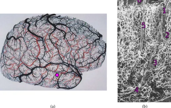
The human brain is one of the most fascinating objects in the living world. Besides keeping us alive by regulating all vital functions, e.g., our heartbeat, respiratory and sleep rhythms, it is involved in all our feelings, thoughts and decisions and sustains our learning processes and creativity. It works reliably for decades with remarkable energy efficiency, consuming thousands of times less than a small supercomputer. (a)
1
2
3
4
5
6
(b)Figure 1.1: The tremendous complexity of the cerebral vascular system. (a) Drawing of the surface (cortical pial) vessels of the right hemisphere. Colored vessels (red, blue and green) represent arteries. Black vessels represent veins. The purple dot correspond to the approximate location of (b). This figure is a modified version of figure 1 in Duvernoy, Delon, and Vannson [1]. (b) Scanning Electron Microscopy image (x50) of intracortical arteries and veins (cortical surface at the top). 1- lntracortical arteriole (small artery) diving from the pial surface. 2-3-4 secondary arterioles branching off from the first one. 5- Intracortical vein ascending to the cortical surface. 6- pial vein at the surface of the brain. The smaller, more tortuous vessels are capillary vessels. This figure is a modified version of figure 29 in Duvernoy, Delon, and Vannson [1]
Central to this formidable machinery, is the cerebral vascular system, i.e. the huge collection of vessel through which the blood flows. Such a system is responsible for the delivery of vital molecules (e.g. oxygen, glucose) and the clearance of metabolic wastes (e.g. carbone dioxide, amyloid) to and from brain cells. In humans, the cerebral vascular system forms a single, gigantic and intricate
modelling the cerebral microvascular system: a problem of scale 5
(a) (b)
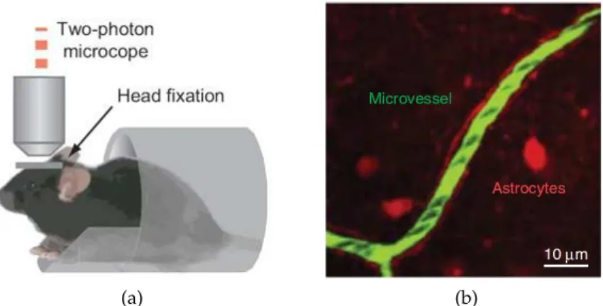
Figure 1.3: In vivo measurement of red blood cell velocity in mouse using two-photon microscopy. (a) Schematics illustrating the stereotype of the experimental set-up. This figure is a modified version of the figure 1b in Komiyama et al. [24]. (b) Snapshot of a capillary in the cerebral cortex (in green) embedded in tissues (red). Each individual black spot represents a single red blood cell flowing through the microvessel. Such snapshot typically can be obtained down to 1 mm below the cortical surface with a resolution neighbouring 1 µm. This figure is a modified version of figure 1c in Shih et al. [21]
cerebral macrocirculation (surface arteries and veins), e.g. see [16,17,18,19]. Consequently, we still know little about this vital system and its mechanisms.
However, the last decades have seen significant advances in imaging techniques, such as multi-photon [20,21] or light sheet [22,23] microscopy, that have enabled the observation of the cerebral microvascular system with an unprecedented level of accuracy. Figure 1.3 shows that it is now possible to measure, in vivo, the velocity of individual red blood cells in mouse capillaries. However, such techniques generate large amounts of data that are difficult to interpret without a proper theoretical and numerical framework.
In this thesis, we address this need by building computationally efficient models that accurately describe blood flow and molecular transport in the cerebral microvascular system. More precisely, we focus on large anatomical networks, similar to the one observed in figure1.1b.
One of the biggest challenge in the resolution of blood flow and molecular transport problems at this scale stems from the size of the anatomical networks. Although they represent only a fraction of the complete cerebral microvascular system, they include tens of thousands of vessels and exhibit highly complex geometries. Consequently, it is hardly concievable to directly solve the local equations governing blood flow and molecular transport using classic numerical methods such as finite volumes or finite elements.
To get around this computational issue, we adopt a modelling approach that aims at simplifying blood flow and molecular transport problem formulations while still capturing the underlying physics. For instance, we use a network model inspired by Pries et al. [25] and Lorthois, Cassot, and Lauwers [26] to describe blood flow. Such a startegy enables the transformation of complex microvascular networks into oriented graphs, thus dramatically reducing the computational costs of the problem. Similarily, for molecular transport, we apply formal homogenisation methods, such as volume averaging [27,28] and multiscale asymptotics [28,29], to transform the local three-dimensional equations within each vessel into effective one-three-dimensional transport equations along the vessel axes. With regard to transport within the brain tissue, we take inspiration from Secomb et al. [30] to make use of Green’s functions to transform the local three-dimensional diffusion-reaction equation into a distribution of sources and dipoles located at the interface between vessel and tissues, thus saving us from meshing the complex tissue space.
6 modelling the cerebral microvascular system: a problem of scale
The advantage of having this approach is that it simplifies the treatment of each vessel, making it possible to model large volumes and to capture the topology of the network. The disadvantage, however, is that its accuracy heavily relies on the quality of the models used to represent each individual vessel and the coupling between vessels and tissue. That is why, throughout this thesis, we systematically validate the models introduced against in vitro and in vivo measurements when they are available, and against reference analytical and numerical solutions otherwise.
Altogether, we believe that the theoretical framework developped in this thesis will strengthen our fundamental understanding of the cerebral microvascular system and its mechanisms. In addition, we argue that modelling blood flow and molecular transport at the scale of microvascular networks can help bridge the gap between models describing the physics at the scale of a single capillary (e.g. see [31,32,33,34]) and models describing the macroscopic behaviour at the scale of the whole brain (e.g. see [19,35,36]). Finally, we are convinced that, combining our theoretical approaches with in vivo imaging techniques will help advance our understanding of the pathologies plaguing the cerebral microvascular system.
With regard to this last point, we have closely collaborated with professors C. B. Schaffer and N. Nishimura at Cornell University, who are experts in the field of in vivo microscopy [21,37,38], to unveil the role played by capillary occlusions during the onset of Alzheimer’s disease. Indeed, it has recently been highlighted that a significant decrease in cerebral blood flow is the earliest biophysical marker of the disease in human patients [39]. Coincidently, professors Schaffer and Nishimura have observed in Alzheimer’s diseased mice that a small yet significant proportion (2% to 4%) of capillary vessels are occluded during the onset of the disease, i.e. before the occurrence of any other marker. They further estimated that such occlusions were reducing the cerebral blood flow by 20% to 30%.
This raises the following questions: can 2% to 4% of capillary occlusions cause the cerebral blood flow to decrease 30% of its baseline value? And if so, what are the consequences with regard to molecular exchanges? Answering such questions using only experiments is challeng-ing since it is tremendously difficult to isolate the contribution of such a biophysical process in vivo. To overcome this difficulty, in this thesis, we propose to use our models to investigate the impact of capillary occlusions on both blood flow and molecular transport in large anatomical microvascular networks. In doing so, we aim at improving our understanding of the mechanisms associated with Alzheimer’s disease, contributing to translate results from animals to humans and, in the long term, improving the diagnosis procedure and the efficacy of new treatment strategies. To summarize, the objective of this thesis is threefold:
1. To develop and validate theoretical models for blood flow and molecular transport that are computationally efficients and physically accurate.
2. To solve the blood flow and molecular transport problems in anatomical networks composed of tens of thousands of vessels.
3. To numerically investigate the impact of capillary occlusions on blood flow and molecular transport and their implications in Alzheimer’s disease.
In line with these objectives, this manuscript is organized as follows:
• In Chapter 2, we present a detailed overview of the mechanisms at play in the cerebral microvascular system, with a specific focus on blood rheology and molecular exchanges, and how they are modelled in the litterature. Doing so allows us to lay down the fundamentals of our modelling strategy. Besides, in this chapter, we describe the numerical framework in which all the following work is implemented.
modelling the cerebral microvascular system: a problem of scale 7
• In Chapter 3, we investigate the impact of capillary occlusions on blood flow in microvascular networks. We start the chapter by using a pore-network approach to model blood flow and present the resolution procedure of the associated problem. We then go on and validate such a model, first in vitro in model networks, then in vivo in mice brain. Finally, we quantify the impact of single and multiple capillary occlusions on cerebral blood flow in mice and humans. • In Chapter 4, we introduce a novel model for molecular transport in microvascular networks. Specifically, in this chapter, we aim at transforming the three-dimensional transport equations within the vessels into one-dimensional effective transport equations by means of averaging operators. In doing so, we show that it is formally possible to derive an exact expression for such a model only in the regime of weak couplings, i.e. when the molecular concentration is much larger in the vessel than in the tissue. Finally, using this exact model, we examine the widespread well-mixed hypothesis, under which the molecular concentration is assumed to be uniform in the microvessel cross-sections, ultimately highlighting the impact of intravascular radial gradients of concentration on the transport of various molecules in microvascular networks. • In Chapter 5, we investigate the impact of capillary occlusions on molecular exchanges in
microvascular networks. During the first half of this chapter, we show that the molecular transport model we derived in chapter 4 in the regime of weak coupling can still be used as an ansatz when the concentration in the tissue is no longer small when compared to the concentration within the vessel. We model molecular transport within tissues using Green’s function and further discuss the case of reactive molecules such as oxygen or glucose. Finally, after having validated our model for molecular transport against reference numerical solutions, we investigate the impact of capillary occlusions on the molecular exchanges in mouse brains.
2
C U R R E N T M O D E L S F O R B L O O D F L O W A N D M O L E C U L A R T R A N S P O R T I N T H E B R A I N M I C R O C I R C U L AT I O N
The first objective of this thesis is to provide computationally efficient models that are adapted to solve blood flow and molecular transport problems in large microvascular networks. Consequently, in this chapter, we present an overview of the main mechanisms at play in the cerebral microvascular system and how they are modelled in the literature.
In section2.1, we present the complex rheology of blood in the microcirculation. We introduce the three main mechanisms that govern the distribution of red blood cells at the scale of microvascular networks, namely the Fåhraeus effect, the Fåhraeus-Lindqvist effect and the phase separation effect. Then, in section2.2, we show that most works in the literature have modelled blood flow using two different approaches, i.e., considering the blood as a suspension of red blood cells or as a monophasic fluid with effective properties. We then compare the benefits of the two approaches.
In section 2.3, we present the main mechanisms involved in the transport of a molecule going from blood vessels into brain tissue. We particularly focus on the most unique feature of molecular transport in the brain: the blood-brain barrier, a powerful filter that facilitates the delivery of vital molecules to brain cells and prevents harmful chemicals from leaving blood vessels.
In section2.4, we then describe the models from the literature capable of portraying such a complex set of mechanisms and delineate the hypotheses made during the derivation of such models.
Finally, in section2.5, we present our modelling decisions as well as the collaborative platform BrainMicroFlow in which all the subsequent work has been implemented.
2.1 blood rheology in microcirculation
The blood is a complex fluid mainly composed of plasma, which is a Newtonian fluid with a density similar to water [40], and red blood cells, which are specialized cells whose primary purpose is to efficiently deliver oxygen to tissues. In healthy human individuals, red blood cells occupy between 37% and 52% of the total blood volume [41], and, as far as modelling blood flow is concerned, 45% is oftentimes cited as a reference value [25, 26, 42]. Such a volume fraction is called the systemic hematocrit and, by extension, the volume fraction occupied by red blood cells within a given vessel is called the tube hematocrit. The blood also contains a marginal proportion (∼0.3%) of white blood cells and platelets [43]. Finally, macromolecules such as fibrinogen, albumin and globulin can also be found in small amounts in plasma [44].
When the blood flows through the macrocirculation, i.e. through vessels with diameters way larger than the size of a red blood cell (d≫8µm), it virtually behaves like a Newtonian fluid [45] and red blood cells can then be considered as a solute. However, this does not hold for the microcirculation. At this scale, red blood cells have a size comparable to and often larger than the diameter of the vessel they flow through. From a mechanical perspective, the red blood cells can then be considered as biconcave disks that can withstand large deformations, so that they can squeeze into vessels with a diameter less than 5 µm [38].
10 current models for blood flow and molecular transport in the brain microcirculation
In the microcirculation, red blood cells induce counterintuitive flow structures, as figure 2.1a
illustrates, including the cell-free layer close to the microvessel walls. Figure2.1bshows that such a cell depleted layer results from the balance between migration mechanisms that tend to concentrate the red blood cells towards the center of the vessel, e.g. boundary exclusion effect (figure 2.1b1 and2.1b4, see also Pries, Secomb, and Gaehtgens [46]) or effects induced by the velocity profile shape (figure2.1b2and2.1b3, see also Coupier et al. [47] and Kaoui et al. [48]) and mechanisms that tend to move cells away from the vessel center, such as shear-induced dispersion (figure2.1b5, see also Leighton and Acrivos [49]).
(a)
(b)
Figure 2.1: The complex rheology of the blood in the microcirculation. (a) Schematics of red blood cells flowing in microvessels, higlighling the existence of a cell-free layer close to the vessel walls and the uneven repartition of red blood cells at bifurcations (phase separation effect). This figure is a modified reproduction of figure 1 in Secomb [45]. (b) Schematics illustrating the migration mechanisms involved in the creation of the cell-free layer depicted in (a). 1- Size exclusion layer. 2-3-Effect of velocity profile with 2- effects of the linear component in the velocity gradient and 3-effects induced by the curvature of the velocity profile. 4- Macromolecular exclusion layer. This effect only occurs in vivo and is caused by a compound called the glycocalyx (for more details see Pries, Secomb, and Gaehtgens [46]). 5- cell-to-cell interactions generated by cell overcrowding. Altogether, 1- 2- 3- 4- move the red blood cells towards the center of the vessel, concentrating the red blood cells and 5- moves the cells away from the center. This figure is a modified reproduction of figure 3 in Secomb [45].
Although the cell-free layer is a great example highlighting the unexpected behaviour of individual red blood cells in microcirculation, it is not unique. Indeed, the physics of individual red blood cells is in itself fascinating, going from the surprising shape adopted by red blood cells when flowing through microvessels [50,51], to the unusual tumbling [52] and tank-treading [53] motions.
However, in this thesis, we aim at modelling blood flow at the scale of anatomical networks, which are much larger than the scale of a single red blood cell. In the following sections we focus on the
2.1 2.1 blood rheology in microcirculation 11
three main effects collectively induced by red blood cells on the blood flow, namely the Fåhraeus effect, the Fåhraeus-Lindqvist effect and the phase separation effect.
2.1.1 The Fåhraeus effect
As summarized above, when flowing through microvessels, red blood cells tend to migrate towards the vessel center, creating a cell-free layer close to the vessel wall. We know that the velocity of a fluid flowing through an impermeable straight pipe is maximal at the centerline and small close to the walls due to viscous effects [54]. Consequently, on average (over time), the collective velocity of red blood cells is higher than the average velocity of the blood (red blood cells + plasma), yielding
hUi
hURBCi
<1, (2.1)
wherehURBCidenotes the average red blood cell velocity andhUithe average velocity of the blood.
Furthermore, we can estimate the tube hematocrit Ht, for a given vessel of length L, as follows
Ht = VRBC V = SRBCL SL = SRBC S , (2.2)
where VRBC is the volume occupied by the red blood cells, V the total vessel volume, SRBC the area
occupied by the red blood cells in the transverse direction and S the vessel cross-section. In fact, SRBC can approximately be seen as S minus the annulus corresponding to the cell-free layer close
to the vessel wall. Starting from this point, the ratio of velocity presented in equation 2.1 can be transformed using equation2.2so that
hUi hURBCi = hUiHtS hURBCiSRBC = QHt QRBC = Ht Hd, (2.3)
where QRBC is the red blood cell flow rate, Q the blood flow rate and Hd = QRBCQ the discharge
hematocrit, which corresponds to the ratio between the two flow rates. Consequently, equation2.1
now reads Ht
Hd
<1, (2.4)
which implies that, if a sample of blood is flowing through a microvessel, the associated tube hematocrit is smaller than if the same sample was at rest.
12 current models for blood flow and molecular transport in the brain microcirculation
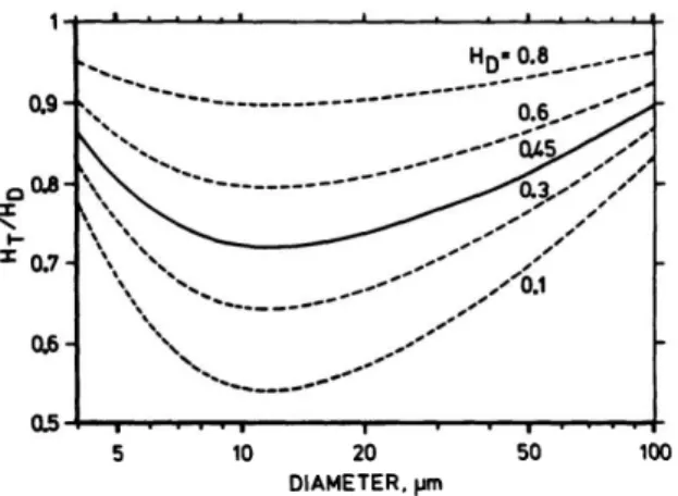
Figure 2.2: The Fåhraeus effect. The ratio Ht
Hd as a function of vessel vessel diameter for different discharge
hematocrit. This figure is a modified reproduction of figure 1 in Pries et al. [25]
This effect is called the Fåhraeus effect, which has been described in the literature using semi-empirical relationships, e.g., see Fahraeus [44], Barbee and Cokelet [55], and Pries et al. [25]. Figure2.2
shows Ht
Hd as a function of vessel diameter for different discharge hematocrits. We can see that the Fåhraeus effect is decreasing with increasing discharge hematocrit. This can be explained since a high discharge hematocrit implies a high number of red blood cells, which creates significant shear induced dispersion [49], ultimately decreasing the size of the cell-free layer and the difference between the average velocity of red blood cells and plasma.
Conversely, starting from large microvessels and decreasing the vessel diameter, we observe an increase in the Fåhraeus effect. That is because, in this range of diameter, the thickness of the cell-free layer is approximately constant [56], so that its relative size increases when decreasing the vessel diameter, causing the average red blood cell velocity to increase compared to the average blood flow velocity. We also note that, for diameters below 12 µm, the Fåhraeus effect starts to decrease. According to Albrecht et al. [57], this transition is mainly due to the size of the red blood cells becoming the same as, or larger than, the vessel they flow through, therefore causing the thickness of the cell-free layer to decrease. This ultimately leads to a decrease in the average red blood cell velocity relatively to the blood average velocity.
2.1.2 The Fåhraeus-Lindqvist effect
Viscous dissipation is larger in very diluted suspensions of solid particles than in monophasic fluids [58]. Such a result still holds for concentrated suspensions of solid particules [59,60] and can be extended to suspension of deformable vesicles [61].
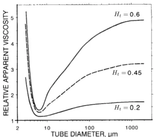
As far as blood is concerned, the impact of enhanced viscous dissipation due to red blood cells is usually represented by means of an effective viscosity that is systematically larger than the viscosity of the plasma [62,63]. Figure2.3shows the apparent relative viscosity of the blood in vitro, defined as the ratio between the effective viscosity and the plasma viscosity, as a function of vessel diameter for various tube hematocrits. It shows that the effective viscosity is an increasing function of the tube hematocrit. This result is intuitive in the sense that a high hematocrit implies a large number of red blood cells, hence a large number of fluid-cell and cell-cell interactions, in turn yielding more viscous dissipation.
Figure2.3also shows that the effective viscosity of the blood is a non monotonous function of the vessel diameter, exhibiting a minimum value around 8 µm. This unexpected behaviour defines the Fåhraeus-Lindqvist effect, for which Kiani and Hudetz [64], Pries, Neuhaus, and Gaehtgens [63], and
2.1 2.1 blood rheology in microcirculation 13
Lipowsky, Usami, and Chien [65] have derived semi-empirical relationships. This complex effect can be interpreted as the result of the interplay between the finite size of vessel diameters and the thickness of the cell-free layer close to the vessel wall.
Figure 2.3: The Fåhraeus-Lindqvist effect. In vitro relative apparent viscosity µeff
µplasma as a function of vessel
diameter for different tube hematocrit (Ht). This figure is a modified reproduction of figure 6 in
Pries, Neuhaus, and Gaehtgens [63].
Loosely speaking, starting from large microvessels and decreasing the vessel diameter, figure2.3
shows that there is a significant decrease in the effective viscosity. This shear-thinning effect is induced by the cell-free layer. As explained before, in this range of diameter, the thickness of the cell-free layer is approximately constant [56], so that its relative size increases when decreasing the vessel diameter. Now, recalling that only plasma flows through the cell-free layer means that there is proportionally less viscous dissipation in smaller vessels, explaining the decrease in the effective viscosity [45].
On the other hand, starting from small microvessels and decreasing the vessel diameter, figure2.3
shows that the effective viscosity sharply increases. In order to flow through such small vessels, red blood cells have to sustain significant deformations. Such a confinement likely generates a considerable amount of cell-wall interactions, increasing viscous dissipation and in turn increasing the effective viscosity. The cell-free-layer and the effect of confinement being antagonists justifies the non monotonous behaviour of the effective viscosity.
2.1.3 The phase separation effect
One of the defining feature of a microvascular network is the presence of diverging bifurcations, where one mother vessel feeds two daughter vessels. As mentioned before, red blood cells in the microcirculation do not behave in a trivial way, which raises the question of how red blood cells distribute at a diverging bifurcation.
Svanes and Zweifach [66] have observed in vivo that red blood cells tend to favor the daughter vessel with the highest flow rate. Such a phenomenon is often referred to as the Zweifach-Fung effect and Doyeux et al. [67] have shown, using rigid particles, that it is caused by the particle depleted layer close to the vessel walls. Similarly, for red blood cells, the Zweifach-Fung effect is a direct consequence of the cell-free layer mentioned previously.
16 current models for blood flow and molecular transport in the brain microcirculation
Going beyond these two categories, Schmid-Schönbein et al. [83] and Obrist et al. [82] have developed an original approach in which they consider each red blood cell as a symbolic particle flowing through a square grid (figure2.5b). Their idea is then to treat the blood flow problem like a cellular automaton, where each red blood cell follows a set of simple rules. For instance, Obrist et al. [82] prescribe that a red blood cell entering a bifurcation always exits by the daughter vessel with the highest flow rate. Similarily, they assume that the additional resistance to flow in a given vessel is proportional to the number of red blood cells in single file.
Even though their model formulation is simple, they are able to recover, at least partially, complex effect such as the phase separation effect. Using this new approach allows for simulating blood flow in large microvascular networks while keeping track of individual red blood cells. The drawback, however, is that it is restricted to microvascular networks composed of vessels with a constant and small diameter so that red blood cells are forced to flow in single file.
Altogether, blood flow models describing individual red blood cells are powerful tools to under-stand the complex dynamics occurring at the cell scale. Further, they can also help bridge the gap between the red blood cell scale and the vessel scale by enabling the derivation of effective laws describing the average behaviour of the blood based on the contributions of individual red blood cell. However, they are either computationally too demanding for simulating blood flow in large microvascular networks [73] or limited to very specific controlled configurations [82].
2.2.2 The blood as a homogeneous fluid
As described above, the blood is mainly a suspension of red blood cells and every red blood cell contributes to the blood flow. However, when investigating blood flow at the scale of a large anatomical network counting tens of thousands of vessels, it is interesting to look at the time-averaged blood flow in each vessel rather than at the instantaneous blood flow, the latter likely being difficult to interpret. Adopting this point of view makes it intuitive to consider the blood as a monophasic fluid with an effective viscosity and the red blood cells as a volume fraction. Doing so makes it easier to simplify microvascular networks into graphs, where each vessel is a segment and each bifurcation is a node.
This modelling approach is widespread in the literature [25,84,85,26,86,87,88,42], as it greatly simplifies the treatment of blood flow in each vessel, making it possible to model large volumes while still capturing the topology of the microvascular networks (figure2.6). In this context, solving the blood flow problem is generally equivalent to solving a stationary non-linear problem yielding the hematocrit, the blood flow rate and the pressure in each vessel and at each bifurcation of the network.
The disadvantage, however, is that this modelling approach heavily relies on the quality of the models used to represent the blood behaviour in each vessel. Indeed, the Fåhraeus effect, Fåhraeus-Lindqvist effect and phase separation effect can no longer emerge from red blood cells interactions with their surroundings and instead have to be prescribed. As mentioned before, semi-empirical relationships describing these effects have been derived by different authors, yielding different formulations. Even though there is no clear consensus, we note that the work of Pries [25,43,63,89] is frequently cited [26,86,90,88], as it provides relationships that are consistent for the entire range of vessel diameters encountered in the microcirculation.
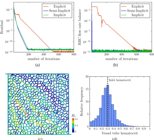
Practically, several strategies have been considered in the literature to solve the blood flow problem using a network approach. The most common one, used by Pries et al. [25], Lorthois, Cassot, and Lauwers [26], and Gould and Linninger [91], solves iteratively the balance of blood flow rates and red blood cell flow rates at each bifurcation, and the balance of momentum in each vessel (a
18 current models for blood flow and molecular transport in the brain microcirculation
derived by Pries to describe the underlying blood behaviour. The step by step derivation of the blood flow model will be presented in chapter3.
2.3 molecular transport in the cerebral microcirculation
The brain is the only organ of the human body that does not possess substantial reserves of energy. Consequently, it heavily relies on the efficacy of the microvascular system to continuously deliver vital molecules to brain cells. Part of this efficacy lies in the geometry of microvascular networks that form a dense mesh perfusing through the brain tissues [93]. Another part lies in the transport mechanisms at the scale of microvessels.
At such a small scale, the transport of a molecule going from the blood to the brain tissue can be divided into three main domains, namely the transport within the vessel (from the vessel lumen to the vessel walls), the transport across the vessel walls and the transport within the tissue (from the vessel walls to the brain cells, e.g. neurons, atrocytes).
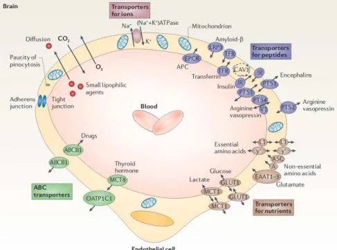
However, this general description of molecular transport is only a stereotype. Indeed, transport mechanisms, particularly in the brain, are molecule dependant, as schematized in figure2.7. This is mainly due to the blood-brain barrier, i.e., the layer of cells forming the walls of microvessels, that acts as a powerful filter facilitating the delivery of vital molecules to brain cells and preventing harmful chemicals from leaving blood vessels [94].
Figure 2.7: The diversity of molecules transported in the cerebral microcirculation. Schematics representing a cross-section of a brain capillary, with a description of various transport mechanisms across the vessel walls. This figure is a modified reproduction of figure 2 in Zlokovic [8]
Furthermore, even though the blood is mainly made out of water (92% of plasma volume [40], 70% of red blood cells volume [95]), there are a lot of other constituants flowing continuously through the microcirculation. This includes ions (e.g. Na+, K+, Ca2+, Cl−) [43], proteins (e.g. fibrinogen, albumin and globulin) [44], blood gasses (e.g. oxygen, carbon dioxide, nitrogen) and glucose. Depending on the context, other consituants, such as tracers used in medical imaging (e.g. gadolinium [96],
2.3 2.3 molecular transport in the cerebral microcirculation 19
radioactive water [97]) or therapeutic drugs that can cross the blood-brain barrier (e.g. ibuprofen, Nordazepam) [98], can also be found.
Finally, there is the particular case of oxygen. Indeed, unlike all other solute, most of the oxygen transported is bound to hemoglobin, a molecule which is exclusively located within the red blood cells. Consequently, oxygen has first to dissociate itself from hemoglobin before being transported from the vessels to the brain tissue. Such a dissociation mechanism is in fact a reversible chemical reaction and Hill [99] has derived a semi-empirical relationship describing the equilibrium between the free and bound oxygen in the blood.
Altogether, the transport of molecules in the brain is both very diverse and very complex, making it difficult to describe it in a comprehensive manner. However, it is still possible to highlight general transport mechanisms, so as to introduce generic models representing the behaviourof classes of relevant molecule. For that purpose, in the remaining of this section, we first focus on the current understanding of the main transport mechanisms at play within the microvessels, across the blood-brain barrier and inside the blood-brain tissue, as deduced from a series of recent experiment.
In section2.4, we go on and present how authors from the literature model such mechanisms at the microvascular scale. Then, we introduce the main simplifying assumptions associated with the different strategies developed in the literature to model and solve the transport of molecules at the scale of microvascular networks.
2.3.1 Molecular transport inside a microvessel
The transport of a molecule within a microvessel is mainly the result of two mechanisms, namely convection and molecular diffusion. The contributions of these two mechanisms, well-established in the case of a monophasic fluid [100], are very challenging to evaluate for the blood. Indeed, as already mentioned several times, the blood is a suspension of red blood cells. In the previous sections, we have shown that red blood cells, when flowing through the microcirculation, sustain significant migration effects, such as shear-induced dispersion [49], that are caused by the cells interacting with each others, the surrounding plasma and the vessel walls. This raises the question of how these interactions affect the transport of solute within the vessels. Indeed, these migration effects likely create secondary flows with consequences on the transport of molecules which are difficult to predict. Although the impact of such migration effects have, to the best of our knowledge, never been measured precisely, several studies have investigated the transport of molecules, and more particularly the transport of oxygen, inside microvessels [7,101–104].
For instance, Parpaleix, Houssen, and Charpak [102] have measured, using in vivo two-photon microscopy, the partial pressure of oxygen at many points between two red blood cells flowing in single file in capillaries, as shown in figure2.8a. To help understand, the partial pressure of oxygen can be seen, using Henry’s law for gas, as the local concentration of oxygen. Precisely, Figure2.8a
shows that there are strong heterogeneities in the partial pressure field, with less oxygen in the plasma between the red blood cells than close to the red blood cell membranes. This can be explained, since most of the oxygen is concealed within red blood cells. Consequently, at this scale, and particularly for the oxygen, the red blood cells have a strong impact on the transport and exchanges of molecules.
2.4 2.4 modelling molecular transport in the cerebral microcirculation 23
the microcirculation is concerned, numerous simplifying assumptions have been made over the past century to reduce this very complex situation to a more generic situation that can be modelled.
Krogh and Erlang [31] were the first to develop a model for the transport of molecules in the microcirculation. Precisely, they studied the transport of oxygen inside a single capillary embedded in an annulus of tissue. They assumed that the blood was a monophasic fluid and the surrounding tissue was a continuous medium. They further assumed that the partial pressure of oxygen inside the vessel cross-section was homogeneous. This simple configuration, now referred to as the Krogh cylinder, has since been widely used in various situations, including the transport of other solutes [115–120] or situations where the red blood cells were described individually [32,121,122,33].
In particular, Hellums [32], by describing the red blood cells individually, was one of the first to highlight that the partial pressure of oxygen, even inside the smallest capillaries, was not uniformly distributed in the vessel cross-sections and exhibited radial gradients that were likely to impact the exchanges with the surrounding tissue, which is consistent with what is observed experimentally nowadays [7,102]. Recently, Lücker et al. [123] have extended the work of Hellums and have gone beyond the Krogh cylinder configuration by simulating, in a small cerebral microvascular network, the transport of oxygen taking into account the corpuscular nature of red blood cells. However, even at the scale of a small microvascular network, the aforementioned studies have focused on the particular case of red blood cells flowing in single file through microvessels and are thus limited to the smallest capillary vessels.
Moreover, in less confined geometries, Kabacaoglu, Quaife, and Biros [124] have numerically inves-tigated the impact of vesicles on the transport of a passive solute in a two-dimensional Couette flow (figure2.11). Their study explored various initial and flow conditions (e.g. initial solute distribution, number of vesicles, Péclet number). Overall, they showed that the vesicles tend to reduce the mixing of the solute when compared to a monophasic Couette flow under the same conditions. To explain such a result, they suggested that vesicles create regions where the solute is trapped and cannot escape easily.
Even though the configuration investigated in Kabacaoglu, Quaife, and Biros [124] is simple and does not correspond to physiological geometries, it constitutes, to the best of our knowledge, one of the only study focusing on the collective effect of vesicles on the transport of a solute.
Altogether, transport models taking into account the corpuscular nature of red blood cells have been, so far, restricted to small networks or very particular configurations, mainly due to computational limitations. Consequently, when reaching larger scales, similarly to the blood flow, the most common approach is to consider the blood as a homogeneous fluid and to describe the transport of solute within microvessels using one-dimensional effective transport equations [30, 125, 126, 127, 42]. However, using such a simplified approach raises the question of how to take into account the heterogeneities in the concentration field within the vessel cross-section (e.g. the radial gradients evidenced by Sakadži´c et al. [7] in arterioles) that are likely to have an impact on the transport of molecules. In chapter4we derive such effective transport equations, and discuss at length the impact of such heterogeneities and how to incorporate them in our model.
24 current models for blood flow and molecular transport in the brain microcirculation
Figure 2.11: Impact of vesicles on the transport of a passive solute in a cylindrical Couette flow. In this exemple, the solute is initially located within the vesicles. This figure is a modified reproduction of figure 1 in Kabacaoglu, Quaife, and Biros [124]
Similarly, a significant problem of scale arises when modelling the transport accross the blood-brain barrier. Indeed, it is possible to simulate the transport of molecules within the blood-brain barrier [98,
128], but only at the scale of a cellular membrane. For instance, Carpenter et al. [98] use Molecular Dynamics (MD), a particle-based method (see section 2.2.1), to simulate the passive transport of various drugs through a simplified version of the blood-brain barrier. Although this method allows for the precise description of the interactions between molecules, it requires heavy computational ressources. Consequently, even at the scale of a single vessel, the blood-brain barrier is generally considered as a thin layer, with a diffusion coefficient different from the ones in blood and tissue [3], or even as a geometric manifold (infinitely thin layer), with an effective permeability [129].
Finally, as far as modelling transport in brain tissue is concerned, given the tremendous complexity of the medium at hand, the general consensus is to consider the brain cells and the interstitial space between them as a continuum where only molecular diffusion and metabolic reaction occur (e.g. see Nicholson [113]). This consensus has been reinforced recently, as Holter et al. [130] have simulated, using highly refined finite elements, convection and diffusion in the extravascular space, taking into account individual brain cells and interstitial space (figure2.12). They concluded that convection in brain tissue was negligible, even when overstimating the pressure gradients around vessels or the volume of the interstitial space. Although impressive, with a mesh made of 84 million tetrahedrons, their simulation was only covering a volume of 64 µm3, which represented less than 7% of the volume occupied by a capillary with characteristic diameter of 5 µm and length of 50 µm. Consequently, it is not possible to describe the tissue, at the scale of a microvascular network, using their approach.
26 current models for blood flow and molecular transport in the brain microcirculation
molecule when being transported from the center of the vessel lumen to the tissue domain. This includes the resistance to diffusion inside the plasma and red blood cells [132] and through the blood-brain barrier.
Similarly to the network approach presented in section2.2.2for the blood flow, the modelling approaches described above have the advantage of reducing the computational costs of the problem. The disadvantage, however, is that they heavily rely on the quality of the effective models used to represent the transport of molecules in each domain. That is particularly true for the effective transport equation inside the vessel. For example, most effective transport models neglect the effective diffusion along the vessel axis [30,125,126,127,131, 42]. In doing so, these models are unable to capture effects such as shear-enhanced dispersion (also known as Taylor’s dispersion) [100] that may have a significant impact on the transport of molecules, especially in fast flowing arterioles [117]. Similarly, some of these models neglect the effect of heterogeneities, such as radial gradients of concentration within the vessel cross-section, on the effective transport [125,126]. In chapter4, we also discuss these kinds of simplifications and propose our own effective transport equation based on homogenisation methods.
Practically, several strategies have been considered in the literature to solve this coupled problem. One of them is to adapt classic direct resolution methods such as finite elements or finite volumes [87,125] to such a simplified representation of microvascular networks. For instance, Linninger et al. [87] have developed a double mesh approach where they discretize independently vessel and tissues and then solve the problem using finite volumes. The vessels are divided into smaller cylindrical segments, and the tissue domain into relatively large thetrahedrons. In this context, each segment is considered a point source in the tissue, and a single thetahedron can host several sources. Although computationally efficient, this approach should be taken with caution as the coupling of several point sources in a finite volume cell is not straightforward [35].
On the other hand, Fang et al. [125] used an hybrid approach based on finite elements where they mesh the volume of tissue and the surface of microvessels using triangles and thetahedrons while still considering the transport inside the vessel as one dimensional. Doing so allows to describe more accurately the complex geometry of the microvascular networks. However, it also involves significantly more mesh cells than the approach proposed by Linninger et al. [87] which can limit the size of problem.
Finally, Hsu and Secomb [133] and then later Secomb et al. [30] proposed and improved a method to describe the concentration within tissue as the superposition of sources and wells distributed at the interface between vessel and tissues using Green’s function. This approach is really interesting as it saves, to some extent, from meshing the volume of tissue. A detailed presentation of this method is given in chapter5.
2.4.2 The molecular transport model derived in this thesis
Based on the above model review, and given the scale, complexity and diversity of molecular transport problems, in this thesis, we propose to take a step back and model a fictitious molecule capable of crossing the blood-brain barrier and to be metabolized by brain tissue, i.e. representing a generic nutrient or a drug.
With regard to the transport of molecule inside microvessels, we will follow the general consensus and consider one-dimensional effective transport equations along each vessel axis. However, as mentioned in the previous section, some simplifications found in the literature need to be adressed. Consequently, we propose to derive a new formulation for the transport of molecules inside microves-sels starting from the three dimensional transport problem and using homogenisation methods such as volume averaging [27,28] and multiscale asymptotics [28,29] (the presentation of these methods
2.5 2.5 modelling decisions and numerical implementations 27
alongside with the derivation of the effective transport equation is done in chapter4) to obtain an effective transport equation.
Similarly to Jain [129], we will approximate the blood-brain barrier by a geometric manifold with an effective permeability. Doing so allows us to easily enforce the selective aspect of the blood-brain barrier.
Finally, following the general consensus, we will consider the cerebral tissue as a continuous medium with homogeneous properties in which only molecular diffusion and metabolic reaction occur. As far as solving the molecular transport problem within tissue is concerned, we will take inspiration from Secomb et al. [30] and use Green’s function to describe the concentration of solute inside the domain of tissue. A more detailed derivation of this model is done in chapter5.
2.5 modelling decisions and numerical implementations
Thoughout this chapter, our primary objective was to present the complex mechanisms at play in the cerebral microvascular system along with the strategies found in the literature to model them. In doing so, we have shown that developing a comprehensive model for such microvascular transport mechanisms, at the scale of microvascular networks, is highly challenging. Consequently, simplifications have to be introduced. In doing so, the main difficulty is to still be able to accurately capture the multiscale and multiphysics nature of the problem, which couples non-linear effects in the rheology of blood [43, 89], molecular transfers across complex biological tissue [94,105] and effects linked to the geometry/topology of the vasculature [93].
As far as our models are concerned, with regard to blood flow, we will use a network approach and consider the blood as a monophasic fluid with an effective viscosity and describe the red blood cells as a volume fraction. Consistent with this vision, we will model the transport of molecules, by means of one-dimensional effective equations along the vessel axes. We will then couple such equations through an effective membrane condition to a three-dimensional continuum representing the tissue.
As already mentioned, the advantage of introducing such simplifications is that we can solve blood flow and molecular transport problems in large volumes while still capturing the detailed microvascular architecture. However, the downside is that this approach heavily relies on the accuracy of each effective models (e.g. the effective transport coefficients, the effective membrane permeability, the semi-empirical relationships describing the blood behaviour).
Consequently, our philosophy throughout this thesis will be to build these effective models, starting from simple mechanisms and elementary configurations, adding one layer of complexity at a time. In doing so, our goal is to be able to evaluate the implications of each new increment in the models. Further, we will validate, as far as possible, the effective models, at each step of their derivation, either against experimental measurements when they are available or against numerical reference solutions otherwise.
2.5.1 The collaborative platform BrainMicroFlow
Even though we have carefully chosen modelling approaches that allow for the simplification of the blood flow and molecular transport problem formulations, solving such problems in large microvascular networks still involves considerable amount of computations.
To address this issue, the work presented in the following chapters has been implemented in the collaborative platform BrainMicroFlow developed at the Institut de Mécanique des Fluides de Toulouse. Initiated by Peyrounette [134], this platform, which has since been deposited at the
28 current models for blood flow and molecular transport in the brain microcirculation
Agence de Protection des Programmes [135] , aims at providing a collection of high performance computational tools to investigate the physics of blood flow and mass transfers in the cerebral microvascular system. The platform itself can be decomposed into the following three parts
• The HybridNetwork-FiniteVolumeCode part aims at simulating blood flow and molecular transport in large vascular networks. It also contains a module dedicated to upscaling the blood flow problem at the scale of the whole brain using a hybrid approach (for more details, see Peyrounette et al. [35] and Peyrounette [134]).
• The FEM_simulations part is dedicated to the upscaling of the molecular transport problem within capillary vessels by means of formal homogenization methods (for more details see Doyeux et al. [136]). It also contains a module for the direct numerical simulations of blood flow and molecular transport in relatively small capillary networks (100 to 1, 000 vessels). This part is based on Feel++, a finite element library adapted to high performance computing (for more details see Prud’homme et al. [137]).
• The Network-Python-Toolbox part focuses on the conversion of different I/O file formats describing microvascular networks. The idea behind this toolbox is to propose a black box capable of converting any element of a list of available formats into any other format of the same list. The toolbox also includes a large collection of pre- and post-processing tools adapted to microvascular networks.
Consequently, we have implemented our blood flow and molecular transport models in the
HybridNetwork-FiniteVolumeCode part. More precisely, we have extended the already existing flow solver to include the non-linearities induced by the red blood cells and developed a new module, made of several new classes, for the description of molecular transport in large microvascular networks.
The HybridNetwork- FiniteVolumeCode itself is written in C++. This compiled language has been selected because of its low level and object-oriented nature. Another argument justifying the choice of C++ is the fact that it is well-suited for the use of the external library PETSc [138]. This library is a suite of data structures and functions designed to make the resolution of partial differential equations easier. It is also a powerful tool that provides efficient routines related to linear algebra. In addition, the PETSc library heavily relies on MPI (Message Passing Interface) standards so that it allows to write easily efficient parallel codes .
As mentioned earlier, BrainMicroFlow is a collaborative platform, which implies that particular programming rules had to be instored for developers in order to maintain the quality and integrity of the structure. One of the most important rules is the “one function one test” rule which states that each method of a given class should be associated to a test. This rule is enforced by means of CxxTests (https://cxxtest.com), which is a testing library adapted to C++. This tool automatically checks all the assertions implemented in the tests and provides a detailed report in case of failure. Moreover, this library allows for the construction of classes, thus the test files themselves can mirror the source files they test. From a developper’s perspective, testing each method involves a considerable amount of prototyping and programming but ultimately leads to more robust codes. For the complete set of programming rules, see Peyrounette [134].
Finally, the platform versioning is done using the free software Gitlab (https://about.gitlab.com). In doing so, the code is protected and its whole history archived. In addition, Gitlab is compatible with CxxTest, so that whenever a developer commits new changes to the platform, all the tests are automatically run and the modifications are accepted if and only if all tests have been successfully passed.
2.5 2.5 modelling decisions and numerical implementations 29
The idea behind the precautions presented above is to ensure the reliability of the BrainMicroFlow platform and to provide a clean environment for the developers. As far as the models presented in this thesis are concerned, they have been implemented and tested according to the patform standards. In addition, throughout the following chapters, they are also validated by direct comparison against in vitro and in vivo measurements when they are available and against reference analytical and numerical solutions otherwise.
Part II R E S U LT S
3
I M PA C T O F C A P I L L A R Y O C C L U S I O N S O N B L O O D F L O W I NM I C R O VA S C U L A R N E T W O R K S
Chapter synopsis:
The early onset of Alzheimer’s disease is associated with a significant cerebral blood flow decrease (up to 30%) that has been recently shown to be caused by a small fraction of capillaries (2% to 4%) being occluded by white blood cells. Experimentally, it is not possible to isolate the effects of such capillary occlusions or to translate results from murine models to humans. In this chapter, we develop a model to solve these issues and study the blood flow in anatomical networks made of tens of thousands of vessels. We start by validating the blood flow model by comparison against experiments performed in controlled in vitro environments. We then go on and validate it using in vivo experiments in mice. The model being validated, we find out that, in mouse, the cerebral blood flow linearly decreases with the fraction of capillaries occluded and that 2% to 4% occlusions yield 5% to 12% cerebral blood flow decrease. We further show that, even though the network geometry mainly controls the impact of a single capillary occlusion at the scale of a bifurcation, it is the network connectivity that drives the cerebral blood flow decrease at the scale of a full microvascular network. This allows us to extrapolate our results from mouse to human, highlighting the potential consequences of such occlusions at early stages of Alzheimer’s disease.
The sections 3.5 to 3.7 of this chapter summarize my contributions to the following two publications:
• Neutrophil adhesion in brain capillaries contributes to cortical blood flow decreases and impaired memory function in a mouse model of Alzheimer’s disease, Nature Neuroscience (2019) [11]. • Brain capillary networks across species: a few simple organizational requirements are sufficient to
reproduce both structure and function, Frontiers in Physiology (2019) [139].
Alzheimer’s disease is the most common form of dementia, and, even though early diagnosis procedures have been greatly improved in recent years [140], there is no treatment available yet. During the past 25 years many studies have focused on amyloid beta plaques as they were believed to be the key actor in Alzheimer’s disease (e.g. see the review by Selkoe and Hardy [141] and references therein). Amyloid beta is a small waste molecule naturally produced by brain cells and is a known neurotoxic [142]. In Alzheimer’s disease, amyloid beta is not correctly cleared and aggregates around cells forming plaques, ultimately leading to the death of the surrounded cells. Unfortunately, the very high rate of clinical failure (≥99%) for drugs removing such plaques tends to show that treating amyloid beta might not be the solution to Alzheimer’s disease [143,144]. Consequently, alternative


![Figure 3.3: Validation of the blood flow model against the in vitro experiments performed by Merlo [ 153 ]](https://thumb-eu.123doks.com/thumbv2/123doknet/2964169.81786/50.892.202.696.102.864/figure-validation-blood-model-vitro-experiments-performed-merlo.webp)