Klebsiella pneumoniae Siderophores Induce Inflammation, Bacterial
Dissemination, and HIF-1
␣ Stabilization during Pneumonia
Victoria I. Holden,aPaul Breen,bSébastien Houle,cCharles M. Dozois,c Michael A. Bachmana,b
Department of Microbiology and Immunology, University of Michigan, Ann Arbor, Michigan, USAa; Department of Pathology, University of Michigan, Ann Arbor,
Michigan, USAb; Institut National de la Recherche Scientifique, Institut Armand-Frappier, Laval, Canadac
ABSTRACT Klebsiella pneumoniae is a Gram-negative pathogen responsible for a wide range of infections, including
pneumo-nia and bacteremia, and is rapidly acquiring antibiotic resistance. K. pneumopneumo-niae requires secretion of siderophores, low-molecular-weight, high-affinity iron chelators, for bacterial replication and full virulence. The specific combination of sidero-phores secreted by K. pneumoniae during infection can impact tissue localization, systemic dissemination, and host survival. However, the effect of these potent iron chelators on the host during infection is unknown. In vitro, siderophores deplete
epithe-lial cell iron, induce cytokine secretion, and activate the master transcription factor hypoxia inducible factor-1␣ (HIF-1␣)
pro-tein that controls vascular permeability and inflammatory gene expression. Therefore, we hypothesized that siderophore secre-tion by K. pneumoniae directly contributes to inflammasecre-tion and bacterial disseminasecre-tion during pneumonia. To examine the effects of siderophore secretion independently of bacterial growth, we performed infections with tonB mutants that persist in
vivo but are deficient in siderophore import. Using a murine model of pneumonia, we found that siderophore secretion by K. pneumoniae induces the secretion of interleukin-6 (IL-6), CXCL1, and CXCL2, as well as bacterial dissemination to the
spleen, compared to siderophore-negative mutants at an equivalent bacterial number. Furthermore, we determined that
siderophore-secreting K. pneumoniae stabilized HIF-1␣ in vivo and that bacterial dissemination to the spleen required alveolar
epithelial HIF-1␣. Our results indicate that siderophores act directly on the host to induce inflammatory cytokines and bacterial
dissemination and that HIF-1␣ is a susceptibility factor for bacterial invasion during pneumonia.
IMPORTANCEKlebsiella pneumoniae causes a wide range of bacterial diseases, including pneumonia, urinary tract infections, and
sepsis. To cause infection, K. pneumoniae steals iron from its host by secreting siderophores, small iron-chelating molecules. Classically, siderophores are thought to worsen infections by promoting bacterial growth. In this study, we determined that siderophore-secreting K. pneumoniae causes lung inflammation and bacterial dissemination to the bloodstream independently of bacterial growth. Furthermore, we determined that siderophore-secreting K. pneumoniae activates a host protein, hypoxia
inducible factor (HIF)-1␣, and requires it for siderophore-dependent bacterial dissemination. Although HIF-1␣ can protect
against some infections, it appears to worsen infection with K. pneumoniae. Together, these results indicate that bacterial sid-erophores directly alter the host response to pneumonia in addition to providing iron for bacterial growth. Therapies that dis-rupt production of siderophores could provide a two-pronged attack against K. pneumoniae infection by preventing bacterial growth and preventing bacterial dissemination to the blood.
Received 8 August 2016 Accepted 18 August 2016 Published 13 September 2016
Citation Holden VI, Breen P, Houle S, Dozois CM, Bachman MA. 2016. Klebsiella pneumoniae siderophores induce inflammation, bacterial dissemination, and HIF-1␣ stabilization
during pneumonia. mBio 7(5):e01397-16. doi:10.1128/mBio.01397-16.
Editor Jeff F. Miller, UCLA School of Medicine
Copyright © 2016 Holden et al. This is an open-access article distributed under the terms of theCreative Commons Attribution 4.0 International license. Address correspondence to Michael A. Bachman, mikebach@med.umich.edu.
K
lebsiella pneumoniae is a Gram-negative bacterium within the Enterobacteriaceae family and is the causative agent of a wide range of infections, including pneumonia, urinary tract infec-tions, wound infecinfec-tions, and bacteremia. As the third-most-common cause of hospital-acquired infections, K. pneumoniae represents a major health care threat (1). Further compounding this concern, K. pneumoniae is rapidly acquiring resistance to all known antibiotics, thus becoming increasingly difficult to treat. In particular, carbapenem-resistant strains of K. pneumoniae are re-sistant to all or nearly all antibiotics and exhibit strikingly high mortality rates of 41% to 50% for bloodstream infections (2, 3).In order to establish infection, K. pneumoniae secretes mole-cules called siderophores that are critical for bacterial growth and
replication (4, 5). Siderophores are small, high-affinity iron-chelating molecules secreted by a wide variety of microorganisms that are critical for virulence in many Gram-negative bacteria (6). Enterobactin (Ent), with the highest known affinity for iron of any molecule, is the prototypic catecholate siderophore and effectively outcompetes host iron-binding proteins for iron (7). To counter the effects of Ent, neutrophils and epithelial cells secrete lipocalin 2 (Lcn2; also known as NGAL, Scn, and 24p3), which binds Ent with subnanomolar affinity (8). In vivo, the presence of Lcn2, by preventing bacterial uptake of Ent, has been shown to be bacterio-static (9, 10). Bacteria have evolved to secrete Lcn2-evasive sidero-phores, such as salmochelin (Sal), a glycosylated Ent that requires the iroA locus for production and transport; the phenolate
on February 20, 2019 by guest
http://mbio.asm.org/
phore yersiniabactin (Ybt); and the citrate-hydroxamate sidero-phore aerobactin (6, 11, 12).
Because iron is critical for the function of many cellular pro-cesses, including DNA replication and oxygen metabolism, and as a cofactor for many cellular reactions, iron chelation by sidero-phores could have significant effects on host cells (13, 14). How-ever, the effects of siderophore-dependent manipulation of host iron homeostasis during bacterial infection are largely unknown. Iron chelation by siderophores in the presence of Lcn2 induces in vitro proinflammatory cytokine secretion of interleukin-8 (IL-8), IL-6, and CCL20 from lung epithelial cells (15, 16). Siderophores also induce the stabilization of the master transcription factor hypoxia inducible factor-1␣ (HIF-1␣) in vitro (15). HIF-1␣ regu-lates the expression of many genes, including those involved in glycolysis, inflammation, and angiogenesis, and is itself regulated by the availability of oxygen or iron within a cell (17–19). In nor-moxia, HIF-1␣ protein is targeted for degradation by prolyl hy-droxylases, a reaction that requires iron (20). However, under conditions of low oxygen or low iron levels, HIF-1␣ protein is stabilized and translocates to the nucleus to activate gene expres-sion (20–22). In addition to roles in adaptation to hypoxia and tumor development, HIF-1␣ activation has recently been associ-ated with innate immunity against infections. Infection with Pseu-domonas aeruginosa in a Caenorhabditis elegans model system identified siderophodependent activation of a hypoxic host re-sponse that was partially protective (23). In a murine urinary tract infection (UTI) model, HIF-1␣ was protective against Escherichia coli infection through host innate immunity modulation (24). These results indicate that siderophores have a broad and inflam-matory effect on host epithelial cells in addition to their role as iron acquisition molecules for bacteria. However, a critical barrier to investigating this phenomenon in vivo is that siderophores al-low bacterial proliferation, and an increase in bacterial CFU may indirectly increase inflammation, tissue damage, and bacterial dis-semination.
Ferric siderophores are actively imported by bacteria through siderophore-specific outer membrane receptors that are depen-dent on the TonB-ExuB-ExuD energy transducing system, and tonB mutants secrete siderophores but cannot utilize them for growth (25). K. pneumoniae tonB mutants retain their antiphago-cytic capsule and serum resistance but are unable to grow under iron-limited conditions (26). Upon inoculation, K. pneumoniae tonB mutants stimulate the host immune response but do not cause a productive infection.
In this study, we tested the hypothesis that K. pneumoniae sid-erophores disrupt iron homeostasis in host cells, leading to altered host responses to pneumonia. To study the effects of siderophore secretion independently of the indirect effects of siderophore-dependent bacterial growth, we employed tonB mutants that are capable of secreting siderophores but are not capable of utilizing them. By dissociating siderophore secretion from bacterial growth, we could directly compare levels of proinflammatory cy-tokine secretion, bacterial dissemination, and HIF-1␣ stabiliza-tion in response to infecstabiliza-tion with isogenic siderophore synthesis mutants at equivalent bacterial numbers.
RESULTS
Siderophores induce bacterial dissemination and lung inflam-mation. To measure the overall contribution of siderophores to
inflammation and bacterial dissemination, mice were infected
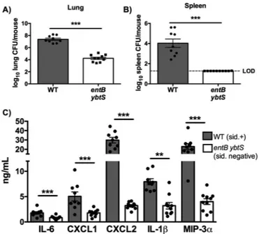
with a wild-type (WT) strain of K. pneumoniae that secretes Ent, Sal, and Ybt or an isogenic siderophore-negative entB ybtS mutant (4, 27). Infection with WT K. pneumoniae resulted in increased lung bacterial load compared to infection with the entB ybtS mu-tant (Fig. 1A). Furthermore, siderophores were required for K. pneumoniae dissemination to the spleen (Fig. 1B). Additionally, siderophores were required for induction of proinflammatory cy-tokines: the WT strain, but not the entB ybtS mutant, induced expression of interleukin-6 (IL-6), CXCL1, CXCL2, IL-1, and macrophage inflammatory protein-3 alpha (MIP-3␣) (Fig. 1C).
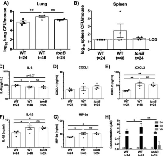
The increase in bacterial dissemination and the inflammatory response during WT infection could be due to direct effects of iron chelation by siderophores on the host or to an indirect effect of siderophores increasing bacterial CFU in the lungs. To distinguish between these possibilities, we utilized a tonB mutant. As ex-pected, the tonB mutant secreted siderophores but was not able to utilize endogenous or exogenous siderophores for bacterial growth (see Fig. S1 in the supplemental material). Because the WT and tonB K. pneumoniae strains produce an antiphagocytic cap-sule, we hypothesized that the tonB mutant would persist in the lung and secrete siderophores but would not replicate (26, 28). To compare infections performed with the WT and tonB K. pneu-moniae strains, C57BL/6 mice were infected with 1⫻ 108CFU tonB K. pneumoniae or 1⫻ 104CFU WT K. pneumoniae for 24 or 48 h. After 24 h, tonB K. pneumoniae persisted in the lung and spleen, with a bacterial load comparable to that seen after 48 h of infection with WT K. pneumoniae (Fig. 2A and B). At 24 h, tonB K. pneumoniae infection also caused levels of induction of IL-6, CXCL1, CXCL2, IL-1, and MIP-3␣ secretion that were compa-rable to those seen after 48 h of infection with WT K. pneumoniae
FIG 1 K. pneumoniae siderophores enhance bacterial growth and are
re-quired for cytokine secretion and bacterial dissemination. C57BL/6 mice (n⫽ 9 to 10 per group) with 1⫻ 104CFU wild-type K. pneumoniae or entB ybtS
K. pneumoniae, a mutant deficient in siderophore production. (A and B) At
day 2, mice were euthanized, and organs were harvested for bacterial load in the (A) lung and (B) spleen. LOD, limit of detection. (C) Lung homogenates were assayed by ELISA for IL-6, CXCL1, CXCL2, IL-1, and MIP-3␣ secre-tion. Statistics were calculated using t-test (A and B) or one-way ANOVA with Fisher’s posttest (**, P⬍ 0.01; ***, P ⬍ 0.001 [as indicated]). sid., siderophore.
Holden et al.
on February 20, 2019 by guest
http://mbio.asm.org/
(Fig. 2C to G). Additionally, we determined the concentrations of siderophores during infection with the tonB and WT K. pneu-moniae strains by performing mass spectrometry (Fig. 2H; see also Fig. S2). Micromolar concentrations of Sal were detected in whole-lung homogenates from all infections. Ybt was detected in lower quantities in all lung samples, but Ent was not detected. Although bacterial growth dynamics differ, these results indicate that tonB K. pneumoniae infections can be used to examine the impact of siderophores on the host at concentrations and bacterial densities that mimic WT K. pneumoniae infection.
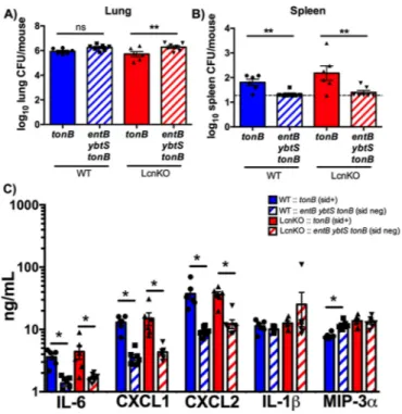
To determine the impact of siderophore secretion on dissem-ination and cytokine responses, C57BL/6 mice were infected with isogenic tonB (siderophore-secreting) or entB ybtS tonB (siderophore-negative) K. pneumoniae. Despite equivalent bacte-rial loads, infection with tonB K. pneumoniae resulted in increased bacterial dissemination and IL-6, CXCL1, and CXCL2 secretion compared to infection with the entB ybtS tonB mutant (Fig. 3). Infection with the tonB strain did not enhance secretion of IL-1 or MIP-3␣ compared to the entB ybtS tonB mutant (Fig. 3C), suggesting that the differences between the level of induction by the WT strain and the level of induction by the entB ybtS strain
were attributable to differences in bacterial density (Fig. 1C). In vitro, Lcn2 enhances induction of proinflammatory cytokines by siderophores (15). To examine the contribution of Lcn2, Lcn2-deficient (LcnKO) mice were infected with tonB or entB ybtS tonB K. pneumoniae. The same pattern of cytokine induction and dis-semination was observed in WT and LcnKO mice, indicating that Lcn2 was not required for siderophore-dependent dissemination and inflammation in vivo (Fig. 3).
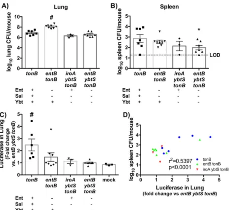
Multiple siderophores are required for bacterial dissemina-tion and cytokine secredissemina-tion. In vitro, Ent and Ybt induced
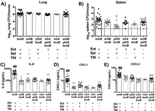
proin-flammatory cytokine secretion, but Sal did not (15). Because WT K. pneumoniae secretes Ent, Sal, and Ybt, it is unclear which sid-erophores are required to induce bacterial dissemination and in-flammation during pneumonia. To test which siderophores were necessary for maximal dissemination and cytokine secretion, we used isogenic siderophore-secreting tonB mutants (Table 1) that secrete one or two siderophores to infect C57BL/6 mice. The iroA locus, required to produce Sal, was disrupted by mutation of the iroB glycosylase gene (9). Importantly, the infections performed with all mutants resulted in equivalent lung bacterial loads (Fig. 4A). Consistently, infection with tonB K. pneumoniae
in-FIG 2 The results of 24-h tonB K. pneumoniae infection are comparable to those of 48-h wild-type infection. C57BL/6 mice (n⫽ 4 per group) were infected with 1⫻ 104CFU wild-type K. pneumoniae or 1⫻ 108CFU tonB K. pneumoniae, a mutant that secretes but cannot take up siderophores. (A and B) Following 24 or 48 h, mice were euthanized, and organs were harvested for bacterial load in the (A) lung and (B) spleen. (C to H) Lung homogenates were assayed for (C) IL-6, (D) CXCL1, (E) CXCL2, (F) IL-1, and (G) MIP-3␣ secretion by ELISA and (H) siderophore quantification by liquid chromatography-tandem mass spectrom-etry (LC-MS/MS). Statistics were calculated using one-way (A to G) or two-way (H) ANOVA with Fisher’s posttest (*, P⬍ 0.05; **, P ⬍ 0.01; ns, P ⬎ 0.05). Elapsed time values represent hours.
on February 20, 2019 by guest
http://mbio.asm.org/
duced significantly more dissemination to the spleen than infec-tion with the entB ybtS tonB mutant (Fig. 4B). No other sidero-phore mutant induced significant dissemination compared to the entB ybtS tonB (siderophore-negative) mutant. These data indi-cate that all siderophores are required in combination for maxi-mal bacterial dissemination to the spleen.
To determine if each individual siderophore is sufficient to induce cytokine secretion, enzyme-linked immunosorbent assays (ELISAs) were performed on lung homogenates taken at 24 h postinfection. Only infection with the tonB mutant was sufficient to induce more secretion of IL-6, CXCL1, or CXCL2 than infec-tion with the entB ybtS tonB (siderophore-negative) mutant, indi-cating that all three siderophores are required for secretion of these cytokines (Fig. 4C to E). Consistent with Fig. 3, siderophore
secretion did not specifically induce IL-1 or MIP-3␣ (see Fig. S3 in the supplemental material). Binding by Lcn2 could mask the effects of Ent on inflammation and dissemination. To test this hypothesis, we compared the levels of cytokine secretion and dis-semination in C57BL/6 and LcnKO mice infected with iroA ybtS tonB (Ent-positive [Ent⫹]) K. pneumoniae (see Fig. S4). LcnKO mice did not display differences in spleen bacterial load or lung inflammation compared to C57BL/6 WT mice, indicating that the presence of Ent is not sufficient to induce dissemination or in-flammation, even in the absence of Lcn2. Together, these results indicate that Sal, Ent, and Ybt are required to induce maximal secretion of IL-6, CXCL1, and CXCL2.
Siderophores secreted by K. pneumoniae stabilize HIF-1␣.
To test the hypothesis that K. pneumoniae siderophores stabilize HIF-1␣ during pneumonia, we utilized a transgenic mouse model that expresses a fusion protein of luciferase with the oxygen-dependent domain (ODD) (ODD-Luc) of HIF-1␣ that is subject to prolyl hydroxylation and becomes stabilized under low oxygen or low iron conditions (29–31). Infection of ODD-Luc mice with WT K. pneumoniae induced increased bioluminescence in the lung compared to the results obtained with a phosphate-buffered saline (PBS) vehicle control (see Fig. S5A in the supplemental material). To determine whether siderophores secreted by K. pneumoniae induce HIF-1␣ stabilization in vivo during infec-tion, ODD-Luc mice were infected with tonB (siderophore-positive) or entB ybtS tonB (siderophore-negative) K. pneu-moniae. Infection with tonB induced greater bioluminescence in the lung than infection with entB ybtS tonB, though infection with entB ybtS tonB did induce some bioluminescence compared to infection with the PBS vehicle control (see Fig. S5B). These results indicate that siderophores secreted in vivo can stabilize the HIF-1␣ transcription factor.
We then sought to determine whether individual siderophores were capable of stabilizing HIF-1␣. To do so, ODD-Luc mice were infected with isogenic siderophore tonB mutants, and bacterial loads in the lung and spleen were quantified (Fig. 5A and B). The tonB mutants had equivalent lung CFU levels but significantly higher spleen CFU levels than the entB ybtS tonB mutants, consis-tent with previous data. Additionally, luciferase expression in the lung homogenate was quantified as fold change compared to the entB ybtS tonB (siderophore-negative) mutant. Infection with the tonB mutant induced significantly more luciferase expression than infection with the other isogenic strains (Fig. 5C). Infection with the iroA ybtS tonB (Ent⫹) strain resulted in CFU counts equivalent to tonB and entB ybtS tonB infection but did not result in significant dissemination to the spleen or induction of
lu-FIG 3 Siderophore secretion by K. pneumoniae results in bacterial
dissemi-nation and IL-6, CXCL1, and CXCL2 secretion in a Lcn2-independent man-ner. C57BL/6 mice (n⫽ 6 to 7 per group) were infected with 1 ⫻ 108CFU tonB or entB ybtS tonB K. pneumoniae. (A and B) Following 24 h, mice were eutha-nized, and organs were harvested for bacterial load in the (A) lung and (B) spleen. (C) Lung homogenates were assayed for IL-1, IL-6, CXCL1, CXCL2, and MIP-3␣ secretion by ELISA. Statistics were calculated using one-way ANOVA with Fisher’s posttest (*, P⬍ 0.05; **, P ⬍ 0.01; ns, P ⬎ 0.05 [as indicated]).
TABLE 1 K. pneumoniae mutants used in this work
Strain Description
Siderophore produced
Reference or source
Ent Ybt Sal
Wild type KPPR1; RifR derivative of ATCC 43816 ⫹ ⫹ ⫹ 27
entB ybtS VK089; KPPR1 entB ybtS ⫺ ⫺ ⫺ 27
tonB KP273; KPPR1 tonB::kan ⫹ ⫹ ⫹ This work
entB ybtS tonB KP281; VK089 tonB::kan ⫺ ⫺ ⫺ This work
entB tonB KP285; VK087 tonB::kan ⫺ ⫹ ⫺ This work
ybtS tonB KP277; VK088 tonB::kan ⫹ ⫺ ⫹ This work
iroA ybtS tonB KP2202; KP20 tonB::hyg ⫹ ⫺ ⫺ This work
iroA tonB KP2227; KP25 tonB::hyg ⫹ ⫹ ⫺ This work
Holden et al.
on February 20, 2019 by guest
http://mbio.asm.org/
ciferase. ODD-Luc mice infected with the entB tonB (Ybt⫹) mu-tant displayed a higher bacterial load in the lung upon infection, confounding comparisons to the other strains. This mutant in-duced increased luciferase expression for a few mice and had cor-respondingly high CFU counts in the spleen. We therefore hy-pothesized that siderophore-dependent HIF-1␣ stabilization in the lung correlates with bacterial load in the spleen and that the outliers in the entB tonB mutant are the exceptions that prove the rule. To examine this hypothesis, spleen bacterial load was graphed as a function of luciferase levels in the lung, and the cor-relation was determined. This plot revealed a positive corcor-relation between spleen bacterial load and luciferase expression during infection with K. pneumoniae across all siderophore mutant geno-types (Fig. 5D). Taken together, these data indicate that HIF-1␣ is consistently stabilized only by tonB K. pneumoniae that produces all three siderophores and that stabilization of HIF-1␣ by K. pneu-moniae secreting siderophores correlates with bacterial dissemi-nation to the spleen.
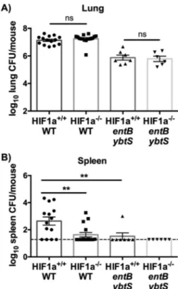
Lung epithelial HIF-1␣ is required for bacterial
dissemina-tion to the spleen. Because HIF-1␣ stabilization correlates with
bacterial dissemination to the spleen, and because HIF-1␣ regu-lates vascular permeability and inflammation, we hypothesized that HIF-1␣ is required for siderophore induction of bacterial dissemination and the host proinflammatory response. To test this hypothesis, we utilized transgenic mice that have an inducible lung epithelial cell-specific Hif1a deletion (55), using either mice induced with doxycycline postnatally (Hif1a⫺/⫺) or uninduced, wild-type littermates (Hif1a⫹/⫹). To test the effect of epithelial
HIF-1␣ on a productive infection with replicative K. pneumoniae, the WT strain and entB ybtS mutant were used instead of their tonB counterparts. Infection with WT K. pneumoniae did not cause HIF-1␣-dependent differences in lung bacterial load (Fig. 6A). However, Hif1a⫺/⫺ mice displayed significantly less bacterial dissemination to the spleen after 24 h, indicating that HIF-1␣ promotes bacterial dissemination to the spleen (Fig. 6B). The siderophore-negative entB ybtS mutant had lower lung and spleen CFU counts than the WT, which was not affected by the absence of lung epithelial HIF-1␣ (Fig. 6). Infection with WT K. pneumoniae induced significantly more IL-6 and CXCL2 than infection with the entB ybtS mutant, consistent with the siderophore-dependent effects observed as described above, and more IL-1, which may be attributable to higher bacterial density (see Fig. S6 in the supplemental material). However, there were no HIF-1␣-dependent differences in lung cytokine secretion, indi-cating that lung epithelial HIF-1␣ is not required to induce IL-1, IL-6, CXCL1, CXCL2, or MIP-3␣ secretion during infection (see Fig. S6). These data suggest a role for epithelial HIF-1␣ stabiliza-tion by siderophores in the inducstabiliza-tion of bacterial disseminastabiliza-tion during K. pneumoniae infection.
DISCUSSION
K. pneumoniae is a Gram-negative bacterium that is rapidly ac-quiring resistance to all known antibiotics, including carbapen-ems. Developing novel therapies to combat antibiotic-resistant infections requires a more complete understanding of disease pathogenesis. To determine the effect of siderophores on the host
FIG 4 Multiple siderophores are required for bacterial dissemination and IL-6, CXCL1, and CXCL2 secretion. C57BL/6 mice (n⫽ 5 to 18 per group) were infected with 1⫻ 108CFU isogenic tonB K. pneumoniae as indicated. (A and B) Following 24 h, mice were euthanized, and organs were harvested for bacterial load in the (A) lung and (B) spleen. (C to E) Lung homogenates were assayed for (C) IL-6, (D) CXCL1, and (E) CXCL2 secretion using ELISA. Statistics were calculated using one-way ANOVA with Fisher’s posttest (*, P⬍ 0.05 [versus entB ybtS tonB]; ##, P ⬍ 0.001 [versus all other conditions]).
on February 20, 2019 by guest
http://mbio.asm.org/
response to infection, we utilized tonB mutants that allowed us to uncouple siderophore secretion from bacterial growth. We show that K. pneumoniae siderophores are a major trigger of the inflam-mation and bacterial dissemination induced during lung infection with K. pneumoniae, independently of their ability to deliver iron to bacteria. Additionally, we show that the induction of bacterial dissemination by siderophores requires master transcription fac-tor HIF-1␣ in lung epithelial cells. These findings represent a novel function for bacterial siderophores in cytokine secretion and bacterial dissemination and a novel function for host master transcription factor HIF-1␣ as a susceptibility factor for the devel-opment of sepsis.
Our data indicate that siderophores induce dissemination through chelation of host cellular iron, leading to inactivation of iron-dependent prolyl hydroxylases and HIF-1␣ stabilization in lung epithelial cells. We have previously shown that purified sid-erophores deplete cellular iron in respiratory epithelial cells and stabilize HIF-1␣ and that activation of HIF-1␣-dependent gene expression is abrogated by iron (15). The siderophore desferriox-amine is a canonical activator of HIF-1␣, indicating that iron che-lation is sufficient for stabilization (24). Accordingly, we demon-strate that the prolyl-containing ODD of HIF-1␣ is stabilized in vivo by siderophore-producing K. pneumoniae. The WT strain secretes Sal and Ybt in vivo, and both are required for maximal HIF-1␣ induction. Siderophore-dependent dissemination is blocked in a lung epithelial HIF-1␣ knockout mouse, indicating that this cell type mediates bacterial spread from the lung to the
spleen. The mechanism is unknown and, since HIF-1␣ is a global transcriptional regulator, may be complex and multifactorial. For example, HIF-1␣ regulates cellular metabolism and survival and can induce vascular permeability and angiogenesis (32, 33) or dis-ruption of epithelial barriers (34).
HIF-1␣ stabilization by siderophore-dependent iron chelation leading to bacteremia contrasts with the protective effect of HIF-1␣ stabilization by a pharmacological molecule, AKB-4924, during murine UTI (24). These contrasting results may be due to differences in both pathogen and model system. For instance, treatment with AKB-4924 prevented internalization of E. coli by uroepithelial cells. K. pneumoniae is not readily internalized by epithelial cells due to its capsule; therefore, preventing the uptake of bacteria through HIF-1␣ stabilization may not be an effective therapy against K. pneumoniae infection (35). Together, these data illustrate the complexity of HIF-pathogen interactions and high-light the importance of evaluating many various bacterial infec-tions and model systems.
Mass spectrometry demonstrated that Sal and Ybt are the main siderophores produced during K. pneumoniae lung infection (Fig. 2H; see also Fig. S2 in the supplemental material). This anal-ysis has been used to quantify Ent, Sal, and aerobactin in chicken air sacs during E. coli infection (36). To our knowledge, these data represent the first published concentrations of siderophores from murine lung homogenates. The measured concentrations were of the same magnitude as the concentrations used in vitro by our group, as well as others, and provide a context for prior findings
FIG 5 Siderophore secretion by K. pneumoniae induces HIF-1␣ stabilization, which correlates to bacterial dissemination to the spleen. ODD-luciferase mice (n⫽ 3 to 8 per group) were infected with 1 ⫻ 108CFU isogenic tonB K. pneumoniae as indicated. (A to C) Following 24 h, mice were euthanized, and organs were harvested for (A) lung bacterial burden, (B) spleen bacterial burden, and (C) luciferase quantification. (D) Correlation curves were plotted comparing spleen CFU counts as a function of the level of luciferase in the lung. Statistics were calculated using one-way ANOVA with Fisher’s posttest (*, P⬍ 0.05 [versus tonB]; #, P⬍ 0.05 [versus all other conditions]) or the Pearson r correlation curve.
Holden et al.
on February 20, 2019 by guest
http://mbio.asm.org/
obtained using purified siderophores (15, 16, 37). Although tonB K. pneumoniae secreted more siderophores in vitro, the concen-trations observed in vivo were equal to or even slightly lower than those seen with the WT strain, indicating that the tonB mutants can assess the impact of siderophores on the host at physiologi-cally relevant concentrations. Whereas micromolar concentra-tions of Sal and high nanomolar amounts of Ybt were detected, we were unable to detect Ent. We propose three possible explanations for this finding: (i) Ent is sequestered by Lcn2, and is therefore undetectable; (ii) bacteria convert all Ent to Sal in vivo to evade Lcn2; or (iii) a combination of our two hypotheses, whereby bac-teria convert the majority of Ent to Sal and the remaining Ent is sequestered by Lcn2. These data suggest that the majority of in-flammation and bacterial dissemination is due to the secretion of Sal but also suggest a role for Ybt. However, we could not test the contribution of Sal in isolation because it is not possible to create mutants that produce Sal without intact Ent synthesis genes, and we were unable to detect iron chelation by purified Sal in vitro (12, 15). In contrast, a mutant making Ent alone (the iroA ybtS tonB mutant) showed no detectable induction of cytokines or dissem-ination, with or without Lcn2. Together, these data implicate Sal and Ybt as significant inducers of inflammation and dissemina-tion during pneumonia.
In addition to inducing dissemination, Ybt and Sal are re-quired for maximal induction of IL-6, CXCL1, and CXCL2, which are all protective against lung infection with K. pneumoniae (38– 40). These results are consistent with prior in vitro data illustrating
that iron chelation by siderophores, as evidenced by depletion of the labile iron pool and induction of the iron starvation marker NDRG1, induces the secretion of proinflammatory cytokines IL-8 and IL-6 from A549 lung epithelial cells (15). Human IL-6 and murine IL-6 act as inflammatory cytokines involved in hepatocyte acute-phase responses and can upregulate hepcidin, an iron ho-meostasis protein (41, 42). Murine CXCL1 and CXCL2 are neu-trophil chemoattractants and are functionally similar to human IL-8 (43). Whereas lung epithelial HIF-1␣ is instrumental in bac-terial dissemination to the spleen, it was not required for the in-duction of cytokines. These results contrast with studies showing that HIF-1␣ regulates IL-6 and that epithelial HIF-1␣ regulates IL-6 and IL-1 secretion in a lung contusion model (44, 45). My-eloid cell HIF-1␣ has been shown to be instrumental in inflam-mation through myeloid cell development, phagocytosis, and an-timicrobial production (46–48). In addition to HIF-1␣ regulation of inflammation, HIF-2␣ can regulate macrophage function in tumor models, eosinophil function in the lung, and IL-6 secretion from endothelial cells (49–51). Therefore, it is possible that an-other cell-specific HIF-1␣ or HIF-2␣ could be responsible for reg-ulating cytokine secretion in response to K. pneumoniae infection. Because HIF knockouts are embryonically lethal, testing other HIF isoforms and cell types would require multiple lineage-specific knockouts.
Although Lcn2 was necessary for siderophore induction of cy-tokines in vitro, it was dispensable for the siderophore-dependent immune response to K. pneumoniae in vivo (15). This may indi-cate differential abilities of human and murine Lcn2 to modulate immune responses. Although they share 62% amino acid identity, murine Lcn2 lacks the ability to form covalent complexes, which may explain this discrepancy (52). Alternatively, redundant sig-naling pathways may be activated during pneumonia such that inflammatory signaling by Lcn2 is dispensable. It may be that two signals are required for the maximal induction of cytokine secre-tion in response to infecsecre-tion with siderophore-secreting bacteria: (i) perturbation of iron homeostasis by siderophores and (ii) sig-naling by an inflammatory protein(s), including Lcn2. In vivo, many proteins could satisfy the requirement of the second signal, such as inflammasome activation or Toll-like receptor signaling activation by capsule and lipopolysaccharide (53, 54).
On the basis of our data, we propose the following model: upon infection and iron starvation, K. pneumoniae produces and se-cretes siderophores. Siderophores serve to acquire host iron and deliver it to the bacteria, resulting in bacterial growth. In addition to supporting bacterial growth, chelation of host cellular iron by siderophores induces cellular stress. One stress response is the stabilization of HIF-1␣, ultimately resulting in bacterial dissemi-nation to the spleen. An opposing stress response is secretion within the lung of the proinflammatory cytokines IL-6, CXCL1, and CXCL2, which are necessary for protection from K. pneu-moniae. These results indicate novel functions for bacterial sidero-phores during infection that are independent of their iron delivery capabilities and present siderophore molecules as a possible target for therapeutic intervention. Additionally, these results indicate a novel role for HIF-1␣ as a susceptibility factor for systemic spread during K. pneumoniae infection and illustrate the complex inter-play between pathogen and host molecules during bacterial infec-tion.
FIG 6 Lung epithelial HIF-1␣ is necessary for siderophore-dependent bac-terial dissemination to the spleen. Hif1a⫹/⫹or Hif1a⫺/⫺mice (n⫽ 6 to 14 per group) were infected with 1⫻ 104CFU wild-type or entB ybtS K. pneumoniae. Following 24 h, mice were euthanized, and organs were harvested for bacterial load in the (A) lung and (B) spleen. Statistics were calculated using one-way ANOVA with Fisher’s posttest (**, P⬍ 0.01; ns, P ⬎ 0.05 [as indicated]).
on February 20, 2019 by guest
http://mbio.asm.org/
MATERIALS AND METHODS
Animal strains and ethics statement. All work was approved by the
Uni-versity of Michigan Institutional Animal Care and Use Committee (IACUC). C57BL/6 lipocalin 2-deficient (LcnKO), ODD-luciferase (ODD-Luc) (29), and conditional alveolar epithelial HIF-1␣-deficient [SP-C-rtTA⫺/tg/(tetO)
7-CMV-Cretg/tg/HIF-1flox/flox] mice were bred
on-site. To induce epithelial cell knockout of HIF-1␣, newborn mice were treated as previously described (56).
Bacterial strains and media. K. pneumoniae KPPR1 and isogenic
mu-tants were cultured in Luria-Bertani broth (LB) at 37°C with shaking or 30°C on agar (Becton, Dickinson and Company, Sparks, MD) supple-mented with kanamycin (25g/ml), rifampin (30 g/ml), or hygromycin (100g/ml) as indicated (57). As noted, descriptions of bacteria were obtained under iron-limited conditions: bacteria were grown overnight in LB; subcultured 1:100 and incubated for 2 h with 10M 2,2=-dipyridyl (DIP) at 37°C; subcultured into M9 media with 108CFU; and incubated
overnight.
Murine pneumonia model. C57BL/6, lipocalin2-deficient (LcnKO)
ODD-luciferase (ODD-Luc) Hif1a⫺/⫺or Hif1a⫹/⫹mice (6 to 10 weeks
old) were infected with 1⫻ 104WT or 1⫻ 108CFU of indicated isogenic
tonB K. pneumoniae mutant grown under iron-limited conditions as
pre-viously described (5). To determine bacterial numbers in tissues, whole lungs and spleens were removed and homogenized into 1 ml Dulbecco’s phosphate-buffered saline (DPBS) containing EDTA-free protease inhib-itor (Roche) and cultured to obtain bacterial counts.
ELISA. Cytokine protein concentrations in lung homogenates were
determined by ELISAs (Duoset kits; R&D Systems) according to the pro-tocols of the manufacturer.
Siderophore quantification in lung homogenates. Whole lungs were
collected and homogenized in 1 ml DPBS with protease inhibitors as described above, passed through a 0.2-M-pore-size syringe filter (EMD Millipore, Darmstadt, Germany) to remove bacteria, and frozen at⫺80°C until analysis. Siderophore concentrations were determined via mass spectrometry as previously described (12, 36); complete experimental de-tails can be found in Text S1 in the supplemental material.
Luciferase assay. ODD-Luc mice were infected and euthanized as
de-scribed above, and lungs and spleens were collected. Lungs were homog-enized with DPBS and protease inhibitor (Roche), and an aliquot was reserved for luciferase quantification. Luciferase cell lysis buffer (New England Biolabs, Ipswich, MA) was added to form a homogenate, and the reaction mixture was incubated at room temperature for 15 min. Protein concentrations were quantified using the bicinchoninic acid (BCA) assay (Thermo, Fisher). Thirty micrograms of protein was added to an opaque Corning 96-well plate (Corning, NY), and luciferase buffer was added using a BioTek Synergy multimode plate reader (BioTek). Luciferase buf-fer was composed of 4.8 ml 0.11 mM Tris (pH 7.8), 50l 100 mM sodium luciferin, 60l 200 mM ATP, and 120 l 0.5 M MgCl2.
Mutant construction. PCR primers specific for conserved regions of
the tonB gene were constructed by comparing DNA sequences from var-ious K. pneumoniae species (see Table S1 in the supplemental material) (9). An internal 0.3-kb tonB PCR fragment was amplified and then cloned into TA-based PCR cloning vector pCR2.1 (Invitrogen, Carlsbad, CA). The tonB fragment was then extracted using a gel extraction kit (Qiagen, Venlo, Limburg, The Netherlands), purified with a PCR cleanup kit (Qia-gen), dephosphorylated, and ligated with a kanamycin-resistant deriva-tive ofpir-dependent suicide vector pGP704 (58). This tonB suicide vector was transformed into E. coli strain BW20767 [ATCC 47084; RP4-2tet::Mu-1kan::Tn7 integrant uidA(DMlu1)::pir⫹recA1 creB510 leu-63 hsdR17 endA1 zbf-5 thi] and subsequently conjugated into the wild-type
strain and entB, ybtS, and entB ybtS mutants of K. pneumoniae to generate
tonB, entS tonB, ybtS tonB, and entB ybtS tonB mutants. Integration of the
suicide vector into the tonB gene was confirmed by generation of a PCR product using one primer on the vector (pGP704 MCS Pst.Xba) and a
tonB-specific primer flanking the insertion site. To generate tonB mutants
in iroA and iroA ybtS mutant backgrounds, Lambda Red mutagenesis of
tonB was performed as previously described (5). The iroA and iroA ybtS
mutants contained a cointegration of pGP704 in the iroB glycosylase gene that disrupts function of the iroA (salmochelin synthesis) locus as previ-ously described (9). Primers are listed in Table S1.
Statistical analysis. Bacterial counts and ELISA data were
log-transformed and analyzed using one-way analysis of variance (ANOVA) models with one mean per group, and pairs of treatments were compared with Fisher’s posttest (GraphPad Software, Inc.). Luciferase assay data were analyzed using one-way ANOVA with Fisher’s posttest. Correlation data were calculated using the Pearson r correlation curve.
Data availability. All data have been summarized in graphs shown in
the main manuscript and supplemental figures. SUPPLEMENTAL MATERIAL
Supplemental material for this article may be found athttp://mbio.asm.org/ lookup/suppl/doi:10.1128/mBio.01397-16/-/DCSupplemental.
Figure S1, TIF file, 1.7 MB. Figure S2, TIF file, 3.1 MB. Figure S3, TIF file, 0.3 MB. Figure S4, TIF file, 0.2 MB. Figure S5, TIF file, 1.1 MB. Figure S6, TIF file, 0.2 MB. Table S1, TIF file, 1.8 MB. Text S1, DOC file, 0.2 MB. ACKNOWLEDGMENTS
We thank Yatrik Shah and Sadeesh Ramakrishnan for ODD-Luc breeding mice and technical support and Krishnan Raghavendran for HIF⫺/⫺ breeding mice. We also thank Bachman laboratory members and Harry Mobley and his research group for helpful discussion.
This study was funded by a Natural Sciences and Engineering Research Council Canada Discovery grant (to C.M.D.).
The funders had no role in study design, data collection and interpre-tation, or the decision to submit the work for publication.
FUNDING INFORMATION
This work, including the efforts of Charles Martin Dozois, was funded by Gouvernement du Canada | Natural Sciences and Engineering Research Council of Canada (NSERC).
REFERENCES
1. Magill SS, Edwards JR, Bamberg W, Beldavs ZG, Dumyati G, Kainer
MA, Lynfield R, Maloney M, McAllister-Hollod L, Nadle J, Ray SM, Thompson DL, Wilson LE, Fridkin SK, Emerging Infections Program Healthcare-Associated Infections and Antimicrobial Use Prevalence Survey Team. 2014. Multistate point-prevalence survey of health
care-associated infections. N Engl J Med 370:1198 –1208.http://dx.doi.org/ 10.1056/NEJMoa1306801.
2. Munoz-Price LS, Poirel L, Bonomo RA, Schwaber MJ, Daikos GL,
Cormican M, Cornaglia G, Garau J, Gniadkowski M, Hayden MK, Kumarasamy K, Livermore DM, Maya JJ, Nordmann P, Patel JB, Paterson DL, Pitout J, Villegas MV, Wang H, Woodford N, Quinn JP.
2013. Clinical epidemiology of the global expansion of Klebsiella
pneu-moniae carbapenemases. Lancet Infect Dis 13:785–796.http://dx.doi.org/ 10.1016/S1473-3099(13)70190-7.
3. Tumbarello M, Viale P, Viscoli C, Trecarichi EM, Tumietto F,
Marchese A, Spanu T, Ambretti S, Ginocchio F, Cristini F, Losito AR, Tedeschi S, Cauda R, Bassetti M. 2012. Predictors of mortality in
blood-stream infections caused by Klebsiella pneumoniae carbapenemase-producing K. pneumoniae: importance of combination therapy. Clin In-fect Dis 55:943–950.http://dx.doi.org/10.1093/cid/cis588.
4. Lawlor MS, O’Connor C, Miller VL. 2007. Yersiniabactin is a virulence factor for Klebsiella pneumoniae during pulmonary infection. Infect Im-mun 75:1463–1472.http://dx.doi.org/10.1128/IAI.00372-06.
5. Bachman MA, Breen P, Deornellas V, Mu Q, Zhao L, Wu W, Cavalcoli
JD, Mobley HL. 2015. Genome-wide identification of Klebsiella
pneu-moniae fitness genes during lung infection. mBio 6:e00775.http:// dx.doi.org/10.1128/mBio.00775-15.
6. Holden VI, Bachman MA. 2015. Diverging roles of bacterial siderophores
Holden et al.
on February 20, 2019 by guest
http://mbio.asm.org/
during infection. Metallomics 7:986 –995.http://dx.doi.org/10.1039/ c4mt00333k.
7. Crumbliss AL, Harrington JM. 2009. Iron sequestration by small molecules: thermodynamic and kinetic studies of natural siderophores and synthetic model compounds. Adv Inorg Chem 61:179 –250.http:// dx.doi.org/10.1016/S0898-8838(09)00204-9.
8. Goetz DH, Holmes MA, Borregaard N, Bluhm ME, Raymond KN,
Strong RK. 2002. The neutrophil lipocalin NGAL is a bacteriostatic agent
that interferes with siderophore-mediated iron acquisition. Mol Cell 10: 1033–1043.http://dx.doi.org/10.1016/S1097-2765(02)00708-6. 9. Bachman MA, Miller VL, Weiser JN. 2009. Mucosal lipocalin 2 has
pro-inflammatory and iron-sequestering effects in response to bacterial enterobactin. PLoS Pathog 5:e1000622. http://dx.doi.org/10.1371/ journal.ppat.1000622.
10. Flo TH, Smith KD, Sato S, Rodriguez DJ, Holmes MA, Strong RK,
Akira S, Aderem A. 2004. Lipocalin 2 mediates an innate immune
re-sponse to bacterial infection by sequestrating iron. Nature 432:917–921. http://dx.doi.org/10.1038/nature03104.
11. Fischbach MA, Lin H, Zhou L, Yu Y, Abergel RJ, Liu DR, Raymond KN,
Wanner BL, Strong RK, Walsh CT, Aderem A, Smith KD. 2006. The
pathogen-associated iroA gene cluster mediates bacterial evasion of li-pocalin 2. Proc Natl Acad Sci U S A 103:16502–16507.http://dx.doi.org/ 10.1073/pnas.0604636103.
12. Bachman MA, Oyler JE, Burns SH, Caza M, Lépine F, Dozois CM,
Weiser JN. 2011. Klebsiella pneumoniae yersiniabactin promotes
respira-tory tract infection through evasion of lipocalin 2. Infect Immun 79: 3309 –3316.http://dx.doi.org/10.1128/IAI.05114-11.
13. Pantopoulos K, Porwal SK, Tartakoff A, Devireddy L. 2012. Mecha-nisms of mammalian iron homeostasis. Biochemistry 51:5705–5724. http://dx.doi.org/10.1021/bi300752r.
14. Gkouvatsos K, Papanikolaou G, Pantopoulos K. 2012. Regulation of iron transport and the role of transferrin. Biochim Biophys Acta 1820: 188 –202.http://dx.doi.org/10.1016/j.bbagen.2011.10.013.
15. Holden VI, Lenio S, Kuick R, Ramakrishnan SK, Shah YM, Bachman
MA. 2014. Bacterial siderophores that evade or overwhelm lipocalin 2
induce hypoxia inducible factor 1alpha and proinflammatory cytokine secretion in cultured respiratory epithelial cells. Infect Immun 82: 3826 –3836.http://dx.doi.org/10.1128/IAI.01849-14.
16. Nelson AL, Ratner AJ, Barasch J, Weiser JN. 2007. Interleukin-8 secre-tion in response to a ferric enterobactin is potentiated by siderocalin. Infect Immun 75:3160 –3168.http://dx.doi.org/10.1128/IAI.01719-06. 17. Palazon A, Goldrath AW, Nizet V, Johnson RS. 2014. HIF transcription
factors, inflammation, and immunity. Immunity 41:518 –528.http:// dx.doi.org/10.1016/j.immuni.2014.09.008.
18. Lu H, Forbes RA, Verma A. 2002. Hypoxia-inducible factor 1 activation by aerobic glycolysis implicates the Warburg effect in carcinogenesis. J B i o l C h e m 2 7 7 : 2 3 1 1 1 – 2 3 1 1 5 . h t t p : / / d x . d o i . o r g / 1 0 . 1 0 7 4 / jbc.M202487200.
19. Pugh CW, Ratcliffe PJ. 2003. Regulation of angiogenesis by hypoxia: role of the HIF system. Nat Med 9:677– 684.http://dx.doi.org/10.1038/ nm0603-677.
20. Huang LE, Gu J, Schau M, Bunn HF. 1998. Regulation of hypoxia-inducible factor 1alpha is mediated by an O2-dependent degradation do-main via the ubiquitin-proteasome pathway. Proc Natl Acad Sci U S A
95:7987–7992.http://dx.doi.org/10.1073/pnas.95.14.7987.
21. Wang GL, Jiang BH, Rue EA, Semenza GL. 1995. Hypoxia-inducible factor 1 is a basic-helix-loop-helix-PAS heterodimer regulated by cellular O2 tension. Proc Natl Acad Sci U S A 92:5510 –5514.http://dx.doi.org/ 10.1073/pnas.92.12.5510.
22. Peyssonnaux C, Zinkernagel AS, Schuepbach RA, Rankin E, Vaulont S,
Haase VH, Nizet V, Johnson RS. 2007. Regulation of iron homeostasis by
the hypoxia-inducible transcription factors (HIFs). J Clin Invest 117: 1926 –1932.http://dx.doi.org/10.1172/JCI31370.
23. Kirienko NV, Kirienko DR, Larkins-Ford J, Wählby C, Ruvkun G,
Ausubel FM. 2013. Pseudomonas aeruginosa disrupts Caenorhabditis
el-egans iron homeostasis, causing a hypoxic response and death. Cell Host
Microbe 13:406 – 416.http://dx.doi.org/10.1016/j.chom.2013.03.003. 24. Lin AE, Beasley FC, Olson J, Keller N, Shalwitz RA, Hannan TJ,
Hultgren SJ, Nizet V. 2015. Role of hypoxia inducible factor-1alpha
(HIF-1alpha) in innate defense against uropathogenic Escherichia coli in-fection. PLoS Pathog 11:e1004818. http://dx.doi.org/10.1371/ journal.ppat.1004818.
25. Noinaj N, Guillier M, Barnard TJ, Buchanan SK. 2010. TonB-dependent
transporters: regulation, structure, and function. Annu Rev Microbiol 64: 43– 60.http://dx.doi.org/10.1146/annurev.micro.112408.134247. 26. Hsieh PF, Lin TL, Lee CZ, Tsai SF, Wang JT. 2008. Serum-induced
iron-acquisition systems and TonB contribute to virulence in Klebsiella
pneumoniae causing primary pyogenic liver abscess. J Infect Dis 197:
1717–1727.http://dx.doi.org/10.1086/588383.
27. Bachman MA, Lenio S, Schmidt L, Oyler JE, Weiser JN. 2012. Interac-tion of lipocalin 2, transferrin, and siderophores determines the replica-tive niche of Klebsiella pneumoniae during pneumonia. mBio 3:e00224-11. http://dx.doi.org/10.1128/mBio.00224-11.
28. Podschun R, Ullmann U. 1992. Klebsiella capsular type K7 in relation to toxicity, susceptibility to phagocytosis and resistance to serum. J Med Microbiol 36:250 –254.http://dx.doi.org/10.1099/00222615-36-4-250. 29. Safran M, Kim WY, O’Connell F, Flippin L, Günzler V, Horner JW,
Depinho RA, Kaelin WG, Jr. 2006. Mouse model for noninvasive
imag-ing of HIF prolyl hydroxylase activity: assessment of an oral agent that stimulates erythropoietin production. Proc Natl Acad Sci U S A 103: 105–110.http://dx.doi.org/10.1073/pnas.0509459103.
30. Xue X, Ramakrishnan S, Anderson E, Taylor M, Zimmermann EM,
Spence JR, Huang S, Greenson JK, Shah YM. 2013. Endothelial PAS
domain protein 1 activates the inflammatory response in the intestinal epithelium to promote colitis in mice. Gastroenterology 145:831– 841. http://dx.doi.org/10.1053/j.gastro.2013.07.010.
31. Zampell JC, Yan A, Avraham T, Daluvoy S, Weitman ES, Mehrara BJ. 2012. HIF-1alpha coordinates lymphangiogenesis during wound healing and in response to inflammation. FASEB J 26:1027–1039.http:// dx.doi.org/10.1096/fj.11-195321.
32. Forsythe JA, Jiang BH, Iyer NV, Agani F, Leung SW, Koos RD,
Semenza GL. 1996. Activation of vascular endothelial growth factor gene
transcription by hypoxia-inducible factor 1. Mol Cell Biol 16:4604 – 4613. http://dx.doi.org/10.1128/MCB.16.9.4604.
33. Weis SM, Cheresh DA. 2005. Pathophysiological consequences of VEGF-induced vascular permeability. Nature 437:497–504.http://dx.doi.org/ 10.1038/nature03987.
34. Shah YM, Ito S, Morimura K, Chen C, Yim SH, Haase VH, Gonzalez FJ. 2008. Hypoxia-inducible factor augments experimental colitis through an MIF-dependent inflammatory signaling cascade. Gastroenterology 134: 2036 –2048.e3.http://dx.doi.org/10.1053/j.gastro.2008.03.009.
35. Sahly H, Podschun R, Oelschlaeger TA, Greiwe M, Parolis H, Hasty D,
Kekow J, Ullmann U, Ofek I, Sela S. 2000. Capsule impedes adhesion to
and invasion of epithelial cells by Klebsiella pneumoniae. Infect Immun
68:6744 – 6749.http://dx.doi.org/10.1128/IAI.68.12.6744-6749.2000. 36. Caza M, Lépine F, Milot S, Dozois CM. 2008. Specific roles of the
iroBCDEN genes in virulence of an avian pathogenic Escherichia coli O78
strain and in production of salmochelins. Infect Immun 76:3539 –3549. http://dx.doi.org/10.1128/IAI.00455-08.
37. Chaturvedi KS, Hung CS, Crowley JR, Stapleton AE, Henderson JP. 2012. The siderophore yersiniabactin binds copper to protect pathogens during infection. Nat Chem Biol 8:731–736.http://dx.doi.org/10.1038/ nchembio.1020.
38. Sutherland RE, Olsen JS, McKinstry A, Villalta SA, Wolters PJ. 2008. Mast cell IL-6 improves survival from Klebsiella pneumoniae and sepsis by enhancing neutrophil killing. J Immunol 181:5598 –5605.http:// dx.doi.org/10.4049/jimmunol.181.8.5598.
39. Cai S, Batra S, Lira SA, Kolls JK, Jeyaseelan S. 2010. CXCL1 regulates pulmonary host defense to Klebsiella infection via CXCL2, CXCL5, NF-kappaB, and MAPKs. J Immunol 185:6214 – 6225.http://dx.doi.org/ 10.4049/jimmunol.0903843.
40. Greenberger MJ, Strieter RM, Kunkel SL, Danforth JM, Laichalk LL,
McGillicuddy DC, Standiford TJ. 1996. Neutralization of macrophage
inflammatory protein-2 attenuates neutrophil recruitment and bacterial clearance in murine Klebsiella pneumoniae. J Infect Dis 173:159 –165. http://dx.doi.org/10.1093/infdis/173.1.159.
41. Wrighting DM, Andrews NC. 2006. Interleukin-6 induces hepcidin ex-pression through STAT3. Blood 108:3204 –3209.http://dx.doi.org/ 10.1182/blood-2006-06-027631.
42. Tanabe O, Akira S, Kamiya T, Wong GG, Hirano T, Kishimoto T. 1988. Genomic structure of the murine IL-6 gene. High degree conservation of potential regulatory sequences between mouse and human. J Immunol
141:3875–3881.
43. Zlotnik A, Yoshie O. 2000. Chemokines: a new classification system and their role in immunity. Immunity 12:121–127.http://dx.doi.org/10.1016/ S1074-7613(00)80165-X.
on February 20, 2019 by guest
http://mbio.asm.org/
44. Yan SF, Tritto I, Pinsky D, Liao H, Huang J, Fuller G, Brett J, May L,
Stern D. 1995. Induction of interleukin 6 (IL-6) by hypoxia in vascular
cells. Central role of the binding site for nuclear factor-IL-6. J Biol Chem
270:11463–11471.http://dx.doi.org/10.1074/jbc.270.19.11463. 45. Suresh MV, Ramakrishnan S, Thomas B, Machado-Aranda D, Bi Y,
Talarico N, Anderson E, Shah YM, Raghavendran K. 2014. Activation of
hypoxia-inducible factor 1␣ in type 2 alveolar epithelial cell is a major driver of acute inflammation following lung contusion. Crit Care Med
42:e642– e653.http://dx.doi.org/10.1097/CCM.0000000000000488. 46. Sickinger S, Maier H, König S, Vallant N, Kofler M, Schumpp P,
Schwelberger H, Hermann M, Obrist P, Schneeberger S, Margreiter R, Troppmair J, Pratschke J, Aigner F. 2013. Lipocalin-2 as mediator of
chemokine expression and granulocyte infiltration during ischemia and reperfusion. Transpl Int 26:761–769.http://dx.doi.org/10.1111/tri.12116. 47. Cramer T, Yamanishi Y, Clausen BE, Förster I, Pawlinski R, Mackman
N, Haase VH, Jaenisch R, Corr M, Nizet V, Firestein GS, Gerber HP, Ferrara N, Johnson RS. 2003. HIF-1alpha is essential for myeloid
cell-mediated inflammation. Cell 112:645– 657.http://dx.doi.org/10.1016/ S0092-8674(03)00154-5.
48. Peyssonnaux C, Datta V, Cramer T, Doedens A, Theodorakis EA, Gallo
RL, Hurtado-Ziola N, Nizet V, Johnson RS. 2005. HIF-1alpha expression
regulates the bactericidal capacity of phagocytes. J Clin Invest 115: 1806 –1815.http://dx.doi.org/10.1172/JCI23865.
49. Imtiyaz HZ, Williams EP, Hickey MM, Patel SA, Durham AC, Yuan LJ,
Hammond R, Gimotty PA, Keith B, Simon MC. 2010.
Hypoxia-inducible factor 2alpha regulates macrophage function in mouse models of acute and tumor inflammation. J Clin Invest 120:2699 –2714.http:// dx.doi.org/10.1172/JCI39506.
50. Proper SP, Saini Y, Greenwood KK, Bramble LA, Downing NJ,
Harkema JR, Lapres JJ. 2014. Loss of hypoxia-inducible factor 2 alpha in
the lung alveolar epithelium of mice leads to enhanced eosinophilic in-flammation in cobalt-induced lung injury. Toxicol Sci 137:447– 457. http://dx.doi.org/10.1093/toxsci/kft253.
51. Endler A, Chen L, Li Q, Uchida K, Hashimoto T, Lu L, Xu GT,
Shibasaki F. 2013. Int6/eIF3e silenced HIF2alpha stabilization enhances
migration and tube formation of HUVECs via IL-6 and IL-8 signaling. Cytokine 62:115–122.http://dx.doi.org/10.1016/j.cyto.2013.01.021. 52. Kjeldsen L, Cowland JB, Borregaard N. 2000. Human neutrophil
gelatinase-associated lipocalin and homologous proteins in rat and mouse. Biochim Biophys Acta 1482:272–283.http://dx.doi.org/10.1016/ S0167-4838(00)00152-7.
53. Hua KF, Yang FL, Chiu HW, Chou JC, Dong WC, Lin CN, Lin CY,
Wang JT, Li LH, Chiu HW, Chiu YC, Wu SH. 2015. Capsular
polysac-charide is involved in NLRP3 inflammasome activation by Klebsiella
pneu-moniae serotype K1. Infect Immun 83:3396 –3409.http://dx.doi.org/ 10.1128/IAI.00125-15.
54. Regueiro V, Moranta D, Campos MA, Margareto J, Garmendia J,
Bengoechea JA. 2009. Klebsiella pneumoniae increases the levels of
Toll-like receptors 2 and 4 in human airway epithelial cells. Infect Immun
77:714 –724.http://dx.doi.org/10.1128/IAI.00852-08.
55. Saini Y, Harkema JR, LaPres JJ. 2008. HIF1alpha is essential for normal intrauterine differentiation of alveolar epithelium and surfactant produc-tion in the newborn lung of mice. J Biol Chem 283:33650 –33657.http:// dx.doi.org/10.1074/jbc.M805927200.
56. Saini Y, Kim KY, Lewandowski R, Bramble LA, Harkema JR, Lapres JJ. 2010. Role of hypoxia-inducible factor 1alpha in modulating cobalt-induced lung inflammation. Am J Physiol Lung Cell Mol Physiol 298: L139 –L147.http://dx.doi.org/10.1152/ajplung.00252.2009.
57. Broberg CA, Wu W, Cavalcoli JD, Miller VL, Bachman MA. 2014. Complete genome sequence of Klebsiella pneumoniae strain ATCC 43816 KPPR1, a rifampin-resistant mutant commonly used in animal, genetic, and molecular biology studies. Genome Announc 2:e00924-14.http:// dx.doi.org/10.1128/genomeA.00924-14.
58. Miller VL, Mekalanos JJ. 1988. A novel suicide vector and its use in construction of insertion mutations: osmoregulation of outer membrane proteins and virulence determinants in Vibrio cholerae requires toxR. J Bacteriol 170:2575–2583.
Holden et al.