Open Archive Toulouse Archive Ouverte (OATAO)
OATAO is an open access repository that collects the work of some Toulouse
researchers and makes it freely available over the web where possible.
This is
an author'sversion published in: https://oatao.univ-toulouse.fr/23074
Official URL :
https://doi.org/10.1213/XAA.0000000000000488
To cite this version :
Any correspondence concerning this service should be sent to the repository administrator: [email protected]
Ferré, Fabrice and Cugnin, Nina and Martin, Charlotte and Marty, Philippe and Bonnevialle,
Nicolas and Kurrek, Matt and Minville, Vincent Regional anesthesia with noninvasive
ventilation for shoulder surgery in a patient with severe chronic obstructive pulmonary disease:
a case report. (2017) A & A Case Reports, 8 (10). 261-264. ISSN 2325-7237
OATAO
O
pen
A
rchive
T
oulouse
A
rchive
O
uverte
I
nterscalene block (ISB) is accepted widely as the pre-ferred anesthetic technique for shoulder surgery1,2;how-ever, a near 100% loss of hemidiaphragmatic activity resulting from phrenic nerve paralysis limits the use of ISB for patients with limited pulmonary reserve3 because of
con-cerns about dyspnea, atelectasis, and respiratory failure.4 In
contrast, the alternative of a general anesthesia with the use of opioids could not just impair “fast tracking,” but by itself impair postoperative ventilation, especially for patients with chronic respiratory failure.5 Therefore, the optimal anesthetic
management for patients with respiratory failure scheduled for shoulder surgery remains to be defined.
Written consent for publication has been obtained from the patient.
DESCRIPTION OF THE CASE
Patient
A 49-year-old 76 kg man was scheduled for an arthroscopic right rotator cuff repair. The patient was assigned an American Society of Anesthesiologists 3 status because of his hypertension and severe chronic obstructive pulmo-nary disease (COPD; grade 4 Modified Medical Research Council Dyspnea Scale). He required supplemental oxygen to maintain adequate oxygen saturation while awake. His last pulmonary function tests showed an obstructive venti-latory pattern, with a forced expiratory volume in 1 second of 2.19 L (64% of predicted), a po2 of 76 mm Hg, a pco2 of
37 mm Hg, and a saturation of 96% on room air.
After discussion of the risks and benefits of general anesthesia versus regional anesthesia, and with informed consent from the patient, it was decided to proceed under regional anesthesia by combining an ISB with a suprascapu-lar nerve block (SSNB) and an axilsuprascapu-lary nerve block (ANB).
Ultrasonographic Diaphragmatic Function
Assessment
Before block placement, we evaluated the patient’s diaphrag-matic function with 2 different sonographic techniques:
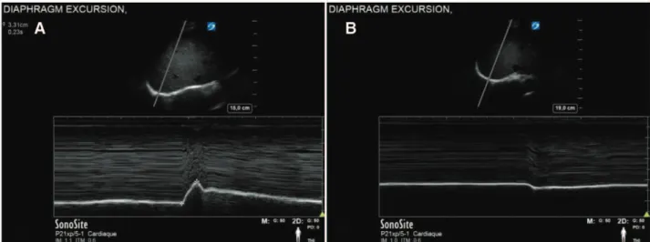
- Diaphragmatic excursion (Figure 1A) during a volun-tary sniff test using a 3.5−5 MHz probe. The diaphrag-matic displacement was measured at 3.3 cm.
- Diaphragmatic thickening during a deep breathing maneuver using a linear high-frequency probe (10–12 MHz). Thickness was measured at 3.8 mm at end-inspiration (Tmax), and 1.7 mm at end-expiration (Tmin). The diaphragmatic thickening fraction (Tmax − Tmin/Tmin) was calculated at 123%, and the thickening ratio (TR) (Tmax/Tmin) at 2.2.
These measurements were considered normal baseline values and were equal on both hemidiaphragms.
Ultrasound-Guided Regional Anesthesia
No sedation or analgesia was administered before/during performance of the blocks. ISB was placed via a posterior approach. Sixteen milliliters of mepivacaine 1% was injected at the level of the fifth and sixth cervical nerve roots.
Then, an in-plane ultrasound-guided SSNB (Figure 2A) was performed6 by locating the brachial plexus and the
departure of the suprascapular nerve (SSN) from the supe-rior trunk under the infesupe-rior belly of the omohyoid muscle by sliding the transducer distally and then injecting 6 mL of ropivacaine 0.475% with 4 mg of dexamethasone added.
For the ANB (Figure 2B),7 the patient was positioned in
the upright sitting position and the neurovascular space bordered by the teres minor muscle, the deltoid muscle, and the shaft of the humerus identified. By the use of an in-plane technique, 6 mL of ropivacaine 0.475% with 4 mg of dexa-methasone were injected.
Interscalene block (ISB) impairs ipsilateral lung function and generally is not used for patients with respiratory insufficiency. We present a 49-year-old man with chronic obstructive pulmonary disease scheduled for shoulder surgery. He was given a regional technique with an ISB (short-acting local anesthetic to minimize duration of diaphragmatic dysfunction) and suprascapular and axillary nerves blocks (long-acting local anesthetic). He was supported with noninvasive ventilation during the time of hemidiaphragmatic paralysis as documented by serial ultrasound examination. A discussion about ISB and its alternatives (general anesthesia versus brachial plexus block versus selective peripheral nerve blocks) always should occur for patients at risk for pulmonary complications.
From the *Department of Anesthesiology and Critical Care Medicine, Purpan University Hospital, Toulouse, France; †Department of Anesthesia, Clinique Médipôle Garonne, Toulouse, France; ‡Department of Orthopedic Surgery, Purpan University Hospital, Toulouse, France; and §Department of Anesthe-sia, University of Toronto, Toronto, Ontario, Canada.
Funding: None.
The authors declare no conflicts of interest.
Address correspondence to Fabrice Ferré, MD, Department of Anesthesiology and Critical Care Medicine, Purpan University Hospital, Hôpital Purpan, Place du Dr Baylac, TSA 40 031, 31059 Toulouse Cedex 9, France. Address e-mail to [email protected].
Regional Anesthesia With Noninvasive Ventilation for
Shoulder Surgery in a Patient With Severe Chronic
Obstructive Pulmonary Disease: A Case Report
Fabrice Ferré, MD,
* Nina Cugnin, MD,* Charlotte Martin, MD,* Philippe Marty, MD,†
Nicolas Bonnevialle, MD, PhD,
‡ Matt Kurrek, MD, PhD,*§ and Vincent Minville, MD, PhD*
Thirty minutes after the placement of the ISB, and complete sensory block, a second ultrasonographic dia-phragmatic function assessment was done, which showed complete ipsilateral hemidiaphragmatic paralysis (TR = 1 and paradoxical downward movement, Figure 1B).
Surgery
The patient was then transferred to the operating room and placed in the beach-chair position. He began to complain of dyspnea, with a respiratory rate of 22/min and a desatura-tion from 97% to 93% despite 2 L/min of oxygen.
We decided to support the patient with noninvasive ventilation (NIV) delivered via a facemask connected to an anesthesia ventilator (Aisys CS2 Datex-Ohmeda Carestation;
GE Healthcare, Little Chalfont, Buckinghamshire, United
Kingdom). The tolerance of this patient’s first experience of NIV was optimized by an appropriate explanation of the procedure and 7.5 μg of sufentanil administered intrave-nously. NIV parameters were adjusted according to patient comfort (inspiratory pressure support 7 cm H2O; positive
end-expiratory pressure 5 cm H2O; fraction of inspired
oxy-gen 30%, titrated to maintain Spo2 ≥ 94%), and allowed a
significant improvement. The case proceeded uneventfully and at the end of the surgery, the patient was transferred to the postanesthetic care unit (PACU).
PACU
In the PACU (2 hours after the ISB), a new diaphragmatic ultrasound evaluation revealed a partial hemidiaphrag-matic paralysis (50% reduction of diaphraghemidiaphrag-matic excursion
Figure 1. Diaphragmatic excursion during a voluntary sniff test before (A) and after (B) an ISB. Diaphragmatic ultrasound was performed using a 3.5–5 MHz phased array probe. The probe was placed immediately below the right costal margin in the right anterior axillary line and was directed medially, cephalad and dorsally, so that the ultrasound beam reached the posterior third of the hemidiaphragm perpendicularly. The 2D mode was used initially to obtain the best view with the liver serving as an acoustic window; the M-mode was then used to display the motion of the anatomical structures along the selected line. In the M-mode, the diaphragmatic excursion (displacement, cm) can be measured during a VS test, for which the patient was asked to forcefully inhale through the nose in a sniffing fashion (A). Note: the paradoxical down-ward movement after ISB (B). This ultrasound pattern (designated negative) indicates a complete hemidiaphragmatic paralysis. ISB indicates interscalene block; 2D, 2-dimensional; VS, voluntary sniff.
Figure 2. A, SSN leaving the superior trunk. The SSN was followed under ultrasound visualization into the subclavian triangle under the inferior belly of the omohyoid muscle. B, AN close to the posterior circumflex humeral artery in the neurovascular space bordered by the teres minor muscle, the deltoid muscle, the triceps muscle, and the humerus. SSN indicates suprascapular nerve; AN, axillary nerve.
DIAPHRAGM EXCURSION DIAPHRAGM EXCURSION
SonoS,te SonoS,te
SUPRASCAPULAR NERVE AXILLARY NERVE
A
Deltoid m. Circumflex a. Subclav,an a. Humerus Sonofae SonoSite ,., '.' .. ·•· . ' ,- '•• , , ' ;, . ' ~..•
'from baseline during voluntary sniff test and TR at 1.6 dur-ing the deep breathdur-ing maneuver). Sensory and motor defi-cit from the ISB was present but diminished.
Four hours after the ISB, a diaphragmatic function assessment revealed a total recovery of the diaphragmatic function, and the NIV was discontinued and only supple-mental oxygen applied (2 L/min by nasal prongs).
The SSN and AN blocks were effective, as judged by the lack of opioid consumption until 24 hours after surgery. No pain (numeric rating scale for pain 0/10) was reported in the PACU after 1 g of intravenous paracetamol and 100 mg of intravenous ketoprofen. Postoperatively, the time to first opioid consumption was 24 hours and the maximal pain reported was 3/10 at the 48th hour. The patient was suc-cessfully discharged from the hospital on day 2.
DISCUSSION
We report the successful anesthetic management of shoulder surgery under regional anesthesia in a patient with COPD. The use of a short-acting local anesthetic (LA) for the ISB limited the duration of hemidiaphragmatic paralysis, and additional nerve blocks (SSNB and ANB) with long-acting LA and added perineural dexamethasone allowed effective postoperative analgesia.
From an anesthetic point of view, the ISB provides excellent analgesia and is considered as the gold standard in the management of postoperative pain after shoulder surgery.2 Respiratory insufficiency, h owever, t
radition-ally has been considered a relative contraindication to performing an ISB because ipsilateral hemidiaphragmatic dysfunction frequently occurs because of the proximity of the phrenic nerve.8 The transient hemidiaphragmatic
paralysis is usually well tolerated in healthy individuals; however, respiratory complications can occur in patients who have preexisting respiratory compromise.9 The
inci-dence of phrenic nerve palsy has been reported as high as 100% after ISB3 and Urmey and McDonald10
demon-strated a 25% decrease in pulmonary function when an ISB was performed. We thought that the use of a short-acting LA could limit the duration of phrenic nerve palsy and would be of major interest in the setting of respiratory insufficiency.
The SSN provides sensory innervation to 70% of the shoulder joint, mainly posteriorly and the AN to the inferior, lateral, and anterior aspects. An isolated SSNB is an alter-native to ISB,11 but its effectiveness appears to be limited,
and an ultrasound-guided ANB combined with a SSNB for arthroscopic rotator cuff repair was shown to provide bet-ter postoperative analgesia when compared with an SSNB alone.12 This strategy could be of major interest in certain
high-risk populations, such as patients with respiratory comorbidities, because it might avoid phrenic nerve paraly-sis and also decrease opioid consumption.
To prolong the analgesic effects of the supplementary SSN and AN blocks, we used long-acting LA with added perineural dexamethasone.13 This allowed good analgesia
and minimized the opioid use, avoiding their side effects, in this patient with COPD.
Opioids have well-known side effects on diaphragmatic and thoracic muscles, leading to a decrease in functional
residual capacity and promoting atelectasis5 and depress
ventilation through their action on the μ-opioid receptor expressed in the brainstem. This can lead to life-threatening complications, and therefore opioids always should be con-sidered as a last-line analgesic treatment in patients with respiratory insufficiency.
Ultrasound is playing and increasing role as an accurate tool to evaluate the diaphragmatic function at the bedside.4
Ultrasonography of the diaphragm at its zone of apposi-tion with the rib cage can be used to measure changes in the thickness of the diaphragm during inspiration. Because thickening of the diaphragm reflects diaphragmatic short-ening, a lack of thickening with inspiration allows the diagnosis of diaphragmatic paralysis. The diaphragmatic thickening fraction (ie, thickness at end-inspiration − thick-ness at end-expiration/thickthick-ness at end-expiration) can be used as an index of diaphragmatic efficiency. A TR of 2.6 can be measured in healthy subjects by dividing the dia-phragmatic thickness during a maximal inspiratory effort by the diaphragmatic thickness while relaxing at functional residual capacity.14 Ultrasonography of the diaphragmatic
dome also has been reported as a method for document-ing diaphragmatic paralysis in adults.14 The values of
dia-phragmatic excursion in healthy individuals were reported to be 2.9 ± 0.6 cm for male patients and 2.6 ± 0.5 cm for female patients during voluntary sniffing.15
Because ultrasonography can distinguish a functioning from a nonfunctioning diaphragm, it can be used to moni-tor recovery of the paralyzed diaphragm.4 In our patient,
respiratory function was supported by NIV until complete recovery of diaphragmatic function was demonstrated by serial ultrasounds.
In summary, this 49-year-old man was able to undergo shoulder surgery under ISB despite severe COPD. An ISB using short-acting LA limited the duration of phrenic nerve palsy and his respiratory status was maintained using NIV for the time needed based on frequent ultrasound monitor-ing of his diaphragmatic function. Supplementary supra-scapular and axillary nerves blocks with long-acting LA and perineural dexamethasone provided good analgesia and minimized the consumption of opioids.
DISCLOSURES
Name: Fabrice Ferré, MD.
Contribution: This author helped perform all measurements and write the manuscript.
Name: Nina Cugnin, MD.
Contribution: This author helped perform all measurements and write the manuscript.
Name: Nicolas Bonnevialle, MD, PhD.
Contribution: This author helped perform the surgical procedure.
Name: Philippe Marty, MD.
Contribution: This author shared his expertise in regional anesthesia.
Name: Charlotte Martin, MD.
Contribution: This author helped perform the intraoperative and postoperative anesthetic management.
Name: Matt Kurrek, MD, PhD.
Contribution: This author helped proofread and edit the manuscript.
Name: Vincent Minville, MD, PhD.
Contribution: This author helped coordinate the investigators,
REFERENCES
1. Lanz E, Theiss D, Jankovic D. The extent of blockade follow-ing various techniques of brachial plexus block. Anesth Analg. 1983;62:55–58.
2. Fredrickson MJ, Krishnan S, Chen CY. Postoperative analgesia for shoulder surgery: a critical appraisal and review of current techniques. Anaesthesia. 2010;65:608–624.
3. Urmey WF, Talts KH, Sharrock NE. One hundred percent incidence of hemidiaphragmatic paresis associated with inter-scalene brachial plexus anesthesia as diagnosed by ultrasonog-raphy. Anesth Analg. 1991;72:498–503.
4. McCool FD, Tzelepis GE. Dysfunction of the diaphragm.
N Engl J Med. 2012;366:932–942.
5. Sasaki N, Meyer MJ, Eikermann M. Postoperative respiratory muscle dysfunction: pathophysiology and preventive strate-gies. Anesthesiology. 2013;118:961–978.
6. Rothe C, Steen-Hansen C, Lund J, Jenstrup MT, Lange KH. Ultrasound-guided block of the suprascapular nerve—a volun-teer study of a new proximal approach. Acta Anaesthesiol Scand. 2014;58:1228–1232.
7. Rothe C, Asghar S, Andersen HL, Christensen JK, Lange KH. Ultrasound-guided block of the axillary nerve: a vol-unteer study of a new method. Acta Anaesthesiol Scand. 2011;55:565–570.
8. Bigeleisen PE. Anatomical variations of the phrenic nerve and its clinical implication for supraclavicular block. Br J Anaesth. 2003;91:916–917.
9. Rose M, Ness TJ. Hypoxia following interscalene block. Reg
Anesth Pain Med. 2002;27:94–96.
10. Urmey WF, McDonald M. Hemidiaphragmatic paresis during interscalene brachial plexus block: effects on pulmonary func-tion and chest wall mechanics. Anesth Analg. 1992;74:352–357. 11. Barber FA. Suprascapular nerve block for shoulder
arthros-copy. Arthrosarthros-copy. 2005;21:1015.
12. Lee JJ, Kim DY, Hwang JT, et al. Effect of ultrasonographically guided axillary nerve block combined with suprascapular nerve block in arthroscopic rotator cuff repair: a randomized controlled trial. Arthroscopy. 2014;30:906–914.
13. Choi S, Rodseth R, McCartney CJ. Effects of dexamethasone as a local anaesthetic adjuvant for brachial plexus block: a sys-tematic review and meta-analysis of randomized trials. Br J
Anaesth. 2014;112:427–439.
14. Matamis D, Soilemezi E, Tsagourias M, et al. Sonographic eval-uation of the diaphragm in critically ill patients. Technique and clinical applications. Intensive Care Med. 2013;39:801–810. 15. Boussuges A, Gole Y, Blanc P. Diaphragmatic motion studied
by m-mode ultrasonography: methods, reproducibility, and normal values. Chest. 2009;135:391–400.