Crystal structure of Enterobacter cloacae 908R class C
b
b-lactamase bound to iodo-acetamido-phenyl boronic acid,
a transition-state analogue
J. Woutersa,*, E. Fonzéb, M.Vermeirec, J.-M. Frèreband P. Charlierb
aInstitut de Recherches Microbiologiques JM Wiame, 1 Av E. Gryzon, 1070 Brussels (Belgium),
Fax: +32 (2) 5276 72 73, e-mail: jwouters@dbm.ulb.ac.be
bCentre d’Ingénierie des Protéines and Laboratoire d’Enzymologie, Institut de Chimie B6, Université de Liège,
Sart-Tilman, 4000 Liège (Belgium)
cInstitut de Physique B5, Université de Liège, Sart-Tilman, 4000 Liège (Belgium)
Received 16 May 2003; accepted 4 June 2003
Abstract. The structures of the class C b-lactamase from Enterobacter cloacae 908R alone and in complex with a
boronic acid transition-state analogue were determined by X-ray crystallography at 2.1 and 2.3 Å, respectively. The structure of the enzyme resembles those of other class C
b-lactamases. The structure of the complex with the
tran-sition-state analogue, iodo-acetamido-phenyl boronic acid, shows that the inhibitor is covalently bound to the
ac-tive-site serine (Ser64). Binding of the inhibitor within the active site is compared with previously determined tures of complexes with other class C enzymes. The struc-ture of the boronic acid adduct indicates ways to improve the affinity of this class of inhibitors. This structure of 908R class C b-lactamase in complex with a
transition-state analogue provides further insights into the mecha-nism of action of these hydrolases.
The expression of b-lactamases is one of the most
com-mon and well-studied forms of antibiotic resistance. Class C b-lactamases are among the most challenging of these
enzymes because they are not significantly inhibited by clinically used b-lactamase inhibitors. Moreover, they have
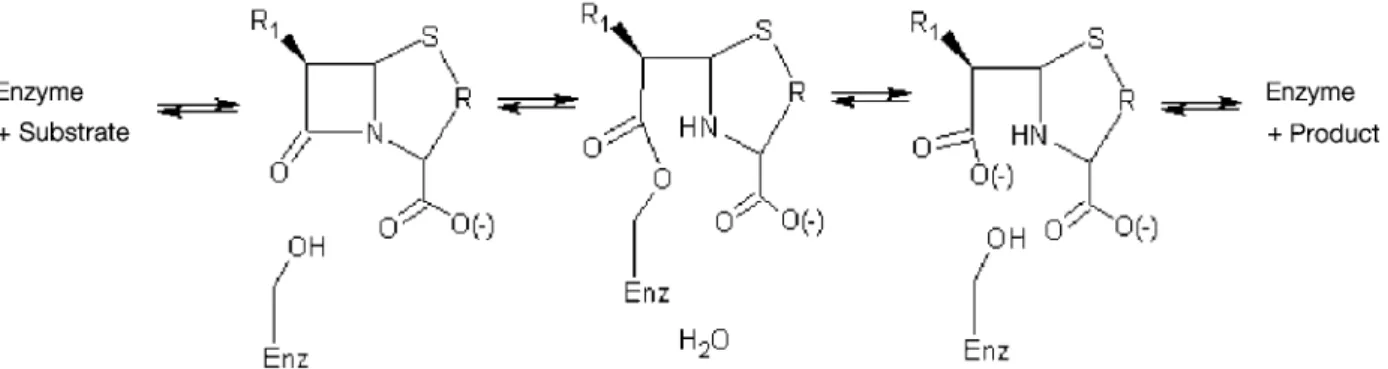
a broad spectrum of action and can hydrolyse a variety of substrates, including the third-generation cephalosporins. Class C b-lactamases hydrolyse their substrates in a
four-step process (fig. 1). Early mutagenesis [1, 3] and struc-tural [4 – 6] studies identified key catalytic residues, in-cluding Ser64, Tyr150, Lys67 and Asn152.
Subsequent X-ray crystallographic studies investigated acyl-adduct complexes of the class C enzymes with
b-lactam inhibitors and poor substrates such as aztreonam (PDB code 1FR6 [4]), moxalactam (PDB code 1FCO [7]), ceftazidime (PDB code 1IEL [8]), imipenem (PDB code 1LL5 [9]) and a cephem sulphone (PDB code 1GA0 [10]). These structures revealed key interactions that contribute to the formation of the acyl-enzyme intermediate. In
Escherichia coli AmpC, one of the class C b-lactamases
that has been extensively studied, binding to imipenem (PDB code 1LL5) differs from its binding to moxalactam (PDB code 1FCO) but is similarly to its binding to TEM1 (PDB code 1 BT5), a class A b-lactamase. The
less-con-strained active-site geometry in class C enzymes thus al-lows different binding modes and, hence, different possi-ble mechanisms of inhibition. For example, imipenem appears to act as an inhibitor because, once it has reacted
*Corresponding author.
Key words. Class-C b-lactamase; Enterobacter cloacae 908R; boronic acid complex; iodo-acetamido-phenyl boronic
with the enzyme, the acyl-enzyme adduct formed under-goes conformational changes that rotate the acyl centre away from the point of hydrolytic attack [9].
Recently, efforts to capture crystal structures of interme-diates (pre-covalent enzyme-substrate and post-covalent enzyme-product) have been undertaken by studying com-plexes between the S64G mutant AmpC in complex with the b-lactam cephalothin in its substrate and product
forms (1KVL, 1KVM [11]). These structures of mile-stones in the reaction pathway of AmpC provide insight into substrate recognition, catalysis, and product release. One major conclusion from these structural and mecha-nistic studies is that the ligand undergoes a dramatic con-formational change as the reaction progresses.
Since the X-ray crystal structures of class C b-lactamases
have been determined, these molecules are attractive tar-gets for novel inhibitor discovery using structure-based methods [12,13]. The E. coli AmpC enzyme is one of the class C b-lactamase that has been extensively studied.
Several classes of non-b-lactam inhibitors have been
iden-tified for this protein. Substituted boronic acids have been prepared leading to irreversible inhibitors with high affinity and specificity [14, 15]. Complexes with transi-tion-state boronic acids [5, 8, 14, 16] and phosphonate in-hibitors [17] confirmed the possibility of different binding modes within the binding site of class C b-lactamases.
One concern with this type of boronic acid inhibitors, also
observed with phosphonates [17], is that they form cova-lent adducts with activated serine nucleophiles, potentially reducing their selectivity versus other serine active en-zymes like serine proteases. In an effort to design non-co-valent, more selective inhibitors of class C b-lactamases,
molecular docking calculation and virtual screening techniques have been used to identify original molecules, and novel non-covalent inhibitors of AmpC have emerged from this structure-based discovery process [18]. The structure of the enzyme with one of these new inhibitors, 3-[(4-chloroanilino)sulphonyl]thiopene-2-carboxylic acid, was determined by X-ray crystallography and closely re-sembled the docking prediction.
As part of this structural approach to class C b-lactamases
giving access to mechanistic and structure-based drug de-sign studies, we report here the crystal structures of the wild-type Enterobacter cloacae 908R class C
b-lacta-mase, alone and in complex with iodo-acetamido-phenyl boronic acid (IAPB; scheme 1).
Material and methods
Crystallisation, data collection, phase determination and structure refinement
Crystals of E. cloacae 908R were prepared as described elsewhere [19]. Crystals of the complex with IAPB, were obtained by soaking a crystal of the apo enzyme in a sat-urated solution of the inhibitor in the mother liquor [0.1 M bicine buffer at pH 9.0 and 30% polyethylene glycol (Mr = 6000)] 30 min before starting data collection. Crys-tals were directly flash-frozen from this solution. X-ray diffraction data were collected at 100 K using a Siemens X1000 area detector and a Rigaku RU-200 rotating anode generator as X-ray source. The detector was at a distance of 130 mm with a 2q angle of 20° and 22° for the
908R-IAPB complex and the free enzyme, respectively. The data collection step size was 0.2° between frames, with a count time of 70 s. per frame. Indexing, integration and scaling of data sets were carried out using the XENGEN 2.0 package [20].
Figure 1. Four-step process for the hydrolysis of substrates by class C b-lactamases (Enz = enzyme).
Scheme 1. The structure of iodo-acetamido-phenyl boronic acid (IAPB).
Apo structure (unliganded form) of E. cloacae 908R class C bb-lactamase
The refined 2.3-Å structure of the E. cloacae P99 enzyme (PDB code 1 BLS [17]) was used as a search model for a molecular replacement solution using data within the res-olution range of 10 – 2.5 Å. The 361-amino-acid sequence of the molecular probe differs by only five mutations. One clear solution for both the rotation and translation func-tions was directly obtained with an R factor and a corre-lation coefficient of 0.28 and 0.804, respectively. The elec-tron density calculated with the best solution was of very good quality and clearly showed the mutations to be in-troduced in the structure of E. cloacae 908R.
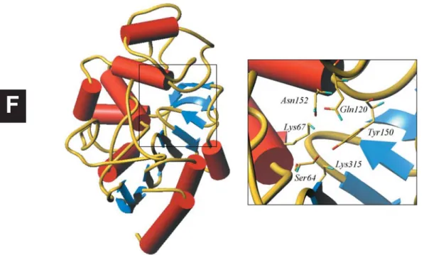
The overall structure of E. cloacae 908R is similar to those previously reported for other class C b-lactamases,
including E. coli AmpC, E. cloacae P99 and Citrobacter
freundii enzymes. It is composed of a mixed a/b
sec-ondary structure with nine-stranded antiparallel b sheets
in the middle flanked by a row of three a helices on one
side, and a helical domain with 11 helices on the other. The primary catalytic residue (Ser64), which is acylated in the course of the enzymatic hydrolysis, is positioned at the N-terminal end of a buried central helix (fig. 2).
Structural similarities are also found between the various class C b-lactamases, class A b-lactamases and
b-lactam-sensitive D-analyl-D-alanine carboxy-peptidase/transpep-tidase (DD-pepcarboxy-peptidase/transpep-tidase) [24].
In the apo structure of E. cloacae 908R, a water molecule occupies the so-called oxyanion hole and is held by two strong hydrogen bonds involving the main chain at residues Ser318 and Ser64 (table 2). The combined strength of both hydrogen bonds increases slightly from class C to class A and could be related to the rates of dea-cylation in the penicillins.
Complex with IAPB
Clear electron density connects the Og atom of the
cat-alytic serine Ser64 to the tetrahedral boron atom of the phenyl boronic acid inhibitor, suggesting that a dative co-valent bond is formed between IAPB and the enzyme (fig. 3). One of the oxygen atoms (O1) of the boron adduct sits in the ‘oxyanion hole’, forming direct hydro-gen bonds to main-chain nitrohydro-gen atoms (table 2), as dis-cussed below.
and graphical modelling using the program Turbo-Frodo [23]. An initial model of the IAPB inhibitor was built and energy minimised using InsightII and Dis-cover (MSI/biosym San Diego). In its isolated con-formation, the boronic acid B(OH)2function is planar,
as expected. In the complex with E. cloacae 908R, a tetra-hedral co-ordination of the boron atom is observed affecting the geometry of the inhibitor. Those modified internal co-ordinates of the inhibitor were translated into 1 – 2 and 1 – 3 distance restraints used in the refine-ment process.
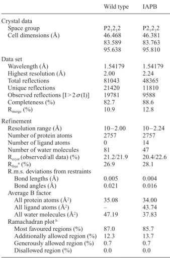
Table 1. Data collection and refinement statistics.
Wild type IAPB
Crystal data Space group P21212 P21212 Cell dimensions (Å) 46.468 46.381 83.589 83.763 95.638 95.810 Data set Wavelength (Å) 1.54179 1.54179 Highest resolution (Å) 2.00 2.24 Total reflections 81043 48365 Unique reflections 21420 11810
Observed reflections [I > 2s(I)] 19781 9588
Completeness (%) 82.7 88.6
Rmerge(%) 10.9 12.8
Refinement
Resolution range (Å) 10 – 2.00 10 – 2.24
Number of protein atoms 2757 2757
Number of ligand atoms 0 14
Number of water molecules 81 47
Rcryst (observed/all data) (%) 21.2/21.9 20.4/22.6
Rfreea(%) 26.9 28.1
R.m.s. deviations from restraints
Bond lengths (Å) 0.005 0.004
Bond angles (Å) 0.021 0.016
Average B factor
All protein atoms (Å2) 35.08 34.00
All ligand atoms (Å2) – 43.74
All water molecules (Å2) 47.19 37.83
Ramachadran plotb
Most favoured regions (%) 87.0 85.7
Additionally allowed region (%) 12.3 13.7
Generously allowed region (%) 0.7 0.7
Disallowed region (%) 0.0 0.0
a Free test subset represents 5% of total unique reflections.
plexed structure, Gln120 is displaced by the inhibitor and no longer interacts with Asn152. The lateral iodo-acetamido chain is further stabilised by a hydrogen bond between the carbonyl of the carboxamido group and Gln120. Apart from these changes within the active site, comparison of the refined structures of 908R, unliganded and in complex with the boronic acid inhibitor, shows that neither backbone nor side-chain conformations change much upon complexation.
Interestingly, the relative orientation of the inhibitor in-side the active site of E. cloacae 908R is distinct from This covalent bond between IAPB and the protein is
con-sistent with the proposed mechanism of inhibition and similar to that observed in other structures of AmpC com-plexed with aryl boronic acid, as discussed further in the text. The IAPB inhibitor makes a number of favourable in-teractions with the protein (table 2). Atoms within hydro-gen-bonding distance of the inhibitor include the main-chain nitrogen of Ser64, and the backbone N and O atoms of Ser318, which interact with one of the boronic oxygens (O1).
In the apo structure, the conformation of Gln120 is such that it hydrogen bonds with Asn152 (table 2). In the
com-Figure 2. Overall structure of the E. cloacae 908R class C b-lactamase, with a closer view of the active site showing the active Ser64 and residues Lys67, Gln120, Asn152, Tyr150 and Lys315.
F
Table 2. Selected geometric features in the active site of E. cloacae 908R class C b-lactamase in its unliganded form or in complex with IABP. Distances (Å) unliganded in complex with IAPB Og_Ser64 – OH_Tyr150 2.84 3.21 OH_Tyr150 – Nz_Lys315 2.81 2.70 OH_Tyr150 – Nz_Lys67 3.26 3.23 Og_Ser64 – Nz_Lys67 3.08 3.49 Og_Ser64 – O_water * 2.58 – N_Ser318 – O_water * 2.50 – O_Ser318 – O_water * 3.11 – N_Ser318 – O1_IAPB – 2.49 O_Ser318 – O1_IAPB – 2.42 Ne2_Gln120 – Od1_Asn_152 2.86 3.38 Ne2_Gln120 – O_IAPB – 2.75
* Oxyanion water molecule. Figure 3. Interactions between IAPB and selected residues of the
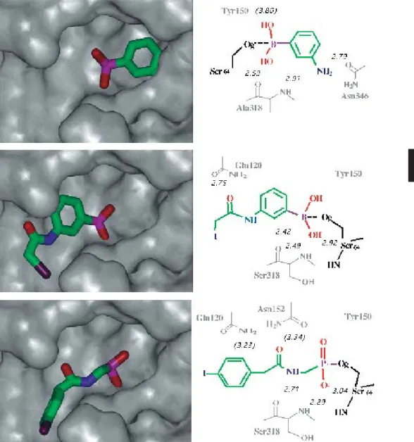
Figure 4. Comparison of the relative orientation of phenyl boronic acids in the active site of class C b-lactamases (molecular surface rep-resentation of the enzyme). Upper: E. coli AmpC with m-aminophenyl boronic acid. Middle: E. cloacae 908R with IAPB. Lower: E.
cloa-cae P99 with N-[(p-iodophenyl)acetyl]aminomethyl phosphate.
F
Two distinct positions of the iodine atom were refined, both with half occupancy.
The structure of the E. cloacae 908R-IAPB complex re-sembles that of other class C b-lactamases in complex
with other small boronic acids. The E. cloacae
908R-to the amides of Gln120 and Asn152. One phosphonyl oxygen atom is hydrogen bonded to main-chain NH groups of Ser318 and Ser64, while the other oxygen is not within hydrogen-bonding distance of any amino acid. Tyr150 and Lys67 are closely associated with Og of
Ser64. This arrangement, similar to that observed in the present structure, was interpreted in terms of the transition state for breakdown of the tetrahedral intermediate in the deacylation step of catalysis. In this mechanism, Tyr150 phenol seems the most likely general acid, and thus the corresponding phenoxide anion would be the gen-eral base catalyst in acylation [17]. This view is in com-plete agreement with recent mechanistic studies per-formed on AmpC [11].
Comparison with other class C bb-lactamases
complexes with aryl boronic acids
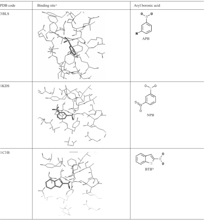
As small aryl boronic acids inhibit class C b-lactamases
at sub-micromolar concentrations, different X-ray crystal structures of complexes with boronic acid inhibitors are available in the PDB data bank. Taking advantage of the large amount of structural data available for these en-zymes, statistical searches in the PDB were performed with the Relibase program to compare the geometries of complexes and identify structural features of the binding site of class C b-lactamases (fig. 5). Complexes
with aryl boronic acids were investigated. From the sta-tistical search with Relibase, apart from the structure of cocaine esterase in complex with phenyl boronic acid (PDB code 1JU3 [25]), aryl boronic acid ligands are ap-parently not found in other structures deposited at the PDB. This does not mean that the aryl boronic fragment is specific for class C b-lactamase but probably reflects
the fact that no one has yet attempted to obtain complexes between these compounds and other (serine active) en-zymes.
Analysis of the structures of complexes between class C
b-lactamases and aryl boronic acids reveals that, except
for the complex with m-aminophenyl boronic acid, orien-tation of IAPB deduced from our structure is similar to that observed with other aryl boronic acid transition-state analogues. Indeed, comparison of the structures in figure 5 suggests that small aryl boronic acid inhibitors bind to a well-defined cleft in the different class C b-lactamases.
This cleft has also been shown to bind the ubiquitous R1 side chain of b-lactams [15, 18]. Much of this cleft is
left unoccupied by the small aryl boronic acids. With the exception of one complex (PDB code 3 BLS), all boronic ligands adopt similar orientations in the protein binding sites. Combination of structure-based design and in-par-allel synthesis allowed rapid exploration, in AmpC, of this binding site whose role in recognition had not been previously explored [15]. The resulting inhibitors differ considerably from b-lactams but nevertheless inhibit the
enzyme well. The crystal structure of ETP [3-(4-benzene-sulphonyl-thiophene-2-sulphonylamino)-phenylboronic acid, Ki 83 nM] in complex with AmpC (PDB code 1GA9) is the first exploration of this highly conserved cleft and provides valuable information for further design against class C b-lactamases.
Comparison of the present structure of 908R complexed to IAPB with complexes with other boronic acid inhibitors and class C b-lactamases further reveals consensus
bind-ing sites. These sites were already identified and charac-terised in previous studies, using a combination of crys-tallographic data and computational approaches [15, 18]. Among these binding sites, the highly conserved oxyan-ion hole recognises one hydroxyl group of the boronic acid. The aryl ring of aryl boronic acids is also stabilised by interactions with Asn152. Interestingly, in complexes with b-lactams, this conserved residue defines a
recogni-tion site that interacts with the carbonyl oxygen in the R1 side chain of b-lactams [18].
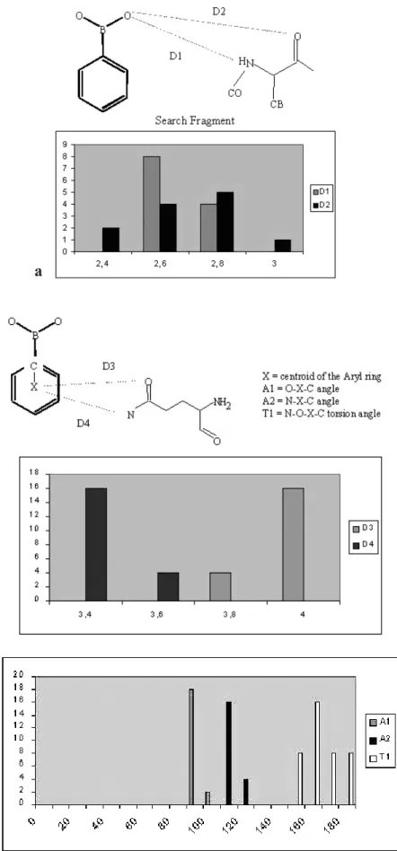
Statistical analysis of the geometry within these sites was performed on the structures retrieved with the Relibase (fig. 6). The geometry of the oxyanion described in this work (table 2) is consistent with the distribution of dis-tances given in figure 6 a (D1 and D2 define the disdis-tances between the hydroxyl group of the boronic inhibitor and the main-chain nitrogen and oxygen atoms, respectively, and correspond to N _Ser318–O1 and O_Ser318–O1 dis-tances in the complex of 908R with IAPB). The geometry of the aryl-amide interaction involving residue Asn152 is given by the parameters D3, A1, A2 and T1 in figure 6 b. These parameters underline contacts (less than 4 Å) be-tween the side chain of residue 152 and the aromatic ring of the boronic inhibitors together with an axial approach (A1 and A2 close to 90° and T1 close to 180°). In the structure with IAPB, the corresponding parameters are: D1 = 3.63 Å, D2 = 3.92 Å, A1 = 130.2°, A2 = 98.7°, T1 = 161.4°. These values fall within the statistical distri-bution.
Conclusion
The structure of the complex between IAPB and E.
cloa-cae 908R is consistent with the status of boronic acids as
transition-state analogues.
Comparison with the structures of complexes with other transition-state boronic acids and b-lactams confirms the
possibility of different binding modes within the binding site of class C b-lactamases. The less-constrained
active-site geometry observed in different class C enzymes po-tentially allows different possible mechanisms of inhibi-tion. Recent mechanistical studies based on X-ray structures on trapped reaction intermediates are in agree-ment with this view. From the point of view of drug de-sign, this opens interesting perspectives for potentially modulating existing inhibitors to make them more selec-tive for class C b-lactamases.
1KDS
NPB
1 C3 B
BTBb
Figure 5. Results of the Relibase search for structures of complexes with aryl boronic acids. aThe same relative orientation of the bind-ing site has been retained, with the catalytic Ser64 at the top, and Tyr150 on the right. Only the structure of the bindbind-ing site in chain A is presented because the ligands in each active site (chains A and B) bind similarly. b The structure of benzothiophene boronic acid has been added to the selection by similarity with the initial search fragment (‘benzene ring’ plus ‘boronic acid’). cNote the two distinct geometries of the B(OH)2groups: one, tetrahedral, covalently bound to the active-site catalytic serine and the other, planar, not forming any covalent bond within the binding site.
Figure 5 (continued).
PDB code Binding sitea Aryl boronic acid
1KDW 4 CB 1KE0 CVB 1KE3 BPBc 1GA9 ETP
Figure 6. Statistical analysis of the geometry of complexes between class C b-lactamases and boronic acid inhibitors. (a) First hydroxyl binding site, (b) Aryl-pi binding site.
a
1 Monnaie A., Dubus A. and Frere J.-M. (1994) The role of lysine-67 in a class C b-lactamase is mainly electrostatic. Biochem. J.
302: 1 – 4
2 Dubus A., Normark S., Kania M. and Page M. G. P. (1994) The role of tyrosine 150 in catalysis of b-lactam hydrolysis by AmpC b-lactamase from Escherichia coli investigated by site-directed mutagenesis. Biochemistry 33: 8577 – 8586
3 Dubus A., Normark S., Kania M. and Page M. G. P. (1995) Role
of asparagine 152 in catalysis of b-lactam hydrolysis by
Escherichia coli AmpC b-lactamase studied by site-directed mutagenesis. Biochemistry 34: 7757 – 7764
4 Oefner C., D’Arcy A., Daly J. J., Gubernator K., Charnas R. L., Heinze I. et al. (1990) Refined crystal structure of a of b -lacta-mase from Citrobacter freundii indicates a mechanism forb -lac-tam hydrolysis. Nature 343: 284 – 288
5 Usher K. C., Blaszczak L. C., Weston G. S., Shoichet B. K.
and Remington S. J. (1998) Three-dimensional structure of
AmpC b-lactamase from Escherichia coli bound to a
transi-tion-state analogue: possible implications for the oxyanion hypothesis and for inhibitor design. Biochemistry 37: 16082 – 16092
6 Lobkovsky E., Moews P. C., Liu H., Zhao H., Frere J.-M. and
Knox J. R. (1993) Evolution of an enzyme activity: crystallo-graphic structure at 2-Å resolution of cephalosporinase from the ampC gene of Enterobacter cloacae P99 and comparison with a class A penicillinase. Proc. Natl. Acad. Sci. USA 90: 11257 – 11261
7. Patera A., Blaszczak L. C., Stoichet B. (2000) Crystal structures of substrate and inhibitor complexes with AmpC b-lactamase: possible implications for substrate-assisted catalysis. J. Am. Chem. Soc. 122: 10504 – 10512
8 Powers R. A., Caselli E., Focia P. J., Prati F. and Shoichet B. K. (2001) Structures of ceftazidime and its transition-state
ana-logue in complex with AmpC b-lactamase: implications for
resistance mutations and inhibitor design. Biochemistry 40: 9207 – 9214
9 Beadle B. M. and Shoichet B. K. (2002) Structural basis for
imipenem inhibition of class C b-lactamases. Antimicrob.
Agents Chemother. 46: 3978 – 3980
10 Crichlow G. V., Nukaga M., Doppalapudi V. R., Buynak J. D. and Knox J. R. (2001) Inhibition of class C b-lactamases: struc-ture of a reaction intermediate with a cephem sulfone. Bio-chemistry 40: 6233 – 6239
11 Beadle B. M., Trehan I., Focia P. J. and Shoichet B. K. (2002) Structural milestones in the reaction pathway of an amide hy-drolase: substrate, acyl, and product complexes of cephalothin with AmpC b-lactamase. Structure 10: 412 – 424
12 Shoichet B. K., McGovern S. L., Wei B. and Irwin J. J. (2002) Lead discovery using molecular docking. Curr. Opin. Chem. Biol. 6: 439 – 446
13 Powers R. and Stoichet B. K. (2002) Structure-based approach for binding site identification on AmpC b-lactamase. J. Med. Chem. 45: 3222 – 3234
14 Caselli E., Powers R. A., Blasczcak L. C., Wu C. Y. E., Prati F. and Shoichet B. K. (2001) Energetic, structural, and antimicro-bial analyses of b-lactam side chain recognition by b -lacta-mases. Chem. Biol. 8: 17 – 31
15 Tondi D., Powers R. A., Caselli E., Negri M.-C., Blázquez J., Costi M. P. et al. (2001) Structure-based design and in-parallel synthesis of inhibitors of AmpC b-lactamase. Chem. Biol. 8: 593 – 610
16 Powers R. A., Blazquez J., Weston G. S., Morosini M. I., Ba-quero F. and Shoichet B. K. (1999) The complexed structure and antimicrobial activity of a non-beta-lactam inhibitor of AmpC beta-lactamase. Protein Sci. 8: 2330 – 2337
17 Lobkovsky E., Billings E. M., Moews P. C., Rahil J., Pratt R. F. and Knox J. R. (1994) Crystallographic structure of a phospho-nate derivative of the Enterobacter cloacae P99 cephalospori-nase: mechanistic interpretation of a b-lactamase transition-state analog. Biochemistry 33: 6762 – 6772
18 Powers R. A., Morandi F. and Shoichet B. K. (2002) Structure-based discovery of a novel, noncovalent inhibitor of Amp C b -lactamase. Structure 10: 1013 – 1023
19 Wouters J., Charlier P., Monnaie D., Frère J.-M. and Fonzé E. (2001) Expression, purification, crystallization and preliminary X-ray analysis of the native class C b-lactamase from
Enter-obacter cloacae 908R and two mutants. Acta Crystallogr. D57:
162 – 164
20. Howard A. J., Gilliland G. L., Finzel B. C., Poulos T., Ohlendorf D. O. and Salemme F. R. (1987) The use of an imaging propor-tional counter in macromolecular crystallography. J. Appl. Crys-tallogr. 20: 383 – 387
21. Navaza J. (1994) AMoRe: an automated package for molecular replacement. Acta Crystallogr. A50: 157 – 163
22 Sheldrick G. M. and Schneider T. R. (1997) SHELXL: high resolution refinement. Methods Enzymol. 277: 319 – 343 23 Roussel A. and Cambillau C. (1992) Turbo-Frodo. Biographics,
AFMB, Marseille, France
24 Knox, J. R., Moews P. C. and Frere J.-M. (1996) Molecular evo-lution of bacterial beta-lactam resistance. Chem. Biol.
3:937 – 947
25 Larsen N. A., Turner J. M., Stevens J., Rosser S. J., Basran A., Lerner R. A. et al. (2002) Crystal structure of a bacterial cocaine esterase. Nat. Struct. Biol. 9: 17 – 21