Science Arts & Métiers (SAM)
is an open access repository that collects the work of Arts et Métiers Institute of Technology researchers and makes it freely available over the web where possible.
This is an author-deposited version published in: https://sam.ensam.eu Handle ID: .http://hdl.handle.net/10985/18615
To cite this version :
Louis CLAVEL, Valérie ATTALI, Isabelle RIVALS, Marie-Cécile NIERAT, Pierantonio LAVENEZIANA, Philippe ROUCH, Thomas SIMILOWSKI, Baptiste SANDOZ - Decreased respiratory-related postural perturbations at the cervical level under cognitive load - European Journal of Applied Physiology - Vol. 120, n°5, p.1063-1074 - 2020
Any correspondence concerning this service should be sent to the repository Administrator : [email protected]
Decreased respiratory‑related postural perturbations at the cervical
level under cognitive load
Louis Clavel1,2 · Valérie Attali1,2,3 · Isabelle Rivals1,4 · Marie‑Cécile Niérat1 · Pierantonio Laveneziana1,5 · Philippe Rouch2 · Thomas Similowski1,6 · Baptiste Sandoz2
Abstract
Purpose In healthy humans, postural and respiratory dynamics are intimately linked and a breathing-related postural pertur-bation is evident in joint kinematics. A cognitive dual-task paradigm that is known to induce both postural and ventilatory disturbances can be used to modulate this multijoint posturo-ventilatory (PV) interaction, particularly in the cervical spine, which supports the head. The objective of this study was to assess this modulation.
Methods With the use of optoelectronic sensors, the breathing profile, articular joint motions of the cervical spine, hip, knees and ankles, and centre of pressure (CoP) displacement were measured in 20 healthy subjects (37 years old [29; 49], 10 females) during natural breathing (NB), a cognitive dual task (COG), and eyes-closed and increased-tidal-volume con-ditions. The PV interaction in the CoP and joint motions were evaluated by calculating the respiratory emergence (REm). Results Only the COG condition induced a decrease in the cervical REm (NB: 17.2% [7.8; 37.2]; COG: 4.2% [1.8; 10.0] p = 0.0020) concurrent with no changes in the cervical motion. The CoP REm (NB: 6.2% [3.8; 10.3]; COG: 12.9% [5.8; 20.7] p = 0.0696) and breathing frequency (NB: 16.6 min-1 [13.3; 18.7]; COG: 18.6 min-1 [16.3; 19.4] p = 0.0731) tended to increase, while the CoP (p = 0.0072) and lower joint motion displacements (p < 0.05) increased.
Conclusion This study shows stable cervical spine motion during a cognitive dual task, as well as increased postural pertur-bations globally and in other joints. The concurrent reduction in the PV interaction at the cervical spine suggests that this “stabilization strategy” is centrally controlled and is achieved by a reduction in the breathing-related postural perturbations at this level. Whether this strategy is a goal for maintaining balance remains to be studied.
Keywords Breathing · Centre of pressure · Cognitive dual task · Joint kinematics · Postural control · Optoelectronic plethysmography · Posturo-ventilatory interaction · Cervical spine
Abbreviations
Amp Signal amplitude AP Antero-posterior BF Breathing frequency
BMI Body mass index CI Confidence interval
COG Cognitive dual-task condition CoM Centre of mass
CoP Centre of pressure CW Chest wall volume Communicated by Lori Ann Vallis.
* Louis Clavel
1 UMRS1158 Neurophysiologie Respiratoire Expérimentale
et Clinique, Sorbonne Université, INSERM, 75005 Paris, France
2 Arts et Metiers Institute of Technology, IBHGC, 151 bd de
l’Hopital, 75013 Paris, France
3 Service des Pathologies du Sommeil (Département R3S),
AP-HP, Groupe Hospitalier Universitaire APHP-Sorbonne Université, Site Pitié-Salpêtrière, 75013 Paris, France
4 Equipe de Statistique Appliquée, ESPCI Paris, PSL Research
University, 75005 Paris, France
5 Service des Explorations Fonctionnelles de la Respiration, de
l’Exercice et de la Dyspnée, Département “R3S”, Assistance Publique des Hôpitaux de Paris, Groupe Hospitalier Pitié-Salpêtrière Charles Foix, 75013 Paris, France
6 Service de Pneumologie, Médecine Intensive et
Réanimation (Département R3S), AP-HP, Groupe Hospitalier Universitaire APHP-Sorbonne Université, Site Pitié-Salpêtrière, 75013 Paris, France
EC Eyes-closed condition
ITV Increased tidal volume condition ML Medio-lateral
NB Natural breathing condition PV Posturo-ventilatory Q1; Q3 First quartile; third quartile REm Respiratory emergence TE Expiratory time TI Inspiratory time TLA Time-locked averaging
Introduction
Postural and respiratory dynamics are intimately linked and exhibit strong temporal relationships in healthy humans (Dally 1908; Gurfinkel et al. 1971; Perry and Carrier 2006). Ribcage movements during breathing disturb an individu-al’s balance by modifying the costo-vertebral articulation position, thoracic spinal curvature and postural alignment (Dally 1908; Attali et al. 2019) and by inducing ribcage-related centre of mass (CoM) displacements. Breathing-related postural perturbations during natural breathing are partially counteracted by phasic contractions of paraverte-bral (Kantor et al. 2001) and pelvic floor muscles (Hodges et al. 2007; Talasz et al. 2011). The interaction between the breathing-related postural perturbations and these counter-acting muscle contractions that occurs to maintain balance is defined as the posturo-ventilatory (PV) interaction. From a stabilometric point of view, the PV interaction is charac-terized by minimal rhythmic displacements of the centre of pressure (CoP—the whole body CoM vertical projection on the ground) induced by natural breathing at rest, whereas this displacement almost disappears during apnoea (Kan-tor et al. 2001; Caron et al. 2004) and increases with CO2-induced hyperventilation (David et al. 2012) or voluntary deep breathing (Hamaoui et al. 2010). The PV interaction is centrally integrated (Gurfinkel et al. 1971), as evidenced by the perturbations that occur in patients with stroke (Manor et al. 2012) or by the anticipated increases—i.e., those involving cortical resources—in the role of the diaphragm in postural control during rapid postural adjustments (Hodges et al. 1997).
The PV interaction acts on a multijoint kinetic chain with several degrees of freedom at the spine, hip, knee and ankle levels (Gurfinkel et al. 1971; Hodges et al. 2002) to con-trol breathing-related postural instability (Kuznetsov and Riley 2012). Compared to other joints in the postural chain, the cervical spine directly supports the head and is highly involved in the regulation of its position in space. It plays a crucial role in the maintenance of balance, as suggested by its role in sit-to-stand tasks (Hamaoui and Alamini-Rodri-gues 2017) and by the presence of impaired stabilometric
parameters in patients with a history of moderate neck trauma (Gandelman-Marton et al. 2016) or neck pain (Quek et al. 2018). In addition, changing the orientation of the head during tracking requires a synergy between the most inferior joints and the cervical spine for an individual to maintain balance (Park et al. 2012). Permanent adjustments of the cervical curvature during standing are needed to preserve an adequate head-to-pelvis alignment, which is essential for balance (Vital and Senegas 1986; Dubousset 1994; Amabile et al. 2018), as well as to maintain the head position and a horizontal gaze (Hasegawa et al. 2017), which are important for the transmission of the appropriate visual, vestibular and neck proprioceptive afferent feedback signals (Bove et al.
2009; Williams et al. 2017; Malmström et al. 2017). These adjustments of the cervical curvature are anticipated during tasks requiring attention, such as complex visual (Boulanger et al. 2017) or motor tasks (Hamaoui and Alamini-Rodrigues
2017), as evidenced by prefrontal cortex recruitment, which indicates their central nature (Jahn et al. 2004; Taube et al.
2008; Mihara et al. 2008). Therefore, even though it is a part of the postural chain involved in the PV interaction, the cervical spine is centrally modulated by predominant “non-respiratory” cortical adjustments, which may compete with the central control of the PV interaction. Indeed, compared to other joints in the postural chain, the cervical spine exhib-its less breathing-related mobility during natural breathing (Hodges et al. 2002; Kuznetsov and Riley 2012) and higher variability when the breathing amplitude increases (Kuznet-sov and Riley 2012).
The dual-task paradigm is known to divert cortical resources and induce an increase in cognitive load (Stelzel et al. 2018), and it has been validated for the investigation of the interactions between cognitive processes and other cortical functions such as postural control (Huxhold et al.
2006; Lacour et al. 2008; Stelzel et al. 2018) and the corti-cal control of breathing (Mador and Tobin 1991; Grassmann et al. 2016). Cognitive load is indeed known to be indepen-dently associated with modulations in postural control (Hux-hold et al. 2006; Lacour et al. 2008; Stelzel et al. 2018) and breathing patterns (Mador and Tobin 1991; Grassmann et al.
2016). To date, no specific modulations in the PV interac-tion under a cognitive load have been described; one study reported reduced postural sway, which was partly related to a reduction in breathing amplitude, in young adults (Hagio et al. 2018). This result suggests that central postural per-turbations induced by a cognitive task may be compensated by a reduction in breathing-related postural perturbations, indicating a strong interaction between cognition and the PV interaction.
Therefore, we hypothesized that cognitive processes can modulate the PV interaction and its distribution along the multijoint postural chain, with less important breathing-related postural perturbations occurring at
the cervical level compared to other joints. To test these hypotheses, we used a cognitive dual-task methodology, and we assessed the global effect of the PV interaction on CoP displacement, as well as on the joint motions in the postural chain, with a focus on the cervical spine. The PV interaction is a delicate phenomenon that is easily altered, and the choice of our cognitive task was driven by the necessity to prevent the occurrence of any respiratory changes or postural perturbations linked to the cognitive task itself, particularly at the cervical level. Hence, motor tasks such as eye tracking (which induces head move-ments) and vocal counting (which involves the respiratory system) were not appropriate. Therefore, purely mental tasks were chosen, while the recall task (to verify that the subjects had truly completed the mental task) was per-formed only when the registration ended. Both cognitive tasks that were used in this study tested working memory, and one task additionally tested the subject’s calculation ability and attentional capacities.
Materials and methods
Subjects
Twenty-one healthy adult subjects (age ≥ 18 years) were included in the study (10 F, age: 37 years [29; 49], BMI: 22.9 kg/m2 [20.6; 25.5], data presented as the median [Q1;
Q3]). None of the subjects had a history of postural or res-piratory disease, and all subjects had normal pulmonary function test results. This study was approved by the eth-ics committee Ile-de-France VI (Paris, France) (CPP IDF VI 06036) and recorded in the ISRCTN registry under the number ISRCTN56129394. All subjects gave their written consent to participate.
Protocol
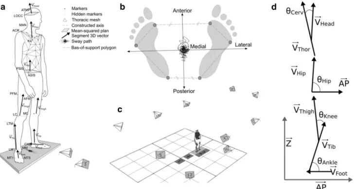
A total of 65 retroreflective markers were placed on the sub-jects; four were placed on the head, 41 were placed on the trunk, seven were placed on each leg, and three were placed on each foot (Fig. 1a). Motion data were captured with an optoelectronic system (Vicon Nexus 1.8.5, Oxford, UK) using 12 cameras (Vero 1 MPa) that were placed around the subject and a sampling rate of 100 Hz (Fig. 1c); the motion data were used to determine the breathing profile and joint motions (cervical spine, hips, knees and ankles). One force plate (BP 4051040-2 k, AMTI, Watertown, USA) measur-ing the subject’s CoP displacements was synchronized with the optoelectronic system at the same sampling frequency. The motion and CoP displacement data were simultaneously recorded with Nexus software.
The subjects were instructed to stand relaxed and barefoot on the force plate with the feet located shoulder-width apart and the arms placed alongside the body. The first set of sev-eral 45-s recordings was taken randomly under four different conditions: (1) a natural breathing reference condition (NB); two cognitive dual-task conditions (COG), which consisted of (2) listening and remembering 8 words, then repeating them once the previous task was completed (COG1) and (3) listening to five multiplication problems, performing them mentally, then giving the answers once the registration had ended (COG2); and (4) an eyes-closed condition (EC). During these conditions, no specific instructions were given regarding breathing. An additional thirty-second recording was performed under an increased voluntary tidal volume (ITV) condition, during which the subjects were asked to increase their tidal volume but not their breathing frequency. For all conditions except for EC, the subjects were instructed to focus on a landmark on the wall to maintain horizontal gaze.
Signal processing CoP displacements
Subject-specific antero-posterior (AP) and medio-lateral (ML) axes were defined from the four foot markers (Fig. 1b). The AP axis was defined as the line joining the middle of the anterior markers and the middle of the posterior markers projected in the horizontal plane. The ML axis was defined as the line perpendicular to the AP axis in the horizontal plane. The output of the force plate allowed the computation of the CoP time series in the AP direction. The amplitude (Amp) of the CoP displacements was estimated by the mean of their linear envelopes.
Breathing profile
From the 41 markers placed on the trunk, the chest wall (CW) volume was computed at each time point as the vol-ume enclosed in the surface of the triangular mesh delim-ited by the spatial marker locations (Fig. 1a), following the method developed in 1996 by Cala et al. (1996) using Green-Ostogradski’s theorem. In the following equation, S is the surface, V is the volume enclosed by Si, Fi is an arbitrary
vector considered as the unit vector (F), ni is the normal unit
vector at the different points of Si, and ∇ is the divergence
operator, which is considered the unit divergence. The ana-lytical expression of the theorem becomes:
∫ S ⃗F.n��⃗idSi = ∫ V ∇⃗FdVi = ∫ V dVi
In the discrete form, the CW volume can be computed by summing the areas of the regions in the K triangle constitut-ing the chest wall mesh, with Ai corresponding to the area
of each region:
With the signal of the variation in the CW volume, the start of each inspiration and expiration event was located. The inspiration/expiration events were defined as the local maxima and minima obtained from the volume signal: the end of an inspiration event corresponded to a local maxi-mum, and the end of an expiration event corresponded to a local minimum. A respiratory cycle was defined as the dura-tion between the start of two successive inspiradura-tion events. The global breathing frequency (BF) was calculated as the ratio between the number of respiratory cycle integers (nC)
and the elapsed time (i.e., 45 s). The mean inspiratory (TI) and expiratory (TE) times were computed over the whole breathing period. The amplitude (Amp.) of variation in the CW volume was estimated in litres by the mean of its linear envelopes. CW = K ∑ i=i ⃗F.n��⃗i.Ai Joint motions
Seven specific 3D vectors were considered in a subject-specific frame (Fsub) and defined with in the AP, ML and
vertical axes. The head (VHead), thoracic (VThor), hip (VHip),
thigh (VThigh), tibial (VTib) and foot frames (VFoot) were then
defined using the local segment markers. VHead and VHip were defined as the normal of the mean-squared plane defined by the four markers on each segment. VThor was defined as the
cross product of the vectors defined; the vector between the right and left acromion markers and the vector defined by the K4-manubrium markers were calculated first, then VThor
was calculated as the cross product of them (more simply, VThor is orthogonal to the two other vectors defined by the acromion marker on one side and the K4 and manubrium markers on another side). VThigh, VTib and VFoot were defined
by the markers located on the proximal and distal parts of each segment. These seven vectors were then projected in the sagittal plane (defined by the AP and Z axes), and the motions at the four joint levels (cervical spine, hip, knee and ankle) were computed as the angles between the pro-jected vectors (Fig. 1d). Cervical spine motion was com-puted as the angle between VHead and VThor, pelvic motion
Fig. 1 a Locations of the retroreflective markers and thoracic mesh
used to define the body segment frame; b definitions of a specific subject’s antero-posterior and medio-lateral axes according to the foot markers; c positions of the cameras located around the subject;
d segment vector projected in the sagittal plane and the definition of
the articular joint angle (ATMP anterior temple bone, LOCC lateral occipital bone, MAN manubrium, ACR acromion, K4 fourth kyphosis
vertebra, ASIS/PSIS anterior/posterior superior iliac spine, AFM/PFM anterior/posterior femoral marker, LC/MC the most lateral/medial point on the border of lateral tibial condyle, LTM lateral tibial marker,
LM/MM tip of the lateral/medial malleolus, CAL calcaneus, MT1/ MT5 first/fifth metatarsal bone, θ angle computed for each joint; AP
was computed as the angle between VHip and the AP axis, knee motion was computed as the angle between VThigh and VTib and ankle motion was computed as the angle between FTib and FFoot. The amplitudes (Amps) of the joint motions
(cervical spine, hip, knee and ankle angles) were estimated by the means of their linear envelopes.
For each kinematic signal, the power spectral density was computed from the signal, which was downsampled to 10 Hz, using Welch’s periodogram over 512 points with a 128-point Hamming window and 66-point overlap (1 point every 0.0196 Hz).
Posturo‑ventilatory interaction
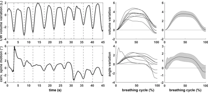
The posturo-ventilatory interaction was estimated by respira-tory emergence (REm) (Hamaoui et al. 2010) and by time-locked averaging (TLA) (Kuznetsov and Riley 2012) vari-ables, which both require a calculation using the respiratory signal and synchronized motion signal as follows. The REm is a measure of the weight of the respiratory component during movement and was assessed following a method that was previously described (Hamaoui et al. 2010). In brief, fast Fourier transformation was performed on the motion signal, the REm was computed in the frequency domain as the ratio of the average power of a band of 0.08 Hz centred on the mean breathing frequency and the overall average power of the motion signal. A REm close to 100% indi-cates that the respiratory component is a major component of the motion signal. In this study, the REm was assessed in the CoP displacement projected on the AP axis and cervi-cal spine, hip, knee and ankle motions in the sagittal plane. For the TLA, each breathing cycle was resampled to obtain
100 equally spaced points. Changes in the signals were then computed over the duration of each breathing cycle as the deviation in a signal from the value at the beginning of the cycle (Fig. 2). For the four joint angles (cervical spine, hip, knee and ankle), the presence of significant movements repeated throughout the breathing cycle was determined fol-lowing a method that was previously described (Kuznetsov and Riley 2012): first, the 95% confidence interval (95% CI) over the breathing cycle (step 1 to 100) was computed; then, a movement was considered significant if for over 20% of the breathing cycle, the value 0 was not included in the 95% CI. Significant movements with a 95% CI entirely less than 0 were considered flexion movements, and those entirely greater than 0 were considered extension move-ments (Fig. 3).
Data analysis
For the knees and ankles, the Amp, TLA and REm values were averaged across the left and right sides. The results for the COG1 and COG2 conditions were also averaged (COG). The majority of the variables were non-normally distributed (Shapiro–Wilk test), and all of them are described by the median and interquartile range [Q1; Q3]. The continuous variables were compared with Wilcoxon’s test, and the pro-portions were compared with Fisher’s exact test on the 2 × 2 corresponding contingency tables, considering a significance level of 0.05. The change in PV interaction along the mul-tijoint postural chain with a cognitive load was assessed by comparing the REm values derived from the CoP displace-ment and cervical spine, hip, knee and ankle motions and the occurrences of changes in the TLA (flexion, extension or no
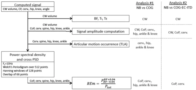
change) for the four joint motions (cervical spine, hip, knee and ankle) between the COG and NB conditions (analysis 1). The change in PV interaction induced by a cognitive load at the cervical level was compared to the change induced by the EC and ITV conditions and was assessed by the REm derived from the cervical spine motion (analysis 2). Figure 4
summarizes the different computed variables and performed analyses in a flowchart.
Results
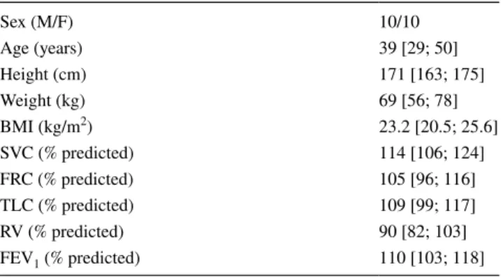
Data from one subject were excluded due to data recording issues, as two pelvic markers were missing. The data from twenty subjects were included in the analysis. The subjects’ baseline characteristics are presented in Table 1.
Modulation of the PV interaction along the multijoint postural chain during a cognitive load (analysis 1)
For COG1 and COG2, 7/8 words [6; 8] and 4/5 multiplica-tion answers [3; 5], respectively, were recalled by subjects. During the COG condition, compared to the NB condi-tion, the BF tended to increase (+ 1.5 min-1 [− 0.6; 4.6], p = 0.0731), the TE decreased (− 0.09 s [− 0.29; 0.09], p = 0.0228), TI (− 0.03 s [− 0.24; 0.19], p = 0.2959) and CW volume variation (− 0.05 L [− 0.97; 042], p = 0.3905) remained unchanged. The COG condition was associ-ated with an increase in the CoP Amp by + 2.2 mm [0.2; 6.3] (p = 0.0072). The amplitude of cervical motion was unchanged (+ 0.02° [− 0.49; 0.73], p = 0.2180), while significant increases in the amplitudes of motion were observed for the knee + 0.08° [0.02; 0.57] (p = 0.0072) and -0.8 -0.4 0 0.4 Flexion 0 50 100 -0.4 0 0.4 (° ) No Change 0 50 100 -0.4 0 0.4 0.8 cerv.spine change (° ) Extension 55% 69%
Breathing cycle (%) Breathing cycle (%) Breathing cycle (%)
cerv.spine change
(°
)
cerv.spine change
0 50 100
Fig. 3 Representative cervical spine changes over a breathing cycle with respect to the beginning of each cycle for three subjects under different conditions, with the mean value shown as the black line and the 95% confidence interval (CI) shown as the grey shaded
region. Left: flexion with the CI entirely less than zero over 55% of the breathing cycle; centre: no significant change with a CI includ-ing zero over the entire breathinclud-ing cycle; right: extension with the CI entirely greater than zero over 69% of the breathing cycle
Fig. 4 Flowchart presenting the computation of each studied vari-able and its use in analyses 1 and 2. (CW volume chest wall volume,
CoP centre of pressure, cerv. spine cervical spine, PSD power
spec-tral density, REm respiratory emergence, BF breathing frequency, TI inspiratory time, TE expiratory time, NB natural breathing, COG cog-nitive tasks, EC eyes closed, ITV increased tidal volume)
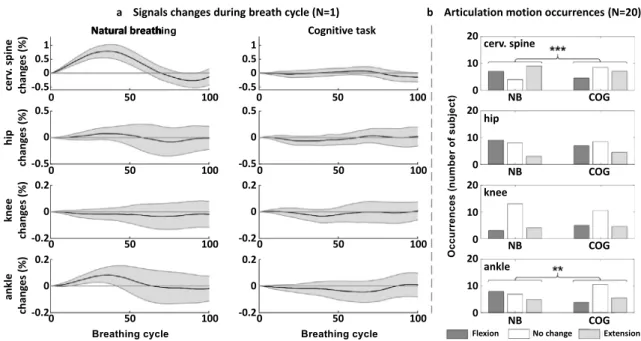
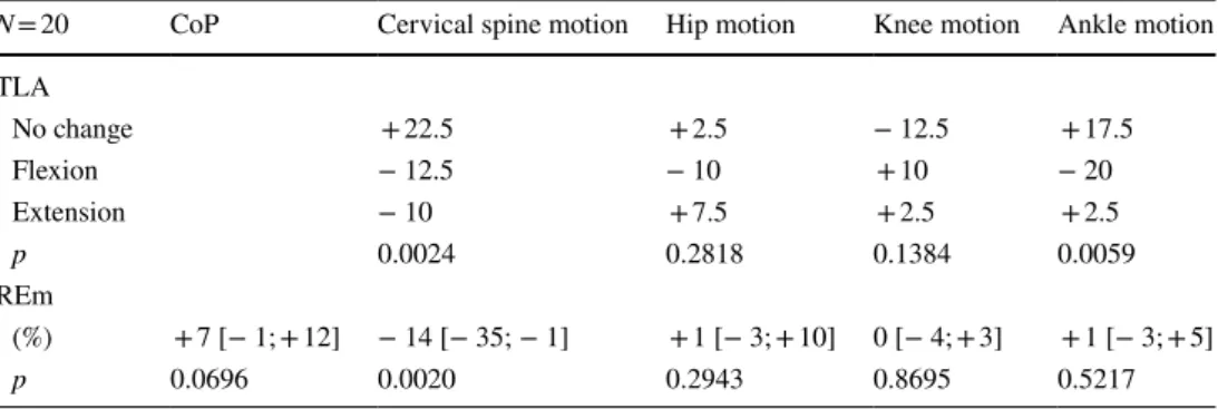
ankle + 0.12° [0.03; 0.60] (p = 0.0152) (Fig. 5). Regarding the PV interaction, the REm increased nonsignificantly for the CoP (p = 0.0696) but significantly decreased at the cervical level (p = 0.002), and it remained unchanged at the other joints (Fig. 6). The TLA analysis showed a sig-nificant decrease in the occurrence of breathing articular motion at the cervical spine and ankle levels but no change at the knee and hip levels. This phenomenon is illustrated in one subject (Fig. 7a), showing that the breathing-related perturbation manifested predominantly by the extension of the cervical spine, which was observed during the NB condition and was counteracted during the COG condi-tion. At the cervical level, breathing-related extension
and flexion movements were reduced globally by a similar magnitude; however, different profiles of cervical motion were observed between subjects (Fig. 7b; Table 2). Table 2
presents changes in the PV interaction between the NB and COG conditions for the REm and TLA analyses. Cervical PV interaction variations by conditions (analysis 2)
With respect to the NB condition, only the COG condi-tion induced a decrease in the breathing-related postural perturbation at the cervical level, as evidenced by a signifi-cant reduction in the REm (p = 0.002) (Table 3). Moreover, only the COG condition altered the ventilatory profile, as evidenced by a decrease in the TE and an increase in the BF (ns), while no change was observed under the ITV and EC conditions (Table 3). The amplitude of the CoP dis-placement increased during the COG and ITV conditions and remained unchanged during the EC condition. The motion of the cervical spine remained unchanged during the COG condition, while it decreased during the EC con-dition and increased during the ITV concon-dition (Table 3).
Discussion
This study shows that a cognitive load induces specific changes in the PV interaction, as attested by a change in the breathing-related postural perturbation along the postural chain, with a reduction at the cervical level only. This result
Table 1 Anthropomorphic characteristics and pulmonary function testing results
The results are presented as the median [Q1; Q3]
SVC slow vital capacity, FRC functional residual capacity measured
by helium dilution, TLC total lung capacity, RV residual volume,
FEV1 forced expiratory volume in one second
Sex (M/F) 10/10 Age (years) 39 [29; 50] Height (cm) 171 [163; 175] Weight (kg) 69 [56; 78] BMI (kg/m2) 23.2 [20.5; 25.6] SVC (% predicted) 114 [106; 124] FRC (% predicted) 105 [96; 116] TLC (% predicted) 109 [99; 117] RV (% predicted) 90 [82; 103] FEV1 (% predicted) 110 [103; 118] CoP 0 2 4 6 8 10 12 14 16 18 20 22 Amp. (mm) NB cog.
cerv.spine hip knee ankle 0 0.5 1 1.5 2 2.5 3 3.5 A m p. (°) NB cog.
**
*
a
b
**
Fig. 5 Variations in the amplitude (Amp.) of a the centre of pressure (CoP) displacement; b joint rotations (cervical spine, hip, knee, ankle) dur-ing the natural breathdur-ing (NB) and cognitive conditions (COG) in the standdur-ing position in N = 20 subjects. (*p < 0.05; **p < 0.01; ***p < 0.001)
was concurrent with an increase in global postural pertur-bations and lower limb motions, while the cervical motion remained unchanged. This result suggests that the reduction in the ventilatory-related perturbation at the cervical level protects the cervical spine from the postural perturbation induced by the cognitive load.
Posturo‑ventilatory interaction and cognition Cognitive dual-task paradigms allow us to investigate inter-actions between cognitive processes and other cortical func-tions, such as postural control (Huxhold et al. 2006; Lacour et al. 2008) or the cortical control of breathing (Grassmann et al. 2016). A cognitive load is known to perturb the control of balance by diverting attentional resources (Huxhold et al.
2006; Lacour et al. 2008). These interactions between the mental process and postural control are mostly modulated by visual or motor stimuli, the difficulty of the cognitive task, postural constraints, sensorimotor expertise (for example, in experts in gymnastics), and ageing (Huxhold et al. 2006). The subjects in our study were all healthy and had no senso-rimotor expertise. They were in a relaxed standing position without any postural stimulation, and the orientation of their gaze was constant across the conditions. Therefore, we may anticipate that the postural impact of a cognitive task on individuals’ balance depends mostly on their age and on the intensity of the cognitive load (Huxhold et al. 2006; Lacour et al. 2008). A cognitive load is generally associated with improved balance parameters in subjects in their twenties (Huxhold et al. 2006; Hagio et al. 2018), with no change or impaired balance in older subjects (Lacour et al. 2008), and/ or during a high cognitive load (Huxhold et al. 2006; Lacour et al. 2008). As expected, considering the median age of the subjects in this study (39 years) and the high cognitive load to which they were subjected, we observed impaired balance during the COG condition. We observed a relative
Fig. 7 a The cervical spine, hip, knee and ankle signal changes over a
breathing cycle during natural breathing (NB) and the cognitive task (COG) in one subject (black line: mean value; grey shaded region: 95% confidence interval); b motion distribution (flexion, no change and extension) measured in the 20 subjects for the cervical spine,
hip, knee and ankle. Comparison between natural breathing (NB) and the cognitive task (COG) (proportions were compared with the exact test on the 2 × 3 corresponding contingency tables: **p < 0.01; ***p < 0.001)
CoP cerv.spine hip knee ankle 0 10 20 30 40 50 60 70 80 90 100 RE m (%) NB cog. **
Fig. 6 Variations in respiratory emergence (REm) for the centre of pressure (CoP) displacement amplitude and joint motions (cervical spine, hip, knee, ankle) during the natural breathing (NB) and cog-nitive conditions (COG) in the standing position in N = 20 subjects. (Wilcoxon’s test: *p < 0.05; **p < 0.01; ***p < 0.001)
increase, though the difference was not significant, in the breathing frequency during the cognitive load, which was also previously reported (Grassmann et al. 2016). This mod-ulation in the breathing pattern induced by a dual task is not surprising, as in addition to automatic commands, cortical commands transmitted from the primary cortex (Similowski et al. 1996), premotor cortex and supplementary motor area (Raux et al. 2007) control human respiration. To the best of our knowledge, however, our study is the first to dem-onstrate a specific modulation in the PV interaction under cognitive load. This result was shown in the subjects in our study by an increase in respiratory emergence close to sta-tistical significance (p = 0.0696) and by significant changes in the TLA. This result indicates the central modulation of the PV interaction and suggests that postural control partly depends on central interactions between the cortical control of breathing and cognition in healthy humans. Therefore, as dual tasks are common in real-life settings, our results lead us to hypothesize that postural control may be less efficient in situations associated with a pathological adap-tive increase in awake cortical respiratory drive, such as in healthy individuals during experimental dyspnoea (Raux et al. 2007) or in some patients with obstructive sleep apnoea syndrome (OSAS) (Launois et al. 2015), chronic obstruc-tive pulmonary disease (COPD) (Nguyen et al. 2018) or hyperventilation syndrome (Dubois et al. 2016). In these settings, the pre-inspiratory potential observed in the motor cortex and the supplementary motor area during resting ventilation (Raux et al. 2007) has been associated with a perturbation of attentional resources (Sharman et al. 2014), which suggests that the balance between cognition, postural control and the control of breathing under cognitive load is impaired. This concept has been demonstrated in healthy subjects by a negative impact of experimental dyspnoea on both cognition and locomotion, as measured by the “timed up-and-go” test and its imagery version (Nierat et al. 2016). This concept has also been shown in patients with OSAS by a reduction in cognitive performance to maintain a bal-ance performbal-ance similar to that of controls (Baillieul et al.
2018), but it remains to be specifically addressed.
Table 2 Change in the posturo-ventilatory interaction during the cognitive load condition
TLA time locked averaging, REm: respiratory emergence, CoP centre of pressure
N = 20 CoP Cervical spine motion Hip motion Knee motion Ankle motion TLA No change +22.5 + 2.5 − 12.5 + 17.5 Flexion − 12.5 − 10 +10 − 20 Extension − 10 + 7.5 + 2.5 + 2.5 p 0.0024 0.2818 0.1384 0.0059 REm (%) + 7 [− 1; + 12] − 14 [− 35; − 1] + 1 [− 3; + 10] 0 [− 4; + 3] + 1 [− 3; + 5] p 0.0696 0.0020 0.2943 0.8695 0.5217
Table 3 Posturo-ventilatory interaction at the cervical level during the cognitive load, eyes-closed and increased tidal volume conditions compared to natural breathing
Bold corresponds to significant p < 0.05
The results are presented as the median [Q1; Q3] and the p value obtained from Wilcoxon’s test. The COG, EC and ITV conditions were compared to the NB condition
BF breathing frequency, TI inspiratory time, TE expiratory time; Amp. Amplitude, REm respiratory emergence, CoP centre of
pres-sure, NB natural breathing condition, COG cognitive condition, EC eyes-closed condition, ITV increased tidal volume condition
N = 20 NB COG EC ITV Ventilation BF (min−1) 16.6[13.3; 18.7] – 18.6 [16.3; 19.4] 0.0731 16.3 [13.8; 18.1] 0.9108 13.5 [10.7; 18.5] 0.5257 TI (s) 1.9 [1.5; 2.2] – 1.8 [1.6; 1.9] 0.2959 2.0 [1.6; 2.3] 0.5016 2.2 [1.9; 2.9] 0.1790 TE (s) 2.0 [1.7; 2.4] – 1.7 [1.5; 1.9] 0.0228 1.9 [1.7; 2.3] 0.3135 2.3 [1.7; 3.4] 0.1169 Amp. (L) 2.19 [1.35; 3.42] – 2.01 [1.22; 2.78] 0.3905 2.40 [1.58; 2.81] 0.8983 9.10 [5.74; 10.93] 8.2*10–5 Cervical spine Amp. (°) 1.14 [0.84; 1.60] – 1.01 [0.62; 1.47] 0.2180 0.82 [0.66; 1.09] 0.0090 6.79 [4.19; 9.76] 0.0001 Rem (%) 17.2 [7.8; 37.2] – 4.2 [1.8; 10.0] 0.0020 20.8 [8.0; 36.3] 0.9702 40.7 [19.1; 59.8] 0.1169 CoP Amp. (mm) 3.29[2.04; 5.28] – 5.74 [3.35; 10.55] 0.0072 3.15 [2.67; 3.63] 0.4553 5.57 [4.74; 7.45] 0.0010 Rem (%) 6.2 [3.8; 10.3] – 12.9 [5.8; 20.7] 0.0696 9.3 [5.5; 14.4] 0.1790 18.3 [12.3; 25.1] 0.0004
Preservation of the cervical spine
from breathing‑related perturbations and cognitive load
Our analysis at the joint level confirmed a significant reduc-tion in the PV interacreduc-tion at the cervical level and no change or increase at the other joints. Our results suggest that the PV interaction in humans cannot be modelled using a sim-ple inverted pendulum model; rather, the model must take into account multiple degrees of freedom, as previously reported (Hodges et al. 2002; Kuznetsov and Riley 2012). Several joints (spine, hip, knee and ankle), each acting as a degree of freedom on the postural body chain, counter-act the breathing-related postural perturbation and provide flexibility in central control (Hodges et al. 2002; Kuznetsov and Riley 2012) . In our study, we confirmed that this pos-tural chain was altered under cognitive load. In addition, our study supports the hypothesis that the maintenance of stability of the neck is related to the protection of the cervi-cal spine from ventilatory perturbations, as attested by the reduction in respiratory emergence at the cervical level. It has been previously reported that the breathing-related component of CoP displacements, i.e., the breathing-related postural perturbation, almost disappears during voluntary apnoea in healthy humans (Caron et al. 2004). Indeed, in healthy humans, during standing, even the breathing-related rhythmic perturbation is continuously counteracted, and this counteraction is not complete, which implies that natural breathing “normally perturbs” the balance along the multi-joint postural chain (Kuznetsov and Riley 2012). Therefore, when a subject voluntarily holds his or her breath, the effect of the suppression of the breathing-related postural perturba-tion on the CoP displacements (Caron et al. 2004), as well as on the cervical spine motion (Hodges et al. 2002), may be linked to a “stabilization strategy”. Similarly, in the subjects in our study, the reduction in the breathing-related perturba-tion, the unchanged amplitude of the motion of the cervical spine during cognitive load, and the increased amplitudes of postural sway and knee and ankle motion probably corre-spond to a “local stabilization strategy” of the cervical spine that may have limited the impairment of balance; however, we acknowledge this postulation remains to be addressed.
Actually, the stabilization of the neck may help maintain the head position and a horizontal gaze (Hasegawa et al.
2017). This compensatory adaptation may stem from the necessity to improve visual, vestibular and neck propriocep-tive afferent feedback efficiency (Bove et al. 2009; Williams et al. 2017; Malmström et al. 2017) during a high-load cog-nitive task (Huxhold et al. 2006; Lacour et al. 2008), which is known to compromise balance by diverting attentional prefrontal resources (Mihara et al. 2008; Stelzel et al. 2018) that are primarily dedicated to the control of balance and anticipatory adjustments of the cervical curvature (Hamaoui
and Alamini-Rodrigues 2017; Boulanger et al. 2017). Con-sequently, the stability of the cervical spine, which is known to play an important role in the maintenance of balance (see also Introduction section) (Gandelman-Marton et al. 2016; Hamaoui and Alamini-Rodrigues 2017), is probably crucial when cortical resources are challenged. Likewise, in COPD patients, who often recruit their respiratory neck muscles, pathologic breathing-related neck stabilization in dual-task settings may represent one of the physiopathological mecha-nisms of their specific postural dysfunction (Janssens et al.
2014). This hypothesis needs to be investigated in future studies.
Methodological considerations and limitations We acknowledge that the instruction to subjects to focus their gaze on a landmark may have encouraged them to sta-bilize their neck. The instruction to stand barefoot on the force plate and focus their gaze on a landmark on the wall was used to minimize the variability of their posture and to avoid additional postural perturbations between subjects and between the NB and COG conditions. Therefore, the instruc-tion’s effect on balance was comparable in the reference NB and COG conditions. In addition, we acknowledge that this instruction may induce an additional cognitive load. Regard-less of whether it induced an additional cognitive load, our results demonstrate that a higher cognitive load during the dual task was accompanied by a reduced respiratory pos-tural perturbation at the neck joint level, no change in the neck motion, and increases in the COP displacement and lower joint motions. This strategy seems specific to a central modulation, as it was not observed during “non-cortical” postural perturbations, such as the EC or ITV conditions, while the breathing-related postural perturbation at the neck level remained unchanged.
In our study, the relatively small number of subjects, par-ticularly that of subjects aged under 25 years (n = 3), limits our interpretation of the age effects present in our results. This factor remains to be addressed in future studies.
Balance in the standing position is a complex phenom-enon that varies with postural alignment (Amabile et al.
2018), postural tone and metabolic cost (van Emmerik and van Wegen 2002; Houdijk et al. 2015). Therefore, some changes in postural sway are not associated with perturba-tions in balance. However, postural sway increases when balance tends to be compromised. This relation is the rea-son why we considered, by analogy, the amplitude of the motions of the cervical spine and other joints to be stability parameters of a given segment. We acknowledge that elec-tromyography of the neck muscles can be useful; however, we are confident that the divergence between the cervical spine and other joints in terms of the breathing-related
postural perturbation supports our “stabilization hypothesis” at the cervical level.
Conclusion
This study shows that the cervical spine is protected from the breathing-related postural perturbation during a cogni-tive dual task, while the postural perturbation occurs glob-ally and at inferior joints. This result represents a cortical adaptation of the posturo-ventilatory interaction to stabilize the neck and possibly to limit the cognitive-induced pos-tural perturbation. This adaptation may eventually become impaired in patients suffering from chronic respiratory dis-ease. More generally, this adaptation supports the existence of a strong interaction between cognition, the control of breathing and postural control.
Author contributions LC, VA, BS and TS contributed substantially to the study design, data analysis and interpretation, and the writing of the manuscript. IR, MCN, PL and PR contributed substantially to the data analysis and interpretation, and the writing of the manuscript. All authors approved the final version of the manuscript and agreed to be accountable for all aspects of the work.
Funding This work was funded by the Chancellerie des Universités de Paris (Grant number: Legs Poix (LEG 1604)); ENS Cachan (Grant number: Ph.D. fellowship); Assistance Publique - Hôpitaux de Paris (Grant number: Grant “poste d’accueil APHP/Arts et Métiers” délé-gation à la Recherche Clinique et à l’Innovation (DRCI)); Paristech (Grant number: BiomeCAM chair).
Compliance with ethical standards
Conflict of interest The authors have no conflict of interest for this study.
References
Amabile C, Le Huec J-C, Skalli W (2018) Invariance of head-pelvis alignment and compensatory mechanisms for asymptomatic adults older than 49 years. Eur Spine J 27:458–466. https ://doi. org/10.1007/s0058 6-016-4830-8
Attali V, Clavel L, Rouch P et al (2019) Compensation of respiratory-related postural perturbation is achieved by maintenance of head-to-pelvis alignment in healthy humans. Front Physiol 10:441. https ://doi.org/10.3389/fphys .2019.00441
Baillieul S, Wuyam B, Pépin J-L et al (2018) Continuous positive air-way pressure improves gait control in severe obstructive sleep apnoea: a prospective study. PLoS ONE 13:e0192442. https ://doi. org/10.1371/journ al.pone.01924 42
Boulanger M, Giraudet G, Faubert J (2017) Interaction between the oculomotor and postural systems during a dual-task: compensa-tory reductions in head sway following visually-induced postural perturbations promote the production of accurate double-step sac-cades in standing human adults. PLoS ONE 12:e0173678. https :// doi.org/10.1371/journ al.pone.01736 78
Bove M, Fenoggio C, Tacchino A et al (2009) Interaction between vision and neck proprioception in the control of stance. Neuro-science 164:1601–1608. https ://doi.org/10.1016/j.neuro scien ce.2009.09.053
Cala SJ, Kenyon CM, Ferrigno G et al (1996) Chest wall and lung volume estimation by optical reflectance motion analysis. J Appl Physiol 81:2680–2689
Caron O, Fontanari P, Cremieux J, Joulia F (2004) Effects of ventila-tion on body sway during human standing. Neurosci Lett 366:6–9.
https ://doi.org/10.1016/j.neule t.2004.04.085
Dally JFH (1908) An inquiry into the physiological mechanism of res-piration with especial reference to the movements of the vertebral column and diaphragm. Proc R Soc 80:93–114
David P, Laval D, Terrien J, Petitjean M (2012) Postural control and ventilatory drive during voluntary hyperventilation and carbon dioxide rebreathing. Eur J Appl Physiol 112:145–154. https ://doi. org/10.1007/s0042 1-011-1954-8
Dubois M, Chenivesse C, Raux M et al (2016) Neurophysiological evidence for a cortical contribution to the wakefulness-related drive to breathe explaining hypocapnia-resistant ventilation in humans. J Neurosci 36:10673–10682. https ://doi.org/10.1523/ JNEUR OSCI.2376-16.2016
Dubousset J (1994) Three-dimensional analysis of scoliotic deformity. In: Weinstein SL (ed) Pediatric spine: principles and practices. Raven Press ltd, NY, pp 479–496
Gandelman-Marton R, Arlazoroff A, Dvir Z (2016) Postural stability in patients with different types of head and neck trauma in com-parison to healthy subjects. Brain Inj 30:1612–1616. https ://doi. org/10.1080/02699 052.2016.11999 04
Grassmann M, Vlemincx E, von Leupoldt A et al (2016) Respiratory changes in response to cognitive load: a systematic review. Neural Plast 2016:1–16. https ://doi.org/10.1155/2016/81468 09
Gurfinkel VS, Kots YM, Paltsev EI, Feldman AG (1971) The compen-sation of respiratory disturbances of the erect posture of man as an example of the organization of interarticular interaction. In: Gelfand IM, S GV, V FS, L TM (eds) Models of the structural-functional organization of certain biological systems, Cambridge, Massachusetts
Hagio K, Obata H, Nakazawa K (2018) Effects of breathing movement on the reduction of postural sway during postural-cognitive dual tasking. PLoS ONE 13:e0197385. https ://doi.org/10.1371/journ al.pone.01973 85
Hamaoui A, Alamini-Rodrigues C (2017) Influence of cervical spine mobility on the focal and postural components of the sit-to-stand task. Front Hum Neurosci 11:129. https ://doi.org/10.3389/fnhum .2017.00129
Hamaoui A, Gonneau E, Le Bozec S (2010) Respiratory disturbance to posture varies according to the respiratory mode. Neurosci Lett 475:141–144. https ://doi.org/10.1016/j.neule t.2010.03.064
Hasegawa K, Okamoto M, Hatsushikano S et al (2017) Standing sagittal alignment of the whole axial skeleton with reference to the gravity line in humans. J Anat 230:619–630. https ://doi. org/10.1111/joa.12586
Hodges PW, Gandevia SC, Richardson CA (1997) Contractions of spe-cific abdominal muscles in postural tasks are affected by respira-tory maneuvers. J Appl Physiol 83:753–760
Hodges PW, Gurfinkel VS, Brumagne S et al (2002) Coexistence of stability and mobility in postural control: evidence from postural compensation for respiration. Exp Brain Res 144:293–302. https ://doi.org/10.1007/s0022 1-002-1040-x
Hodges PW, Sapsford R, Pengel LHM (2007) Postural and respiratory functions of the pelvic floor muscles. Neurourol Urodyn 26:362– 371.https ://doi.org/10.1002/nau.20232
Houdijk H, Brown SE, van Dieën JH (2015) Relation between pos-tural sway magnitude and metabolic energy cost during upright
standing on a compliant surface. J Appl Physiol 119:696–703.
https ://doi.org/10.1152/jappl physi ol.00907 .2014
Huxhold O, Li S-C, Schmiedek F, Lindenberger U (2006) Dual-tasking postural control: aging and the effects of cognitive demand in conjunction with focus of attention. Brain Res Bull 69:294–305.
https ://doi.org/10.1016/j.brain resbu ll.2006.01.002
Jahn K, Deutschländer A, Stephan T et al (2004) Brain activation pat-terns during imagined stance and locomotion in functional mag-netic resonance imaging. Neuroimage 22:1722–1731. https ://doi. org/10.1016/j.neuro image .2004.05.017
Janssens L, Brumagne S, McConnell AK et al (2014) Impaired postural control reduces sit-to-stand-to-sit performance in individuals with chronic obstructive pulmonary disease. PLoS ONE 9:1–5. https :// doi.org/10.1371/journ al.pone.00882 47
Kantor E, Poupard L, Le Bozec S, Bouisset S (2001) Does body stabil-ity depend on postural chain mobilstabil-ity or stabilstabil-ity area? Neurosci Lett 308:128–132. https ://doi.org/10.1016/S0304 -3940(01)01986 -3
Kuznetsov NA, Riley MA (2012) Effects of breathing on multijoint control of center of mass position during upright stance. J Mot Behav 44:241–253. https ://doi.org/10.1080/00222 895.2012.68889 4
Lacour M, Bernard-Demanze L, Dumitrescu M (2008) Posture con-trol, aging, and attention resources: models and posture-analysis methods. Neurophysiol Clin Neurophysiol 38:411–421. https :// doi.org/10.1016/j.neucl i.2008.09.005
Launois C, Attali V, Georges M et al (2015) Cortical drive to breathe during wakefulness in patients with obstructive sleep apnea syn-drome. Sleep 38:1743–1749. https ://doi.org/10.5665/sleep .5156
Mador MJ, Tobin MJ (1991) Effect of alterations in mental activity on the breathing pattern in healthy subjects. Am Rev Respir Dis 144:481–487. https ://doi.org/10.1164/ajrcc m/144.3_Pt_1.481
Malmström E-M, Fransson P-A, Jaxmar Bruinen T et al (2017) Dis-turbed cervical proprioception affects perception of spatial orien-tation while in motion. Exp Brain Res 235:2755–2766. https ://doi. org/10.1007/s0022 1-017-4993-5
Manor BD, Hu K, Peng CK et al (2012) Posturo-respiratory synchroni-zation: effects of aging and stroke. Gait Posture 36:254–259. https ://doi.org/10.1016/j.gaitp ost.2012.03.002
Mihara M, Miyai I, Hatakenaka M et al (2008) Role of the prefrontal cortex in human balance control. Neuroimage 43:329–336. https ://doi.org/10.1016/j.neuro image .2008.07.029
Nguyen DAT, Boswell-Ruys CL, McBain RA et al (2018) Inspiratory pre-motor potentials during quiet breathing in ageing and chronic obstructive pulmonary disease. J Physiol 596:6173–6189. https :// doi.org/10.1113/JP275 764
Nierat M-C, Demiri S, Dupuis-Lozeron E et al (2016) When breathing interferes with cognition: experimental inspiratory loading alters timed up-and-go test in normal humans. PLoS ONE 11:e0151625.
https ://doi.org/10.1371/journ al.pone.01516 25
Park E, Schöner G, Scholz JP (2012) Functional synergies underlying control of upright posture during changes in head orientation. PLoS ONE 7:e41583. https ://doi.org/10.1371/journ al.pone.00415 83
Perry SF, Carrier DR (2006) The coupled evolution of breathing and locomotion as a game of leapfrog. Physiol Biochem Zool 79:997– 999.https ://doi.org/10.1086/50765 7
Quek J, Treleaven J, Clark RA, Brauer SG (2018) An exploratory study examining factors underpinning postural instability in older adults with idiopathic neck pain. Gait Posture 60:93–98. https ://doi. org/10.1016/j.gaitp ost.2017.11.016
Raux M, Straus C, Redolfi S et al (2007) Electroencephalographic evi-dence for pre-motor cortex activation during inspiratory loading in humans. J Physiol 5782:569–578. https ://doi.org/10.1113/jphys iol.2006.12024 6
Sharman M, Gallea C, Lehongre K et al (2014) The cerebral cost of breathing: an fMRI case-study in congenital central hypoventila-tion syndrome. PLoS ONE 9:e107850. https ://doi.org/10.1371/ journ al.pone.01078 50
Similowski T, Straus C, Coïc L, Derenne JP (1996) Facilitation-inde-pendent response of the diaphragm to cortical magnetic stimu-lation. Am J Respir Crit Care Med 154:1771–1777. https ://doi. org/10.1164/ajrcc m.154.6.89703 69
Stelzel C, Bohle H, Schauenburg G et al (2018) Contribution of the lateral prefrontal cortex to cognitive-postural multitasking. Front Psychol 9:1075. https ://doi.org/10.3389/fpsyg .2018.01075
Talasz H, Kremser C, Kofler M et al (2011) Phase-locked paral-lel movement of diaphragm and pelvic floor during breathing and coughing-a dynamic MRI investigation in healthy females. Int Urogynecol J 22:61–68. https ://doi.org/10.1007/s0019 2-010-1240-z
Taube W, Gruber M, Gollhofer A (2008) Spinal and supraspinal adaptations associated with balance training and their functional relevance. Acta Physiol 193:101–116. https ://doi.org/10.111 1/j.1748-1716.2008.01850 .x
van Emmerik REA, van Wegen EEH (2002) On the functional aspects of variability in postural control. Exerc Sport Sci Rev 30:177–183 Vital JM, Senegas J (1986) Anatomical bases of the study of the con-straints to which the cervical spine is subject in the sagittal plane. A study of the center of gravity of the head. Surg Radiol Anat 8:169–173
Williams K, Tarmizi A, Treleaven J (2017) Use of neck torsion as a specific test of neck related postural instability. Musculoskelet Sci Pract 29:115–119. https ://doi.org/10.1016/j.msksp .2017.03.012
Publisher’s Note Springer Nature remains neutral with regard to jurisdictional claims in published maps and institutional affiliations.