T
T
H
H
È
È
S
S
E
E
En vue de l'obtention du
D
D
O
O
C
C
T
T
O
O
R
R
A
A
T
T
D
D
E
E
L
L
’
’
U
U
N
N
I
I
V
V
E
E
R
R
S
S
I
I
T
T
É
É
D
D
E
E
T
T
O
O
U
U
L
L
O
O
U
U
S
S
E
E
Délivré par l'Université Toulouse III - Paul Sabatier Discipline ou spécialité : Biogéochimie
JURY
Professeur Bruno LARTIGES, Université Paul Sabatier, Toulouse, président
Professeur Christophe DUPRAZ, professeur assistant, Université du Connecticut, Storrs, rapporteur Docteur Bénédicte MENEZ, chargée de recherche CNRS, IPGP, Paris, rapporteur
Docteur Steeve BONNEVILLE, Université de Gree, Bruxelles, examinateur
Docteur Pascale BENEZETH, directeur de recherche CNRS, GET, Toulouse, directeur de thèse Docteur Oleg POKROVSKY, chargé de recherche CNRS, GET, Toulouse, directeur de thèse
Ecole doctorale : Sciences de l’Univers, de L’Environnement et de l’Espace (SDU2E) Unité de recherche : Géosciences Environnement Toulouse (GET)
Directeur(s) de Thèse : Pascale BENEZETH, directeur de recherche CNRS, GET, Toulouse Oleg POKROVSKY, chargé de recherche CNRS, GET, Toulouse
Présentée et soutenue par Irina A. BUNDELEVA
Le 24 juin 2011
Titre :
Modélisation expérimentale de la précipitation des minéraux carbonatés lors de l'activité bactérienne.
1
Remerciements
Cette thèse est le résultat de trois ans de recherche au laboratoire Géosciences Environnement Toulouse (GET) (jusqu'à janvier 2011 Laboratoire des Mécanismes et Transferts en Géologie (LMTG)). Ce travail m’a été proposé par Pascale Bénézeth et Oleg Pokrovsky, que je remercie chaleureusement. J’ai pu grâce à eux me perfectionner dans des disciplines qui n’étaient pas – initialement – mes disciplines de prédilection, en particulier la géologie, géochimie, minéralogie. Je suis reconnaissant à mes encadrants de m’avoir guidé dans les moments difficiles, en particulier le début de thèse. Je remercie tout particulièrement Jacques Schott d’avoir veillé sur moi durant cette période d’adaptation.
Je remercie Christophe Dupraz et Bénédicte Menez d’avoir accepté de réaliser le rapport de ce manuscrit. J’adresse également mes remerciements à Steeve Bonneville qui a accepté de participer au Jury de ma soutenance. Je suis reconnaissante à Bruno Lartiges d’avoir présidé ce Jury.
Je tiens également à remercier mon amie Liudmila Shirokova, qui m’a énormément aidé sur la partie microbiologique. Je voudrais dire «Merci» à Vasileios Mavromatis pour son aide précieuse lors des analyses isotopique du magnésium.
Beaucoup de techniciens œuvrent sans relâche dans ce laboratoire et sont un soutien irremplaçable pour nous, doctorants. Je pense à Carole Causserand, Thierry Aigouy, Michel Thibaut, Stéphanie Balor, Sophie Gouy, Manuel Henry, Jérôme Chmeleff, Jonathan Prunier, Stéphanie Mounic, Philippe Besson. Merci pour leur chaleur humaine et leur accueuil.
Un grand merci à tous les doctorant-e-s et post-doctorant-e-s du labo pour avoir égayé mes dures journées par leur sourire, fou-rire, blagues et même avec des discussions scientifiques sérieuses (Thomas et Marianne, Emilie et Fabrice, Julien et Sylvie, Elena et
Laurent, Ekaterina, Svetlana, Alisson, Quentin, Nico, Sylvaine, Aude, Jean-Sébastien,
Joaquin, ). Merci aux occupants successifs du bureau F161 (Camille Truche, Sophie Demouy, Guilhem Hoareau, Jérémy Masbou, Alexandre Bellefleur, Anh-Tuan Nguyen) d’avoir assuré une ambiance incroyable tout au long de ces trois ans.
Je suis très reconnaissant à ma famille (mes parents Alexandre et Tatiana, mes sœurs Liudmila et Alexandra) et mes amies russes (Anna, Evguenia, Irina, Galina, Natalia, Evgueni, Lusia, Anton) de m’avoir aidé dans toutes les situations. J’adresse également mes remerciements à famille D’Abzac (Louis, Brigitte, François-Xavier, Alexandre) qui m’a aidé à bien m’intégrer dans ma nouvelle vie, à amélioré mon français.
2
Enfin, ce travail n’aurait pu aboutir sans le soutien et aide de mon cher ami François-Xavier. FX, merci à toi. Merci.
Je vous souhaite à tout(e)s la bonne continuation, bonne chance et bonne courage. Soyez heureux !
Abstract
3
Abstract
Microbially-induced mineralization is considered as one of the main natural processes
controlling CO2 levels in the atmosphere and a major structural and ecological player, in the
modern and in the past ecosystems. In this study are presented the data of laboratory
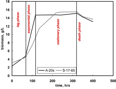
experimental work on CaCO3 precipitation with pure cultures of two anoxygenic phototrophs
bacteria (APB): haloalcaliphilic Rhodovulum steppens A-20s and neutrophilic halophilic
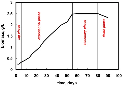
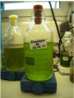
Rhodovulum sp. S-17-65; and cyanobacteria Gloeocapsa sp.. These bacteria represent two
important groups of photosynthetic organisms in the past and at present time. APB is the oldest microorganism which could be dominant during the anoxygenic period of Earth’s life (approximately 4 billon years ago) whereas the origin of oxygen evolving microorganisms (cyanobacteria) is placed at about 3.5 billion years ago as based on oxidation records of the Earth’s crust. In modern ecosystems, cyanobacteria are the dominant primary producers. Nonetheless, the potential of APB are abundant in the modern microbial mats and stromatolites and thus may represent a considerable fraction of the standing biomass. However, biomineralization induce by these bacteria has not been thoroughly studied up to now.
In this context, the aim of this thesis is to characterize the process of biological CaCO3
precipitation and to assess the existence of metabolic processes protecting studied bacteria against carbonate mineralization on their surfaces. For this, kinetic experiments, SEM and TEM imaging, EDX and XRD analyses, zeta-potential measurements and Ca adsorption into bacterial surface were carried out.
The result of this study demonstrates the participation of studied bacteria in CaCO3
precipitation. Zeta-potential measurements suggest the existence of a cells protection mechanism for studied APB, based on the metabolic maintenance of a positive surface charge
at alkaline pH, preserving active bacteria against Ca2+ adsorption and subsequent carbonate
precipitation on their surfaces. The existence of the same mechanism for Gloeocapsa sp. was not confirmed.
Overall, the results of this study show two different mechanisms of CaCO3-nucleation:
an unspecific supersaturation by APB and a specific nucleation at the cell wall by cyanobacteria Gloeocapsa sp..
4
Key words: Anoxygenic phototrophic bacteria, Rhodovulum sp., Cyanobacteria, Gloeocapsa sp., electrophoresis, zeta potential, calcium, bicarbonate, adsorption, calcite,
calcium carbonate, precipitation, kinetics.
Résumé
La minéralisation induite par l’activité microbienne joue un rôle majeur dans le fonctionnement des écosystèmes passés et présents. Cette étude présente les données
expérimentales de précipitation de CaCO3 à partir de cultures pures de deux types de bactéries
anoxygéniques phototrophiques (APB) : Rhodovulum steppens A-20s haloalcaphilique et
Rhodovulum sp. S-17-65 neutrophilique halophilique, ainsi que de cyanobactéries Gloeocapsa
sp.. Ces bactéries représentent deux groupes importants d’organismes photosynthétiques depuis les temps les plus anciens jusqu’à nos jours. Les APB sont des microorganismes dominants durant la période anoxygénique de la Terre (il y a environ 4 milliards d’années) tandis que l’origine des microorganismes évoluant grâce à l’oxygène (cyanobactéries) se situe à environ 3.5 milliards d’années, en se basant sur les enregistrements d’oxydation de la croûte terrestre. Au sein des écosystèmes modernes, les cyanobactéries sont les producteurs primaires dominants. Cependant, le potentiel des APB est important de part leur abondance dans les biolfilms microbiens et les stromatolites modernes, représentant ainsi une fraction considérable de la biomasse effective. La biomineralisation induite par ces bactéries a toutefois été très peu étudiée jusqu’à présent.
Dans ce contexte, cette thèse a pour objectif principal de caractériser les processus
biologiques de précipitation de CaCO3 et d’évaluer l’existence d’un processus métabolique
protégeant les bactéries étudiées contre la minéralisation de carbonates à leur surface. Pour cela, des expériences cinétiques, des mesures de potentiel zeta et d’adsorption de Ca à la surface bactérienne, couplées à des observations par Microscopie Electronique à Balayage (MEB), en Transmission (MET) et des analyses chimiques par émission et diffraction de rayons X (EDX et XRD) ont été menées.
Les résultats de cette étude démontrent que les bactéries étudiées prennent une part
active dans la précipitation de CaCO3. Les mesures de potentiel zeta suggèrent l’existence
d’un mécanisme de protection de la cellule pour les APB étudiées, basé sur le maintien métabolique d’une charge de surface positive à pH alcalin, préservant les bactéries actives de
Abstract
5
l’adsorption de Ca2+ et de la précipitation subséquente de carbonates à leur surface.
L’existence d’un tel mécanisme n’est pas confirmée pour Gloeocapsa sp..
Ainsi, deux mécanismes différents de nucléation de CaCO3 peuvent être mis en
évidence : un premier mettant en jeu une sursaturation non-spécifique pour les bactéries anoxygéniques phototrophiques (APB), et un deuxième par nucléation spécifique au niveau de la membrane cellulaire pour les cyanobactéries Gloeocapsa sp..
Mots-clés : Bactéries anoxygéniques phototrophique, Rhodovulum sp., Cyanobactéries, Gloeocapsa sp., électrophorèse, potentiel zeta, calcium, bicarbonate, adsorption, calcite, précipitation, cinétique
Table of contents
7
Table of contents 9 REMERCIEMENTS... 1 ABSTRACT... 3 RESUME... 4 TABLE OF CONTENTS ...7 INTRODUCTION...13 RESUME DE L’INTRODUCTION EN FRANÇAIS... 15 INTRODUCTION... 19 CHAPTER I ...27 DESCRIPTION OF ANOXYGENIC PHOTOTROPHIC BACTERIA AND CYANOBACTERIA ...27 1. Anoxygenic phototrophic bacteria (APB) ... 29 1.1. Anoxygenic photosynthesis... 30 1.2. Diversity of Anoxygenic Phototrophic Bacteria... 31 1.2.1. Rhodovulum steppense sp. nov. A‐20s... 35 1.2.2. Rhodovulum sp. S‐17‐65 ... 37 2. Cyanobacteria... 39 2.1. Oxygenic Photosynthesis of Bacteria ... 40 2.2. Diversity of cyanobacteria... 41 2.2.1. Cyanobacteria Gloeocapsa sp. ... 45 2.2.2. Cyanobacteria Synecochoccus sp. ... 46 CHAPTER II ...49 MATERIALS AND METHODS...49 1. Bacterial growth and cultivation ... 51 1.1. Bacterial growth... 51 1.2. Anoxygenic phototrophic bacterial (APB) growth and cultivation ... 53 1.2.1. Bacteria growth medium preparation and conditions of cultivation... 53 1.2.2. Characterization of APB bacterial growth... 53 1.3. Cyanobacteria growth and cultivation ... 55 1.3.1. Bacterial growth medium, preparation and conditions of cultivation... 55 1.3.2. Characterization of Gloeocapsa sp. growth ... 55 2. Electrophoretic Measurements... 56 2.1. Electrical double layer (EDL)... 57 2.2. Stern theory of the Diffuse Double Layer... 58 2.3. Zeta (ξ) potential of bacterial cells ... 59 2.4. Isoelectric point (IEP) ... 61 2.5. Electrophoresis method... 61 2.6. Zeta potential measurements... 62
10 2.6.1. Preparation of the cells for zeta potential measurements ... 63 2.6.2. Procedure of zeta potential measurements ... 64 3. Calcium adsorption ... 65 3.1. Isotherm of adsorption ... 65 3.2. Metal adsorption onto bacterial surfaces ... 65 3.3. Experimental procedure of Ca adsorption on cell surfaces... 68 4. Kinetics experiments ... 69 4.1. Kinetics experiments conditions with anoxygenic phototrophic bacteria ... 69 4.2. Kinetics experiments conditions with cyanobacteria Gloeocapsa sp... 71 4.3. Liquid samples preparation and analysis ... 72 4.3.1. pH and cells biomass mesurements... 72 4.3.2. Ca, Alkalinity and DIC analyses... 74 5. Solid phase analyses ... 74 5.1. Precipitation experiments... 74 5.2. SEM and XRD analyses ... 75 5.3. Transmission Electron Microscope (TEM) ... 78 CHAPTER III ...83
ZETA‐POTENTIAL OF ANOXYGENIC PHOTOTROPHIC BACTERIA AND CA ADSORPTION AT THE CELL SURFACE ...83 RESUME EN FRANÇAIS... 85 CHAPTER IV...99 CALCIUM CARBONATE PRECIPITATION BY ANOXYGENIC PHOTOTROPHIC BACTERIA ...99 RESUME EN FRANÇAIS... 101 RESUME EN FRANÇAIS... 101 ABSTRACT... 105 1. Introduction ... 106 2. Materials and Methods ... 108 2.1. Anoxygenic phototrophic bacteria (APB) cultures ... 108 2.2. Growth and preparation of bacteria ... 108 2.3. Experimental procedure and analyses. ... 110 2.4. Rate calculation... 113 3. Results and discussion ... 115 3.1. Bacterial biomass development... 115 3.2. Ca incorporation by APB cells ... 116 3.3. Mineral precipitation in biotic and abiotic experiments ... 117 3.4. Characterization of solid phases ... 118 3.5. Three stages of CaCO3 precipitation by APB ... 122 3.6. Kinetics of calcium carbonate precipitation by APB... 128
Table of contents
11
3.7. APB cell protection against CaCO3 incrustation? ... 131
CONCLUSIONS... 134
ESM 1. SOLUBILITY OF CALCITE IN NUTRIENT MEDIA OF APB... 135
CHAPTER V... 139
EXPERIMENTAL MODELING OF CALCIUM CARBONATE PRECIPITATION BY CYANOBACTERIA GLOEOCAPSA SP. ... 139 RESUME EN FRANÇAIS... 141 ABSTRACT... 145 1. Introduction ... 146 2. Materials and Methods ... 148 2.1. Gloeocapsa sp. cultures ... 148 2.2. Growth and preparation of Gloeocapsa sp. ... 148 2.3. Experimental procedure and analyses ... 149 2.3.1. Ca adsorption on cell surfaces ... 149 2.3.2. Ca and DIC uptake during cell growth... 151 2.3.3. Mineral precipitation experiments ... 151 2.3.4. Solid phase’s analysis... 152 3. Results and discussion ... 153 3.1. Calcium adsorption ... 153 3.2. Ca and DIC uptake during bacterial growth ... 155 3.3. Kinetic of CaCO3 precipitation in the presents of Gloeocapsa sp... 156 3.4. Solid phase characterization ... 163 4. Discussion: rates and mechanisms of calcium carbonate formation by Gloeocapsa sp.... 168 5. Conclusions ... 172 CONCLUSIONS AND PERSPECTIVES ... 175 RESUME EN FRANÇAIS DE LA CONCLUSION... 177 CONCLUSIONS AND PERSPECTIVES... 181 BIBLIOGRAPHIC LIST ... 187 APPENDIX 1... 207
MG ISOTOPE TRACING OF HYDROUS MAGNESIUM CARBONATE PRECIPITATION IN ALKALINE LAKE WITH CYANOBACTERIAL STROMATOLITES ... 207
APPENDIX 2... 251
MAGNESIUM ISOTOPE FRACTIONATION DURING HYDROUS MAGNESIUM CARBONATE PRECIPITATION WITH AND WITHOUT CYANOBACTERIA ... 251
12
SUMMARY OF KINETIC EXPERIMENTS WITH APB ... 289 APPENDIX 4... 303 SUMMARY OF KINETIC EXPERIMENTS WITH GLOEOCAPSA SP. ... 303
Introduction
15
Résumé de l’introduction en français
La biominéralisation est le processus de formation des minéraux au cours de l'activité des organismes vivants. En comparaison avec les minéraux produits de manière inorganique, les biominéraux ont souvent des propriétés spécifiques de forme, de taille et de cristallinité conduisant à la notion de biosignature. D’autre part les compositions isotopiques et en éléments en trace sont différentes de celles des minéraux abiotiques (Lowenstam and Weiner, 1989).
La minéralisation microbienne est un acteur majeur des écosystèmes passés et présents. Les stromatolites ("structures organo-sédimentaire principalement accrétées par le piégeage des sédiments, leur liaison, et/ou la précipitation in situ à la suite de la croissance et de l'activité métabolique des micro-organismes" (Papineau et al., 2005)) sont présents dans les relevés géologiques et sont des bio-signatures importantes de la Terre primitive et pour la recherche de vie extraterrestre. Les plus anciens exemples de fossiles de stromatolites préservés datent d'environ 3,5 milliards d'années et proviennent d’Australie occidentale et d'Afrique du Sud (Lowe, 1980). Les stromatolites se développent par accrétion et par précipitation de minéraux à leur surface, où le développement microbiologique est également le plus remarquable, et se traduit communément par des biofilms finement stratifiés (Reid et al., 2003).
Dans cette étude sont présentées les données des travaux expérimentaux effectués sur des cultures de deux souches de bactéries anoxygéniques phototrophiques (APB) pourpres non-sulfatées et une souche de cyanobactéries Gloeocapsa sp .. Ces bactéries ont été choisies pour plusieurs raisons. Elles représentent deux groupes majeurs d'organismes photosynthétiques tout on long de l’histoire de la Terre. Les APB sont les plus anciens micro-organismes probablement dominants au cours de la période anoxygénique de la Terre (il y a environ 4 milliards d’années). Les premiers organismes évoluant avec l’oxygène (cyanobactéries) datent d’environ 3,5 milliards d'années, selon les enregistrements géologiques d'oxydation de la croûte terrestre (Xiong et Bauer, 2002). Dans les écosystèmes modernes ces cyanobactéries sont les principaux producteurs primaires. Néanmoins, les APB sont abondantes dans les biofilms microbiens et les formations de stromatolites modernes et peuvent donc représenter une fraction considérable de la biomasse effective (Papineau et al. 2005). Il existe de grandes différences entre le métabolisme des ABP et celui des cyanobactéries. Une description détaillée de la diversité des bactéries étudiées et leur
16
métabolisme est développée dans le Chapitre 1. Brièvement, les bactéries anoxygéniques phototrophiques sont connues pour utiliser une grande variété de substrats comme donneurs d'électrons dans la photosynthèse, dont l'hydrogène, le soufre, mais aussi de petites molécules organiques (Bosak et al., 2007). Les cyanobactéries quand à elles utilisent les électrons de l’eau pour la production d’oxygène. Cette grande différence entre ces bactéries pourrait expliquer des mécanismes distincts de précipitation des carbonates.
Plusieurs hypothèses ont été avancées pour décrire les mécanismes responsables de la précipitation du carbonate de calcium par des bactéries. Elles peuvent être classées en deux groupes: biologiquement induite et biologiquement contrôlée (Lowenstam, 1989). Dans le premier cas, les minéraux précipitent car les organismes modifient le micro-environnement chimique de la couche d'eau adjacente à la cellule. Dans le second cas, le rôle de l'organisme va au-delà du rôle de la seule augmentation de la sursaturation locale: il contrôle le processus de minéralisation par l'intermédiaire d'une matrice organique constituée de macromolécules qui passent de la membrane externe des cellules à la solution (Dittrich et Obst, 2004). Par exemple, la précipitation de carbonate de calcium sur des substances polymériques extracellulaires (EPS) de bactéries hétérotrophes sulfato-réductrices a été observée par Aloisi et al. (2006).
Par conséquent, mon étude a été orientée autour de trois grandes questions: 1. Quel est le rôle de la surface des bactéries étudiées dans la précipitation de carbonate de calcium ?
2. Quel est le rôle de ces différentes bactéries dans cette précipitation ? 3. Quel est le mécanisme de formation de ces minéraux carbonatés ?
Afin d’apporter des réponses à ces questions, différentes techniques expérimentales ont été utilisées et sont détaillées dans le Chapitre 2. Des mesures du potentiel zeta ont été
réalisées en fonction du pH, de la force ionique, des concentrations en Ca2+ et bicarbonate
(HCO3-), et avec ou sans lumière afin de caractériser la charge surfacique des bactéries dans
des environnements différents. A ces mesures s’ajoutent des expériences d'adsorption de calcium. Ces travaux sont réunis sous la forme d’un article sous presse dans Journal of
Colloid and Interface Science (Chapitre 3). Par ailleurs, des expériences cinétiques ont été
effectuées afin de quantifier le rôle des bactéries sur les vitesses de précipitation des carbonates. Les mécanismes biologiques actifs ont été étudiés en comparant les cinétiques de
précipitation de CaCO3 abiotiques et biotiques. Les résultats obtenus, en particulier le rôle
Introduction
17
CaCO3 sont décrits dans les Chapitres 4 et 5 pour APB et Cyanobactéries Gloeocapsa sp.,
respectivement (sous forme de manuscrits : un soumis à Chemical Geology et l’autre qui sera
soumis à Geobiology, respectivement). La possibilité de précipitation microbienne de CaCO3
sur la surface et les exopolysaccharides (EPS) des cellules bactériennes a été examinée par microscopie électronique à transmission (MET). Cette technique permet de répondre à la troisième question et notamment de comprendre le mécanisme de précipitation déclenché par différents types de bactéries. Les caractéristiques morphologiques des minéraux précipités ont été déterminées par microscopie électronique à balayage (MEB) et diffraction des rayons X (XRD).
L’ensemble des résultats obtenus dans cette étude permet : 1) de mieux comprendre les mécanismes de formation des carbonates aussi bien en contexte paléontologique (passé)
que dans les temps présents (séquestration géologique du CO2, cycles biogéochimiques) et 2)
d’apporter de précieuses informations sur l'évolution de la vie microbienne dans les premières périodes de l'histoire biologique ainsi que sur la formation des écosystèmes bactériens contemporains (contexte écologique).
Introduction
19
Introduction
Biomineralization is the process of mineral formation during activity of living organisms. Biominerals, in contrast to organominerals, often have their own specific properties of shape, size, crystallinity, isotopic and trace element compositions (Lowenstam and Weiner, 1989), leading to the general notion of biosignature.
Microbially-induced mineralization is considered as one of the main natural processes
controlling CO2 levels in the atmosphere and a major structural and ecological player, in the
modern and in the past ecosystems (Dupraz et al., 2009). Stromatolites (“organo-sedimentary structure predominantly accreted by sediment trapping, binding, and/or in situ precipitation as a result of the growth and metabolic activity of microorganisms” (Papineau et al., 2005)) are found throughout the geological record and are important biosignatures of the early Earth and in the search for extraterrestrial life. The oldest examples of preserved fossil stromatolites in the geological record are about 3.5 billion years old and have been found in Western Australia and South Africa (Lowe, 1980). Stromatolites grow by accretion and precipitation of material at the outer surface layer, where the most conspicuous microbiology also occurs, commonly as a thinly stratified microbial mat (Reid et al., 2003). In modern natural systems, biological
CaCO3 precipitation occurs in different forms, such as (Dupraz et al., 2009):
• CaCO3 precipitation in travertine platforms of specific hotspring mats in
Yellowstone (Farmer, 2000; Fouke et al., 2000);
• Dolomite production in saline lagons like Lagoa Vermelha Brazil (Vasconcelos et al., 2006) ;
• Microbialite formation in hypersaline and/or alkaline lakes (Arp et al., 2003; Dupraz et al., 2004) as well as in freshwater rivers and lakes (Freytet and Verrecchia, 1999);
• Open marine stromatolites in the Bahamas (Reid et al., 2003) and in the hypersaline coastal Shark Bay (Ried et al., 2003; Burns et al., 2004).
Numerous works have addressed calcium carbonate formation via cyanobacterial activity (Thompson and Ferris, 1990; Hartley et al., 1995; Douglas and Beveridge, 1998; Obst and Dittrich, 2006; Dittrich and Sibler, 2010; Kranz et al., 2010; Martinez et al., 2010;), algal and coral (Ries, 2010) with a number of studies devoted to mineral precipitation via
sulphate-20
reducing (Vasconselos et al., 1995; Warthmann et al., 2000; Van Lith et al., 2003; Bontognali et al., 2008), methanogenic archae (Kenward et al., 2009) and heterotrophic ureolitic (Ferris et al., 2004; Mitchell and Ferris, 2005, Dupraz et al., 2009) and aerobic halophilic (Sánchez-Román et al., 2011) bacteria. Whereas these studies are certainly helpful for understanding contemporary settings of microbial calcification, the deciphering of past biocalcification processes is still at the very beginning.
There are several characteristics which make bacteria as ideal nucleating agents for mineral precipitation. Due to their small size, bacteria as a group have the highest surface area-to-volume ratio of any group of living organisms and this, together with the presence of charge chemical groups on their cell surface, is responsible for the potential mineral-nucleating ability of these cells (Douglas and Beveridge, 1998).
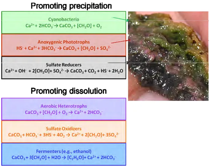
The understanding of microbial-mineral interactions is critical for interpretation of the mineral formation mechanism. In modern and historical ecosystems microbiological life is presented in the form of community, i.e. microbial mats. Microbial mats are widely regarded as the Earth’s earliest ecosystem (Tice and Lowe, 2006) and have been present on Earth since 3 billion years (Schopf, 2006). The typical arrangement of microbial groups is a vertical organized structure, where the lamination is determined by the light quantity and quality (Dupraz et al., 2009). According to the study of Dupraz et al. (2009), depending on the mat type, five to seven key groups of microorganisms can enter into the microbial mat composition: 1. photolithoautotrophs (i.e. cyanobacteria); 2. aerobic (chemoorgano-) heterotrophs; 3. fermenters; 4. anaerobic heterotrophs (predominantly sulfate-reducing bacteria); 5. sulfide oxidizers. 6. anoxyphototrophs (i.e. purple and green (non) sulfur bacteria)); 7. methanogens.
The typical microbial mat structure and combined metabolic-geochemical reactions leading to carbonate precipitation and dissolution are presented in Figure 1.
Introduction
21
Fig. 1. Metabolic-geochemical reactions in a microbial mat leading to carbonate precipitation and dissolution (modified from Dupraz et al., 2009).
In this figure the six major guilds of microorganisms that compose a typical microbial mat are arranged by their respective effects on the precipitation process. Associated equations combine metabolic and geochemical reactions (Dupraz et al., 2009). All these processes are detailed in the study of Visscher and Stolz (2005). Photosynthesis and sulfate reduction are known to increase alkalinity (promoting carbonate precipitation), whereas aerobic respiration, sulfide oxidation and fermentation are more likely to induce dissolution. When oxygen-depending metabolisms stop during the night, anaerobic heterotrophy such as sulfate reduction prevails. The net carbonate precipitation depends on the balance between the different metabolic activities as well as their temporal and spatial variations (Dupraz et al., 2009).
From the early Proterozoic and certainly since Precambrian-Cambrian boundary until the end of the Cretaceous, calcifying cyanobacteria frequently occur in normal marine
22
environments (Ries, 2010). After the end of the Cretaceous, however, they seem to be restricted to non-marine settings. At the present time, calcification by cyanobacteria occurs almost exclusively in freshwater, alkaline and hypersaline or brackish water (Merz, 1992). Although anoxygenic photosynthesis does not dominate primary production in the modern stromatolites, this metabolism may have been crucial for the growth of Archean and some Palaeoproterozoic stromatolites (Bosak et al., 2007). It has been suggested that anoxygenic photosynthesis could determine primary productivity in shallow marine environments before the rise of oxygenic photosynthesis and the widespread atmospheric oxygenation (Olson and Blankenship, 2004). Thus, biofilms formed by anoxygenic photosynthetic microorganisms would have helped building stromatolites even before cyanobacteria became the dominant primary producers in Precambrian reefs (Bosak et al., 2007).
In order to better understand the role of each group of bacteria in carbonate precipitation, the individual metabolic reactions of the guilds outlined above must be considered (Dupraz et al., 2009). Then, individual laboratory experimental study of each group must be conducted in the context of carbonate precipitation.
The aim of this thesis is to characterize the process of biological CaCO3 precipitation
and to assess the existence of metabolic processes protecting studied bacteria against carbonate mineralization on their surfaces. More precisely, this study focused on three important questions:
• What is the role of surface of studied bacterial in calcium carbonate precipitation?
• What is the role of these different bacteria in this precipitation?
• What is the mechanism of the biological carbonates mineral formation?
To address these questions, two types of bacteria were used: pure cultures of two strain of purple non-sulfur anoxygenic phototrophs bacteria (APB) and one strain of cyanobacteria
Gloeocapsa sp..These bacteria were chosen for the following reasons. They represent two
important groups of photosynthetic organisms in the past and at present time. APB is the oldest microorganism which could be dominant during the anoxygenic period of Earth’s life (approximately 4 billon years ago). The origin of oxygen evolving microorganisms (cyanobacteria) is placed at about 3.5 billion years ago as based on oxidation records of the Earth’s crust (Xiong and Bauer, 2002). In modern ecosystems cyanobacteria are the dominant primary producers. Nonetheless, the potential of APB are abundant in the modern microbial
Introduction
23
mats and stromatolites and thus may represent a considerable fraction of the standing biomass (Papineau et al. 2005). However, biomineralization induce by these bacteria has not been thoroughly studied up to now.
As there are great differences between the metabolism and diversity of APB and cyanobacteria, a detailed description of these bacteria is presented in the Chapter 1. Briefly APB are known to use a wide variety of substrates as electron donors in photosynthesis, including hydrogen, sulfide, and small organic molecules (Bosak et al., 2007). Cyanobacteria
use electrons from H2O with O2 production. This great difference between these bacteria
could be the reason for different mechanisms of carbonate precipitation.
A large variety of mechanisms were proposed to explain calcium carbonate precipitation by bacteria. They can be classified onto two groups: biologically induced and biologically controlled (Lowenstam, 1989). In the first case, the mineral precipitates because the organisms change the chemical microenvironment of the water layer adjacent to the cell. In the biologically controlled process, the role of organism goes beyond the role of merely increasing local oversaturation: the organism control the processes of mineralization via an organic matrix consisting of macromolecules that reach out from the outer cell membrane into the solution (Dittrich and Obst, 2004). As an example of the second mechanism, calcium carbonate precipitation on extracellular polymeric substances (EPS) of heterotrophic sulphur reducing bacteria was observed by Aloisi et al. (2006).
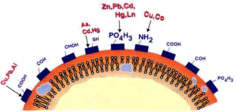
Microbial cell surface and excreted extracellular polymeric substances (EPS), which
carry a net negative electric charge and have the capacity to bind Ca2+ ions, are frequently
cited as being the sites of carbonate nucleation (Aloisi et al., 2006). The architecture of membranes differs for various cell types and with that also the specificity of the interaction between lipids and peptides\proteins. Membrane of positive as well as of Gram-negative bacteria express a high amount of Gram-negative charged lipids at the outer leaflet of the membrane which is in direct contact with the extracellular environment (Hagge et al., 2006)
thus capable of adsorbing Ca2+ ions.
Different experimental techniques were used as detailed in Chapter 2. Zeta-potential
measurements were performed under different conditions (pH, ionic strength, various [Ca2+]
and [HCO3-] concentrations, with/without light) to characterize the bacterial cell surface
charge in different solution environments and to determine the degree to which bacteria metabolically control their surface potential equilibria. These measurements were reinforced by Ca adsorption experiments as presented in Chapter 3. Moreover, kinetic experiments were
24
performed to quantify the role of bacteria on the rate of carbonate precipitation. The active biological processes were investigated by comparing kinetics of microbiological and chemical
(abiotic) CaCO3 precipitation. In chapters 4 and 5 are presented the physical and
biochemical evidences of bacterial mineral precipitation, in which microbial metabolic activities (photosynthesis) plays an important role. Sanning Electron Microscopy and X-ray diffraction have been used to characterize the nature and the forms of the precipitated minerals, whereas Transmission Electron Microscopy (TEM) has been used to localize the microbial precipitation of carbonates on/or near the surface of bacterial cells. This technique helps to answer the third question and understands the mechanism of precipitation driven by different type of bacteria. The results and interpretation are presented in Chapter 4 and 5.
Overall, the results of this study can help to answer important questions about i)
carbonate formations in past (paleontological context) and present time (context of CO2
storage and biogeochemical cycle); ii) evolution of microbial life in the earliest periods of biological history (biological context); and iii) bacterial ecosystems formations under contemporary land surface conditions (ecological context).
Introduction
Chapter
I
Description
of
Anoxygenic
phototrophic bacteria and
Cyanobacteria
Description of APB and Cyanobacteria
29
Plants, algae, and cyanobacteria are known as oxygenic photoautotrophs because they synthesize organic molecules from inorganic materials, convert light energy into chemical energy, use water as an electron source, and generate oxygen as an end product of photosynthesis. Unlike the oxygenic plants, algae, and cyanobacteria, anoxygenic phototrophs do not use water as an electron source and, therefore, do not produce oxygen during photosynthesis. The electrons come from compounds such as hydrogen gas, hydrogen sulfide, and reduced organic molecules. This chapter will give a short introduction into the diversity of anoxygenic phototrophic bacteria (APB) and of cyanobacteria, list some important properties of the species, and indicate important physiological features. Two strains of anoxygenic phototrophs (purple nonsulfur anoxygenic phototrophic bacteria) and oxygenic phototrophs cyanobacteria Gloeocapsa sp. were studied in this work.
1.
Anoxygenic phototrophic bacteria (APB)
Anoxygenic phototrophic bacteria have always attracted scientists because of their position at the beginning of life evolution and their ability to perform photosynthesis in the absence of air and without producing oxygen. Despite the common feature of these bacteria, there are significant variations in their morphological, physiological and molecular properties, including molecular structures of the photosynthetic pigments and the photosynthetic apparatus (Blankenship et al, 2004).
The literature contains significant information on the geochemical activity, physiology and distribution of APB in earth surface environments. Places of mass dwelling of APB usually divide into three types: thermal sources, shallow water reservoirs (salty and fresh), the stratified water body such as lakes reservoirs. However, in small amounts APB are also present in practically all aquatic systems and flooded soils (Gorlenko, 1977).
30
1.1. Anoxygenic photosynthesis
Anoxygenic photosynthetic bacteria differ from oxygenic organisms in that each species has only one type of reaction center (photosynthetic reaction center is a complex of several proteins, pigments and other co-factors assembled together to execute the primary energy conversion reactions of photosynthesis) (Blankenship et al., 1995). In some photosynthetic bacteria the reaction center is similar to photosystem II and in others it is similar to photosystem I (Fig. 1). However, neither of these two types of bacterial reaction
center is capable of extracting electrons from water, so they do not evolve O2. Many species
can only survive in environments that have a low concentration of O2. To provide electrons
for the reduction of CO2, anoxygenic photosynthetic bacteria must oxidize inorganic or
organic molecules available in their environment. For example, the purple bacterium
Rhodobacter sphaeroides can use succinate to reduce NAD+ by a membrane-linked reverse electron transfer that is driven by a transmembrane electrochemical potential. Although many
photosynthetic bacteria depend on Rubisco and the Calvin cycle for the reduction of CO2,
some are able to fix atmospheric CO2 by other biochemical pathways (Huzisige and Ke,
1993).
Despite these differences, the general principles of energy transduction are the same in anoxygenic and oxygenic photosynthesis. Anoxygenic photosynthetic bacteria contain bacteriochlorophyll, a family of molecules that are similar to the chlorophyll, that absorb strongly in the infrared region between 700 and 1000 nm. The antenna system consists of bacteriochlorophyll and carotenoids that serve as reaction center where primary charge separation occurs (Blankenship et al., 1994). The electron carriers include quinone (e.g., ubiquinone, menaquinone) and the cytochrome bc complex, which is similar to the cytochrome bf complex of oxygenic photosynthetic apparatus. As in oxygenic photosynthesis, electron transfer is coupled to the generation of an electrochemical potential that drives
phosphorylation by ATP synthase and the energy required for the reduction of CO2 is
Description of APB and Cyanobacteria
31
Fig. I. 1. Schematic of photosystems I and II.
1.2. Diversity of Anoxygenic Phototrophic Bacteria
There are 7 groups of APB: filamentous anoxygenic phototrophs (Fig. 2A), phototrophic purple nonsulfur bacteria (Fig. 2B), green sulfur bacteria (Fig. 2C), phototrophic sulfur bacteria, heliobacteria (Fig. 2D), thermophilic anoxygenic phototrophs, aerobic anoxygenic phototrophs (Blankenship et al, 2004). To illustrate the diversity of APB we will consider some the most interesting examples.
Filamentous anoxygenic phototrophs are a diverse group of photosynthetic bacteria that are of particular evolutionary significance. The best known species is the thermophilic
Chloroflexus aurantiacus (Fig. 2A). This organism is a prominent member of hot spring
microbial mat communities. Because it has an interesting combination of characteristics found in very different and diverse groups of phototrophic prokaryotes, it is of particular significance in addressing questions of evolutionary importance (Frigaard and Bryant, 2004).
There are several other strains of filamentous photosynthetic bacteria from a wide range of environments that are substantially different from Cf. aurantiacus, yet have enough similarity in fundamental photosynthetic features to be likely relatives. Sequence data (16S rRNA) are needed to define the phylogenetic range of the family Chloroflexaceae.
32
Fig. I. 2. Anoxygenic phototrophic bacteria: A. Chloroflexus (green non-sulfur bacteria, Chloroflexaceae), B. Rhodospirillum (purple non-sulfur bacteria, Rhodospirillaceae), C. Chlorobium (green sulfur bacteria, Chlorobiaceae), D.
Heliobacterium (Gram-positive, Heliobacteriaceae).
The physiology of Cf. aurantiacus is intriguing in several aspects. The recently
described autotrophic CO2 fixation pathway involving 3-hydroxypropionate is unlike any
other known autotrophic mechanism. C. aurantiacus is also unique among all groups of phototrophs in lacking the capacity for nitrogen fixation. The regulation of pigment synthesis in response to changing growth conditions is particularly interesting due to the presence of two different photosynthetic pigments located in different sub-cellular environments. The fact that Cf. aurantiacus is a thermophile provides another dimension of complexity to its physiology. It is also quite resistant to UV radiation. Some of its characteristics may be relicts from Precambrian ancestors (Blankenship et al, 2004).
Other interesting example of APB is heliobacteria (Fig. 2D). Heliobacteria are anoxygenic phototrophs that contain bacteriochlorophyll g as their sole chlorophyll pigment. These organisms are primarily soil residents and are phylogenetically related to Gram-positive bacteria, in particular to the endospore-forming Bacillus/Clostridium line. Some species of
heliobacteria produce heat resistant endospores containing dipicolinic acid and elevated Ca2+
levels. Heliobacteria can grow photoheterotrophically on a limited group of organic substrates and chemotrophically (anaerobically) in darkness by pyruvate or lactate fermentation. They are also active nitrogen-fixers. Their photosynthetic system resembles that of photosystem I of green plants but is simpler, containing a small antenna closely associated with the reaction center located in the cytoplasmic membrane; no chlorosomes; typical of the green sulfur bacteria; or differentiated internal membranes, typical of purple bacteria, are found in the heliobacteria. These bacteria are apparently widely distributed in rice soils and occasionally
Description of APB and Cyanobacteria
33
found in other soils. The ecology of heliobacteria may be tightly linked to that of rice plants, and the ability of heliobacteria to produce endospores probably has significant survival value in the highly variable habitat of rice soils. The unique assemblage of properties shown by the heliobacteria has necessitated creation of a new taxonomic family of anoxygenic phototrophic bacteria, the Heliobacteriaceae, to accommodate organisms of this type (Madigan and Ormerod, 1995)
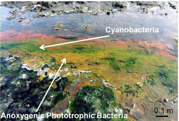
It is apparent that very few species of anoxygenic phototrophs occur or grow at high temperatures, particularly when compared to species numbers for thermophilic Archaea and non-photosynthetic Bacteria. Chloroflexus spp. are the most thermotolerant (up to ~70 °C) (Fig. 3), but none of the APB are in the hyperthermophilic category. Recognizing that there may be some endemic populations of anoxygenic phototrophic bacteria that have not been dispersed among geographically disparate geothermal sites, the major factors affecting the distribution of these bacteria are temperature, pH, and concentration of sulfide. Oxygen may have an effect on the vertical distribution and the diel vertical migration of some species within mats. Facultative aerobic metabolism appears to be a property of many of the anoxygenic phototrophs (but not Chlorobium or Heliobacillus) in these dynamic habitats. Light quantity and quality are affected by the diversity of pigmentation within the vertically stratified communities and adaptation to low photon fluence rates is a necessity for many species (Castenholz and Pierson, 2004).
Phototrophic sulfur bacteria are characterized by oxidizing various inorganic sulfur compounds for use as electron donors in carbon dioxide fixation during anoxygenic photosynthetic growth. These bacteria are divided into the purple sulfur bacteria (PSB) and the green sulfur bacteria (GSB). They utilize various combinations of sulfide, elemental sulfur, and thiosulfate and sometimes also ferrous iron and hydrogen as electron donors (Frigaard and Dahl, 2009). Phototrophic sulfur bacteria often form mass developments in aquatic environments, either planktonic or benthic, where anoxic layers containing reduced sulfur compounds are exposed to light. From the point of view of population dynamics, the abundance of these organisms is the consequence of a certain balance between growth and losses. Both specific growth rates and specific rates of loss through several processes are analyzed in several environments, in an attempt to generalize on the growth status of blooms of phototrophic sulfur bacteria. The available information indicates the existence of an upper limit for the production of these bacteria in nature, and seems to suggest the existence of an upper limit for biomass based in the balance between growth and losses (Hell et al., 2008)
34
Fig. I. 3. APB and cyanobacteria in the hot spring (40-60°C), Kamchatka, Russia.
During the last 15 years, more than 20 new strains of aerobic anoxygenic phototrophs bacteria which possess bacteriochlorophyll (BChl) a have been identified (Blankenship et al, 2004). They are distinguished from typical anaerobic (anoxygenic) phototrophs in that they
synthesize BChl only under aerobic conditions and cannot grow without O2 or other oxidants,
even under light. In some species, photosynthetic activities have been demonstrated. Reaction centers and light-harvesting complexes isolated from some species were shown to be similar to those of typical purple photosynthetic bacteria. The regulatory mechanism of synthesis of pigments and proteins of the photosynthetic apparatus are apparently opposite with respect to
O2 pressure compare to that of typical anoxygenic phototrophs. The low content of BChl,
unique composition of carotenoids and presence of non-photosynthetic carotenoids in most strains are other marked characteristics of these aerobic bacteria. Phylogenetically, they are not classified into single group. Species so far described are distributed rather widely within the α-subclass of Proteobacteria (purple bacteria) in which most of the purple nonsulfur bacteria as well as many non-photosynthetic bacteria are included. Apparently, these aerobic BChl-containing bacteria represent an evolutionary transient phase from anaerobic phototrophs to aerobic non-phototrophs. However, some characteristic features distinct from
Description of APB and Cyanobacteria
35
anaerobic phototrophs suggest that most of them are in an evolutionary stable state (Yurkov and Beatty, 1998).
Our experimental study of calcium carbonate biomineralization during bacterial activity deal with two strain of purple nonsulfur APB: Rhodovulum steppense sp. nov. A-20s and Rhodovulum sp. S-17-65.
1.2.1. Rhodovulum steppense sp. nov. A-20s
Rhodovulum steppense (step.pen'se, N.L. n. steppum steppe; L.neut. suff. -ense suffix
used with the sense of pertaining to; N.L. neut. adj. steppense pertaining to the steppe,
widespreadin steppe soda lakes). The type strain, A-20sT (=VKM B-2489T =DSM 21153T).
The 16S rRNA gene sequences of A-20s strain were deposited in GenBank under the
following accession numbers: EU741680–EU741684, EU918391, FJ895099 (Kompantseva et
al., 2009).
Strains of purple nonsulfur bacteria Rhodovulum steppense sp. nov. A-20s were
isolated from the shallow-water steppe soda lake Khilganta (Zabaikal'skii Krai, southern
Siberia,Russia) (Fig.4A) in the cryoarid zone of CentralAsia (Kompantseva et al., 2010).
Fig. I. 4. (A) Lake Khilganta, Zabaikal'skii Krai, southern Siberia, Russia; (B) Hypersaline water body in Crimea steppe.
Cells are ovoid to rod-shaped, 0.3–0.8 µm wideand 1.0–2.5 µm long (Fig. 5A), mobile
by means of polar flagella, multiply by binary fission, and contain vesicular internal
photosynthetic membranes. Ultrathin sections of cells of strain A-20s showed a
Gram-negative type of cell wall. The internal photosynthetic membranes formedmultiple vesicles
36
Fig. I. 5. (A,B) TEM images of APB A-20s. Nutrient solution. 10 days old culture. (B) Vesicles in the internal photosynthetic membranes of APB A-20s.
Cell suspensions are yellow to brown in anoxic conditions (Fig. 6A) and red in the
presence of O2 when grown aerobically (Fig. 6B).
Fig. I. 6. Bacterial suspensions of A-20s cultivated in anoxic conditions (brown color) (A) and in the presents of oxygen (red color) (B).
The photosynthetic pigments of this APB strain are bacteriochlorophyll a and carotenoids of the spheroidene series (Kompantseva et al., 2009). In vivo absorption spectra show maxima at 461, 483, 515, 592, 803 and 861 nm. Growth occurs anaerobically in the light (photoheterotrophically) or aerobically in the dark (chemoheterotrophically). Growth is
Description of APB and Cyanobacteria
37
best with propionate, butyrate, valerate, pyruvate, lactate and glycerol. Asparagine,acetate,
caprylate, malate, succinate, formate, fumarate, glutamate, mannose, ethanol, casein
hydrolysate and yeast extract are also used. Alanine, arginine, aspartate, ornithine, proline,
threonine,valine, citrate, benzoate, tartrate, arabinose, glucose, maltoseand fructose support
slow growth. No growth occurs with sucrose, sorbitol, mannitol or methanol. No anaerobic
respiration takesplace with nitrite, nitrate or fumarate. Growth at the expenseof fermentation
does not occur. Sulfide, sulfur and thiosulfate support photolithoautotrophic and
chemolithoautotrophic growth,in the course of which they are oxidized to sulfate. Sulfideas
electron donor is oxidized via temporary deposition of extracellular elemental sulfur. No
growth occurs with hydrogen as electron donor. Ammonium salts are used as the nitrogen
source. Sulfate,thiosulfate, elemental sulfur and cysteine are used as the sulfursource. Three
vitamins (biotin, thiamine, niacin) are neededas growth factors. Obligately haloalkaliphilic:
growth occurswithin a wide range of salinity (0.3–10 %, w/v) and pH(7.5–10) (Kompantseva
et al., 2010). Growth optima are at 1–5 % NaCl, pH 8.5and 25–35 °C. No growth occurs in
the absence of NaCland/or at pH 7. The major quinone is ubiquinone Q-10. This APB strain
is resistant to amikacin, ampicillin, chlortetracycline, bacitracin, vancomycin, gentamicin,
nystatin,nalidixic acid, novobiocin, penicillin, rifampicin, kanamycin,lincomycin, neomycin
and streptomycin, but sensitive to tetracycline,benzylpenicillin and polymixin B.
Anaerobically in the light and in the presence of organic compounds,the strain A-20s
reduced selenite and tellurite (at initial concentrationsof up to 10 mM) to elemental selenium
and tellurium, respectively (Kompantseva et al., 2010).
1.2.2. Rhodovulum sp. S-17-65
Bacteria Rhodovulum sp. S-17-65 were isolated a long time ago and originally described as Rhodobacter euryhalinus, renamed later in Rhodovulum euryhalinum. In light of new data this species includes only typical strain, but not Rhodovulum sp. S-17-65. Thereby, Rhodovulum sp. S-17-65 is likely to be another species, which have been not described yet. In this subsection we provide the old description of bacteria S-17-65.
Rhodobacter euryhalinus (Gr. adj. eurus, wide, broad; Gr. n. hals halos, salt; L. suff. -inus -a -um, suffix used with the sense of belonging to; N.L. neut. adj. euryhalinum, living in
a wide range of salinity). Type strain: strain Kompantseva KA-65 = KA-65 = DSM 4868. GenBank/EMBL/DDBJ accession number for the 16S rRNA gene sequence of the type strain: D16426 (Kompantseva, 1989).
38
Fig. I. 7. TEM images of cells S-17-65. Nutrient solution. 10 days old culture.
Strains of purple nonsulfur bacteria Rhodovulum sp. S-17-65 isolated from the hypersaline water body in Crimea steppe (Fig. 4B). It was shown that the main factors determining efficiency of growth were: increase salt content, sulphide content in silt deposits, unstable character of anaerobic conditions in bottom water (Gorlenko et al., 1983).
The form of cells is coccus or short rod-shaped, the size is 0.7-0.1.0 µm wideand 1.0–
3.0 µm long (Fig. 7), the cells are mobile by means of polar flagella, multiply by binary fission. Cell suspensions are brown as shown in Fig. 8.
Fig. I. 8. Cells suspension of APB S-17-65.
The photosynthetic pigments are bacteriochlorophyll a and carotenoids of the
Description of APB and Cyanobacteria
39
microaerophilically in the dark on organic substrate. The cultures can growth
photoototrophicaly with sulphide. S-17-65 is obligately halophilic with growth optima of 12
% NaCl, pH 7-8 and 25–35 °C. The source of nitrogen is ammonium, source of sulfur are
sulfide, cysteine. These bacteria need four vitamins: B1, B5, biotin, n-aminobenzoate
(Kompantceva, 1985).
2.
Cyanobacteria
A cyanobacterium (also known as blue-green algae, blue-green bacteria, and Cyanophyta) is a phylum of bacteria that obtain their energy through photosynthesis. The name "cyanobacteria" comes from the color of the bacteria (Greek: κυανός (kyanós) = blue).
The ability of cyanobacteria to perform oxygenic photosynthesis is thought to have converted the early reducing atmosphere into an oxidizing one, which dramatically changed the composition of life forms on Earth by stimulating biodiversity and leading to the near-extinction of oxygen-intolerant organisms (Smith, 1982).
Cyanobacteria can be found in almost every conceivable environment, from oceans and fresh water to bare rock and soil. They can occur as planktonic cells or form phototrophic biofilms in fresh water and marine environments. They occur in damp soil or even on temporarily moistened rocks in deserts. A few are endosymbionts in lichens, plants, various protists, or sponges and provide energy for the host. Some live in the fur of sloths, providing a form of camouflage (Madigan et al., 2000).
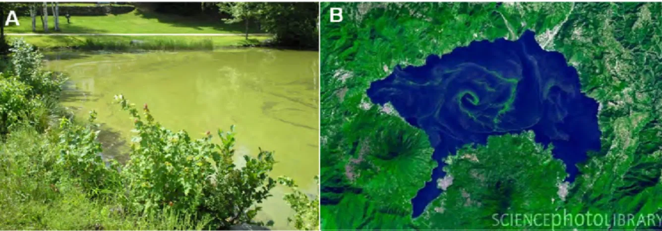
Aquatic cyanobacteria are probably best known for the extensive and highly visible blooms that can form in both freshwater and the marine environment and can have the appearance of blue-green paint or scum (Fig. 9). The association of toxicity with such blooms has frequently led to the closure of recreational waters when blooms are observed. Marine bacteriophages are a significant parasite of unicellular marine cyanobacteria. When they infect cells, they lyse them, releasing more phages into the water (Rippka et al., 1979).
40
Fig. I. 9. Blooms of cyanobacteria. (A) Lake in New England; (B) Lake Atitlán, Guatamala highlands, satellite image, November 22, 2009.
2.1. Oxygenic Photosynthesis of Bacteria
The photosynthetic process in all plants and algae as well as in certain types of
photosynthetic bacteria involves the reduction of CO2 to carbohydrate and removal of
electrons from H2O, which results in the release of O2. In this process, known as oxygenic
photosynthesis, water is oxidized by the photosystem II reaction center, a multisubunit protein located in the photosynthetic membrane. Years of research have shown that the structure and function of photosystem II is similar in plants, algae and certain bacteria, so that knowledge gained in one species can be applied to others. This homology is a common feature of proteins that perform the same reaction in different species. This homology at the molecular level is important because there are estimated to be 300,000-500,000 species of plants (Huzisige and Ke, 1993).
Cyanobacteria are photosynthetic prokaryotic organisms that evolve O2 (Bryant,
1994). Fossil evidence indicates that cyanobacteria existed over 3 billion years ago and it is thought that they were the first oxygen evolving organisms on earth (Wilmotte, 1994).
Cyanobacteria are presumed to have evolved in water in an atmosphere that lacked O2.
Initially, the O2 released by cyanobacteria reacted with ferrous iron in the oceans and was not
released into the atmosphere. Geological evidence indicates that the ferrous Fe was depleted
around 2 billion years ago, and earth's atmosphere became aerobic. The release of O2 into the
Description of APB and Cyanobacteria
41
The photosynthetic apparatus of cyanobacteria is similar to that of chloroplasts. The main difference is in the antenna system. Cyanobacteria depend on chlorophyll a and specialized protein complexes (phycobilisomes) to gather light energy (Sidler, 1994). They do not contain chlorophyll b. As in chloroplasts, the chlorophyll a is located in membrane bound proteins. The phycobilisomes are bound to the outer side of the photosynthetic membrane and act to funnel exciton energy to the photosystem II reaction center. They are composed of phycobiliproteins, protein subunits that contain covalently attached open ring structures known as bilins that are the light absorbing pigments. Primary photochemistry, electron transport, phosphorylation and carbon reduction occur much as they do in chloroplasts. Cyanobacteria have a simpler genetic system than plants and algae that enable them to be easily modified genetically. Because of this, cyanobacteria have been used as a model to understand photosynthesis in plants. By genetically altering photosynthetic proteins, researchers can investigate the relationship between molecular structure and mechanism (Barry et al., 1994).
Over the past three decades several types of oxygenic bacteria known as prochlorophytes (or oxychlorobacteria) have been discovered that have light harvesting protein complexes that contain chlorophyll a and b, but do not contain phycobilisomes (Palenik and Haselkorn 1992, Urbach et al., 1992; Matthijs et al., 1994). Because prochlorophytes have Chlorophyll a/b light harvesting proteins like chloroplasts, they are being investigated as models for plant photosynthesis.
2.2. Diversity of cyanobacteria
The cyanobacteria were traditionally classified by morphology into five sections, referred to by the numerals I-V. The first three - Chroococcales, Pleurocapsales, and Oscillatoriales - are not supported by phylogenetic studies. However, the latter two - Nostocales and Stigonematales - are monophyletic, and make up the heterocystous cyanobacteria. The members of Chroococales are unicellular and usually aggregate in colonies. The classic taxonomic criterion has been the cell morphology and the plane of cell division. In Pleurocapsales, the cells have the ability to form internal spores (baeocytes). The rest of the sections include filamentous species. In Oscillatoriales, the cells are uniseriately arranged and do not form specialized cells (akinetes and heterocysts). In Nostocales and
42
Stigonematales the cells have the ability to develop heterocysts in certain conditions. Stigonematales, unlike Nostocales, includes species with truly branched trichomes (Smith, 1982).
Cyanobacteria include unicellular and colonial species. Colonies may form filaments, sheets or even hollow balls. Some filamentous colonies show the ability to differentiate into several different cell types: vegetative cells, the normal, photosynthetic cells that are formed under favorable growing conditions; akinetes, the climate-resistant spores that may form when environmental conditions become harsh; and thick-walled heterocysts, which contain the enzyme nitrogenase, vital for nitrogen fixation. Heterocysts may also form under the appropriate environmental conditions (anoxic) when fixed nitrogen is scarce. Heterocyst-forming species are specialized for nitrogen fixation and are able to fix nitrogen gas into
ammonia (NH3), nitrites (NO2−) or nitrates (NO3−) which can be absorbed by plants and
converted to protein and nucleic acids (atmospheric nitrogen cannot be used by plants directly). Rice crops utilize healthy populations of nitrogen-fixing cyanobacteria in some rice paddy fertilizers (Scanlan and Nyree, 2002). Rippka (Rippka et al., 1979) divides the cyanobacteria into five sections. She describes first two sections, I and II, as "Unicellular; cells single or forming colonial aggregates held together by additional outer cell wall layers". Her other three sections, III to V, she describes as "Filamentous; a trichome (chain of cells) which grows by intercalary cell division". The paper by Rippka et al. (1979) contains many micrographs of cell morphologies typical of these genera.
Anabaena (Fig. 10) is a genus of filamentous cyanobacteria, found as plankton. It is
known for its nitrogen fixing abilities, and they form symbiotic relationships with certain plants, such as the mosquito fern. They are one of four genera of cyanobacteria that produce neurotoxins, which are harmful to local wildlife, as well as farm animals and pets. Production of these neurotoxins is assumed to be an input into its symbiotic relationships, protecting the plant from grazing pressure (Mishra et al. 2009).
Oscillatoria (Fig.11) is another genus of filamentous cyanobacterium which is named
for the oscillation in its movement. Filaments in the colonies can slide back and forth against each other until the whole mass is reoriented to its light source. It is commonly found in watering-troughs waters, and is mainly blue-green or brown-green. Oscillatoria is an organism that reproduces by fragmentation. Oscillatoria forms long filaments of cells which can break into fragments called hormogonia. The hormogonia can grow into a new, longer filament. Breaks in the filament usually occur where dead cells (necridia) are present. Each
Description of APB and Cyanobacteria
43
filament of oscillatoria consists of trichome which is made up of rows of cells. The tip of the trichome oscillates like a pendulum (Madigan et al. 2000).
Fig. I. 10. Cyanobacteria Anabaena (Scientific classification: Kingdom: Bacteria, Phylum: Cyanobacteria, Order: Nostocales, Family: Nostocaceae, Genus: Anabaena).
Fig. I. 11. Cyanobacteria Oscillatoria (Scientific classification: Kingdom: Bacteria, Phylum: Cyanobacteria, Class: Cyanophyceae, Order: Oscillatoriales, Family:
44
Some of cyanobacterial organisms contribute significantly to global ecology and the oxygen cycle. Although Prochlorococcus (Fig.12) is the smallest known phototroph it contributes 30-80% of primary production in the world's oligotrophic oceans, and is consequently plays a significant role in the global carbon cycle and the Earth's climate. This tiny marine unicellular cyanobacterium was discovered in 1986 and accounts for more than half of the photosynthesis of the open ocean (Scanlan et al. 2002).
Fig. I. 12. Cynobacteria Prochlorococcus (Scientific classification: Kingdom: Bacteria, Phylum: Cyanobacteria, Order: Synechococcales, Family: Synechococcaceae,
Genus: Prochlorococcus)
Each individual cell of a cyanobacterium typically has a thick, gelatinous cell wall. They lack flagella, but some species may move by gliding along surfaces. Many of the multi-cellular filamentous forms of Oscillatoria are capable of a waving motion; the filament oscillates back and forth. In water columns some cyanobacteria float by forming gas vesicles, like in archaea. These vesicles are not organelles as such. They are not bounded by lipid membranes but by a protein sheath (Smith, 1982).
In our experimental study of calcium carbonate biomineralization cyanobacteria
Gloeocapsa sp. was studied in details for determinate its role in precipitation and the
Description of APB and Cyanobacteria
45
2.2.1. Cyanobacteria Gloeocapsa sp.
Scientific classification: Kingdom: Bacteria, Class/Phylum: Cyanobacteria, Order: Chroococcales, Family: Chroococcaceae.
Gloeocapsa (from the Greek gloia (glue) and the Latin capsa (box)) may be
unicellular or made up of small groups of cells grouped together within concentric mucilage envelopes (Fig. 13). The individual colonies are usually spherical, microscopic, and enclosed within larger masses of mucilage. The cells are oval-shaped or ellipsoidal, and hemispherical after dividing. Each cell has a rounded, firm, inner mucilaginous sheath surrounded by older sheath material from the parent cell, revealing the pattern of cell division. The sheaths are colorless or vivid shades of yellow, brown, red, orange, blue, or violet, and may be affected by changes in pH. The cells are usually bright blue-green or olive green and do not have distinct gas vesicles (Smith, 1982).
Gloeocapsa cells divide along three perpendicular planes during successive
generations. Each daughter cell grows to the size and shape of the parent cell before dividing again. The parent colonies disintegrate to form new daughter colonies (Madigan, 2000).
Fig. I. 13. Cyanobacteria Gloeocapsa (cells surrounded by a capsule). The black scale bars are 10 µm.
Most species of Gloeocapsa live in freshwater lakes, on wet stony substrates, on tree bark, or in terrestrial environments such as moist soils. Many grow on wet rocks or mountain walls, while some species are restricted to calcareous or acidic rocky surfaces. Some live
46
within porous rocks in hot desert regions or can be seen as black bands on high intertidal seacoast rocks. Some species of Gloeocapsa are symbiotic with fungi, forming lichens (Smith, 1982).
Cyanobacterium Gloeocapsa sp. used in this study is usually made up of small number of cells grouped within concentric mucilage envelopes. The individual colonies are spherical, microscopic, and enclosed within larger masses of mucilage. Gloeocapsa sp. f-6gl was provided from the culture collection of the Institute of Microbiology RAS (Moscow), isolated from a hot spring (30-40°C) in Kamchatka (Pokrovsky et al., 2008).
2.2.2. Cyanobacteria Synecochoccus sp.
Scientific classification: Kingdom: Bacteria, Phylum: Cyanobacteria, Order: Synechococcales, Family: Synechococcaceae, Genus: Synechococcus.
Synechococcus (from the Greek synechos (in succession) and the Greek kokkos
(berry)) is a unicellular cyanobacterium that is very widespread in the marine environment. Many freshwater species of Synechococcus have also been described (Madigan et al., 2000).
A Synechococcus cyanobacterium was isolated from the surface of coastal stromatolites sampled in February 2008 from the depth of 1 m at 50 m from the shoreline of the Salda Lake, SW Turkey (Fig. 14).
Description of APB and Cyanobacteria
47
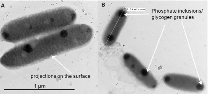
The cultures of Synechococcus sp. were purified on the agar BG-11 or Pratt media and individual colonies was grown in synthetic, cyanobacteria BG-11 Freshwater Solution for 3 weeks until the stationary growth phase was reached. Continuous illumination at 2000 lx was provided from fluorescent lamps. Cyanobacteria Synechococcus typically consists of isolated elongated cells, without significant amount of mucilage. Its size varies from 0.8 µm to 1.5 µm (Fig. 15). They are gram negative cells with highly structured cell walls that may contain projections on their surface. Electron microscopy frequently reveals the presence of phosphate inclusions, glycogen granules and more importantly highly structured carboxysomes (Smith, 1982) (Fig.15).
Fig. I. 15. TEM images of cyanobacteria Synechococcus sp. (A) Projections on their surface; (B) Phosphate inclusions / glycogen granules.
Laboratory modeling experiments with cyanobacteria Synechococcus sp. were performed in order to characterize hydrous magnesium carbonate precipitation in alkaline lake (stromatolites formation) and to assess the range and characterize the mechanisms of Mg isotope fractionation in lacustrine environment. The results of this study are described in the articles Mavromatis et al. (2011) and Shirokova et al. (2011). These works are presented in Appendices 1 and 2.