HAL Id: dumas-01782595
https://dumas.ccsd.cnrs.fr/dumas-01782595
Submitted on 7 Jun 2018HAL is a multi-disciplinary open access
archive for the deposit and dissemination of
sci-L’archive ouverte pluridisciplinaire HAL, est destinée au dépôt et à la diffusion de documents
Large granular lymphocyte leukemia and pulmonary
hypertension : an unusual association
Brieuc Chérel
To cite this version:
Brieuc Chérel. Large granular lymphocyte leukemia and pulmonary hypertension : an unusual asso-ciation. Life Sciences [q-bio]. 2017. �dumas-01782595�
N° d'ordre : ANNÉE 2017
THÈSE D'EXERCICE / UNIVERSITÉ DE RENNES 1
sous le sceau de l’Université Bretagne Loire
Thèse en vue du
DIPLÔME D'ÉTAT DE DOCTEUR EN MÉDECINE
présentée par
Brieuc CHEREL
Né le 01/02/1988 à Ploërmel
Leucémie à grands
lymphocytes
granuleux et
hypertension
pulmonaire : une
association rare
Thèse soutenue à Rennes le 25/10/2017
devant le jury composé de :
Marc HUMBERT
Professeur – APHP - Hôpital de Bicêtre /
rapporteur
Céline CHABANNE
Praticienne hospitalière – CHU Rennes /
rapporteur
Stéphane JOUNEAU
Professeur – CHU Rennes / examinateur
Mohamed HAMIDOU
Professeur – CHU Nantes / examinateur
Pascal GODMER
Praticien hospitalier – CH Vannes / examinateur
Olivier DECAUX
Professeur - CHU Rennes / président du jury
Thierry LAMY
N° d'ordre : ANNÉE 2017
THÈSE D'EXERCICE / UNIVERSITÉ DE RENNES 1
sous le sceau de l’Université Bretagne Loire
Thèse en vue du
DIPLÔME D'ÉTAT DE DOCTEUR EN MÉDECINE
présentée par
Brieuc CHEREL
Né le 01/02/1988 à Ploërmel
Leucémie à grands
lymphocytes
granuleux et
hypertension
pulmonaire : une
association rare
Thèse soutenue à Rennes le 25/10/2017
devant le jury composé de :
Marc HUMBERT
Professeur – APHP - Hôpital de Bicêtre / rapporteur
Céline CHABANNE
Praticienne hospitalière – CHU Rennes / rapporteur
Stéphane JOUNEAU
Professeur – CHU Rennes / examinateur
Mohamed HAMIDOU
Professeur – CHU Nantes / examinateur
Pascal GODMER
Remerciements
Monsieur le Professeur Olivier DECAUX, je vous remercie de m’avoir fait l’honneur d’accepter de présider ce jury. Merci de vos nombreux conseils, de votre disponibilité et de votre accessibilité. J’en profite également pour vous remercier pour votre aide et votre soutien pour le CORECT.
Monsieur le Professeur Marc HUMBERT, je vous remercie d’avoir accepté de juger mon travail. Votre présence est pour moi un très grand honneur.
Monsieur le Professeur Mohamed HAMIDOU, je vous remercie de m’avoir fait l’honneur d’accepter de juger mon travail. Merci de votre disponibilité et de votre accueil lors de mon passage à Nantes.
Monsieur le Professeur Stéphane JOUNEAU, je vous remercie de m’avoir fait l’honneur d’accepter de juger mon travail.
Madame le Docteur Céline CHABANNE, je vous remercie de votre disponibilité et de votre précieuse aide tout au long de ce travail. Je suis honoré que vous ayez accepté de juger mon travail.
Monsieur le Docteur Pascal GODMER, je vous remercie de m’avoir fait l’honneur d’accepter de juger mon travail. Je me réjouis de rejoindre votre équipe d’ici peu.
Monsieur le Professeur Thierry LAMY, je vous remercie de m’avoir fait l’honneur de me confier ce sujet d’étude concluant l’apprentissage de cette spécialité passionnante. Je vous remercie pour votre pédagogie au cours des stages en hématologie. Merci de votre précieux soutien et de vos relectures attentives. Veuillez trouver ici l’expression de mon plus profond respect.
Messieurs les Docteur LIFERMANN, Professeur ZAMBELLO, Docteur LEOCIN, Professeur LOUGHRAN Jr, Docteur LEBLANC et Professeur MONTANI, je vous remercie de votre aide pour ce travail.
Merci à mes parents, à mes frères (IRL ou non) pour leur soutien et leur patience pendant ces nombreuses années d’études.
Merci Marie pour ton aide et tes relectures précieuses.
Merci à mes amis notamment aux enfants du 2Pavie et à Jérôme et Rémi, j’ai hâte de vous retrouver prochainement dans les mêmes circonstances pour vous.
Merci Laure pour ton soutien durant ce dernier semestre et tes remarques avisées lors de tes relectures. Merci à mes co-internes, aux praticiens hospitaliers, aux chefs de clinique ainsi qu’aux équipes paramédicales du service. Cela a été un plaisir de travailler et de m’enrichir à vos côtés durant ces années d’internat.
10
Table des matières
Titre………...11
Résumé Français………..12
Résumé Anglais………...13
Abréviations………14
Introduction.………15
Matériels et Méthodes……….16
Résultats………..17
Discussion………...18
Bibliographie………...21
Appendice………25
Tableau 1.………...26
Tableau 2.………...27
Tableau 3.………...28
Figure 1....………..29
Figure 2………..30
Leucémie à grands lymphocytes granuleux et hypertension
pulmonaire : une association rare
Large
granular
lymphocyte
leukemia
and
pulmonary
hypertension: an unusual association
Brieuc Cherel1, Renato Zambello2, Matteo Leoncin2, François Lifermann3, Thomas P.
Loughran Jr4, Francis LeBlanc4, Mohamed Hamidou5, Marc Humbert6, David Montani6, Olivier Decaux7, Cedric Pastoret8, Céline Chabanne9 and Thierry Lamy1, 10.
1Departement of Hematology, Pontchaillou University Hospital, Rennes, France
2Departement of Medicine Hematology and Clinical Immunology, Padua School of Medicine,
Padova, Italy
3Departement of Internal Medicine, Dax Hospital, France 4University of Virginia Cancer Center, Charlottesville, VA, US
5Departement of Internal Medicine, Hotel-Dieu University Hospital, Nantes, France
6Reference Center of pulmonary arterial hypertension, INSERM U999, Bicêtre Hospital
AP-HP, Paris, France
7Departement of Internal Medicine, Hopital Sud University Hospital, Rennes, France 8Laboratory of Hematology, Pontchaillou University Hospital, Rennes, France
9Departement of Cardiology and vascular diseases, Pontchaillou University Hospital, Rennes,
France
12
Résumé Français
La leucémie à grands lymphocytes granuleux (LGL) est un syndrome lymphoprolifératif rare caractérisé par une expansion de cellules T/NK associée à des cytopénies et d’éventuelles pathologies auto-immunes. Cette étude internationale rétrospective présente 9 cas de patients atteints d’une hypertension pulmonaire prouvée associée à une leucémie LGL. Sur les 5 patients réévalués par cathétérisme cardiaque droit, 4 présentent une amélioration de l’hypertension pulmonaire sous traitement immunosuppresseur spécifique de la leucémie LGL. Avec un suivi médian de 88 mois, tous les patients sont vivants. Les mécanismes pathogéniques liant les deux pathologies sont discutés. Cette étude suggère de compléter le bilan initial d’une HTAP par la recherche d’une population clonale LGL en cas de perturbations hématologiques évocatrices. Si le diagnostic de leucémie LGL est confirmé, l’institution d’un traitement immunosuppresseur peut être proposée.
Abstract English
Large granular lymphocyte (LGL) leukemia is a rare lymphoproliferative disorder characterized by a clonal expansion of T/NK cells associated with cytopenia and immune diseases. This retrospective international cohort reports 9 cases of patients presenting with pulmonary hypertension (PH) proved by right heart catherization (RHC) and LGL leukemia. Among the 5 patients revalued by RHC, 4 patients showed hemodynamic PH improvement following immunosuppressive treatment of LGL Leukemia. With a median follow-up of 88 months, all patients are alive. Pathogenesis mechanisms uniting both diseases are discussed. These data suggest completing the initial PAH work-up with T cell clonality detection and to propose starting immunosuppressive therapy if LGL leukemia is concomitantly diagnosed.
Key words: Large granular lymphocyte (LGL) leukemia, Pulmonary hypertension (PH),
Pulmonary arterial hypertension (PAH), Right heart catheterization (RHC), Endothelial cells, Immunosuppressive agents
14
Abbreviations:
6WMT: 6-minutes walking test
AIHA: Auto-immune hemolytic anemia ANA: Antinuclear antibodies
ANC: Absolute neutrophil count CI: Cardiac index
CLPD-NK: Chronic lymphoproliferative disorder of natural killer cells
CR: Complete response
DISC: Death-inducting signaling complex ECs: Endothelial cells
ERS: European respiratory society ESC: European society of cardiology FPHN: French Pulmonary Hypertension Network
HIV: Human immunodeficiency viral KIR: Killer immunoglobin-like receptor LGL: Large granular lymphocyte
MHC: Major histocompatibility complex NK: Natural Killer
NKRs: Natural Killer Receptors NO: Nitric oxide
PAH: Pulmonary arterial hypertension PAPm: Mean pulmonary arterial pressure PAWP: Pulmonary artery wedge pressure PCR: Polymerase chain reaction
PH: Pulmonary hypertension PR: Partial Response
PTRV: Peak tricuspid regurgitation velocity
PVR: Pulmonary vascular resistance REVEAL: Registry to Evaluate Early and Long-Term Pulmonary Arterial Hypertension Disease Management
RHC: Right heart catheterization TCR: T-cell receptor
TNF: Tumor necrosis factor
TTE: Transthoracic echocardiography WHO: World health organization WU: Woods units
Introduction
Large granular lymphocyte (LGL) leukemia belongs to the chronic mature lymphoproliferative disorders of the T/natural killer (NK) lineage1. According to the WHO classification, there are three types of LGL leukemia2. The T-LGL leukemia, representing 85% of the cases, and the chronic lymphoproliferative disorder of NK cells (CLPD-NK), which are indolent diseases and share the same clinical and biological presentation3. While aggressive NK cell leukemia, is rarer and characterized by a poor clinical outcome4. Overall survival of T and NK LGL leukemia at 10 years is about 70%5 and their frequency is estimated at 2% to 5% of the chronic lymphoproliferative diseases in North America and up to 5% to 6% in Asia6. The diagnosis of LGL leukemia requires the presence of a T or NK LGL clonal expansion, with an LGL count > 0.5x10⁹/L lasting more than six months7. The immunophenotyping characteristics of T-LGL leukemia are those of activated T-cells: CD3+,
TCR αβ+, CD4-, CD5dim, CD8+, CD16+, and CD57+. The CLPD-NK LGL display a CD2+, sCD3-,TCR αβ-, CD4-, CD8+, CD16+et CD56+ phenotype8. Evidence of the T-LGL clonality is assessed using T-cell receptor (TCR) polymerase chain reaction (PCR) and Vβ TCR repertory analysis9. Because NK cells do not express TCR, restricted expression of Killer
immunoglobulin-like receptor (KIR) is used as marker for monoclonal expansion of NK-LGL. Constitutive activation of Stat3 via a gain-of-function mutation is demonstrated in up to 75% of T-LGL leukemia and 50% of CLPD-NK10. Other recurrent mutations have been
reported such as Stat5b11, TNFAIP3, and correlation between the presence of a Stat3 mutation and auto-immune manifestations have been suggested12. Clonal LGL expansion arises from chronic antigenic stimulation which induces dysregulation of apoptosis, mainly due to constitutive activation of survival pathways5. Clinical presentations are mainly related to recurrent bacterial infections associated to neutropenia and/or anemia. B signs and splenomegaly are also reported. Only one third of patients are asymptomatic at diagnosis. Associated diseases, such as auto-immune cytopenia, B cell lymphoma, myelodysplastic syndrome, vasculitis and auto-immune disease, are common. Rheumatoid arthritis occurs in 10 to 18% of patients13. Few observations of pulmonary hypertension (PH) has been reported14-15-16-17-18. This association is further remained controversial.
PH is a syndrome based on diverse etiologies and pathogenesis, defined as an increase in mean pulmonary arterial pressure (PAPm) ≥ 25 mmHg assessed by right heart catheterization (RHC). PH is classified into 5 major groups. In the group 5 defined, as “unclear and/or multifactorial mechanisms”, PH may be associated with hematological disorders such as chronic anemia, myeloproliferative disorders and splenectomy19. However, lymphoproliferative disorders are not mentioned.
This study reports on a descriptive cohort of 9 patients presenting with PH and LGL leukemia and supports a proposal for pathogenesis.
16
Materiel & Methods
Cases have been compiled retrospectively from 4 French centers (Dax Hospital, Nantes Hotel-Dieu University Hospital, Bicêtre Hospital AP-HP and Rennes Pontchaillou University Hospital) based on the national French LGL registry, from the Italian LGL registry (Azienda Ospedaliera Universita, Padova) and from American LGL registry (University of Virginia Cancer Center, Charlottesville, VA).
Inclusion criteria: - LGL Leukemia:
Patients fulfilled the usual LGL leukemia diagnostic criteria3-6-20, with more than 0.5 x 10.9/L
of circulating LGL. LGL Leukemia must have been proved by typical immunophenotyping on peripherical blood (CD3+ CD8+ CD57+ for T-LGL and CD3- CD8+ CD16- for CLPD-NK) with T-LGL clonality assess by TCR. Patients had bone marrow aspirate, Vβ TCR repertory analysis and Stat3 mutation detection based on Sanger sequencing. Associations with auto-immune disorders or cancer were notified.
- Pulmonary Hypertension:
Criteria for the PH diagnosis were those recently published21. PAPm ≥ 25 mmHg assessed by RHC was required. According to the European society of cardiology (ESC) and European respiratory society (ERS) guidelines, pulmonary arterial hypertension (PAH) refers to a group of PH patient who presents a pre-capillary PH characterized by a pulmonary artery wedge pressure (PAWP) ≤15 mmHg and a pulmonary vascular resistance (PVR) > 3 Woods units (WU) in the absence of others causes of pre-capillary PH. To eliminate any other causes of PH, an initial checkup was performed. It usually included an electrocardiogram, an echocardiography, a pulmonary function test, a high-resolution tomography with pulmonary angiography, a ventilation/perfusion lung scan, an abdominal ultrasound scan and blood tests including arterial blood gazes, thyroid function, immunology (antinuclear antibodies (ANA), antiphospholipid antibodies, anticardiolipin antibodies and lupus anticoagulant), hemolysis search and HIV serology.
Response to therapy: - LGL Leukemia:
Response to treatment was evaluated by blood count after 4 months of treatment. Hematologic complete response (CR) was considered achieved when blood counts returned to normal ranges (eg, platelets>150 G/L, absolute neutrophil count (ANC) > 1500/mm3, hemoglobin > 12 g/dL, and lymphocytes < 4G/L) and circulating LGL in the normal range. In addition, complete molecular remission was considered achieved when T-cell clone was undetectable using PCR. Partial response (PR) was defined as an improvement in blood counts (eg, ANC > 500 or decreasing transfusion requirements) in the absence of CR6-22.
- Pulmonary hypertension:
Response in pulmonary hypertension was assessed by clinical examination (signs of right heart failure, syncope and WHO functional class), functional test as 6-minutes walking test (6MWT), cardiopulmonary exercise testing with oxygen consumption (VO2) measurement, biological test as BNP/NT-pro-BNP plasma levels, imaging via echocardiography or cardiac magnetic resonance (Right atrial measure and presence of pericardial effusion) and hemodynamics test assessed by RHC (Right atrial pressure, cardiac index (CI) and mixed venous oxygen saturation)21. Transthoracic echocardiography (TTE) provided an estimate of systolic pulmonary arterial pression in the absence of RHC evaluation during follow-up but should not be considered as a robust substitute. On TTE, systolic pulmonary arterial pression
is estimated from the peak tricuspid regurgitation velocity (PTRV) considering right atrial pressure as described by the simplified Bernoulli equation23.
According ESC/ERS, these data allowed classifying patients into 3 risk groups; low risk, intermediate risk and high risk. The estimated 1-year mortality was < 5%, 5-10% and > 10% respectively21. The « Registry to Evaluate Early and Long-Term Pulmonary Arterial Hypertension Disease Management » (REVEAL) risk score was also calculated. Five risk groups were obtained through score calculation; low risk (score 1-7), average risk (score = 8), moderate high risk (score = 9), high risk (score = 10-11) and very high risk (score > 12). The estimated 1-year mortality was > 95%, 90-95%, 85-90%, 70-85% and < 70% respectively24-25.
Main treatment goal in patients with PH is to achieve a low-risk status. Recent guidelines adopt also as treatment objective an improvement of 6MWT with distance > 440 m26.
Results
This international cohort collected 13 patients, among them 4 patients were excluded due to lack of PH confirmation by RHC. The median age at diagnostic was 53 years and the sex ratio was 2/7. The median follow-up for LGL Leukemia and PH was 88 months. The clinical and biological characteristics of the population at diagnostic are described in Table 1.
Initial presentation: - LGL leukemia:
8 patients had T-LGL Leukemia and 1 patient had CLPD-NK. 3 patients suffered from an associated immune pathology; Behçet’s disease, auto-immune hemolytic anemia (AIHA) and arthritis. LGL leukemia was diagnosed before PH for 5 patients and both diseases were concomitantly detected for 2 patients.
- Pulmonary Hypertension:
Among the 9 patients assessed by RHC, 7 patients met PAH criteria. 2 patients (cases 2 and 3) were classified PH because their PVR measures were < 3 WU and presented different hemodynamic characteristics with elevated cardiac index (4.15 and 4.6 L/min/m², respectively). The initial research of secondary causes of PH noticed moderate interstitial lung pneumonia associated to Methotrexate for a patient and positive anticardiolipin antibodies for 2 patients. Three patients presented positive antinuclear antibodies but without specificity. At diagnostic, median PAPm was 43 mmHg (27-63), median PVR was 7.5 mmHg (2.3-12.7) and median cardiac index was 2.6 L/min/m² (2.17-4.6). About clinical consequences of PH, the median 6MWT was 360 m (285-544). Right heart cavity dilatation at TTE was viewed at PH diagnosis for 7 patients. One patient showed pericardial effusion.
Response to therapy:
Hemodynamic, clinical and LGL characteristics available at diagnosis and at last news are reported in Table 2.
- LGL Leukemia:
The median line of hematological treatment was 2 (0-3). Immunosuppressive therapy included methotrexate (n=6), ciclosporin (n=4) and cyclophosphamide (n=5) with an overall
18
- Pulmonary Hypertension
At last news, all patients included were alive. Oxygen withdrawal was obtained for the 2 initially oxygen-dependent patients. Right heart cavity dilatation regression was achieved for 3 of the 7 patients and stability for 3 others. Among the 5 evaluable patients assessed by RHC during follow-up, 4 (Cases 2, 5, 6 and 7) showed PH hemodynamic improvement following hematological treatment. Only one patient achieved a complete hemodynamic normalization. Immunosuppressive therapy included cyclophosphamide in 3 cases and ciclosporin in one case. Two responder patients received also PH specific therapy, one bosentan then sildenafil and the other ambrisentan. The patient, who was stable on sildenafil, presented PH amelioration after ciclosporin initiation. The non-responder patient was diagnosed with CLPD-NK and did not receive any immunosuppressive therapy but combination of bosentan, sildenafil and nifedipine. About the 4 patients without RHC assessment during follow-up, 3 (Cases 1, 3 and 4) showed clinical or TTE improvement. Overall 7 of 9 patients showed a clinical, TTE or hemodynamic improvement of PH on immunosuppressive treatment.
- Associated response:
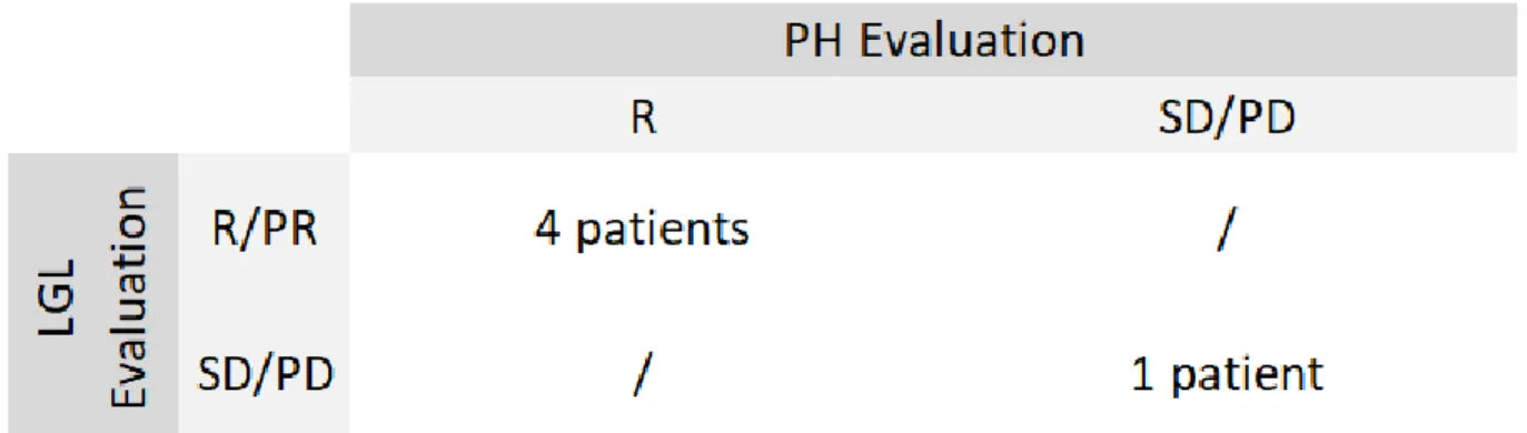
Associated response of LGL Leukemia and PH on immunosuppressive treatment was observed in 4 patients, as detailed in Table 3 and illustrated in Figure 1 (cases 1 and 2). No patient showed hemodynamical improvement of PH without a LGL leukemia response. Safety:
No toxicity was reported for ciclosporin. An ANC decrease with bacterial dermo-hypodermitis was related to cyclophosphamide leading to treatment cessation. Methotrexate was associated with an immune-allergic interstitial lung disease with treatment cessation requirement and in two cases with hepatitis leading to dosage adjustment.
Discussion
We report on a series of 9 patients suffering from concomitant LGL leukemia and PH. To our knowledge, this has been previously described in 7 sporadic cases14-15-16-17-18, making a total
number of 15 cases. One patient of our cohort was initially reported in a case report15. Considering the rarity of both diseases, it is tempting to postulate a possible link between LGL clonal expansion and the development of PH.
Based on the clinical characteristics of these 9 PH patients, we may consider that they rather belong to the group 5 of PH classifying. The main causes of PH have been excluded: heritable, drugs, portal hypertension, HIV infection, connective tissue disease, congenital or left heart disease and chronic thromboembolic disorders. However, we cannot exclude the implication of underlying causes in 5 out of the 9 cases. Patient 2 developed interstitial pneumonitis on methotrexate which could result in PH. Nevertheless, PH persisted after pneumonitis resolution and finally decreased following further immunosuppressive therapy. Patient 5 showed a right-left shunt by patent oval foramen but it was considered as consequence and not causality of PH by cardiologic staff. Patients 7 and 8 had positive anticardiolipin antibodies but without thromboembolic disorders signs on CT pulmonary angiogram and ventilation/perfusion scan and we assume that this was not related to the development of PH. AIHA was reported in patient 4, hemolytic disorders can induce PH via significant reduction in nitric oxide (NO) availability due to free hemoglobin and accompanying platelet activation27 but no active hemolysis was objectified at PH diagnosis
The benefit from specific therapy of LGL leukemia led to significant hemodynamical PH improvement in 4 of the 5 RHC evaluable patients. In addition, three patients showed clinical improvement with NYHA score decrease or 6MWT enhancement but without significant hemodynamic change. None of the patients died with a median follow-up of 88 months. If we are focusing on the 7 patients with PAH associated to LGL leukemia, their clinical evolution and survival appeared to be different from PAH group 1 patients. Group 1 includes idiopathic and heritable PAH, PAH associated with human immunodeficiency viral infection (HIV), schistosomiasis, congenital heart defect, connective tissue diseases, portal hypertension and drug induced PAH21. The most important American cohort REVEAL
showed a 1-year survival at 91% for patients from group 124. The 1-year survival was < 70% for very high-risk patients. 40% and 26.2% of patients received combination PAH therapies or an intravenous prostacyclin analog. In the French cohort « French Pulmonary Hypertension Network » (FPHN), also including group 1 patients, the 1-year survival was 88%28. Our cohort had favorable initial hemodynamics and clinical features than those of REVEAL and FPHN cohorts. However, the PAH evolution following immunosuppressive therapy and the absence of PAH related mortality over a long-term follow-up suggests a different condition. Of note, we cannot exclude that 4 patients could have benefit from concomitant administration of PAH specific therapy with LGL leukemia treatment. In two cases, the PAH response did not appear to be correlated with PAH therapy; patient 4 could achieve dosage reduction of epoprostenol during methotrexate therapy and patient 6, who was stable on sildenafil, showed PAH improvement after ciclosporin initiation, suggesting a potential role of immunosuppressive therapy on PAH.
PAH is considered as a vasculopathy with endothelial dysfunction leading to abnormal vascular homeostasis. Dysregulated levels of vasodilator molecule as NO and vasoconstrictor molecule as Endothelin-1, associated with cell survival and proliferation constitute constrictive lesion, which include medial hypertrophy and intimal and adventitial thickening, and complex lesion as plexiform lesion, very characteristic of PAH29. This pathological vascular remodeling leads to an increase in pulmonary artery pressures and eventual right ventricular (RV) dysfunction. We suggest that LGL leukemia is responsible for PAH development. In fact, LGL leukemia seems to precede PAH development and PAH improvement seems to be correlated to LGL leukemia response to immunosuppressive therapy. Furthermore, improvement of hemodynamic data allowing PAH therapeutic de-escalation is quite unusual due to the supposed irreversibility of PAH histological lesions. Pathogenesis mechanism proposal is described in Figure 2.
A direct cytotoxicity of CD3+ T cells on pulmonary endothelial cells (ECs) is suggested. This cytotoxicity is controlled by balance between activating Natural Killer Receptors (NKRs) and inhibiting NKRs recognizing major histocompatibility complex (MHC) class I proteins30. Leukemic LGLs, expressing elevated levels of activating NKRs, acquire the ability to directly lyse pulmonary artery ECs30. Activating bonding of NKRs drives a cytoplasmic pathway leading a direct cytotoxicity via migration of lytic granules containing perforin and granzyme B toward the contacted cell like CEs31-32. Because the activation of NKRs leads on Ras/MAPK signaling pathway, a farnesyltransferase inhibitor Tipifarnib, was tested in a patient who presented association between LGL leukemia and PAH16. After 4 courses of
20
expression36. LGLs express surface FasL and secrete high level of soluble FasL, considered as a potential cause of auto-immune symptoms and neutropenia in LGL leukemia37. ECs express
also Fas and FasL promoting the inhibition of leukocyte extravasation and protecting vascular endothelium from local inflammation. ECs have also an elevated expression of c-FLIP, inhibiting the Fas/FasL mediated apoptosis38-39-40. However, local expression of cytokines such as TNFα, expressed in high level in LGLs environment41, decreases the ECs expression of FasL making ECs susceptible to cytotoxicity42. A study reported 2 patients with association of LGL leukemia and PAH, who expressed high level of circulating FasL18. Lung biopsy described LGL cells infiltration expressing FasL with pneumocyte apoptosis mimicking pathological changes of graft-versus-host disease. Patients clinical improvement was correlated with decreased of circulating FasL levels on LGL leukemia therapy.
In murine model, ECs-Fas mediated apoptosis induces PAH typical vascular lesion43. This
could explain why hemodynamic normalization could not be observed in patients with PAH and LGL leukemia. The only patient who achieved hemodynamic normalization was classified PH because of PVR < 3WU, pleading for two distinct mechanisms of PH associated with LGL leukemia. Firstly, vascular remodeling induced by LGL leads to elevated vascular resistance characterizing PAH. Secondly, moderate vascular resistance in the absence of definitive vascular lesions, and elevated cardiac index linked to significative anemia, cause reversible PH.
The association between LGL leukemia and PH does not appear fortuitous regarding their respecting low incidence and the unusual association of both diseases. Moreover, the improvement of PH on immunosuppressive therapy strengthens the relationship between these both diseases. It suggests supplementing the current recommendations for initial work-up with a T clonality search in context of clinic-biological abnormalities evoking an underlying LGL leukemia. A better understanding of pathogenesis mechanisms is required to adapt the optimal therapeutic option for such patients. In absence of gravity signs of PH, we propose to initiate only immunosuppressive therapy with a close monitoring of PH evolution.
21
References:
1. Loughran TP, Kadin ME, Starkebaum G, Abkowitz JL, Clark EA, Disteche C, et al.
Leukemia of large granular lymphocytes: association with clonal chromosomal abnormalities and autoimmune neutropenia, thrombocytopenia, and hemolytic anemia. Ann Intern Med. 1985 Feb;102(2):169-75.
2. Swerdlow SH, Campo E, Pileri SA, Harris NL, Stein H, Siebert R, et al. The 2016
revision of the World Health Organization classification of lymphoid neoplasms. Blood. 2016 May 19;127(20):2375-90.
3. Lamy T, Loughran TP. Clinical features of large granular lymphocyte leukemia.
Semin Hematol. 2003 Jul;40(3):185-95.
4. Suzuki R, Suzumiya J, Nakamura S, Aoki S, Notoya A, Ozaki S, et al. Aggressive
natural killer-cell leukemia revisited: large granular lymphocyte leukemia of cytotoxic NK cells. Leukemia. 2004 Apr;18(4):763-70.
5. Lamy T, Moignet A, Loughran TP. LGL leukemia: from pathogenesis to treatment.
Blood. 2017 Mar 2;129(9):1082-94.
6. Lamy T, Loughran TP. How I treat LGL leukemia. Blood. 2011 Mar
10;117(10):2764-74.
7. Loughran TP. Clonal diseases of large granular lymphocytes. Blood. 1993 Jul
1;82(1):1-14.
8. Lundell R, Hartung L, Hill S, Perkins SL, Bahler DW. T-cell large granular
lymphocyte leukemias have multiple phenotypic abnormalities involving pan-T-cell antigens and receptors for MHC molecules. Am J Clin Pathol. 2005 Dec;124(6):937-46.
9. Langerak AW, van Den Beemd R, Wolvers-Tettero IL, Boor PP, van Lochem EG,
Hooijkaas H, et al. Molecular and flow cytometric analysis of the Vbeta repertoire for clonality assessment in mature TCRalphabeta T-cell proliferations. Blood. 2001 Jul 1;98(1):165-73.
10. Fasan A, Kern W, Grossmann V, Haferlach C, Haferlach T, Schnittger S. STAT3
mutations are highly specific for large granular lymphocytic leukemia. Leukemia. 2013 Jul;27(7):1598-600.
11. Rajala HLM, Eldfors S, Kuusanmäki H, van Adrichem AJ, Olson T, Lagström S, et al.
Discovery of somatic STAT5b mutations in large granular lymphocytic leukemia. Blood. 2013 May 30;121(22):4541–50.
12. Loughran TP, Zickl L, Olson TL, Wang V, Zhang D, Rajala HLM, et al.
Immunosuppressive therapy of LGL leukemia: prospective multicenter phase II study by the Eastern Cooperative Oncology Group (E5998). Leukemia. 2015 Apr;29(4):886-94.
13. Zhang R, Shah MV, Loughran TP. The root of many evils: indolent large granular
lymphocyte leukaemia and associated disorders. Hematol Oncol. 2010 Sept;28(3):105-17.
14. Rossoff LJ, Genovese J, Coleman M, Dantzker DR. Primary pulmonary hypertension
in a patient with CD8/T-cell large granulocyte leukemia: amelioration by cladribine therapy. Chest. 1997 Aug;112(2):551-3.
15. Grossi O, Horeau-Langlard D, Agard C, Haloun A, Lefebvre M, Neel A, et al.
Low-dose methotrexate in PAH related to T-cell large granular lymphocyte leukaemia. Eur Respir J. 2012 Feb;39(2):493-4.
16. Epling-Burnette PK, Sokol L, Chen X, Bai F, Zhou J, Blaskovich MA, et al. Clinical
improvement by farnesyltransferase inhibition in NK large granular lymphocyte leukemia associated with imbalanced NK receptor signaling. Blood. 2008 Dec 1;112(12):4694-8.
17. Howard LSGE, Chatterji S, Morrell NW, Pepke-Zaba J, Exley AR. Large granular
lymphocyte leukaemia: a curable form of pulmonary arterial hypertension [corrected]. Hosp Med. 2005 Jun;66(6):364-5.
18. Lamy T, Bauer FA, Liu JH, Li YX, Pillemer E, Shahidi H, et al. Clinicopathological
features of aggressive large granular lymphocyte leukaemia resemble Fas ligand transgenic mice. Br J Haematol. 2000 Mar;108(4):717-23.
19. Simonneau G, Gatzoulis MA, Adatia I, Celermajer D, Denton C, Ghofrani A, et al.
Updated clinical classification of pulmonary hypertension. J Am Coll Cardiol. 2013 Dec 24;62(25 Suppl):D34-41
20. Semenzato G, Zambello R, Starkebaum G, Oshimi K, Loughran TP. The
lymphoproliferative disease of granular lymphocytes: updated criteria for diagnosis. Blood. 1997 Jan 1;89(1):256–60.
21. Galiè N, Humbert M, Vachiery J-L, Gibbs S, Lang I, Torbicki A, et al. 2015 ESC/ERS
Guidelines for the diagnosis and treatment of pulmonary hypertension: The Joint Task Force for the Diagnosis and Treatment of Pulmonary Hypertension of the European Society of Cardiology (ESC) and the European Respiratory Society (ERS): Endorsed by: Association for European Paediatric and Congenital Cardiology (AEPC), International Society for Heart and Lung Transplantation (ISHLT). Eur Heart J. 2016 Jan 1;37(1):67-119.
22. Zhang D, Loughran TP. Large granular lymphocytic leukemia: molecular
pathogenesis, clinical manifestations, and treatment. Hematology Am Soc Hematol Educ Program. 2012;2012:652-9.
23. Rudski LG, Lai WW, Afilalo J, Hua L, Handschumacher MD, Chandrasekaran K, et
al. Guidelines for the echocardiographic assessment of the right heart in adults: a report from the American Society of Echocardiography endorsed by the European Association of Echocardiography, a registered branch of the European Society of Cardiology, and the Canadian Society of Echocardiography. J Am Soc Echocardiogr. 2010 Jul;23(7):685-713; quiz 786-788.
24. Benza RL, Miller DP, Gomberg-Maitland M, Frantz RP, Foreman AJ, Coffey CS, et
23
25. Benza RL, Gomberg-Maitland M, Miller DP, Frost A, Frantz RP, Foreman AJ, et al.
The REVEAL Registry risk score calculator in patients newly diagnosed with pulmonary arterial hypertension. Chest. 2012 Feb;141(2):354-62.
26. McLaughlin VV, Gaine SP, Howard LS, Leuchte HH, Mathier MA, Mehta S, et al.
Treatment goals of pulmonary hypertension. J Am Coll Cardiol. 2013 Dec 24;62(25 Suppl):D73-81.
27. Mathew R, Huang J, Wu JM, Fallon JT, Gewitz MH. Hematological disorders and
pulmonary hypertension. World J Cardiol. 2016 Dec 26;8(12):703-18.
28. Humbert M, Sitbon O, Chaouat A, Bertocchi M, Habib G, Gressin V, et al. Pulmonary
arterial hypertension in France: results from a national registry. Am J Respir Crit Care Med. 2006 May 1;173(9):1023-30.
29. Santos-Ribeiro D, Mendes-Ferreira P, Maia-Rocha C, Adão R, Leite-Moreira AF,
Brás-Silva C. Pulmonary arterial hypertension: Basic knowledge for clinicians. Arch Cardiovasc Dis. 2016 Oct;109(10):550-61
30. Chen X, Bai F, Sokol L, Zhou J, Ren A, Painter JS, et al. A critical role for DAP10
and DAP12 in CD8+ T cell-mediated tissue damage in large granular lymphocyte leukemia. Blood. 2009 Apr 2;113(14):3226-34
31. Djeu JY, Jiang K, Wei S. A view to a kill: signals triggering cytotoxicity. Clin Cancer
Res. 2002 Mar;8(3):636-40.
32. Bielawska-Pohl A, Crola C, Caignard A, Gaudin C, Dus D, Kieda C, et al. Human NK
cells lyse organ-specific endothelial cells: analysis of adhesion and cytotoxic mechanisms. J Immunol. 2005 May 1;174(9):5573-82.
33. Lamy T, Liu JH, Landowski TH, Dalton WS, Loughran TP Jr. Dysregulation of
CD95/CD95 ligand-apoptotic pathway in CD3(+) large granular lymphocyte leukemia. Blood. 1998 Dec 15;92(12):4771-7.
34. Krammer PH. CD95’s deadly mission in the immune system. Nature. 2000 Oct
12;407(6805):789-95.
35. Liu JH, Wei S, Lamy T, Li Y, Epling-Burnette PK, Djeu JY, et al. Blockade of
Fas-dependent apoptosis by soluble Fas in LGL leukemia. Blood. 2002 Aug 15;100(4):1449-53.
36. Yang J, Epling-Burnette PK, Painter JS, Zou J, Bai F, Wei S, et al. Antigen activation
and impaired Fas-induced death-inducing signaling complex formation in T-large-granular lymphocyte leukemia. Blood. 2008 Feb 1;111(3):1610-6.
37. Liu JH, Wei S, Lamy T, Epling-Burnette PK, Starkebaum G, Djeu JY, et al. Chronic
neutropenia mediated by fas ligand. Blood. 2000 May 15;95(10):3219-22.
38. Richardson BC, Lalwani ND, Johnson KJ, Marks RM. Fas ligation triggers apoptosis
in macrophages but not endothelial cells. Eur J Immunol. 1994 Nov;24(11):2640-5.
39. Wang X, Wang Y, Zhang J, Kim HP, Ryter SW, Choi AMK. FLIP protects against
hypoxia/reoxygenation-induced endothelial cell apoptosis by inhibiting Bax activation. Mol Cell Biol. 2005 Jun;25(11):4742-51.
40. Stoneman VEA, Bennett MR. Role of Fas/Fas-L in vascular cell apoptosis. J
41. Shvidel L, Duksin C, Tzimanis A, Shtalrid M, Klepfish A, Sigler E, et al. Cytokine
release by activated T-cells in large granular lymphocytic leukemia associated with autoimmune disorders. Hematol J. 2002;3(1):32-7.
42. Sata M, Walsh K. TNFalpha regulation of Fas ligand expression on the vascular
endothelium modulates leukocyte extravasation. Nat Med. 1998 Apr;4(4):415-20.
43. Goldthorpe H, Jiang J-Y, Taha M, Deng Y, Sinclair T, Ge CX, et al. Occlusive lung
arterial lesions in endothelial-targeted, fas-induced apoptosis transgenic mice. Am J Respir Cell Mol Biol. 2015 Nov;53(5):712-8.
25
1 2 3 4 5 6 7 8 9 53 59 55 52 82 58 33 39 38 F F M F F F F F M T-LG L Le uk emi a T-LG L Le uk emi a T-LG L Le uk emi a T-LG L Le uk emi a T-LG L Le uk emi a T-LG L Le uk emi a T-LG L Le uk emi a CL PD -N K T-LG L Le uk emi a + + + + + + + -+ + + + + + + + - ( di m) + + -+ +/ -+ + + -+ -+ -+/ -+/ -+ + + + -+ -al ph a/ be ta al ph a/ be ta al ph a/ be ta al ph a/ be ta al ph a/ be ta al ph a/ be ta al ph a/ be ta Pl oy cl on al al ph a/ be ta y Po ly cl on al M on oc lo na l M on oc lo na l M on oc lo na l M on oc lo na l Po ly cl on al M on oc lo na l Po ly cl on al NA Y6 40 G NA D 66 1Y NA W ild ty pe Y6 40 F D 56 6N e x 19 W ild ty pe NA Ye s Ye s Ye s Ye s Ye s Ye s Ye s Ye s No No No No Te le ng ec ta si a + A IH A No No Se ro ne ga ti ve A rth ri ti s M ig ra nt pa lin dr omi c ar th ri ti s Be hç et' s 311 115 10 209 8 88 265 19 5 2 2 1 1 2 3 3 0 2 Tu mo ra l s yn dr ome N eu tr op en ia N eu tr op en ia N eu tr op en ia + B si gn s + PH + PA H + PA H PA H PH PH PA H PA H PA H PA H PA H PA H ry a ng io gr am N or ma l IL P M TX -a ss oc ia te d N or ma l N or ma l N or ma l N or ma l N or ma l N or ma l N or ma l er fu si on S ca n N or ma l NA NA NA N or ma l NA N or ma l N or ma l N or ma l N or ma l N or ma l N or ma l N or ma l N or ma l NA N or ma l N or ma l NA ltr as ou nd N or ma l NA N or ma l N or ma l N or ma l N or ma l N or ma l N or ma l N or ma l A N A (1 /1 28 0) A N A (1 /1 00 ) N or ma l N or ma l A N A (1 /3 20 ) N or ma l A nti ca rd io lip in + A nti ca rd io lip in + NA 49 88 7 229 8 90 179 141 51 No No No Ye s No Ye s Ye s Ye s No 9 6 3 8 5 6 6 7 NA M od er ate ly h ig h Lo w Lo w A ve ra ge Lo w Lo w Lo w Lo w NA H ig h ri sk In te rme di ate ri sk In te rme di ate ri sk H ig h Ri sk In te rme di ate ri sk In te rme di ate ri sk Lo w ri sk In te rme di ate ri sk H ig h Ri sk N eu tr op en ia PA H Re cu rr en t i nf ec ti on Sy mp toma ti c A ne mi a No
27
Table 2: PH and LGL leukemia characteristics available at diagnosis then on treatment at last news, n=9.
Patient n°
LGL At diagnosis On treatment At diagnosis On treatment At diagnosis On treatment At diagnosis On treatment At diagnosis On treatment Hemoglobin (g/dL) 12,2 14 13,3 12,9 10,1 9,1 9,9 12,9 12,8 13 Neutrophils (/mm3) 350 790 380 4300 960 1300 800 1720 3780 3000 Lymphocytes (/mm3) 1790 650 3750 945 4600 3400 2000 1000 4150 2570 LGL (%) 39 NA 90 NA 45 NA 9 0 25 18 LGL therapy 1st Line 2nd Line 3nd Line Follow-up (months)
Pulmonary Hypertension At diagnosis On treatment At diagnosis On treatment At diagnosis On treatment At diagnosis On treatment At diagnosis On treatment Right Heart Catheterisation
PAPm (mmHg) 27 NA 34 12 33 NA 57 NA 35 26 PCWP (mmHg) 7 NA 10 NA 11 NA 10 NA 3 NA PVR (WU) 4,4 NA 2,4 1,5 2,3 NA 12,7 NA 4,4 4 Cardiac Index (L/min/m²) 2,97 NA 4,15 3,4 4,6 NA 2,2 NA 2,2 2,78 TTE
PTRV (m/S) 3,7 3,3 3,7 NA 3,5 3 4,1 4,5 3,1 NA Estimated PAPs (mmHg) 65 56 62 39 65 39 81 88 45 NA Right cavity dilatation Yes No Yes No Yes No Yes Yes No No Clinical Impact
Dyspnea NYHA Score 4 2 2 1 2 1 4 1 3 3 Right heart failure signs No No No No No No No No No No 6MWT (meters) 285 357 300 420 544 NA 290 475 420 395 Ambient Air PaO2 (mmHg) 62 93 NC NA 62 NA 63 NA 69 NA O2-therapy Withdrawal PH Therapy 1st Line 2nd Line Follow-up (months) Patient n°
LGL At diagnosis On treatment At diagnosis On treatment At diagnosis On treatment At diagnosis On treatment Hemoglobin (g/dL) 12,5 12,9 8,3 11 15 NA 13,3 10,6 Neutrophils (/mm3) 400 3000 1050 600 5780 NA 0 10 Lymphocytes (/mm3) 5330 3320 2960 4060 1530 NA 16500 4600 LGL (%) 80 15 70 69 22 NA 65 NA LGL therapy 1st Line 2nd Line 3nd Line Follow-up (months)
Pulmonary Hypertension At diagnosis On treatment At diagnosis On treatment At diagnosis On treatment At diagnosis On treatment Right Heart Catheterisation
PAPm (mmHg) 55 37 46 36 63 52 43 NA PCWP (mmHg) 11 NA 10 NA 12 NA 10 NA PVR (WU) 8,3 5,8 9,2 4,2 7,5 NA 9,4 NA Cardiac Index (L/min/m²) 2,98 3 2,6 4,3 2,17 3,6 2,15 NA TTE
PTRV (m/S) 4 2,5 2,6 3,6 5,1 NA NA NA Estimated PAPs (mmHg) 70 35 42 60 115 NA NA NA Right cavity dilatation Yes Yes Yes Yes Yes NA NA NA Clinical Impact
Dyspnea NYHA Score NA 2 NA 1 NA 2 NA NA Right heart failure signs No No No No Yes No Yes NA 6MWT (meters) 450 400 NA 708 NA 595 NA NA Ambient Air PaO2 (mmHg) NA NA NA NA NA NA NA NA O2-therapy Withdrawal PH Therapy 1st Line 2nd Line Follow-up (months) NA Methotrexate Cyclophosphamide 5 No Treatement 51 Ambrisentan 179 No need No treatement 19
B o sentan + Sildenafil + Nifedipine
141 No need Methotrexate Cyclophosphamide Ciclosporine 88 Bosentan No need No Treatement Methotrexate Cyclophosphamide 8 8 Yes No Treatement 49 Methotrexate 209 Epoprostenol 229 115 88 No need Ciclosporine 12 No Treatement 7 Yes 6 7 8 9 Methotrexate Cyclophosphamide No need No Treatement Cyclophosphamide 311 Sildenafil 90 No need Methotrexate Ciclosporine Cyclophosphamide 265 1 2 3 4 5 Ciclosporine
Abbreviations: 6MWT, 6-minutes walking test; LGL, large granular lymphocyte; NA, no available; PAPm: mean pulmonary arterial pressure; PCWP, pulmonary arterial wedge pressure; PH, pulmonary hypertension; PTRV, Peak tricuspid regurgitation velocity; PVR, pulmonary vascular resistance; TTE, transthoracic echocardiography; WU; Wood units.
Table 3: Association of LGL leukemia and PH response on immunosuppressive treatment.
Abbreviations: CR, complete response; LGL, large granular lymphocyte; NA, No available; PD, progression disease; PH, pulmonary hypertension; PR, partial response; R, response; SD, stable disease.
29
Red arrow indicates time on cyclophosphamide. Abbreviations: 6MWT, 6-minutes walking test; LGL, large granular lymphocyte; PAH, pulmonary arterial hypertension; PTRV, Peak tricuspid regurgitation velocity.
Figure 1: Example for 2 patients (cases 1 and 2) of LGL leukemia and PH evolution on immunosuppressive treatment
Figure 2: Two potential mechanisms of endothelial cell lysis by LGL cells.
NKR Pathway: Activating NKR such as NKG2D, KIR2DS, KIR3DS, NKG2C, NKp44, NKp46, FcR III (CD16), TCR, and NKp30 act through 3 adaptor proteins either in a homodimeric or heterodimeric form (DAP12, DAP10, and CD3). These signals lead to PI3K/MEK/ERK activation then granule redistribution and movement. Granule exocytosis releases Granzyme B who activates caspases in the target cell.
Fas/FasL Pathway: the trimerization of Fas (CD95) induced by interaction with Fas-L leads formation of the DISC. First, the adaptor FADD binds via its own death domain to the death domain in CD95. FADD recruits procaspase-8 (also known as FLICE) into the DISC. Next, procaspase-8 is activated proteolytically and active caspase-8 is released from the DISC into the cytoplasm in the form of a heterotetramer of two small subunits and two large subunits. Active caspase-8 cleaves various proteins in the cell including procaspase-3, which results in its activation and the completion of the cell death programme. Another pathway called intrinsic, may be induced. Caspase-8 cuts the Bcl-2 family member Bid; T-Bid 'activates' the mitochondria who release pro-apoptotic molecules such as cytochrome C. Cytochrome C with the apoptosis protease-activating factor Apaf-1 and procaspase-9 in the cytoplasm form the Apoptosome, the second initiator complex of apoptosis who leads again to apoptosis.
Abbreviations: DISC, Death-inducting signaling complex; FADD, Fas-associated death domain protein; LGL, large granular lymphocyte; NKR, Natural Killer Receptor; T-bid, Truncated Bid.