Université de Montréal
Longitudinal evaluation of sleep-related breathing
disorders in an orthodontic population
SDB and orthodontics in adolescents
par Manuela Mandu
Département de santé buccale Faculté de médecine dentaire
Mémoire présenté à la Faculté des études supérieures et postdoctorales en vue de l’obtention du grade de Maîtrise
en sciences en médecine dentaire option orthodontie
© Manuela Mandu, 2011 Université de Montréal
Faculté des études supérieures et postdoctorales
Ce mémoire intitulé:
Longitudinal evaluation of sleep-related breathing disorders in an orthodontic population
Présenté par : Manuela Mandu
a été évalué par un jury composé des personnes suivantes :
Dre Athena Papadakis, présidente-rapportrice Dr Jack Turkewicz, directeur de recherche
Dre Belinda Nicolau, co-directeur Dre Nelly Huynh, membre du jury
Résumé
Introduction: Les troubles respiratoires du sommeil (TRS), qui représentent une
préoccupation croissante pour la santé, ont des effets significatifs sur la santé, le comportement et la performance académique chez l’enfant. Les malformations craniofaciales, l’hypertrophie adéno-amygdalienne et l'obésité, représentent des facteurs de risque importants dans le développement de cette condition. Les symptômes des TRS ont été étudiés dans une étude prospective chez les enfants et adolescents durant leur traitement orthodontique dans un milieu universitaire. Cette étude a cherché à décrire la prévalence et les facteurs de risque principaux des TRS, ainsi que l'impact des différentes interventions orthodontiques sur les symptômes TRS.
Matériel et méthodes: dans une étude cohorte prospective, un groupe de 168 sujets
âgés de 12 à 21 ans ont été soumis, quatre ans après la prise de données initiale, à un examen craniofacial en plus d'être administré des questionnaires qui ont recueilli des données sur la situation socio-démographique, le bruxisme et les troubles d’ATM, le sommeil et le comportement diurne, et les facteurs neuropsychologiques.
Résultats: l'indice de masse corporelle a été augmenté mais est demeurée dans la
même catégorie aux deux moments de l'enquête. Il ya eu une augmentation du serrement des dents et des symptômes de l'ATM, une diminution de la taille des amygdales, et une augmentation de la somnolence diurne. La prévalence des TRS n'a pas changé entre l’étude initiale et l’étude de suivi. Aucune intervention orthodontique s'est avérée avoir un effet cliniquement significatif sur les voies aériennes supérieures.
Conclusions: la prévalence des symptômes TRS était constante par rapport aux
valeurs de base pour la population étudiée, mais a augmenté si rapportée à la population générale. Les traitements orthodontiques ne montrent aucun effet sur les TRS.
Mots-clés : apnée du sommeil, craniofacial, prévalence, ronflement, traitement orthodontique, voies aériennes supérieures
Abstract
Introduction: Sleep-disordered breathing (SDB), a growing health concern, has
significant effects on a child’s health, behaviour, and scholastic performance. Craniofacial malformations, along with adenotonsillar hypertrophy and obesity, represent important risk factors in the development of this condition. SDB symptoms in children and adolescents followed for orthodontic treatment in a university setting have been investigated in this prospective study. The aims of this study were to describe the prevalence and main risk factors of SDB and the impact of different orthodontic interventions on the SDB symptoms.
Materials and methods: in a prospective cohort study, four years following an initial
evaluation, a group of 168 subjects aged 12-21 years underwent a craniofacial examination in addition to being administered self-completed questionnaires that collected information on socio-demographic and psychosocial factors, bruxism and temporo-mandibular joint (TMJ) disorders, sleep and daytime behaviour, and neuropsychological factors.
Results: Body mass index (BMI) was slightly increased but remained in the same
category at the two time points of investigation. There was an increase in tooth clenching and TMJ symptoms, a decrease in tonsils’ size, and an increase in daytime sleepiness. Prevalence of SDB did not change between baseline and follow-up studies. No orthodontic treatment intervention proved to have any clinically significant impact on the upper airway.
Conclusions: SDB symptoms prevalence was constant when compared to the
baseline values for the studied population, but increased if reported to the general population. Regular orthodontic treatment didn’t show any effect on SDB symptoms.
Keywords : craniofacial, orthodontic treatment, prevalence, sleep apnea, snoring, upper airway
Table of contents
1 Chapter 1 ... 1
1.1 Introduction ... 1
1.2 Literature and methodology review ... 1
1.2.1 Historical perspective ... 1
1.2.2 Classification of SDB ... 2
1.2.3 Definitions ... 2
1.2.4 Epidemiology of SDB and the economic impact of sleep apnea ... 7
1.2.5 Anatomy and physiology of upper airway obstruction ... 8
1.2.6 Snoring ... 10
1.2.7 Obstructive sleep apnea ... 11
1.2.8 Clinical signs and symptoms of SDB ... 16
1.2.9 Diagnosis of SDB ... 18
1.2.10 Consequences of SDB ... 19
1.2.11 Treatment of SDB ... 20
2 Chapter 2 ... 27
2.1 Rational and objectives ... 27
3 Chapter 3 ... 28
3.1 Abstract ... 30
3.2 Introduction ... 30
3.3 Material and methods ... 33
3.3.1 Study population ... 33
3.3.2 Instruments: questionnaires, clinical evaluation, and history of orthodontic treatment... 33
3.3.3 Data analysis ... 35
3.4 Results ... 35
3.4.1 Interexaminer and intraexaminer reliability ... 35
3.4.3 Medical and dental history ... 36
3.4.4 Bruxism and TMD ... 37
3.4.5 Sleep and daytime behaviour ... 37
3.4.6 Orthodontic morphometric evaluation and craniofacial morphology ... 39
3.4.7 Impact of orthodontic treatment on UA ... 39
3.5 Discussion ... 40
3.6 Limitations of the present study ... 42
3.7 Conclusions ... 44
3.8 Acknowledgements ... 44
4 Chapter 4 ... 45
4.1 Summary ... 45
5 Chapter 5 ... 47
5.1 Conclusions and future research directions ... 47
6 Chapter 6 ... 49
6.1 Cumulative Reference List... 49
List of tables
Table I: Sleep-Related Breathing Disorders……….... 3 Table II: Factors predisposing to collapse of the upper airway and
the development of OSA ………15 Table III: Clinical features of sleep apnea ……….……17 Table IV. Indications and contraindications for Surgery in
Sleep-Disordered Breathing ………...…23 Table V. Demographic data of the study group ……….…36 Table VI. Reported bruxism and TMD habits from the Patient
Questionnaire ………..37 Table VII. Reported sleep and daytime behaviour ………..…..38 Table VIII. Distribution of orthodontic treatment procedures in the
study population ……….40 Table IX. Effect of individual orthodontic treatment on SDB symptoms………..40
List of figures
Figure 1 BMI-for-age girls 2 to 20 years……….……….5 Figure 2 BMI-for-age boys 2 to 20 years………..………6 Figure 3 Segments of the upper airway as seen on midsagittal magnetic
resonance imaging……….…..9
Figure 4. The spectrum of sleep disordered breathing………..……..12 Figure 5. Mechanism of upper airway occlusion and its prevention
by nasal continuous positive airway pressure (CPAP)………..22 Figure 6. Sutural opening after RPE ………..………...…….25
List of abbreviations
% Percent
ACCO Acrylic Cervical Occipital Appliance
AHI Apnea-Hypopnea Index
AJODO American Journal of Orthodontics and Dentofacial Orthopedics. BMI Body Mass Index
CDC Centers for Disease Control and Prevention
CI Confidence Interval
CNS Central Nervous System
CPAP Continuous Positive Airway Pressure
CT Computed Tomography
ECG Electrocardiography EDS Excessive Daytime Sleepiness EEG Electroencephalography EMG Electromyography EOG Electro-oculography
GAHMS Genioglossus Advancement, Hyoid Myotomy, and Suspension GER Gastroesophageal reflux
h Hour(s)
ICSD International Classification of Sleep Disorders kg Kilogram(s)
m Meter(s) mm Millimeter(s)
MMA Maxillomandibular Advancement MMO Maxillary and Mandibular Osteotomy MPA Mandibular Plane Angle MRI Magnetic Resonance Imaging OB Overbite
OSA Obstructive Sleep Apnea
OR Odds Ratio
PAP Positive Airway Pressure
PetCO2 Partial Pressure of End-Tidal Carbon Dioxide
PtcCO2 Partial Pressure of Transcutaneous Carbon Dioxide
PSG Polysomnography RDI Respiratory Disturbance Index
RERA Respiratory Effort–Related Arousal RPE Rapid Palatal Expansion
SaO2 Oxygen Saturation
SARPE Surgically Assisted Rapid Palatal Expansion
SB Sleep Bruxism SD Standard Deviation SDB Sleep-Disordered Breathing TC Tooth Clenching TMJ Temporomandibular Joint TMD Temporomandibular Disorder UA Upper Airway
UARS Upper Airway Resistance Syndrome UPPP Uvulo-Palatopharyngoplasty
Acknowledgements
First and foremost, I would like to thank my family, to both my husband Ghita and to my son Victor, for the incommensurable support they provided while I was pursuing my specialized training in Orthodontics. Only deep love can catalyze such an outstanding support and I assure you of reciprocity of this feeling.
I would like to recognize the excellent advice, direction, and assistance provided by both of my supervisors, Dr. Jack Turkewicz and Dr. Belinda Nicolau. Particularly, I thank Dr. Turkewicz for his continuous encouragement and support throughout my training period and for the minutia of editing this present work. I am also grateful to Belinda Nicolau for the understanding that she manifested when I became overloaded with orthodontics and had to reduce my research contribution in her group.
I would like to acknowledge the expert assistance of Dr. Nelly Huynh during preparation of my thesis, in both statistical advice and manuscript redaction. Thank you also to Mr. Pierre Rompré for deciphering and helping me to make sense of a large amount of data.
I would also like to thank Dr. Athena Papadakis for encouraging me to embark on this research project and helping with the logistics. Many thanks are extended to Dr. Paul Morton for his assistance in my research project and for the valuable supervision in the orthodontic clinic.
Last but not least, my thanks go out to Dr. Claude Remise for giving me the chance to evolve into an orthodontist proud to have received her education in a prolific scientific and clinical environment.
Preface and thesis outline
This Masters’ thesis is prepared as a manuscript-based thesis and contains five chapters. Chapter 1 provides a brief literature review as pertaining to sleep-disordered breathing (SDB). Chapter 2 of the thesis deals with the rationale and objectives of the study. Chapter 3 presents an article in preparation, to be submitted to the American Journal of Orthodontics and Dentofacial Orthopedics. Chapter 4 summarizes the results of the present research, and Chapter 5 presents the conclusions of this study and provides future directions of the research.
The research article presented in Chapter 3 is:
Mandu-Hrit M, Huynh N, Morton P, Papadakis A, Rompré PH, Turkewicz J, Nicolau B. Longitudinal evaluation of sleep-related breathing disorders in an orthodontic population. AJODO, 2011. In preparation.
Contribution of authors
In the above mentioned article, the M.Sc. candidate had a major contribution in the design of the study, data collection, data analysis, and preparation of the manuscript. All the other authors reported as co-authors contributed significantly to the research. Dr. Jack Turkewicz and Dr. Belinda Nicolau are the research advisors and coordinated all the studies of the current Masters’ project.
1 Chapter 1
1.1 Introduction
Sleep-disordered breathing (SDB) represents a growing health concern. It comprises a group of disorders distinguished by abnormal respiration during sleep. These cessations in breathing rhythm (called apneas) or sustained reductions in the breath amplitude (hypopneas) can cause arterial hypoxemia and hypercapnia, transient arousals from sleep and sleep fragmentation throughout the night, with a variety of consequences on daily activities and overall health of the individuals.
1.2 Literature and methodology review
1.2.1 Historical perspective
Although the relationship between breathing and sleep has only recently been investigated by the medical community, excellent literary descriptions of what we know now to be the sleep apnea syndrome were made long ago. Obstructive sleep apnea (OSA) syndrome was described in 1836, not by a clinician but by the novelist Charles Dickens. In a series of papers entitled the “Posthumous Papers of the Pickwick Club” Dickens described Joe, a boy who was obese, a loud snorer, and always excessively sleepy. The first electrophysiological sleep recordings of “Pickwickian” patients and the understanding of the syndrome as disordered breathing in sleep were made only decades later, during the late 1950s and 1960s (Bickelmann et al., 1956). Extensive research has been conducted thereafter in the field of pathophysiology of sleep and breathing in the late 1970s through early 1980s, and subsequently a first population study has been conducted (the Wisconsin Sleep Cohort), showing a significant prevalence of sleep apnea in a middle-aged, nonclinical population (Young et al., 1993).
From the mid 1990s to the present, we witnessed an explosion of basic, clinical, and population research investigating the prevalence, causes, consequences, and treatment of this widespread sleep problem whose importance has only been recently appreciated. Given its high prevalence and potential carryover to daytime pathology, sleep apnea has provided great impetus to the growth of sleep medicine as a clinical and research specialty. Hundreds of sleep medicine clinics have been built throughout the western world during the past decades, with the majority of their business concerned with the diagnosis and treatment of sleep apnea. It seems that the two main reasons for sleep apnea syndrome being overlooked for so long have been misdiagnosis of patients with sleep apnea as having narcolepsy and skepticism regarding the validity of excessive daytime somnolence (EDS) as a clinical sign.
1.2.2 Classification of SDB
Sleep-related breathing disorders correspond to a range of nonspecific respiratory disturbances during sleep, including snoring, hypopnea, upper airway resistance, and full-blown apnea.
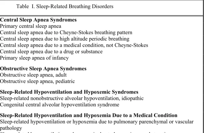
Cessation of breathing during sleep can result from obstruction of the upper airway (obstructive apnea), absence of inspiratory effort (central apnea), or a combination of the two. The second edition of the International Classification of Sleep Disorders (ICSD-2) (American Academy of Sleep Medicine, 2005) classifies sleep-related breathing disorders into five categories: central sleep apnea syndromes, obstructive sleep apnea syndromes, sleep-related hypoventilation and hypoxemic syndromes, sleep-related hypoventilation and hypoxemic syndromes, and other sleep-related breathing disorders (Table I).
1.2.3 Definitions
In order to better understand the complex pathophysiologic mechanism of sleep-related breathing disorders, more specifically OSA, some definitions are first presented.
Apnea, literally, means “no breathing”. It represents a cessation of respiratory airflow that lasts 10 seconds or longer. Apnea can be a central event (when respiratory
efforts are absent), an obstructive event (when respiratory efforts are present), or a mixed event (initial central apnea followed by obstructive apnea).
Table I. Sleep-Related Breathing Disorders Central Sleep Apnea Syndromes
Primary central sleep apnea
Central sleep apnea due to Cheyne-Stokes breathing pattern Central sleep apnea due to high altitude periodic breathing
Central sleep apnea due to a medical condition, not Cheyne-Stokes Central sleep apnea due to a drug or substance
Primary sleep apnea of infancy
Obstructive Sleep Apnea Syndromes Obstructive sleep apnea, adult
Obstructive sleep apnea, pediatric
Sleep-Related Hypoventilation and Hypoxemic Syndromes Sleep-related nonobstructive alveolar hypoventilation, idiopathic Congenital central alveolar hypoventilation syndrome
Sleep-Related Hypoventilation and Hypoxemia Due to a Medical Condition
Sleep-related hypoventilation or hypoxemia due to pulmonary parenchymal or vascular pathology
Sleep-related hypoventilation or hypoxemia due to lower airways obstruction
Sleep-related hypoventilation or hypoxemia due to neuromuscular or chest wall disorders Other Sleep-Related Breathing Disorder
Sleep apnea or sleep-related breathing disorder, unspecified
(Courtesy of the American Academy of Sleep Medicine, Chicago, Ill.)
Hypopnea is a reduction of respiratory airflow of at least 50% from baseline lasting at least 10 seconds, plus oxygen desaturation of at least 4%, caused by the lack of thoracic and abdominal effort (Madani and Madani, 2007a).
Apnea-Hypopnea index (AHI) is the number of apneic plus hypopneic episodes that occur per hour during sleep and is used as a measure of the severity of sleep apnea.
Respiratory effort–related arousal (RERA): A CNS arousal terminating obstructive breathing events that do not meet the criteria for apnea or hypopnea
Respiratory disturbance index (RDI): The number of apneas, hypopneas, and RERAs per hour of sleep. Unfortunately, the terms AHI and RDI are used interchangeably in much of the literature, and it is difficult to assess which abnormal breathing events have been scored.
Somnolence represents the difficulty in maintaining alert wakefulness so that a person falls asleep if not actively aroused.
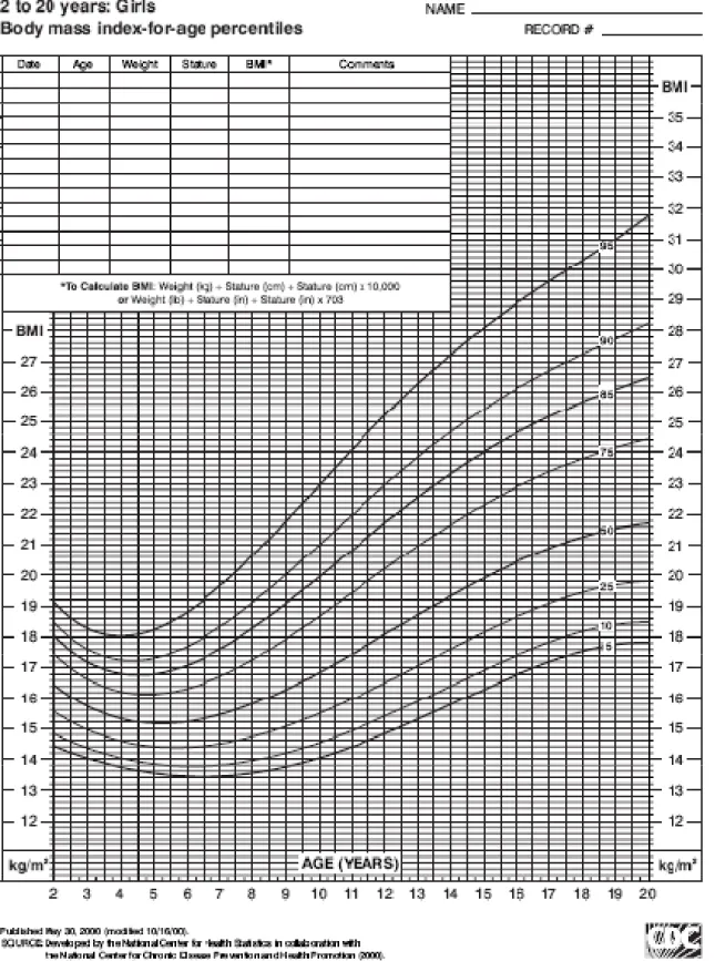
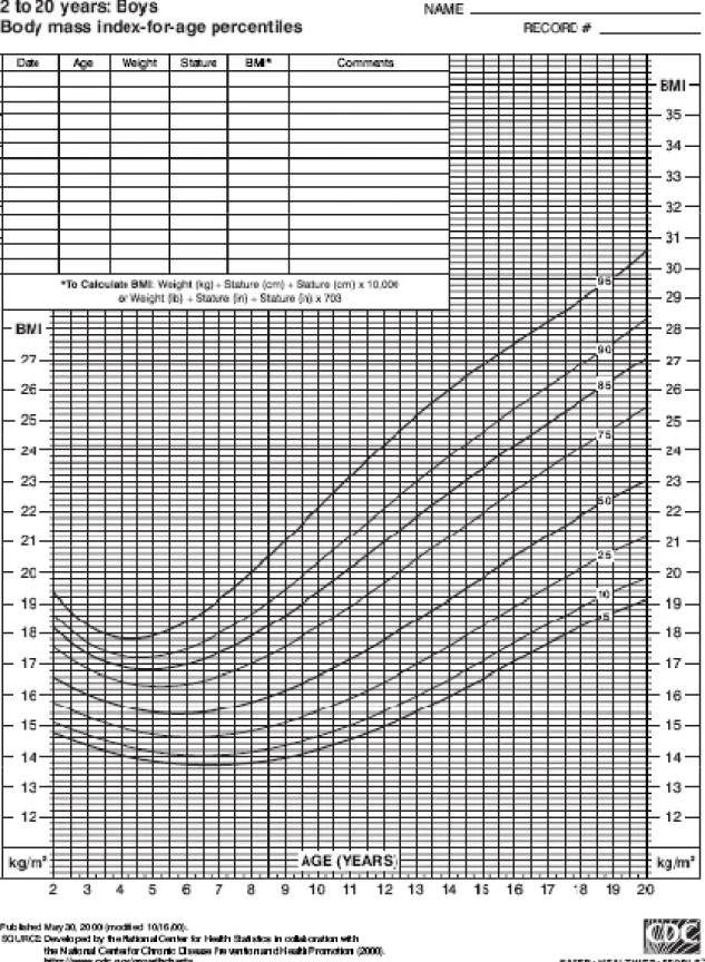
Body Mass Index (BMI) is a measure of weight compared with height, calculated as weight in kilograms divided by height in meters squared. The categories of body mass index for adults are as follows (CDC, 2010):
- Underweight: BMI <18.5 kg/m2
- Healthy weight: BMI between 18.5 and 24.9 kg/m2 - Overweight: BMI between 25 and 29.9 kg/m2 - Obese: BMI over 30 kg/m2
In growing children and adolescent patients, BMI is age-specific and sex-specific and are calculated using BMI-for-age curves. Using percentiles on such curves developed by the Center for Disease Control (Figures 1 and 2), the BMI-for-age weight status categories are as follows:
- Underweight: 5th percentile
- Healthy weight: 5th to 84th percentile
- Overweight: 85th to 94th percentile - Obese: ≥95th percentile (Barlow, 2007)
Polysomnography (PSG) is a mechanical recording of a patient’s sleep using many criteria, such as the amount of oxygen in the bloodstream, pulse, brain waves, and eye movement, among others. PSG continuously and simultaneously records physiologic variables during sleep. One can record EEG, EOG, EMG, ECG, airflow, snoring, thoracic and abdominal movement, and SaO2 using PSG. Other sensors that may be used during PSG
include esophageal pressure monitors, PetCO2, PtcCO2, PAP level, additional EEG channels
for evaluation of suspected nocturnal seizures, video-monitoring for evaluation of suspected parasomnias or seizures, and esophageal pH sensors for evaluation of suspected GER.
PSG is indicated for diagnosis of SDB, PAP titration for SDB, follow-up after UA surgery or dental devices for OSA, diagnosis of narcolepsy and periodic limb movement disorder, evaluation of atypical or injurious parasomnias or of suspected nocturnal seizures.
1.2.4 Epidemiology of SDB and the economic impact of sleep apnea
The reported frequency of disordered breathing varies depending on the population studied, the methods used to detect apnea/hypopnea, or the threshold used to define abnormalities. Several epidemiological studies estimate that 2% to 5% of middle-aged adults are affected by OSA (Young et al., 1993), but many cases might actually be undiagnosed. Snoring, in contrast, is reported as affecting as much as 40% of the adult population (Kryger et al., 2005).
Upper airway obstruction with snoring or obstructive sleep apnea is also frequently seen in children of all ages. Snoring is common, occurring in about 10-12% of young children (O'Brien et al., 2004), with a decreased frequency after the age of 9 years. Interestingly, the prevalence of snoring tends to increase threefold during pregnancy (Lavigne et al., 2009).
A study conducted at the Université de Montréal on an orthodontic population reported that 10.9% of 13 year old children were usual snorers, 17.7% breathed heavily or loudly during sleep, and 5.3% were loud snorers, whereas observed apneas were noted in just 1.8% of patients (Morton, 2008). These results agree with those of other studies
investigating SDB in children (Gislason and Benediktsdottir, 1995;Redline et al., 1999). Childhood OSA has a minimum prevalence of 2 to 3% but some studies reported up to 10 to 20% for habitual snoring children (Young et al., 2002).
The American Academy of Sleep Medicine estimated that 25 million Americans suffer from sleep apnea and that 38,000 persons die each year from complications of sleep apnea (Madani and Madani, 2007b). As many as 2.5 million patients are investigated in sleep laboratories every year and at least half of them are treated by continuous positive airway pressure. The United States Department of Transportation reported that at least 50,000 individuals each year are involved in motor vehicle accidents because of sleep apnea. Putting a dollar figure on everything, it is estimated that the minimum cost of sleep apnea to the US economy is $75 billion each year (Madani and Madani, 2007b).
1.2.5 Anatomy and physiology of upper airway obstruction
Upper airway (UA) airflow is determined by (1) the difference between upstream (i.e., nasal) and downstream pressure, and (2) airway resistance. As a result, airflow is greater with increased upstream pressure, decreased downstream pressure, and decreased airway resistance.
The normal function of the UA requires the capacity for both patency and closure. Pharyngeal airway patency is maintained by a balance between the factors that maintain airway opening (e.g., action of the upper airway dilator muscles) and those that promote airway closure (e.g., negative intraluminal extrathoracic pressure, Bernoulli forces). Airway size is also influenced by lung volume, which decreases during sleep.
The UA includes the extrathoracic trachea, larynx, pharynx, and nose. Of these, the pharynx is the only collapsible segment and can be divided into four sections (Figure 3.):
A. the nasopharynx (from the nasal turbinates to the hard palate), B. the velopharynx (from the hard palate to the tip of the uvula),
D. hypopharynx (from the tip of the epiglottis to the level of the vocal cords).
Figure 3 Segments of the upper airway as seen on midsagittal magnetic resonance imaging (adapted from Kryger’s Principles and Practice of Sleep Medicine, 5th edition)
The most common site of UA collapse in OSA patients is the velopharynx or retropalatal region, although the collapse can extend to other sites or even begin at other locations within the upper airway (Lavigne et al., 2009). An additional site often cited as a primary narrowing location is the retroglossal region (Launois et al., 1993).However, most patients with OSA have more than one site of narrowing.
Abnormal UA anatomy and possibly abnormal neural control during sleep can lead to pharyngeal airway collapse in patients with OSA, who have already been shown to have a narrowed, more collapsible pharyngeal airway.
Repetitive UA obstruction due to reduced activity of UA dilating muscles during sleep is associated with:
- episodic falls in SaO2
- snoring (alternating with periods of silence)
- arrhythmias (relative bradycardia during airway obstruction followed by tachycardia during termination of apnea)
nasopharynx velopharynx oropharynx hypopharynx
- arousal at the termination of the event
- increased BP in the immediate post-apneic period.
1.2.6 Snoring
Before the 1970s, snoring was thought to be simply an acoustic nuisance. Presently, with the understanding of snoring and obstructive sleep apnea as a continuum of sleep-disordered breathing, its main clinical significance is that it is a marker of obstructive sleep apnea syndrome and upper airways resistance syndrome (Lugaresi et al., 1983).
The ICSD defines snoring as a “respiratory sound generated in the upper airway during sleep that typically occurs during inspiration but may also occur in expiration.” (American Academy of Sleep Medicine, 2005) Snoring is not caused by any specific, localized abnormality within the airway; any membranous part of the upper airway that lacks cartilaginous support can vibrate (soft palate, uvula, faucial pillars, or pharyngeal walls). There are a number of alternate names for nonapneic snoring, including “primary snoring,” “simple snoring,” “benign snoring,” “habitual snoring,” and “heavy snoring.”
The polysomnographic criteria to differentiate nonapneic snoring from snoring that is associated with adverse consequences are not yet fully defined. Nevertheless, there is a consensus that estimates that from the number of patients with habitual snoring, approximately 2-3% have clinically relevant disease, which, given the prevalence of 10 to 20% for habitual snoring in children, brings us to a ratio of habitual snoring to OSA of about 3:1 to 5:1 (Schechter, 2002).
Snoring implies incomplete obstruction of the UA and patients who snore are considered to be at increased risk of cardiovascular disease, especially if snoring is associated with OSA. About 20% of patients who snore may also suffer from obstructive sleep apnea (Lavigne et al., 2009). Other commonly cited adverse health consequences associated with snoring are excessive daytime sleepiness (EDS) (Kryger et al., 2005) behavioural and cognitive deficits (attention, language, memory and executive function), and
mood disorders (O'Brien et al., 2004). All children should be screened for snoring, given the gravity of health problems associated with this condition.
Methods of measuring snoring vary, most often being a combination of objective sound measurements (maximum sound intensity, percentage of sleep time spent above a threshold level) and subjective assessment with questionnaires, preferably with input from the bed partner (Dalmasso and Prota, 1996). Snoring should be differentiated from OSA, UARS, or laryngospasm (Lavigne et al., 2009).
1.2.7 Obstructive sleep apnea
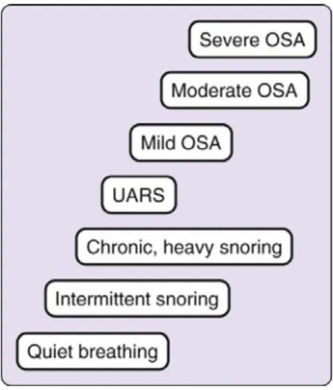
OSA represents a spectrum of disease, ranging from upper airway resistance syndrome (UARS) to OSA syndrome (Lavigne et al., 2009), which is the most common sleep-related breathing disorder and is considered a public health disease. In adults, it is characterized by the repetitive, complete or partial, collapse of the pharyngeal airway during sleep and the need to arouse to restart ventilation. Sleep is thus disrupted, yielding waking somnolence and diminished neurocognitive performance. Pediatric sleep apnea is similar to its adult counterpart, but cortical arousals might not occur, possibly because of a higher arousal threshold (Kryger et al., 2005).
In the current ICSD (2005), UARS is classified under OSA, although traditionally UARS was considered an intermittent form of SBD, between snoring and frank OSA (Figure 4.), characterized by a partial collapse of the upper airway, without the occurrence of obstructive apneas and hypopneas, or an AHI lower than 5 events per hour (Guilleminault et al., 1993). Only a polysomnographic examination can distinguish frank OSA from UARS.
Pathogenesis of OSA
The pathogenesis of OSA is not yet fully understood, but there are certain fundamental characteristics of respiratory airway modifications in patients with OSA which are well described in the literature. For instance, it has generally been shown that in OSA patients the size of the upper airway is smaller, rendering it more susceptible to collapse, and
the long axis of the upper airways tends to be oriented in an anteroposterior direction rather than laterally (Lavigne et al., 2009). The magnitude of the extraluminal pressure plays an equally important role in the collapsibility of the upper airway, and this is determined by the interaction of the volume of soft tissue within the upper airway and the size of the bony compartment.
Figure 4. The spectrum of sleep disordered breathing (Kryger et al., 2005)
Imaging studies using MRI, cephalometry or CT have shown that the volume of soft tissue structures is larger in OSA patients, whereas the bony structures tend to be diminished or retropositioned (smaller mandible, retropositioned maxilla, inferiorly positioned hyoid bone). It appears, therefore, that patients with OSA have anatomically compromised upper airways resulting from skeletal abnormalities, soft tissue abnormalities, or a combination of these factors (Ryan and Bradley, 2005).
Several imaging studies have identified a number of craniofacial factors that can predispose individuals to compromised airway space and the development of OSA:
- retruded maxilla and mandible in relation to cranial base - increased mandibular plane angle and anterior facial height - inferior displacement of the hyoid bone
- reduced length of the mandible
- narrowed posterior airway space and longer soft palate - increased tongue size and/or
- increased craniocervical angulation.
In children, OSA is usually related to enlarged tonsils and/or adenoids, and more recently, with childhood obesity (Dayyat et al., 2009). In a study conducted at the Université de Montréal on a childhood orthodontic population, SDB symptoms have been primarily associated with adenotonsillar hyperthrophy, several craniofacial morphological characteristics (dolichofacial, high mandibular plane angle, narrow palate, severe crowding of the maxilla and mandible), allergies, frequent colds, and habitual mouth breathing (Morton, 2008). In this particular study, obesity per se was not shown to be related to an increased prevalence of SDB. Recent studies (Lee et al., 2010) also show a different balance between craniofacial structures and obesity in patients with OSA of different ethnicities, with increased obesity having a bigger impact on the severity of OSA for a Chinese population as compared to Caucasian populations.
Reduced nasal breathing during wakefulness has long been related to craniofacial abnormality and a cause-and-effect relationship has been proposed (Linder-Aronson, 1970;Woodside et al., 1991). Mouth breathing due to increased nasal resistance alters craniofacial growth thus increasing SDB severity. It has been demonstrated, in both animal and human studies, that mouth breathing leads to an altered pattern of muscle recruitment in the oral and nasal capsule, resulting in skeletal changes (Lavigne et al., 2009). Children with adenotonsillar hypertrophy and mouth breathing have an extended posture of the head, a retrognathic mandible, larger anterior facial height, steeper mandibular plane, lowered position of the hyoid bone, and anteroinferior posture of the tongue when compared to normal children (Woodside et al., 1991).
The soft tissue stretch theory (Solow and Kreiborg, 1977), also developed in an attempt to elucidate the relationship between mouth breathing and craniofacial growth,
postulates that mouth breathing leads to postural changes (3-5° extended craniocervical posture), an altered pattern of muscle function, and concurrent skeletal changes. As the removal of the adenoids has been shown to reverse craniocervical angulation, increased nasal resistance has been been considered a trigger to the extended craniocervical posture.
Recent reports from large epidemioligical studies (the Third National Health and Nutrition Examination Survey) conducted in the United States challenge these previously reported findings that showed a relationship between respiratory function and malocclusion (Lavigne et al., 2009). After controlling for age, race, gender, orthodontic treatment, and socioeconomic status, no significant difference has been found in the prevalence of respiratory disease and/or allergy in individuals with posterior crossbite, negative overjet, open bite, or excessive overjet compared to the prevalence in controlled subjects without these malocclusions. Further studies need to be conducted in order to elucidate this controversial issue.
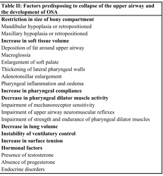
There is also a series of nonanatomic factors that are implied in the pathogenesis of OSA, and these are related to the activity of the pharyngeal dilator muscles and the central control of ventilation. Table II summarizes the anatomic and nonanatomic factors implicated in the pathophysiology of OSA.
There is strong clinical evidence for a significant familial aggregation for symptoms of OSA in children and adults (Redline et al., 1994;Redline and Tishler, 2000;Taheri and Mignot, 2002). Genetics may determine upper airway anatomy, neuromuscular activity, ventilatory control stability, sleep-wake patterns, obesity predisposition, or can explain the occurence of OSA in particular ethnic groups. A brachycephalic head form, measured by anthropometry, is often found in association with reduced upper airway dimensions. This head form is also associated with a small but significant increased risk of OSAH in whites, and it also identifies families at risk for both OSAH and sudden infant death (Cakirer et al., 2001). Genetic studies consistently estimate heritability for AHI to be between 35% and 40% (Larkin et al., 2008).
Table II: Factors predisposing to collapse of the upper airway and the development of OSA
Restriction in size of bony compartment Mandibular hypoplasia or retropositioned Maxillary hypoplasia or retropositioned Increase in soft tissue volume
Deposition of fat around upper airway Macroglossia
Enlargement of soft palate
Thickening of lateral pharyngeal walls Adenotonsillar enlargement
Pharyngeal inflammation and oedema Increase in pharyngeal compliance
Decrease in pharyngeal dilator muscle activity Impairment of mechanoreceptor sensitivity
Impairment of upper airway neuromuscular reflexes
Impairment of strength and endurance of pharyngeal dilator muscles Decrease in lung volume
Instability of ventilatory control Increase in surface tension Hormonal factors
Presence of testosterone Absence of progesterone Endocrine disorders
(Adapted from G. Lavigne et al, Sleep Medicine for Dentist, 2009 Quintessence Publishing) Risk factors for OSA
The presence of certain risk factors can strengthen the clinical suspicion of OSA. The strongest risk factors are obesity and age older than 65 years (Grunstein et al., 1993). There has been shown to be an association between OSA and android-type obesity (fat deposition predominantly in the neck and abdomen), in contrast to the gynecoid-type obesity (with fat deposition in hips and legs) (Guilleminault et al., 1988). A positive family history increases the risk of sleep-disordered breathing by twofold to fourfold (Redline et al., 1995). Several
studies have shown that SDB is also exacerbated by alcohol ingestion, especially around bedtime.
Additional risk factors include ethnicity, craniofacial abnormalities such as Marfan syndrome, Down syndrome, the Pierre-Robin syndrome, and other congenital craniofacial anomalies. Specific craniofacial and oropharyngeal features include: increased neck circumference (> 17 inches in men, >16 inches in women), nasal narrowing or congestion, macroglossia, low-lying soft palate, enlarged tonsils and adenoids (especially in children), mid-face hypoplasia, retrognathia, micrognathia or mandibular hypoplasia, and tracheal stenosis and laryngomalacia.
Adenotonsillar enlargement represents the most important risk factor in children. However, size of the tonsils and adenoids is not predictive of OSA in individual patients. Other risk factors in children are a positive family history, excess body weight, chronic nasal obstruction (e.g., allergies, choanal atresia or stenosis), and craniofacial abnormalities
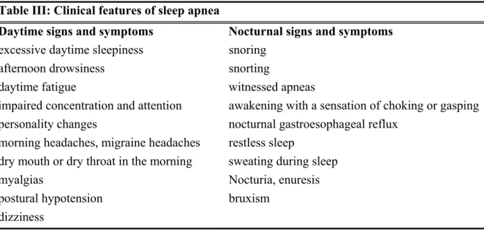
1.2.8 Clinical signs and symptoms of SDB
Patients with SDB manifest a variety of nocturnal and diurnal signs and symptoms (Table III). Almost all patients with OSA and many UARS patients snore. The bed partner usually reports chronic, heavy snoring, snorting, difficulty breathing during sleep, or witnessed breathing pauses through sleep.
Excessive daytime sleepiness (EDS) is the most common complaint, but its severity doesn’t correlate closely with AHI. The manifestations of sleepiness can have subtle consequences (mid-afternoon drowsiness during a group meeting or an occasional nap), severe consequences (falling asleep while eating or talking), or catastrophic consequences (falling asleep while driving).
Table III: Clinical features of sleep apnea
Daytime signs and symptoms Nocturnal signs and symptoms excessive daytime sleepiness snoring
afternoon drowsiness snorting
daytime fatigue witnessed apneas
impaired concentration and attention awakening with a sensation of choking or gasping personality changes nocturnal gastroesophageal reflux
morning headaches, migraine headaches restless sleep
dry mouth or dry throat in the morning sweating during sleep
myalgias Nocturia, enuresis
postural hypotension bruxism
dizziness
(Adapted from Kryger,M., Roth,T., & Dement,W. Principles and practice of sleep
medicine, 2005)
Obesity is another hallmark of sleep apnea, especially increased neck circumference and central obesity. Although patients with OSA are usually obese older men, about 30% of patients with sleep apnea are nevertheless not obese (Malhotra and White, 2002) and the presence of other clinical signs and symptoms must trigger an OSA diagnosis. Hypertension, crowded pharynx, and retrognathia are also usual signs of SDB.
Contrary to adult OSA, in children with sleep apnea the clinical features are less distinguished, with many signs and symptoms inconstantly present. EDS, for instance, is reported as being between 7% (Carroll et al., 1995) and 40%-50% (Chervin et al., 2006), depending on the type of questionnaire used to interogate subjects. EDS should be considered in any child older than 5 years of age who continues to nap during the day, especially if unplanned, or sleeps at least 2 hours more on weekends than on weekdays (‘weekend oversleep’).
Other common EDS features in children are:
- behavioural problems (inattentiveness, irritability, hyperactivity or impulsiveness)
- cognitive problems or academic difficulties - changes in mood (depression or anxiety) - fatigue and lethargy.
Nevertheless, with an increased prevalence of pediatric obesity, we have witnessed a concomitant increase in childhood OSA, the clinical manifestations of which increasingly tend to resemble those of adult OSA.
1.2.9 Diagnosis of SDB
A variety of tools exist to evaluate SDB patients, including questionnaires, clinical examination, or diagnostic imaging, but the gold standard for the diagnosis of SDB is overnight polysomnography (PSG), that includes recordings of airflow, ventilatory effort, oxygen saturation, and body position, as well as ECG, EMG, and EEG. Given the need for the patient to spend a night in a sleep laboratory for a PSG recording and the expenses associated with that, more recently, home sleep-testing devices have been developed (Sunwoo and Kuna, 2010). These new devices are less sensitive than the attended laboratory PSG and are only indicated for patients with a high clinical suspicion for SDB.
There are two schemes to assess severity of OSA. The more conservative one considers that for an adult an AHI of 5 to 20 is mild OSA, 21 to 50 is moderate, and more than 51 is severe OSA (Madani and Madani, 2007a). The second and more liberal one, which is more widely used, judges OSA as being mild for an AHI between 5 and 15 events per hour, moderate for AHI of 15-30 and severe for more than 30 obstructive breathing events per hour (Kryger et al., 2005). In children, an AHI of 1 event or more per hour of sleep is considered abnormal.
Other factors that influence the clinical severity of OSA, apart from AHI, include the degree of EDS, the nadir of SaO2, the extent of sleep fragmentation, the presence of
Lateral cephalometric radiographs, routinely used by orthodontists, have long been considered valuable diagnostic tools for the upper airway obstructions, as they can provide a good assessment of the skeletal and dental maxillary and mandibular relationships, as well as soft tissue relationships of the palate, tonsils, adenoids, and posterior pharynx. More recently, with the advent of the CBCT technology that provides a three-dimensional view of the upper airways, the shortcomings of previously used two-dimensional radiographs have been surmounted.
Questionnaires are important in detecting possible sleep breathing disorders and a multitude of questionnaires have been developed for both adult and pediatric populations. Questionnaires such as the Berlin Questionnaire, the Pittsburgh Sleep Quality Index, the Epworth Sleepiness Scale, or the Sleep Apnoea Quality of Life Index are all validated screening instruments used for adults (Buysse et al., 1989a;Chasens et al., 2009;Chervin et al., 1997;Lacasse et al., 2002;Netzer et al., 1999;Sagaspe et al., 2010). Pediatric sleep questionnaires (Acebo et al., 2005;Kushida et al., 2001;Montgomery-Downs et al., 2006) have also been developed and used in the past three decades, but low sensitivity and specificity have been noted related to OSA diagnosis, most probably caused by the difficulty of differentiating primary snoring from OSA (Carroll et al., 1995).
Several studies questionned the validity of child sleep behaviour information provided by parents who do not necessarily share the room with their children (Chervin et al., 2007;Lumeng and Chervin, 2008); even so, given the expenses associated with SDB diagnosis based on PSG, questionnaires and clinical examinations remain the main instruments used for epidemiological screening of sleep disorders.
1.2.10 Consequences of SDB
There is an increasing body of evidence that points to OSA as a feature of the metabolic syndrome. The metabolic syndrome is a collection of closely related symptoms that together induce an increased risk of cardiovascular disease. Visceral obesity, hypertension, insulin resistance, hyperglycemia and dyslipidemia, are all symptoms of the
metabolic syndrome, with the visceral adiposity being considered the major determinant of this condition. Sleep fragmentation, common to OSA, has been linked to a decrease in insulin sensitivity (Tasali et al., 2008), whereas oxyhemoglobin desaturation has correlated with serum triglyceride and LDL cholesterol levels (Savransky et al., 2008).
Patients with sleep apnea demonstrate variable degrees of neurocognitive deficiencies such as slowed thought processes, forgetfulness, delayed reaction time responses, and inability to concentrate. Both sleep fragmentation and its consequence of EDS have been invoked as causative factors of these neurocognitive impairments. EDS also has an impact on neurobehavioural performance, with many reports of OSA patients manifesting personality changes, irritability or aggressiveness.
The major consequencesof pediatric OSA involve neurobehavioural, cardiovascular, somatic growth, endocrine and metabolic systems. The criteria for the metabolic syndrome (insulin resistance, dyslipidemia, hypertension and obesity) are not yet entirely defined in children; nevertheless, increased evidence points to elevated circulation levels of leptin, an adipokine that regulates appetite, metabolic homeostasis, sleep, and respiratory control, in both adults and children (Tauman et al., 2007). Similar to adult OSA, pediatric OSA has now been associated with an increased risk for cardiovascular morbidities, although with a reduced severity of these manifestations (Bhattacharjee et al., 2009). However, it is anticipated that the frequency and severity of the cardiovascular consequences of pediatric OSA will be amplified by the simultaneous increase in obesity and diabetes among children. Sleep disruption through a period of rapid neurological development may also lead to important neurocognitive and behavioural deficiencies such as hyperactivity, poor school performance, learning or memory impairments, depression, mood changes, and overall problems with quality of life and self-esteem.
1.2.11 Treatment of SDB
Obstructive sleep apnea can be a lethal disease if not treated. There is increasing evidence showing an elevated risk of cardiovascular complications and death for patients
with moderate and severe OSA (Young et al., 2008), independent of age, sex, or obesity status, in particular for those who manifest both snoring and excessive daytime sleepiness (Lindberg et al., 1998).
Treatment options for non-apneic snoring and OSA are basically similar, divided into nonsurgical and surgical approaches. Medical approaches, including risk modification (manipulation of body position during sleep; avoidance of alcohol and sedating medications), weight loss, and positive airway pressure (PAP) modalities are usually the first avenues of treatment. Patients who are unable or unwilling to comply with medical management may be candidates for surgery.
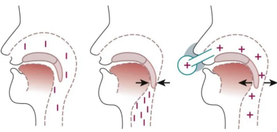
CPAP therapy
Continuous positive airway pressure (CPAP) is the gold standard of treatment for moderate to severe OSA; it may also be appropriate treatment for mild but symptomatic cases. Magnetic resonance imaging studies showed that CPAP increases airway volume and airway area and reduces lateral pharyngeal wall thickness and the upper airway edema that result from chronic vibration and occlusion of the airway (Schwab et al., 1996). Figure 5 shows the mechanism of UA occlusion and its prevention by nasal CPAP. When the patient is awake (left), muscle tone prevents collapse of the upper airway during inspiration. During sleep, the tongue and soft palate are sucked against the posterior oropharyngeal wall (middle). CPAP with low pressure provides a pneumatic splint and keeps the upper airway open (right).
PAP therapy reduces mortality and sleepiness associated with OSA, increases sleep quality, and reduces the frequency of arousals, and increases the overall quality of life. The main limitations of CPAP use are the patient's acceptance and tolerance of treatment.
Although the gold standard in treatment of adult OSA is CPAP, using nasal CPAP in children has proven to be extremely difficult, with a low rate of compliance (Marcus et al., 2006). Moreover, extended use of nasal CPAP in growing individuals has been incriminated in mid-face hypoplasia (Abad and Guilleminault, 2009). The treatment of choice for pediatric OSA is tonsillectomy and adenoidectomy,with or without turbinate surgery, as the
hypertrophy of these structures is the primary cause of childhood SDB (Abad and Guilleminault, 2009). Children with craniofacial abnormalities resulting in maxillary or mandibular insufficiency may benefit from palatal expansion or maxillary/mandibular surgery. PAP therapy may be used for children who are not surgical candidates or if surgery fails.
Figure 5. Mechanism of upper airway occlusion and its prevention by nasal continuous positive airway pressure (CPAP). Adapted from (Sullivan et al., 1981)
Oral appliances
Oral appliances are an established treatment option for snoring and mild obstructive sleep apnea–hypopnea in adults. It is a relatively simple and reversible approach to treatment. They appear to work by increasing upper airway space, stabilizing the anterior position of the mandible, advancing the tongue or soft palate, or both, and possibly by changing upper airway muscle activity.
Most patients report improved sleep quality and less EDS with oral appliance therapy. Additionally, there is growing evidence of improvements in other important health outcomes, including neurocognitive function and cardiovascular health. Oral appliance therapy is generally well-tolerated, and short-term side effects are usually minor and are
related to excessive salivation, jaw and tooth discomfort, and occasionally joint discomfort. These symptoms usually improve over time.
Children with OSA and a small and/or retrognathic mandible can benefit from treatment of their Class II malocclusion with orthopaedic appliances. Activator-headgear therapy followed by fixed appliance treatment has been shown to have the potential to increase pharyngeal airway dimensions and to maintain this change in UA on a long term basis, thus reducing the risk of developing long-term impaired respiratory function (Hanggi et al., 2008).
Surgical therapy
Most common surgical interventions employed to provide or facilitate maintenance of a patent upper airway during sleep are tracheotomy, nasal reconstruction (septal or bony intranasal reconstruction, alar valve or alar rim reconstruction, and turbinectomy), uvulopalatopharyngoplasty (UPPP), tongue reduction, genioglossus advancement-hyoid myotomy and suspension (GAHMS), bimaxillary advancement, or maxillary and mandibular osteotomy (MMO). There are strict indications and contraindications for surgery for the OSA patient and they are listed in Table IV.
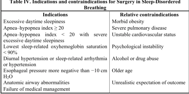
Table IV. Indications and contraindications for Surgery in Sleep-Disordered Breathing
Indications Relative contraindications Excessive daytime sleepiness Morbid obesity
Apnea–hypopnea index ≥ 20 Severe pulmonary disease Apnea–hypopnea index < 20 with severe
excessive daytime sleepiness
Unstable cardiovascular status Lowest sleep-related oxyhemoglobin saturation
< 90%
Psychological instability Diurnal hypertension or sleep-related arrhythmia
or hypertension
Alcohol or drug abuse Esophageal pressure more negative than −10 cm
H2O
Older age
Anatomic airway abnormalities Unrealistic expectation of outcome Failure of medical management
When craniofacial disharmony is causative of sleep apnea or chronic snoring, maxillary and/or mandibular advancement surgery can be a valid treatment option. Maxillary advancement can be performed orthopedically (Kilinc et al., 2008) in a growing patient, or surgically in an adult patient. There are not many studies that investigated the effect of these two procedures on upper airway; there are reports (Samman et al., 2002) of increased nasopharyngeal depth following surgical advancement of the maxilla, but lack of controlled studies make such findings inconclusive.
Surgical mandibular advancement is usually performed in conjunction with genioglossus advancement and addressesupper airway obstruction at the base of the tongue. It helps to stabilize the tongue base along with the associatedpharyngeal dilatators.
Maxillomandibular advancement (MMA) osteotomy is usually used for patients with significant OSA who cannot or will not use CPAP or who have frank mandibular deficiency. MMA is perhaps the most effective surgery for improving OSA when performed on appropriately selected patients. Studies report a reduction in the postoperative respiratory disturbance index (RDI) of atleast 50%, with an average improvement of greater than 85%, in approximately 90% of patients (Won et al., 2008).
Orthodontic therapy
Given the common finding of craniofacial abnormalities in patients suffering from OSA, it has been hypothesized that improvement and normalization of the dentofacial complex may have an impact on respiratory problems and OSA. The three major anatomic regions of potential collapse during sleep in patients with SDB include the nose, palate, and tongue base. Each region can be surgically or orthopedically reconstructed on its own or in combination as necessary.
A narrow maxilla has been associated with an increased prevalence of nasal obstruction and OSA symptoms in children. Maxillary constriction may increase nasal resistance and alter the tongue position, leading to narrowing of the retroglossal airway. Consequently, several studies have investigated the effects of maxillary expansion, a
common orthopaedic treatment for maxillary constriction, on the volume of the nasal cavity and as a result on OSA symptoms.
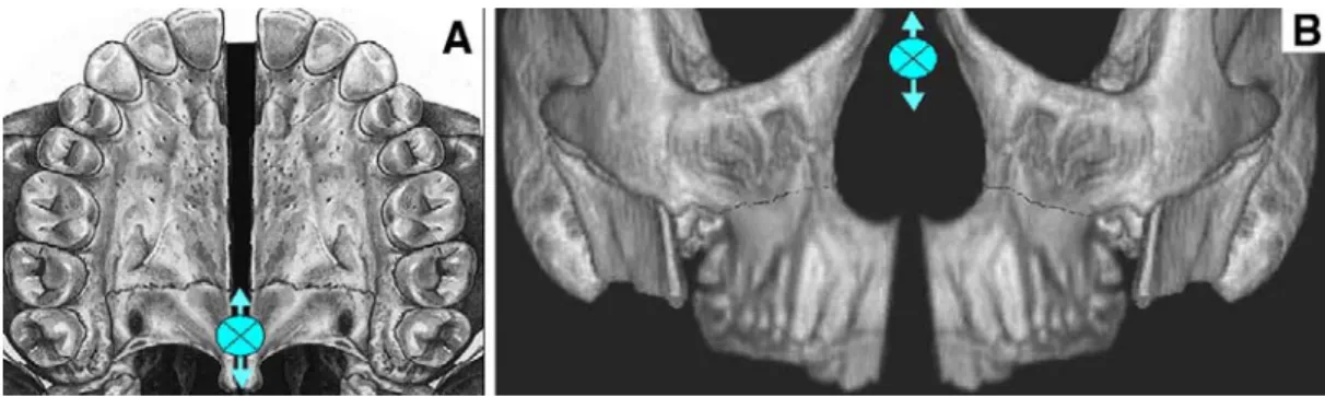
It has been found that rapid palatal expansion (RPE) widens the nasal fossa and releases the septum (see Figure 6), thus restoring normal nasal airflow and resolving OSA in children with a case history of oral breathing, snoring and night time apneas (Pirelli et al., 2005).
Figure 6. Sutural opening after RPE. A, horizontal view: V-shaped opening of palatum durum after RME, with the rotational center near the third molar; B, Frontal view: V-shaped rotation of the maxilla after RME with the rotational center near the frontonasal suture. Adapted from (Deeb et al., 2010)
Similar results have been documented in adults with narrow maxilla and OSA who had RPE or surgically assisted RPE (SARPE); major reductions in snoring, OSA, and EDS were found following these procedures (Cistulli et al., 1998). Conversely, a recent study (Langer et al., 2011) investigated the long-term effects of RPE on nasal resistance in children but couldn’t detect significant differences in nasal airway resistance 30 months after RME. As the long-term stability of the procedures that involve widening of the maxilla is questionable, further studies are needed to evaluate the consequent stability of respiratory improvements following RPE/SARPE. Moreover, a cause-and-effect relationship between OSA and the width of the naso-maxillary complex needs to be established.
Conversely, there are some orthodontic treatments routinely used by orthodontists that were incriminated in inducing or exacerbating SDB symptoms. Headgear therapy, for example, has been shown to possibly contribute to the occurrence of OSA when a strong predisposition, such as mandibular retrognatia, already exists (Godt et al., 2011;Pirila-Parkkinen et al., 1999). Similarly, extraction treatment can be speculated to reduce available tongue space and consequently impinge on the upper airway, worsening SDB symptoms. A study has recently been designed (Valiathan et al., 2010) to evaluate the effect of four-premolar extraction therapy versus non-extraction therapy on upper airways, but no statistically significant oropharyngeal airway volume changes were found between the two groups, despite expected changes in incisor angulations and position.
Given the scarcity of, and sometimes contradictory results in the literature regarding the effects of common orthodontic therapy on SDB symptoms, further evidence and research is needed on the prevalence and evolution of SDB symptoms in orthodontic patients, as well as the impact of all orthodontic interventions on upper airways.
2 Chapter 2
2.1 Rational and objectives
An epidemiological study was conducted at the screening clinic of the graduate orthodontic department of the Université de Montréal on 604 subjects recruited between 2006 and 2007 (Huynh N et al., 2011;Morton, 2008). The aims of that study were (1) to determine the prevalence of SDB and associated morphological and health-related factors in an orthodontic population and (2) to determine the relationship between patient characteristics implicated in reduced UA dimensions and OSA symptoms reported from a pediatric sleep questionnaire.
The current study is established as a follow-up of the initial study conducted at the orthodontic screening clinic of the Université de Montréal.
The aims of the present study were: (1) to describe the prevalence and main risk factors of sleep-disordered breathing (SDB) in children and adolescents having undergone orthodontic treatment, and to compare them with the baseline values; (2) to investigate to what extent different orthodontic treatment procedures affect the UA, influencing SDB symptoms; (3) to provide knowledge transfer by dissemination and communication to peer researchers through conference presentations and publications in peer-reviewed journals.
In particular, we hypothesize that the prevalence of SDB symptoms do not change during childhood and adolescence, and that certain orthodontic interventions have an impact on the UA, influencing SDB symptoms in this particular population.
3 Chapter 3
A previous study of our group investigated, in a cross-sectional manner, the prevalence of SDB symptoms and their associated craniofacial traits in a child and adolescent population seeking orthodontic treatment (Huynh N et al., 2011;Kryger et al., 2005). The prevalence of these symptoms generally mirrored those previously reported in the literature. The craniofacial features that were identified as SDB risk factors by this study were the dolichofacial, high MPA, narrow palate, predominantly mouth-breathing patient, who also had severe crowding of the maxilla and mandible. In this particular population, obesity was not identified as a risk factor for SDB.
After an interval of four years, a follow-up study was conducted and a subgroup of the initial population was subjected to the same investigations as in the baseline study. Most of the participant subjects have had an orthodontic intervention in the meantime, and are now in retention phase, or are still in an active phase of orthodontic treatment. The study, as well as the orthodontic treatment of the subjects, was carried out in the graduate orthodontic department of the Université de Montréal.
Results of the aforementioned follow-up study are presented in the current thesis under the format of a manuscript in preparation for publication in the American Journal of Orthodontics and Dentofacial Orthopedics.
Manuscript in preparation to be submitted for publication in the AJODO
Longitudinal evaluation of sleep-related
breathing disorders in an orthodontic
population
Authors:
Mandu-Hrit Ma,b, Huynh Na, Turkewicz Ja, Morton Pa, Papadakis Aa, Rompré PHa, Nicolau Bb
aFaculté de médecine dentaire, Université de Montréal bFaculty of Dentistry, McGill University
3.1 Abstract
Introduction: Sleep-disordered breathing (SDB) has significant effects on a child’s health, behaviour, and performance. Craniofacial malformations represent important risk factors in the development of this condition. SDB signs and symptoms in children and adolescents followed for orthodontic treatment in a university setting have been investigated in this prospective study. The aims of this study were to describe the prevalence and main risk factors of SDB and the impact of different orthodontic interventions on the SDB symptoms for this specific population.
Materials and methods: 168 subjects aged 12 to 21 years (mean age ± SD was 16.48±2.19) underwent a clinical craniofacial examination in addition to being administered self-completed questionnaires that collected information on socio-demographic and psychosocial factors, bruxism and temporo-mandibular joint (TMJ) disorders, sleep and daytime behaviour, and neuropsychological factors.
Results: Body mass index (BMI) was slightly increased but remained in the same category at the two time points of investigation. There was an increase in clenching and TMJ symptoms, a decrease in tonsil size, and an increase in daytime sleepiness. Prevalence of SDB was constant at baseline and follow-up studies. No orthodontic treatment intervention proved to have a statistically and clinically significant impact on upper airway.
Conclusions: SDB signs and symptoms prevalence was constant when compared to the baseline values for the studied population, but increased when compared to the general population. Regular orthodontic treatment didn’t show any effect on SDB symptoms.
Key words: craniofacial, orthodontic treatment, prevalence, sleep apnea, snoring, upper airway
3.2 Introduction
Sleep-disordered breathing (SDB) represents a growing health concern. Cessation of breathing during sleep can result from obstruction of the upper airway (obstructive sleep
apnea - OSA), absence of inspiratory effort (central apnea), or a combination of the two. Several epidemiological studies estimate that 2% to 5% of middle-aged adults are affected by obstructive sleep apnea (OSA) (Young et al., 1993), but many cases might actually be undiagnosed. Snoring, in contrast, is reported as affecting as much as 40% of the adult population (Kryger et al., 2005).
Childhood OSA has a minimum prevalence of 2 to 3%, but some studies report up to 10 to 20% for habitual snoring children (Young et al., 2002). A study conducted at the Université de Montréal on an orthodontic population found that 10.9% of 13 year old children were usual snorers, 17.7% breathed heavily or loudly during sleep, and 5.3% were loud snorers, whereas observed apneas were noted in just 1.8% of patients (Huynh N et al., 2011;Morton, 2008). These results agree with those of other studies investigating SDB in children (Gislason and Benediktsdottir, 1995;Redline et al., 1999). Major consequencesof pediatric OSA affect neurobehavioural, cardiovascular, somatic growth, endocrine and metabolic systems.
Abnormal upper airway (UA) anatomy and possibly abnormal neural control during sleep can lead to pharyngeal airway collapse in patients with OSA, which have been shown to have a narrowed, more collapsible pharyngeal airway. Several imaging studies have identified a number of craniofacial factors that can predispose individuals to compromised airway space and the development of OSA: retruded maxilla and mandible in relation to cranial base, increased mandibular plane angle and anterior facial height, inferior displacement of the hyoid bone, reduced length of the mandible, narrowed posterior airway space and longer soft palate, increased tongue size, increased craniocervical angulation (Bixler et al., 2009;Huynh N et al., 2011;Riha et al., 2005).
Adenotonsillar enlargement represents the most important risk factor in children. However, size of the tonsils and adenoids is not predictive of OSA in individual patients. Other risk factors in children are a positive family history, excess body weight, chronic nasal obstruction (e.g., allergies, choanal atresia or stenosis), and craniofacial abnormalities. A previous report by our research group demonstrated statistically significant associations
between clinical signs of malocclusion and reported symptoms of SDB. According to Huynh et al, 2011, a pediatric patient that is at increased risk of developing SDB is dolichofacial, has a high mandibular plane angle, a narrow palate, is predominantly a mouth-breather, and demonstrates severe crowding of the maxilla and mandible.
Given the common finding of craniofacial abnormalities in patients suffering from OSA, it has been hypothesized that improvement and normalization of the dentofacial complex may have an impact on respiratory problems and OSA. Several studies demonstrated that rapid palatal expansion (RPE) widens the nasal fossa and releases the septum, thus restoring normal nasal airflow and resolving OSA in children with a case history of oral breathing, snoring and night time apneas (Pirelli et al., 2005). Conversely, there are some treatment regimens routinely used by orthodontists that were incriminated in inducing or exacerbating SDB symptoms. Headgear therapy, for example, has been shown to possibly contribute to the occurrence of OSA when a strong predisposition, such as mandibular retrognathia, already exists (Godt et al., 2011;Pirila-Parkkinen et al., 1999). Similarly, extraction treatment can be speculated to reduce available tongue space and consequently impinge on the upper airway, worsening SDB symptoms, although a recent study (Valiathan et al., 2010) found no statistically significant oropharyngeal airway volume changes between four-premolar extraction and non-extraction treatment groups, despite expected changes in incisor angulations and position.
The current study is established as a follow-up of an initial study conducted at the orthodontic screening clinic of the Université de Montréal with the purpose of determining the prevalence of SDB and associated morphological and health-related factors in an orthodontic population and the relationship between patient characteristics implicated in reduced UA dimensions and OSA symptoms.
The aims of the present study were: (1) to describe the prevalence and main risk factors of SDB in children and adolescents having undergone orthodontic treatment and to compare them with their baseline values and (2) to investigate how different orthodontic treatment procedures affect the UA.
3.3 Material and methods
3.3.1 Study population
The present study was of a longitudinal cohort design, representing the follow-up of an initial study conducted in 2006-2008 at the screening clinic of the graduate orthodontic program of the Université de Montréal (Huynh N et al., 2011;Morton, 2008). That study investigated 604 children and adolescents with respect to SDB prevalence and craniofacial risk factors of SDB. The study collected information on an array of social, psychological, life style and sleep quality and quantity parameters through the use of questionnaires completed by the patient and parent. A clinical head and neck examination was also completed with the goal of identifying upper airway and craniofacial abnormalities that may be related to SDB.
For the present follow-up study 168 subjects responded to the invitation to participate and all were active or retention orthodontic patients still followed in the university orthodontic clinic. The study was conducted in accordance with the university’s ethical standards. All subjects and their parents or guardians gave their written consent to participate in the study.
3.3.2 Instruments: questionnaires, clinical evaluation, and history of
orthodontic treatment
In the present study, participants underwent a clinical examination in addition to being administered self-completed questionnaires. The self-administered questionnaires collected updated information on socio-demographic and psychosocial factors, bruxism and TMJ disorders, sleep and daytime behaviour, as well as neuropsychological factors. All these questionnaires are routinely administered to the orthodontic patients and/or their parents at the Université de Montréal and they are modified and verified French translation of the: (1) medical and dental history questionnaire, (2) bruxism and TMD habits questionnaire, (3) sleep and daytime behavior (Pediatric Sleep Questionnaire), and (4) sleep
duration and quality (Pittsburgh Sleep Quality Index) (Blais et al., 1997;Buysse et al., 1989b;Carrier et al., 2005).Most of the questions were of the dichotomous type (yes/no); in selected cases with multiple variables, the data was reduced into a dichotomous variable (e.g., “never” and “rarely” into “no”, and “often” and “always” into “yes”)
All subjects also undertook a clinical head and neck examination with the goal of identifying features such as retrognathia, tonsillar hypertrophy, enlarged tongue or soft palate, and maxillary and mandibular retroposition, which can narrow upper airway dimensions and promote the occurrence of apneas and hypopneas during sleep. These measurements are also routinely registered for the patients treated in our orthodontic department. The standard orthodontic clinical evaluation was performed by an orthodontic resident (Manuela Mandu-Hrit) following a calibration session with the orthodontist that performed the clinical examination in the baseline study (Athena Papadakis). Details of the clinical examination performed were given in a previous publication of the same study group (Huynh N et al., 2011).
We recorded the subject’s profile (convex, straight, or concave), facial frontal pattern (brachyfacial, mesofacial, or dolicofacial), mandibular plane angle, palatal vault shape, asymmetries of the dental midlines to the facial midline, type of respiration (mouth, nasal breathing, or both), tongue size, and tonsil size according to the Brodsky scale (Brodsky, 1989). TMJs and lateral mandibular movements were assessed, as well as the amount of overjet (OJ) and overbite (OB). The dental components of the malocclusion were evaluated clinically as molar and canine Angle classification, crowding of dental arches, curves of Spee and Monson.
In addition to questionnaire-based investigation and clinical examination, we also collected data on the type of orthodontic treatment received by each subject in the study. We noted if the treatment involved extractions or not, and what kind of extractions (four premolar extractions, two upper premolar extractions, two lower premolar extractions, lower incisor extraction, or other extraction patterns); whether the treatment involved orthognathic surgery or not (mandibular advancement only in this sample population); if palatal