Université de Montréal
Selective inhibition of inducible nitric oxide synthase prevents lipid peroxidation in cartilage from patients with osteoarthritis.
L'inhibition sélective de l'oxyde nitrique synthétase inductible inhibe la péroxidation lipidique dans le cartilage humain arthrosique.
par Mireille Bentz
Département de Pharmacologie, Université de Montréal Faculté de médecine.
Mémoire présenté à la Faculté des études supérieures
en vue de l’obtention du grade de maîtrise (M.Sc.) en Pharmacologie
Université de Montréal
Faculté des études supérieures et postdoctorales
Ce mémoire est intitulé :
L'inhibition sélective de l'oxyde nitrique synthétase inductible inhibe la péroxidation lipidique chez les patients souffrant de l'ostéoarthrose.
Présenté par : Mireille Bentz
a été évalué par un jury composé des personnes suivantes : Dr Moutih Rafei, président-rapporteur
Mohamed Benderdour, directeur de recherche Dr Jonathan Brouillette, membre du jury
Résumé
INTRODUCTION: De nouvelles données indiquent que l’oxyde nitrique (NO), que l’on retrouve en quantité accrue au niveau du cartilage de patients atteints d’ostéoarthrose (OA), joue un rôle dans la production de 4-hydrynonénal (HNE) via la formation de péroxynitrite (ONOO-). Le HNE est considéré comme étant un des produits les plus réactifs de la péroxidation lipidique (LPO). Notre laboratoire a rapporté des niveaux de HNE plus élevés que normal dans le liquide synovial provenant de genoux de patients OA comparativement aux sujets normaux. Nous avions aussi démontré que le HNE peut induire la production des médiateurs inflammatoires et cataboliques connus pour leurs implications dans la dégradation du cartilage dans l’OA. Le but de la présente étude est de vérifier si un inhibiteur sélectif pour la NO synthétase inductible (iNOS), soit le L-NIL (L-N6-(L-Iminoethyl)Lysine), peut empêcher la formation du HNE via l’inhibition de la
production du NO dans des chondrocytes de patients OA.
MÉTHODES: Les cellules ont été traitées soit avec ou sans un générateur du NO (SIN ou interleukine-1beta (IL-1β)) soit avec ou sans du HNE pendant 48 h en présence ou en absence du L-NIL. L’expression protéique de l’iNOS et de la sous-unité de la NADPH oxydase (NOX), la p47 (phox), a été vérifiée par Western blot. La génération du HNE a été révélée par ELISA, Western blot et immunohistochimie. Les niveaux de prostaglandine E2 (PGE2), gluthation-s-transférase (GST) et de la MMP-13 ont été mesurés par des kits commerciaux. La quantité de NO a été évaluée par la méthode de la réaction de Griess. La mesure des niveaux d'espèces réactives d’oxygène (ROS) a été effectuée par fluorescence en utilisant un kit commercial.
RÉSULTATS: Le L-NIL inhibe la stimulation de la production de NO, de HNE ainsi que l’expression d’iNOS au niveau protéique et de l’ARNm par IL-1β. Le L-NIL bloque aussi la production de HNE indépendamment de la production de NO. La production des ROS et l’activation de p47 (phox) ont été inhibées par L-NIL. Fait intéressant, le L-NIL empêche la production de la PGE2, de la cyclooxygénase-2 (COX-2) et de la MMP-13 induite par le HNE.
CONCLUSION: les résultats obtenus semblent démontrer l’effet bénéfique du L-NIL dans l’OA via la prévention de la production du HNE de manière dépendante ou indépendant du NO.
MOTS CLÉS : Arthrose; Chondrocytes; Péroxydation Lipidique; 4-Hydroxynonénal; Oxyde Nitrique; Inflammation; Catabolisme; L-N6-(L-Iminoethyl)Lysine; Cartilage; Interleukine; Stress Oxydatif; Cible Thérapeutique; Oxyde Nitrique Synthétase Inductible; Espèces réactives d’Oxygène; Inhibition sélective
Abstract
INTRODUCTION: Emerging evidence indicates that nitric oxide (NO), which is increased in osteoarthritic (OA) cartilage, plays a role in 4-hydroxynonenal (HNE) generation through peroxynitrite formation. HNE is considered as the most reactive product of lipid peroxidation (LPO). We have previously reported that HNE levels in synovial fluids are more elevated in knees of OA patients compared to healthy individuals. We also demonstrated that HNE induces a panoply of inflammatory and catabolic mediators known for their implication in OA cartilage degradation. The aim of the present study was to investigate the ability of inducible NO synthase (iNOS) inhibitor, L-NIL (L-N6-(L-Iminoethyl)Lysine), to prevent HNE generation through NO inhibition in human OA chondrocytes.
METHOD: Cells and cartilage explants were treated with or without either an NO generator (SIN or interleukin 1beta (IL-1β)) or HNE in absence or presence of L-NIL. Protein expression of both iNOS and free-radical-generating NOX subunit p47 (phox) were investigated by western blot. iNOS mRNA detection was measured by real-time RT-PCR. HNE production was analysed by ELISA, Western blot and immunohistochemistry. S-nitrosylated proteins were evaluated by Western Blot. Prostaglandin E2 (PGE2) and metalloproteinase 13 (MMP-13) levels as well as glutathione S-transferase (GST) activity were each assessed with commercial kits. NO release was determined using improved Griess method. Reactive oxygen species (ROS) generation was revealed using fluorescent microscopy with the use of commercial kits.
RESULTS: L-NIL prevented IL-1β-induced NO release, iNOS expression at protein and mRNA levels, S-nitrosylated proteins and HNE in a dose dependent manner after 24h of incubation. Interestingly, we revealed that L-NIL abolished IL-1β-induced NOX component p47phox as well as ROS release. The HNE-induced PGE2 release and both cyclooxygenase-2 (COX-2) and MMP-13 expression were significantly reduced by L-NIL addition. Furthermore, L-NIL blocked the IL-1β induced inactivation of GST, an HNE-metabolizing enzyme. Also, L-NIL prevented HNE induced cell death at cytotoxic levels.
CONCLUSION: Altogether, our findings support a beneficial effect of L-NIL in OA by preventing LPO process in NO-dependent and/or independent mechanisms.
KEYWORDS : Osteoarthritis; Chondrocyte; Lipid peroxydation; 4-Hydroxynonenal; Catabolism; Inflammation; Nitric Oxide; L-N6-(L-Iminoethyl)Lysine; Cartilage; Interleukin; Oxydative Stress; Therapeutic target; Inducible Nitric Oxide Synthase; Reactive Oxygen Species; Selective Inhibition
Table of content
Résumé ... i
Abstract ... ii
Table of content ... iii
List of figures ... v
List of abbreviations ... vi
Acknowledgements ... xi
CHAPITER 1: Introduction ... 1
1.1ARTICULAR CARTILAGE MORPHOLOGY ... 2
1.1.1 Structure and composition ... 2
1.1.2 Metabolic and biochemical characteristics... 4
1.2OSTEOARTHRITIS (OA) ... 7
1.2.1 Definition and classification ... 7
1.2.2 Epidemiology of OA ... 9
1.2.2.1 Prevalence and incidence of OA ... 9
1.2.2.2 Risk factors ... 10
1.3PATHOPHYSIOLOGY OF OA ... 11
1.3.1 Roles of cytokines in OA ... 14
1.3.1.1 IL-1β ... 14
1.3.1.2 TNF-α ... 17
1.3.1.3 Other proinflammatory cytokines ... 18
1.3.1.4 NF-κB pathway ... 20
1.3.1.5 COX-2 and PGE2 ... 23
1.3.2 Matrix metalloproteinases (MMPs) and aggrecanases ... 24
1.3.3 Reactive oxygen species (ROS) ... 26
1.3.4 Nitric oxide (NO) and inducible NO synthase (iNOS) ... 27
1.44-HYDROXYNONENAL (HNE) ... 29
1.4.1 Synthesis and characteristics of HNE ... 29
1.4.2 HNE adducts and target proteins ... 33
1.4.3 Metabolism of HNE ... 36
1.5INDUCIBLE NITRIC OXIDE SYNTHASE (INOS) INHIBITORS ... 38
1.5.1 L-N6- (l-iminoethyl)lysine (L-NIL) ... 39
1.6OBJECTIVES AND HYPOTHESIS ... 41
CHAPTER 2: Article ... 42
2.1ABSTRACT ... 45
2.2INTRODUCTION ... 46
2.4RESULTS ... 55
L-NIL prevents NO and iNOS production in OA chondrocytes ... 55
L-NIL blocks HNE production through NO and peroxynitrite inhibition ... 57
Immunohistochemistry of HNE and GSTA4-4 ... 59
L-NIL abolishes ROS generation and p47 NOX phosphorylation ... 61
HNE-induced cell death is blocked by L-NIL ... 63
HNE-induced PGE2 and MMP-13 production are abrogated by L-NIL ... 65
2.5DISCUSSION ... 66
2.6ACKNOWLEDGEMENTS ... 72
2.7REFERENCES ... 73
CHAPTER 3: Discussion ... 79
NO as therapeutic target ... 80
iNOS inhibition modulates LPO product generation ... 80
Inhibition of iNOS blocks pathophysiological effects of HNE ... 81
L-NIL reduces oxidative stress by preventing ROS generation ... 82
CHAPTER 4: Conclusion ... 84
List of figures
Figure 1. Articular cartilage composition ... 3
Figure 2. Structural organisation of articular cartilage ... 3
Figure 3. Articular cartilage composition ... 6
Figure 4. Normal and osteoarthritic knee at early and late stages ... 8
Figure 5. Osteoarthritis and matrix homeostasis ... 13
Figure 6. Interleukine-1beta (IL-1β) signal transduction ... 16
Figure 7. Tumor Necrosis Factor-α (TNF-α) signal transduction ... 19
Figure 8. Nuclear factor-kappaB (NF-κB) cytokine activated pathway ... 22
Figure 9. Diagram of cyclooxygenase (COX) role in eicosanoid metabolism ... 24
Figure 10. Role of inducible nitric oxide synthase (iNOS) in OA... 28
Figure 11. Molecular equations of lipidic peroxidation ... 30
Figure 12. Lipid peroxidation ... 32
Figure 13. 4-Hydroxynonenal (HNE) molecule ... 33
Figure 14. 4-Hydroxynonenal (HNE) common reactions ... 34
Figure 15. Effect of 4-Hydroxynonenal (HNE) signalling on cell function ... 35
Figure 16. 4-Hydroxynonenal (HNE) metabolism ... 37
Figure 17. Nitric oxide (NO) synthesis by nitric oxide synthase (NOS) ... 39
Figure 18. L-arginine analogs ... 40
Figure 19. Overview the cellular pathways of studied molecules ... 83
Figure 20. Overview of L-N6- (l-iminoethyl)lysine effect on 4-hydroxynonenal (HNE) ... 86
List of abbreviations
AA Arachidonic Acid
ADAMTS A Disintegrin And Metalloprotease With Thrombospondin Motifs ADAMTS-4 Aggrecanase-1
ADAMTS-5 Aggrecanase-2 AP Activator Protein
APAF-1 Apoptotic Protease Activating Factor-1 BMPs Bone Morphogenetic Proteins
CC Carbon-Carbon CD40L CD40 Ligand CO Carbonyl Group
COL2A1 Type II Collagen Gene A1 Coll II Type 2 Collagen
COX Cyclooxygenase COX-1 Cyclooxygenase 1 COX2 Cyclooxygenase-2 CYS Cysteine CytoC Cytochrome-C DHN 1,4-Dihydroxynonene DNA Deoxyribonucleic Acid ECM Extracellular Matrix
eNOS Endothelial Nitric Oxide Synthase ERK Extracellular Signal-Regulated Kinases FasL Fas Ligand
GSH Glutathione
GST Glutathione S-Transferase H2O2 Hydrogen Peroxide HIS Histidine
HNA Hydroxynonenoic Acid HNE 4-Hydroxynonenal Hsp-72 Heat Shock Proteins 72 Hsp-90 Heat Shock Proteins 90
ICE Interleukin-1 Beta Converting Enzyme Or Caspase-1 IGF-I Insulin-Like Growth Factor I
IKK Inhibitors Of Kappa B Kinase IL-1 Interleukin-1
IL-10 Interleukin-10 IL-17 Interleukin-17 IL-18 Interleukin-18
IL-1R Interleukin-1 Receptor
IL-1Ra Interleukin-1 Receptor Antagonist
IL-1R-AcP Interleukin-1 Receptor Accessory Protein IL-1RI Interleukin-1 Receptor Type I
IL-1RII Interleukin-1 Receptor Type II IL-1α Interleukin-1 Alpha
IL-1β Interleukin-1 Beta IL-6 Interleukin-6 IL-8 Interleukin-8
iNOS Inducible Nitric Oxide Synthase iNOS Inducile Nitric Oxide Synthase IκB Inhibitor Of Keppa B
IκBα Inhibitor Of Keppa B Alpha IκBβ Inhibitor Of Keppa B Beta IκBγ Inhibitor Of Keppa B Gamma IκBε Inhibitor Of Keppa B Epsilon JNK Jun NH2-Terminal Kinase
JUNK1 Jun NH2-Terminal Kinase -1 L• Lipid Radical
LH Unsaturated Lipids L-NIL L-N6-(L-Iminoethyl)Lysine
L-NMA NG-Monomethyl-L-Arginine
L-NNA NG-Nitro-L-Arginine
LOO• Lipid Peroxyl Radical LOOH Hydroperoxides LPO Lipid Peroxidation
LTα Lymphotoxin-Alpha LYS Lysine
MAPK Mitogen-Activated Protein Kinases MDA Malondialdehyde MMP-1 Matrix Metalloproteinase 1 MMP-13 Matrix Metalloproteinase 13 MMP-2 Matrix Metalloproteinase 2 MMP-3 Matrix Metalloproteinase 3 MMP-8 Matrix Metalloproteinase 8 MMP-9 Matrix Metalloproteinase 9 MMPs Matrix Metalloproteinases
NADPH Nicotinamide Adenine Dinucleotide Phosphate NFκB Nuclear Factor Kappa B
nNOS Neuronal Nitric Oxide Synthase NO Nitric Oxide
NOS Nitric Oxide Synthase NOX Nadph Oxidase
O2 Oxygen O2• Superoxide Anion OA Osteoarthritis OH- Hydroxyl Radical ONOO- Peroxynitrite PG Prostaglandin PGE2 Prostaglandin E2 PGG2 Prostaglandins G2 PGHS Prostaglandin H2 Synthase PLA2 Phospholipase A2
PUFAs Polyunsaturated Fatty Acids RA Rheumatoid Arthritis
RANKL Receptor-Activator Of Nuclear Factor Kappa B Ligand RNA Ribonucleic Acid
RNS Reactive Nitrogen Species ROS Reactive Oxygen Species
sIL-6R Soluble Interleukin-6 Receptor
sTNRF-1 Soluble Tumor Necrosis Factor Receptor I sTNRF-2 Soluble Tumor Necrosis Factor Receptor II TGF-β Transforming Growth Factor Beta
TIMP Tissue Inhibitor Of Metalloproteins
TNF-RI Tumour Necrosis Factor Alpha Receptor Type I TNF-RII Tumour Necrosis Factor Alpha Receptor Type II TNF-α Tumour Necrosis Factor Alpha
TX Thromboxane
Education is what remains after one has forgotten what one has learned in school.
Acknowledgements
First and foremost I would like to thank my supervisor, Mohamed Benderdour, for everything, especially his patience. His help and support have been essential to the completion of this dissertation.
I would also like to thank Qin Shi, Marwan Osman, Charlotte Zaouter, Julio C. Fernandes, Guy Rousseau, Mélanie Wellman, Khadije Jizi and others from the Centre de recherche de l'Hôpital du Sacré-Cœur de Montréal for their help and insight. I have learned so much from them.
Lastly, I would like to thank my family and friends for supporting me through everything. Special thanks to dad and JF for your help.
1.1 Articular cartilage morphology
1.1.1 Structure and composition
Articular cartilage is made of hyaline cartilage and is found at bone extremities(Marieb 2005). It is both aneural and avascular, thus requiring the diffusion of nutrients contained within the synovial fluid as the main source of nourishment for its cells (Woessner Jr & Howell 1993; Eckstein et al. 2006). It is composed of an extracellular matrix (ECM), which is mostly composed of water (±70%), collagen (15%) and aggrecans (9%), as well as chondrocytes (3%), cartilage specific cells which are embedded in the ECM (see Figure 1)(Poole et al. 2001; Bollet & Nance 1966). The primary function of the articular cartilage is to provide a lubricated and even joint surface that enables smooth articulation (Poole et al. 2001). To do so, the cartilage cushions against compressive forces using hydrostatic pressurization while also protecting against tensile and shearing forces thanks to the presence of collagen(Marieb 2005; Eckstein et al. 2006; Poole et al. 2001). It thus protects the subchondral bone against abrasions and other stress-induced damages. Hyaline cartilage has a very low coefficient of friction which provides joints with an almost frictionless surface during motion(Krishnan et al. 2003; Naka et al. 2005).
Adult articular cartilage is made up of four anisotropic zones: superficial or tangential, middle or transitional, deep or radial and calcified zone (see Figure 2)(Hunziker et al. 2002; Buckwalter et al. 2005). Collagen fibers are arranged in a complex three-dimensional network which differs depending on cartilage zone(Poole et al. 2001; Krishnan et al. 2003; Wu & Herzog 2002).
Figure 1. Articular cartilage composition
Water accounts for 70% of articular cartilage. Of the other 30%, collagen, mostly type II, constitutes half of the structural components. While aggrecans represent 9% of the articular cartilages wet weight, chondrocytes only represent 3% (Bronner & Farach-Carson 2007).
Figure 2. Structural organisation of articular cartilage
Articular cartilage is organized into zones and is mainly composed of water, type 2 collagen, proteoglycans and chondrocytes. Collagen fiber orientation, proteglycan distribution and chondrocyte organization all change according to cartilage zone. Histologically, adult articular cartilage consists of four zones: superficial or tangential, middle or transitional, deep or radial and calcified zone(Buckwalter et al. 2005). The superficial surface provides a highly efficient lubrication mechanism with an extremely low coefficient of friction(Kumar et al. 2001). Deeper layers are largely responsible for the mechanical properties of the articular cartilage, especially its compressive and tensile properties(Kumar et al. 2001). The anisotropic structural organization of the hyaline cartilage seems to be essential for its functional integrity(Hunziker et al. 2002).
Water 70% Other, 3% Cells, 3% Aggrecans, 9% Collagen, 15% Type II, 12% Other types, 3%
Within this network are the proteoglycans, named « aggrecans », which are negatively charged under physiological conditions thus making them hydrophilic and producing rod-like configurations called « proteoglycan aggregates » (Grodzinsky 1983). The mechanical properties of the articular cartilage depend on the interaction between the collagen network, hydrophilic proteoglycan aggregates and water(Quinn et al. 2001). Indeed, experiments have shown that the low hydraulic permeability and high swelling tendency of proteoglycans as well as their confinement within the collagen fiber network are responsible for the joint cartilage’s functional integrity(Poole et al. 2001).
1.1.2 Metabolic and biochemical characteristics
Cartilage function and integrity are both maintained by a sole cell type: the chondrocyte. Chondrocytes are highly differentiated cells whose primary purpose is to produce and maintain the ECM in articular cartilage(Muir 1995). Since cartilage is avascular, chondrocytes need to be able to function under very low oxygen conditions because concentration in deep layers can be as low as 1%(Spencer et al. 1990; Henrotin et al. 2005). As such, chondrocytes prefer the anaerobic pathway resulting in lactate production even in aerobic environments(Stockwell 1979; Marcus & Sokoloff 1973). Chondrocytes produce a great variety of molecules to maintain articular cartilage homeostasis. Cytokines, such as Interleukin-1 (IL-1), Interleukin-10 (IL-10), tumour necrosis factor alpha (TNF-α) to name only a few, which have pro- or anti- inflammatory properties through many different signalling pathways are synthesized by cartilage cells(Iannone et al. 2000; Fernandes et al. 2002). These cells are usually long-lived, lasting as long as their owners, and hardly ever divide in adults even though they retain capacity to do so if ECM integrity is degraded(Muir 1995).
The collagen produced by chondrocytes accounts for 50% to 90% of the articular cartilage dry weight(Muir 1995). Collagen is an extracellular protein comprised of three
polypeptide α-chains which each have a tripeptide sequence with glycine in every third position and proline or 4-hydroxyproline frequently in first and/or second position(Budavari 1989). These chains twist tightly into a helix; many molecules combine to form extended rope-like structures named « fibrils »(Muir 1995). In articular cartilage, type 2 collagen (Coll II) represents 80% of the collagen(Bronner & Farach-Carson 2007). Articular collagen has a very long half-life resulting in gradual changes and declined tensile strength over the years(Muir 1995).
Chondrocytes also produce the aggrecans which form the proteoglycan aggregates found embedded in the collagen network. To form these aggregates, many aggrecan molecules will bind to a single chain of hyaluronan, another EMC component synthesized by chondrocytes (see Figure 3)(Muir 1995). The size of the proteoglycan aggregates is key in their immobilisation within the collagen network; bigger sized aggregates more effectively lodge themselves making them immovable(Muir 1995). The affinity of aggrecans for hyaluronan is an important factor in the size of the proteoglycan aggregates and is enhanced when they are processed from a low affinity form to a high affinity form(Sandy et al. 1989). Studies have shown that the processing of aggrecans slows with age(Sandy et al. 1989)
Though articular cartilage has great durability, sometimes providing normal joint function for 80 years or more, it has a limited ability to repair itself and its capacity to do so decreases with age(Buckwalter et al. 2005; Buckwalter & Mankin 1998). This lack of an effective repair response leads to risk of progressive degeneration of the tissue in many individuals older than 40 years (Buckwalter et al. 2000).
Figure 3. Articular cartilage composition
a. Collagen network consisting of mostly type II collagen. b. Chain of hyaluronan to which aggrecans bind to
1.2 Osteoarthritis (OA)
1.2.1 Definition and classification
Osteoarthritis (OA), also known as degenerative arthritis or hypertrophic arthritis, is a degenerative joint disorder(Arden & Nevitt 2005).The American College of Rheumatology defines OA as an “heterogeneous group of conditions that lead to joint symptoms and signs which are associated with defective integrity of articular cartilage, in addition to related changes in the underlying bone at the joint margins.”(Bronner & Farach-Carson 2007). It most often affects knee, hip as well as hand joints and is mainly characterized by a progressive and irreversible destruction of cartilage. This causes joint space narrowing, secondary synovial membrane inflammation, osteophytes, subchondral sclerosis, formation of cysts and abnormalities around bone extremities
(see Figure 4)(Arden & Nevitt 2005). OA can be categorized into two different types: either primary (or idiopathic), if its origin is not linked to a pathology (usually caused by aging), or secondary, if it can be associated with a specific cause such as a predisposing pathology(Bronner & Farach-Carson 2007).
OA diagnosis is usually assessed using radiography through detection of the above described characteristics(Arden & Nevitt 2005; Spector et al. 1993). For almost fifty years, the Kellgren and Lawrence system has been used by most physicians to determine the severity and stage of the disease(Arden & Nevitt 2005; Spector et al. 1993).This qualitative global grading scale characterizes OA in five grades, from 0, no OA, to 4, severe OA(Arden & Nevitt 2005; Schiphof et al. 2008).
Figure 4. Normal and osteoarthritic knee at early and late stages
In early osteoarthritis, destruction of cartilage (1) and synovial membrane inflammation (2) can be observed. In late or advanced stage, pathology causes osteophyte formation (3), subchondral sclerosis (4), formation of cysts (5) and abnormalities around the bone extremities (6).
There is currently no cure for the disease(Felson, Lawrence, Hochberg, et al. 2000). Indeed, treatment for OA consists of managing and controlling pain to improve both function and quality of life while avoiding therapeutic toxicity(Felson, Lawrence, Hochberg, et al. 2000). The ultimate and definitive treatment when all else has failed or when OA is too severe is the surgical removal of the affected joint(Felson, Lawrence, Hochberg, et al. 2000). However this is used as a last resort since it is highly invasive, costly and has limited durability with patients who keep the prostatic joint for more than 20 years or participate in high-demand activities(Felson, Lawrence, Hochberg, et al. 2000).
Nevertheless, research is being done to find new ways to slow or even reverse OA pathophysiological effects. Indeed, promising research on the use of inflammatory regulatory pathway decoys, such as NF-κB decoys, in rheumatoid arthritis has shown new potential treatment for OA (Miagkov et al. 1998; Makarov 2000). Also being researched is the regenerative capabilities of adipose tissue-derived stromal cells. It has
been shown, both in vitro and in vivo, that these cells can differentiate into chondrocytes and produce characteristic cartilage matrix molecules which suggest their potential in cartilage tissue regeneration (Awad et al. 2004; Erickson et al. 2002).
1.2.2 Epidemiology of OA
1.2.2.1 Prevalence and incidence of OA
Even though great progress has been made in many fields regarding age- related tissue alteration and diseases, few has been made in articular cartilage changes caused by aging(Martin & Buckwalter 2002). This seems surprising since these changes can increase the risk of synovial joint degeneration with loss of structure and function that can potentially lead to OA. It affects the majority of people by age 65 (60% of men and 70% of women) and 80% of people over 75(Buckwalter et al. 2005; Bronner & Farach-Carson 2007; Arden & Nevitt 2005). According to the Arthritis Foundation, OA affects some 27 million Americans. It is the second most common diagnostic and the number one invalidity cause in elderly people(Peat et al. 2001). In fact, OA is the most common of all joint disorders and accounts for more hospitalizations than rheumatoid arthritis (RA) each year in the United States(Arden & Nevitt 2005). There are few age related diseases that impose a greater burden on the health care system as does OA(Praemer et al. 1992). As mentioned earlier, surgery is the only definitive treatment for OA; this poses a problem because of operation availability. In Canada alone, hip and knee replacement surgeries have more than doubled between 1996 and 2006 with a total of almost 69 000 in 2006(Canadian Institute for Health Information 2008). Waiting times for these surgeries in Canada go from 76 days to 1 year depending on the province and what joint needs replacing(Canadian Institute for Health Information 2006). All of these make OA a major socio-economic problem. Since we are faced with unprecedented rate of population ageing, median age is expected to increase from 26.6 years in 2000 to 37.3 years in 2050, this problem will only get worse unless a solution is found(Arden & Nevitt 2005; United Nations 2002; Steven et al. 2007; Lutz et al. 2008).
1.2.2.2 Risk factors
Risk factors for OA are female gender, exposure to oxidants, overweight/obesity, genetic factors, joint injury, age, increased bone mineral density and intense physical activities(Bronner & Farach-Carson 2007; Arden & Nevitt 2005; Felson et al. 1997; Felson, Lawrence, Dieppe, et al. 2000). Surprisingly, it seems it is not the “wear and tear” that makes aging a risk factor since daily usage appears to have a protective effect on the articular cartilage(Bronner & Farach-Carson 2007). The main problem of aging might rather be linked to a higher rate of cellular senescence, causing problems in EMC replication, repair or maintenance(Bronner & Farach-Carson 2007). As mentioned earlier, the tensile strength of articular collagen changes and decline over the years and processing of aggrecans, giving them higher affinity for hyaluronan, slows with age(Muir 1995; Sandy et al. 1989). Joint injury, overweight/obesity and intense physical activities all add biomechanical stress on the articular cartilage and have been shown in a great number of studies to be important risk factors for knee OA(Blagojevic et al. 2010). Unexpectedly, some studies have shown that smoking might have a small protective effect, though these findings are controversial and are subjects to debate (Blagojevic et al. 2010; Felson et al. 1989).
OA’s etiopathogenesis is still relatively unclear; many concepts have been postulated and though biomechanics appear to play a significant role, biochemistry seems as important(Martin & Buckwalter 2002; Dijkgraaf et al. 1995).
1.3 Pathophysiology of OA
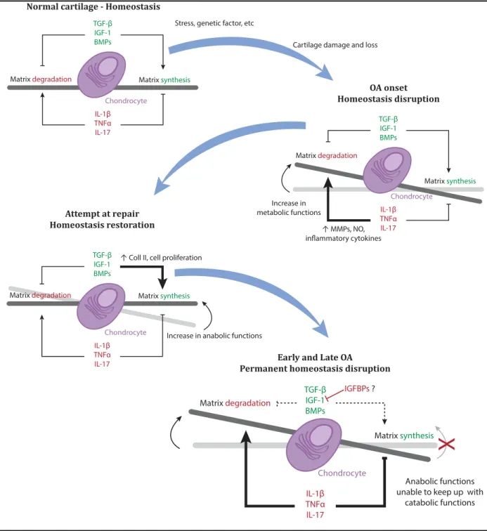
Regardless of what causes it initially, OA starts with an imbalance in the synthesis and degradation of the ECM by chondrocytes (Dijkgraaf et al. 1995). In healthy articular cartilage, chondrocytes are responsible for maintaining ECM homeostasis(Martin & Buckwalter 2002). The anabolic functions of chondrocytes are usually controlled by cytokines and growth factors(Sandell & Aigner 2001). These factors include transforming growth factor beta (TGF-β), insulin-like growth factor I (IGF-I) and bone morphogenetic proteins (BMPs)(Sandell & Aigner 2001). In the beginning of OA, osteoarthritic cartilage shows increased water content, early loss of proteoglycans as well as alterations in both size and arrangement of collagen fibers(Dijkgraaf et al. 1995; Goldring 2000). Repair attempts lead to accelerated cartilage turnover which means increased cell proliferation and matrix synthesis(Dijkgraaf et al. 1995).
The chondrocytes repair response is also thought to be increased: (1) by greater diffusion on growth factors cartilage due to its loss of integrity, (2) by release of previously ECM component-bound growth factors and (3) by chondrocyte upregulation of growth factor expression, such as IGF-1(Dijkgraaf et al. 1995). Unfortunately, as the pathology evolves, the increased synthesis cannot sufficiently compensate for the increased degradation resulting in tissue degeneration (see Figure 5)(Dijkgraaf et al. 1995; Sandell & Aigner 2001).
In early OA, the cytokine/growth factor balance responsible for normal homeostasis is disrupted and an increase in both inflammatory and catabolic cytokines can be measured in OA synovial fluids(Sandell & Aigner 2001; Goldring 2000). Proinflammatory cytokines, such as IL-1, TNF-α, IL-17 and IL-18, are known to cause an important increase in proteinase activity. In OA, they are produced by two types of cells: activated synovial cells, leading to proinflammatory cytokine presence in synovial fluid thus acting on cartilage resident cells in a paracrine manner, as well as chondrocytes, which leads to autocrine activation(Fernandes et al. 2002; Sandell & Aigner 2001; Goldring 2000). They are responsible for greater matrix metalloproteinases (MMPs) synthesis, an increase in aggrecanases, a decrease in MMP enzyme inhibitors and in ECM production(Mort & Billington 2001; Henrotin et al. 2002; Dean 1991). Proinflammatory cytokines also induce cyclooxygenase-2 (COX2) and inducible nitric oxide synthase (iNOS) through nuclear factor kappa B (NFκB) activation which lead to an increased release of both prostaglandin E2 (PGE2) and nitric oxide (NO)(Roman-Blas & Jimenez 2006; Amin et al. 1997). TNF-α also stimulates synthesis of phospholipase A2 (PLA2) by chondrocytes.
Figure 5. Osteoarthritis and matrix homeostasis
In normal cartilage, chondrocytes maintain matrix homeostasis. But when joint suffers cartilage damage and/or loss due to one of many factors (mechanical stress, genetic factors, etc.), the fragile equilibrium between matrix synthesis and degradation is disrupted. This onset of OA leads to upregulation of matrix metalloproteinases (MMPs) and inflammatory cytokines (interleukine 1β (IL-1β), IL-17 and tumour necrosis factor-α (TNF-α)) which increases nitrous oxide (NO) generation(Sandell & Aigner 2001; Goldring 2000). This is followed by attempts to repair cartilage by increasing anabolic functions, thanks to growth factors (transforming growth factor beta (TGF-β), insulin-like growth factor I (IGF-I) and bone morphogenetic proteins (BMPs)) which results in type II collagen (Coll II) synthesis and reorganization as well as in chondrocyte proliferation(Dijkgraaf et al. 1995). Unfortunately, as OA progresses, anabolic functions are unable to keep up with tissue degradation.
Stress, genetic factor, etc
Cartilage damage and loss
TGF-β IGF-1 BMPs IL-1β TNFα IL-17
Matrix degradation Matrix synthesis
Normal cartilage - Homeostasis
OA onset Homeostasis disruption TGF-β IGF-1 BMPs IL-1β TNFα IL-17 Matrix degradation Matrix synthesis Increase in metabolic functions ↑ MMPs, NO, inflammatory cytokines Attempt at repair Homeostasis restoration TGF-β IGF-1 BMPs IL-1β TNFα IL-17
Matrix degradation Matrix synthesis
Increase in anabolic functions ↑ Coll II, cell proliferation
Early and Late OA
Permanent homeostasis disruption
TGF-β IGF-1 BMPs IL-1β TNFα IL-17 Matrix degradation Matrix synthesis IGFBPs ? Anabolic functions unable to keep up with
1.3.1 Roles of cytokines in OA
Proinflammatory cytokines are cell-signaling molecules that are responsible for inducing inflammation mediators such as iNOS, COX-2, PLA2, etc(Dinarello 2000; Feldmann & Saklatvala 2001). It is a widespread belief that proinflammatory cytokines play a crucial role in the genesis and development of OA(Poole et al. 2001; Kobayashi et al. 2005; Hedbom & Häuselmann 2002). There are many proinflammatory cytokines, including many ILs, TNFs, interferons and colony-stimulating factors, but among these, two appear to be most directly implicated in the pathological processes of OA, IL-1β and TNF-α(Dinarello 2000; Kobayashi et al. 2005). Through activation of many transcription factors, such as NF-κB and the activator protein (AP)-1, these two act synergistically and can induce synovial cells and chondrocytes to synthesize other proinflammatory cytokines including IL-8 and IL-6(Fernandes et al. 2002).
1.3.1.1 IL-1β
The IL-1 gene family consists of three genes: IL-1α, IL-1β and IL-1 receptor antagonist (IL-1Ra)(Dinarello 1996). The two former are agonists while the latter is a specific receptor antagonist(Dinarello 1996). IL-1α represents only about 10% of IL-1 agonist proteins produced by LPS-activated human monocytes and its processing and release remain to be understood(Feldmann & Saklatvala 2001). IL-1β accounts for about 90%(Feldmann & Saklatvala 2001). Both exist in two forms: mature (17,5 kDa) and precursor (pro-IL-1α or pro-IL-1β) (31 kDa)(Fernandes et al. 2002; Goldring 2000). In the case of IL-1β, the mature (or active) form is generated through pro-IL-1β cleavage by IL-1β converting enzyme (ICE also known as caspase-1) and can be found in synovial membrane, synovial fluid and cartilage(Fernandes et al. 2002; Feldmann & Saklatvala 2001).
Even though there are two types of cell surface receptors (IL-1R), namely type I (IL-1RI) and type II (IL-1RII), it appears IL-1β induces inflammation solely as a result of its association to the first(Feldmann & Saklatvala 2001). Indeed, signal transduction of
IL-1 can only be achieved through IL-IL-1α or IL-IL-1β binding to IL-IL-1RI(Dinarello IL-1996) (see Figure 6a). Type II IL-1R (IL-1RII) has been described as being a “decoy” receptor for IL-1β since it binds IL-1β tightly but, due to a short signal transducing cytosolic domain, cannot lead to cell signaling(O’Neill & Dinarello 2000; Colotta et al. 1994) (see Figure 6b). Both IL-1RI and IL-1RII can be found in soluble form (IL-1sRI and IL-1sRII) in body fluids(Dinarello 1996). While 1RI has greater affinity with 1α than with 1β, IL-1RII binds IL-1β with a lot more affinity than IL-1α(Dinarello 1996). IL-IL-1RII, in both cell-bound and soluble form, seems to be an efficient IL-1β trap due to high affinity and almost irreversible binding caused by a very slow dissociation rate (2 hours)(Dinarello 1996).
An IL-1R accessory protein (IL-1R-AcP) has also been identified and seems to be vital to IL-1β signaling(Feldmann & Saklatvala 2001). Indeed, IL-1β signal transduction necessitates the formation of a IL-1RI/ IL-1R-AcP/ IL-1β complex which probably leads to dimerization of the IL-1RI and IL-1R-AcP cytosolic domains (see Figure 6a)(Feldmann & Saklatvala 2001; O’Neill & Dinarello 2000; O’Neill et al. 2003). IL-1β appears to first bind IL-1RI with low affinity forming a IL-1β/IL-1RI complex which allows IL-1R-AcP to dock to the newly formed complex whose binding affinity is now high(Dinarello 1996). IL-1 activation results in nuclear translocation of NF-κB and AP-1(Dinarello 2000). The biochemical events induced by IL-1 take place within a few minutes of its activation; among these is phosphorylation of PLA2 AP which leads to a rapid release of arachidonic acid (AA) (Dinarello 2000).
Figure 6. Interleukine-1beta (IL-1β) signal transduction
a. Interleukine-1beta 1β) binds its receptor, IL receptor type I 1RI) with low affinity. IL-1R accessory protein
(IL-1R-AcP) then associates with the IL-1β/ IL-1RI complex with a high affinity. Formation of the IL-1RI/ IL-1R-AcP/ IL-1β complex leads to signal transduction through dimerization of cytosolic domains.
b. IL-1β binds its receptor, IL receptor type II (IL-1RII) with high affinity. However, due to a short signal transducing
cytosolic domain no signal can be transduced.
c. IL-1R antagonist (IL-1Ra) avidly binds to IL-1RI. IL-1R-AcP cannot associates with the IL-1Ra/ IL-1RI complex, thus
leading to no signal transduction.
CELL
IL-1RI IL-1RI IL-1RAcP
IL-1ß IL-1RI IL-1RAcP Signal transduction Signal transduction CELL
IL-1RI IL-1RI IL-1RAcP
IL-1Ra IL-1RI IL-1RAcP IL-1ß CELL IL-1RII IL-1RII No Signal transduction
Another related protein is the IL-1Ra which is a natural inhibitor of IL-1(Kobayashi et al. 2005). It acts by binding to IL-1R without inducing signal transduction since IL-1R-AcP cannot bind the IL-1Ra/ IL-1RI complex(Feldmann & Saklatvala 2001) (see Figure 6c). IL-1Ra is a soluble 22 kDa glycosylated protein which can also be found in a non-glycosylated 17kDa form(Kobayashi et al. 2005). It is under this last form that IL-1Ra binds IL-1R(Dinarello 1996). IL-1Ra has 26% homology with IL-1β (which is greater than the homology between IL-1β and IL-1α) (Dinarello 1996). However, contrary to IL-1β, IL-1Ra has much more affinity with IL-1RI than with IL-1R2(Dinarello 1996). IL-1RI occupancy by IL-1Ra very effectively prevents IL-1 signal transduction(Dinarello 1996).
1.3.1.2 TNF-α
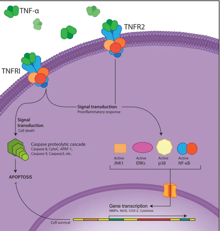
The TNF superfamily comprises many trimeric cytokines and cell surface proteins (at least 19) such as Fas Ligand (FasL), lymphotoxin-α (LTα), receptor-activator of NF-κB ligand (RANKL), CD40 ligand (CD40L), TNF-α, etc.(Baud & Karin 2001; Popa et al. 2007). In mammalian cells, the TNF superfamily plays an essential role in a broad array of biological processes: inflammation, cell activation, apoptosis, host defense, autoimmunity, etc (Keystone & Ware 2010). TNF-α can bind two structurally distinct receptors, type I (TNF-RI) and type II (TNF-RII)(Tracey et al. 2008). The pleiotropic cytokine TNF-α has a complex role in inflammation which is very similar to IL-1 since they share many pathways (Popa et al. 2007). In fact, both cytokines will often synergize for NO induction, resulting in cell death (Dinarello 2000).
TNFR1 activation leads to two distinct signaling pathways: the primary one activates both NF-κB and AP-1 through inhibitors of κB kinase (IKK) and Jun NH2 -terminal Kinase (JNK) while the second induces apoptosis in a caspase-8 and caspase-3 manner(Baud & Karin 2001; Tracey et al. 2008). As for TNFR2, its activation always results NF-κB and AP-1 signaling pathways(Baud & Karin 2001). Activation of NF-κB suppresses the apoptosis pathway, as long as it is not compromised; thus TNF-α signaling will usually lead to a proinflammatory response (see Figure 8)(Tracey et al. 2008). The extracellular domain fragments of each receptor can be shed and they are then said to be in soluble form: soluble TNFR-1 and -2 (sTNRF-1 and -2)(Sharma & Anker 2002). Though there function is yet unknown, at high concentrations, sTNFRs inhibit TNF activity as they trap TNF-α(Sharma & Anker 2002).
1.3.1.3 Other proinflammatory cytokines
Many other proinflammatory cytokines have been described and are thought to play a role in OA, namely IL-8, IL-17 and IL-18(Fernandes et al. 2002). All three enhance matrix degradation and ROS generation through either upregulation of IL-1β and/or TNF-α or suppression of aggrecan synthesis(Fernandes et al. 2002; Bronner & Farach-Carson 2007). IL-6 also seems to play a role in OA as it causes an increase in IL-1Ra, sTNFR, and tissue inhibitor of metalloproteins (TIMP) as well as enhances immune cell function and inflammation(Bronner & Farach-Carson 2007) [. On the other hand, when IL-6 interacts with its soluble receptor (sIL-6R), it leads to upregulation of MMPs and A disintegrin and metalloprotease with thrombospondin motifs (ADAMTS) as well as downregulation of Coll II gene (COL2A1) and aggrecan(Bronner & Farach-Carson 2007).
Figure 7. Tumor Necrosis Factor-α (TNF-α) signal transduction
Tumor Necrosis Factor-α (TNF-α) can bind either TNF Receptor 1 (TNFRI) or 2 (TNFRII). TNFRI activation induces to the Caspase proteolytic cascade through activation of Caspase 8 which leads in Cytochrome-C (CytoC) release and activation of Apoptotic Protease Activating Factor-1 (APAF-1). In turn, APAF-1 will recruit Caspase 9 and activate Caspase 3 which ultimately results in apoptosis. TNFRI activation also activates Jun NH2-terminal Kinase -1 (JUNK1), Mitogen-Activated Protein Kinases (MAPK) p38 and Extracellular signal-Regulated Kinases (ERK) and Nuclear Factor –KappaB (NF-κB); all of these result in a proinflammatory response (Activation of Metalloproteinases (MMPs), induction on inducible Nitrous Oxide (iNOS), cyclooxygenase-2 (COX-2) and many protinflammatory cytokines). The proinflammatory response suppresses cell death. TNFRII activation always leads to the proinflammatory response(Baud & Karin 2001; Popa et al. 2007; Keystone & Ware 2010; Tracey et al. 2008; Sharma & Anker 2002).
Signal transduction Signal transduction APOPTOSIS NF-κB Active p38 Active ERKs Active JNK1 Active
TNFRI
TNF-α
TNFR2
Caspase proteolytic cascade Caspase 8, CytoC, APAF-1, Caspase 9, Caspase3, etc.
Cell survival Cell death
1.3.1.4 NF-κB pathway
The NF-κB family of transcription factors play a central role in the signaling pathways of stress-induced apoptosis, inflammation and immune signaling through innate and adaptive immune responses(Roman-Blas & Jimenez 2006). This family has five members that associate to form a variety of homo- and hetero- dimers: RelA (p65), ReloB, c-Rel, NF-κB1 (p50/p105) and NF-κB2 (p52/p100)(Roman-Blas & Jimenez 2006; Tak & Firestein 2001). The RelA (p65) and NF-κB1 (p50/p105) heterodimer is the most widespread activated form of NF-κB(Tak & Firestein 2001).
In OA, it mediates key events in the inflammatory response by chondrocytes. These events eventually lead to the progressive ECM destruction characteristic of OA(Agarwal et al. 2004). NF-κB is known to regulate more than 150 genes; these include genes encoding cytokines, for example TNF-α, IL-1β and IL-6, inducible enzymes, such as COX-2 and iNOS, and both pro- and anti- apoptotic molecules(Roman-Blas & Jimenez 2006). It is present in the cytoplasm in an inactive form(Roman-Blas & Jimenez 2006; Tak & Firestein 2001). In this state, the NF-κB dimer is associated with regulatory proteins known as inhibitors of κB (IκB): IκBα, IκBβ, IκBε and IκBγ(Roman-Blas & Jimenez 2006). NF-κB can be activated by many stimuli, but in OA, the most significant ones are cytokines, including TNF-α and IL-1β, and free radicals(Roman-Blas & Jimenez 2006). These trigger signalling pathways that ultimately lead IκB phosphorylation by IKK(Roman-Blas & Jimenez 2006; Tak & Firestein 2001). Though the exact mechanism through which the IKK complex is activated by cytokines remains unknown, when activated, IKK initiates IκBα phosphorylation at specific serine residues(Roman-Blas & Jimenez 2006; Tak & Firestein 2001). NF-κBs nuclear localization signal is the revealed and the dimers are then free to translocate into the nucleus, to bind to κB enhancer elements of target genes and stimulate gene transcription (see Figure 8) (Roman-Blas & Jimenez 2006; Tak & Firestein 2001).
NF-κB is implicated in many inflammatory diseases such as OA, multiple sclerosis, asthma, type 2 diabetes, etc(Roman-Blas & Jimenez 2006). In OA patients, IKK is constitutively expressed and its activation by TNF-α and IL-1β is crucial for NF-κB mediated cytokine, collagenase, iNOS and COX-2 and induction(Roman-Blas & Jimenez 2006; Tak & Firestein 2001).
Thought NF-κB activation through IL-1β and TNF-α activation is well known, it has been shown that biomechanical signals can be converted into biochemical signals by cells and therefore also activate NF-κB(Agarwal et al. 2004). In experiments conducted with rabbit articular cartilage grown on flexible membranes, it has been shown that high magnitude cyclic tensile strain (15-18% equibiaxial strain) have the same effect on NF-κB activation than IL-1β since it induced rapid NF-κB p50-p65 heterodimer nuclear translocation, thus upregulating proinflammatory gene expression, matrix degradation and decreasing matrix synthesis(Agarwal et al. 2004). NF-κB is also known to be redox-sensitive(Henrotin et al. 2003).
Taking into account that
OA is an age-related disease; chondrocyte senescence leads to mitochondrial degeneration, therefore to an increase in free radical production, such as superoxide anion (O2•) thus adding redox stress to the articular joint. On the other hand, mechanical strain in known to be an important risk factor for OA. This indicates that both redox and biomechanical signal activation pathways may greatly contribute in OA pathogenesis(Martin & Buckwalter 2002).
Figure 8. Nuclear factor-kappaB (NF-κB) cytokine activated pathway
Proinflammatory cytokines bind to their receptors which lead to an unknown signal transduction that result in inhibitors of κB kinase (IKK) activation through its phosphorylation. IKK activation initiates inhibitors of κB (IκB) phosphorylation which releases NF-κB dimer. NF-κB then translocates into the nucleus to stimulate gene transcription (Activation of Metalloproteinases (MMPs), induction on inducible Nitric Oxide (iNOS), cyclooxygenase-2 (COX-cyclooxygenase-2) and many protinflammatory cytokines)(Dean 1991; Sharma & Anker cyclooxygenase-200cyclooxygenase-2).
Proinflammatory cytokines
Signal transduction
leading to IKK phosphorylation
Proinflammatory cytokines receptors IκB IKK IKK Inactive NF-κB Inactive p65 p50 IκB NF-κB Active p65 p50 Active
1.3.1.5 COX-2 and PGE
2Cyclooxygenase (COX) is the enzyme responsible for prostaglandin (PG) synthesis. Also called prostaglandin H2 synthase (PGHS), COX converts AA into prostaglandins G2 (PGG2), then PGH2 that can subsequently be converted to a variety of eicosanoids, more specifically prostanoids, which include PGE2, PGF2a, PGI2 and thromboxane (TX) A2 (see Figure 9)(Vane et al. 1998; Cryer & Feldman 1998). Though it was long thought that COX existed in only one form, two COX isoforms exist: COX-1 and COX-2(Dubois et al. 1998). Both have the same enzymatic activity and are very similar in structure(Vane et al. 1998). What differentiates the two is that the first is constitutive and present in almost all type of cells while the latter is inducible and normally absent from cells(Dubois et al. 1998). Therefore COX-1 is responsible of producing homeostatic levels of PGs allowing regulation of physiological effects such as platelet aggregation (blood clot formation)(Dubois et al. 1998). Synthesis of COX-2 can be stimulated by a great number of growth factors and proinflammatory cytokines, such as IL-1β and Tα, through NF-κB inhibition(Amin et al. 1997; Gupta & DuBois 2001). In fact, PGs most important action is their role in inflammation; more importantly, PGE2 release is a key event in the swelling/redness, pain and fever caused by inflammation(Clària 2003). COX-2 has been shown to be responsible for PG synthesis in inflamed joint tissues and its induction has been observed in OA cartilage and synovial tissue(Clària 2003). These observations that have led searchers to investigate COX-2 selective inhibitors as an anti-inflammatory treatment in OA (Clària 2003).
Figure 9. Diagram of cyclooxygenase (COX) role in eicosanoid metabolism
Formation of prostaglandin (PG) from arachidonic acid. Cyclooxygenase (COX) -1 and -2 oxygenate arachidonic acid to form PGG2 which will be reduced to PGH2. PGH2 will then be transformed into various prostaglandins by their
respective synthase(Vane et al. 1998).
1.3.2 Matrix metalloproteinases (MMPs) and aggrecanases
MMPs are a family of degradative enzymes which target ECM components and are believed to have a central role in tissue remodelling and repair but, when in excessive expression or in lack of inhibition, they are thought to greatly contribute to OA pathogenesis(Sandell & Aigner 2001; Shapiro 1998). They are produced in an pro-enzyme form and are activated by a proteolytic cleavage by cell surface associated plasmin or by other MMPs(Knäuper et al. 1996; MURPHY et al. 1999). There are many MMPs, such as 1,2,3,7,8,13 and 14, only to name a few, and they consist of different collagenases, stromelysins, gelatinases and matrilysin(Shapiro 1998; Shlopov et al. 1997).
Arachidonic Acid
Cyclooxygenase-1
Cyclooxygenase-2
PGH
2PGG
2PGE
2PGF
2APGI
2TXA
2PGD
2In OA, levels of MMP-3 (a stromelysin) MMP-1, MMP-8 and MMP-13 (all three collagenases) are elevated (Sandell & Aigner 2001; Goldring 2000; Shlopov et al. 1997). In fact, though all three collagenases seem to play a key role in OA, Coll II degradation seems to be mainly caused by MMP-13(Shlopov et al. 1997; Billinghurst et al. 1997). IL-1β stimulation can enhance MMP-13 transcription because of the parallel activation of kinase cascades regulating NF-κB and AP-1(Tak & Firestein 2001). MMP-3 is responsible for degrading many ECM molecules, including proteoglycan and Coll II(Takahashi et al. 1999).
Some enzymes, named « aggrecanases », are responsible of aggrecan proteolytic degradation; it is the case of some members of the ADAMTS family namely ADAMTS-4 (also known as aggrecanase-1) and ADAMTS-5 (aggrecanase-2). Although both ADAMTS-4 and ADAMTS-5 can degrade aggrecans and affect articular cartilage integrity, studies with ADAMTS-4 and/or -5 knock-out mice seem to show that only the latter is associated with increased susceptibility to OA(Tang 2001; Majumdar et al. 2007; Glasson et al. 2004; Glasson et al. 2005).
The activation of MMPs can be inhibited by TIMPs. TIMPs do so by forming a high affinity complex(Pelletier et al. 1990). It seems that conditions in OA tissues favor MMP synthesis and activation over inhibition. Indeed, a lack of sufficient TIMP to counter increased MMP production might be a contributing factor in OA articular cartilage degradation(Pelletier et al. 1990).
1.3.3 Reactive oxygen species (ROS)
Cytokines as well as mechanical stress can both stimulate reactive oxygen species (ROS) production by chondrocytes(Henrotin et al. 2005). ROS interact strongly and quickly with many molecules, both organic and inorganic, irreversibly changing or even destroying them(Bedard & Krause 2007). These include various proteins, lipids and nucleic acid(Bedard & Krause 2007). O2- and NO are the two main ROS generated by articular cartilage cells(Henrotin et al. 2005). Members of the nicotinamide adenine dinucleotide phosphate (NADPH) Oxidase (NOX) family reduce oxygen (O2) to O 2-making them big ROS producers(Bedard & Krause 2007). Both IL-1β and NO can activate NOX(Kaur et al. 2004). O2- and NO play an important role in pathophysiology since derivative radicals such as hydrogen peroxide (H2O2) and proxynitrite (ONOO-) as well as hydroxyl radical (OH-) are created when in presence of iron(Henrotin et al. 2003; Henrotin et al. 2005). In OA, production of ROS is such that antioxidants capacity to detoxify them is insufficient, causing oxidative stress to articular cartilage, thus damaging it. ROS have been shown to be involved in cartilage degradation through intracellular signaling regulation and through direct attack of ECM molecules, proteoglycan and collagen molecules in particular(Henrotin et al. 2003). Various transcription factors, including NF-κB, are redox-sensitive through modification of conserved cysteines(Henrotin et al. 2003). ROS can also modify protein degradation by inhibiting the ubiquitin pathway(Henrotin et al. 2003). One family of ROS which has been shown to be very implicated in OA is the reactive nitrogen species (RNS) family.
1.3.4 Nitric oxide (NO) and inducible NO synthase (iNOS)
RNS formation in chondrocytes starts with NO which will then lead to ONOO- (Henrotin et al. 2005)[17]. NO is synthesized by its enzyme, NO synthase (NOS), which has three isoforms, neuronal NOS (nNOS), endothelial NOS (eNOS) and inducile NOS (iNOS) (Henrotin et al. 2005). Only the two latter are present in chondrocytes. While eNOS is expressed constitutively, iNOS is, as its name suggests, an inducible form. As for ROS stimulation, iNOS expression can be induced by either mechanical stress or cytokines(Henrotin et al. 2005). Since OA is characterized by an overproduction of cytokines and an increase in mechanical stress at the articular cartilage level, it is not surprising that many studies has shown increased levels of iNOS in OA cartilage when compared to normal cartilage(Pelletier et al. 2001). Indeed, OA cartilage produce large amounts of NO(Henrotin et al. 2005; Henrotin et al. 2003; Pelletier et al. 2001; Pelletier et al. 1998). NO combines with O2- to form ONOO-(Henrotin et al. 2005). NO and ONOO -play an important role in OA. They affect cartilage matrix synthesis by being responsible for inhibition of aggrecan and COLL II synthesis and they increase the IL-1β inhibitory effect on EMC component synthesis(Henrotin et al. 2003). They also contribute to cartilage matrix breakdown by inducing chondrocyte apoptosis, increasing MMP-3 and MMP-13 mRNA levels in chondrocytes while inhibiting TIMPs(Henrotin et al. 2003). Since they upregulate proinflammatory cytokines, their production causes an amplification of cartilage catabolism, and all subsequent problems such as inflammation, swelling and pain, through a feedback loop (see Figure 10)(Pelletier et al. 2001).
Figure 10. Role of inducible nitric oxide synthase (iNOS) in OA
Proinflammatory cytokines lead to inducible nitric oxide synthase (iNOS) expression. iNOS stimulates reactive oxygen species (ROS) production such as nitric oxide (NO), superoxide anion (O2-) and proxynitrite (ONOO-)(Henrotin et al.
2003). NO, O2- and ONOO- contribute to cartilage matrix degradation by inhibiting aggrecan and type II collagen (COLL
II) synthesis, increasing the interleukine-1β (IL-1β) inhibitory effect on matrix component synthesis, inducing chondrocyte apoptosis, increasing metalloproteinase (MMP)-3 and MMP-13 mRNA levels in chondrocytes while inhibiting tissue inhibitor of metalloprotease (TIMPs) (Henrotin et al. 2003). They also upregulate proinflammatory cytokines, which causes an amplification of cartilage catabolism, and stimulate lipidic peroxidation of cell membrane(Pelletier et al. 2001; Halliwell & Chirico 1993).
Proinflammatory cytokines NADPH oxidase Proinflammatory cytokines receptors Gene transcription MMPs iNOS COX-2 Cytokines iNOS Uncoupled iNOS ↓ Aggrecan synthesis ↓ Coll II synthesis
↑ IL-1β inhibitory effect on EMC component synthesis ↑ Chondrocyte apoptosis ↑ MMP-3 mRNA level ↑ MMP-13 mRNA level ↓ TIMPs activity ↑ Membrane lipidic peroxydation NO O2-• ONOO
-The iNOS enzyme itself might also contribute to ROS formation. Indeed, limitation of substrate availability leads to uncoupling of NOS and ultimately to ROS formation (Gielis et al. 2011). When NOS is in its uncoupled form, electron transfer can result reduction of molecular oxygen, and NO which can react together to form ONOO- (Griffith & Stuehr 1995). ONOO- is also known for lipid peroxidation (LPO) of the cell membrane, composed of polyunsaturated fatty acids (PUFAs)(Radi et al. 1991). LPO produces lipid peroxides through a free radical chain reaction which will in turn be decomposed to produce epoxides, ketones, acids and aldehydes (see Figure 11 and Figure 12) (Halliwell & Chirico 1993).
1.4 4-Hydroxynonenal (HNE)
1.4.1 Synthesis and characteristics of HNE
One of the many effects of oxidative stress caused by the ROS and RNS family is LPO of the polyunsaturated fatty acids contained in the cell’s membrane (Radi et al. 1991). The cell membrane is a lipid bilayer whose main function is to control the metabolite and electrolyte concentration gradient between the cell and it’s surrounding through modulation of the membrane’s permeability. Lipid damage can thus affect these gradients and affect cell function(Hogg & Kalyanaraman 1999). Of course, cells have the ability to repair or replace damaged lipids, but since LPO is a rapid chain reaction, normal lipid turnover can be insufficient(Hogg & Kalyanaraman 1999). Moreover, products of LPO, such as hydroperoxides (LOOH), alcohols and aldehydes can affect both cellular functions and protein expression [88]. PUFAs, such as arachidonic acid, are more sensitive to oxidation because their methylene hydrogens are more susceptible to hydrogen abstraction by ROS and RNS than those from saturated lipids(Hogg & Kalyanaraman 1999).
Figure 11. Molecular equations of lipidic peroxidation
Overview of aldehyde generation through lipidic peroxidation (Hogg & Kalyanaraman 1999; Herdegen & Delgado-Garcia 2004).
Equations 1 and 2 show how ONOO- and •OH remove hydrogen from unsaturated lipids (LH) to form a lipid radical (L•)(Hogg & Kalyanaraman 1999; Herdegen & Delgado-Garcia 2004). The lipid radical produced subsequently reacts with oxygen to generate a lipid peroxyl radical (LOO•), which is key to the propagation of LPO through the cell membrane (eq.3) (Hogg & Kalyanaraman 1999). The lipid peroxyl radical produced will then remove a hydrogen to the neighboring unsaturated lipid to regenerate a lipid radical, which will lead to the LPO chain reaction, and LOOH which, due to its unstableness, will quickly fragment into reactive aldehydes (eq. 4) (see Figure 11 and Figure 12)(Hogg & Kalyanaraman 1999). Aldehydes produced by LPO are believed to be the main culprits in cell damages caused by oxidative stress(Esterbauer et al. 1991). Because aldehydes are more stable than free radicals, they can diffuse in and out of cells and target cells and structures far from their point of origin(Dalle-Donne et al. 2006). In these cases, aldehydes are secondary messengers to primary reactions(Dalle-Donne et al. 2006). The most important aldehydes generated by LPO are
LH
+
ONOO
-→
L•
+ [ONOOH] →
L•
+ NO
2+
•OH
LH
+
•OH
→
L•
+ H
2O
L•
+ O
2→
LOO•
LOO•
+
LH
→ [
LOOH
]
+
L•
→
ALDEHYDES
+
L•
Chain reaction
(1)
(2)
(3)
malondialdehyde (MDA) and 4-Hydroxynonenal (HNE), the former being the most abundant lipid aldehyde while the latter is the one who which is most responsible for the cytopathologic effects of oxidative stress(Uchida 1999). As many other aldehydes, these two can react with various biomolecules such as phospholipids, proteins and even ribonucleic and deoxyribonucleic acids (RNA and DNA)(Dalle-Donne et al. 2006).
HNE is an amphiphilic compound since it has both hydrophilic and lipophilic properties, though it has greater affinity for lipids than for water(Poli et al. 2008). It is known as a highly reactive molecule due to its three functional groups: a hydroxyl group (OH), a carbon-carbon (CC) double bond as well as a carbonyl group (CO) (see Figure 13)(Poli et al. 2008). Together, these three groups frequently act synergistically so that HNE can react to a great number of biomolecules, especially those with amino residues (cysteine, lysine, histidine and rarely arginine) or thiol groups.
Figure 12. Lipid peroxidation
Lipid peroxidation is a chain reaction in which a reactive oxygen (or nitrogen) species react with unsaturated lipids of the cell membrane and produce reactive aldehydes (adapted from Tim Vickers/ Wikimedia Commons / CC-BY-SA-3.0).
+
O 2 H R H R H R H+
ONOO -ONOOH OH NO2+
Initiation Propagation Lipid hydroperoxide ( LOOH )Lipid peroxyl radical ( LOO• )
Unsaturated lipid from cell membrane (arachidonic or linoleic acid )
R Lipid radical ( L• ) R OOH R OO Cell membrane O2 Reactive aldehydes
HNE
PropagationFigure 13. 4-Hydroxynonenal (HNE) molecule
The HNE molecule has three functional groups : a. a hydroxyl group (OH), b. a CC double bond and c. CO carbonyl group(Poli et al. 2008).
1.4.2 HNE adducts and target proteins
Important reactions for HNE’s biochemical effects, as well as its metabolism, are Schiff base formation and Michael additions (see Figure 14) (Schaur 2003). Interaction between HNE and cell proteins has often been described; many HNE-binding proteins have been found, including heat shock proteins 72 and 90 (Hsp-72 & Hsp-90), CytoC, Ca2+ ATPase, albumin and many more (Carbone et al. 2004; Carbone et al. 2005; Isom et al. 2004; Akaishi et al. 2004; Aldini et al. 2006; Moreau et al. 2005). In addition, growing evidence supports a role for HNE as a physiologic modulator of signal transduction and of posttranslational modification(Poli et al. 2008). Our laboratory has also shown HNE’s ability to induce transcriptional and posttranscriptional modifications of MMP-13 and Coll II(Morquette et al. 2006).
OH
Figure 14. 4-Hydroxynonenal (HNE) common reactions
a. Formation of Schiff base is made possible by HNE’s carbonyl base(Poli et al. 2008; Schaur 2003). b. Michael addition
reaction between thiol or amino group and the CC double bond in HNE(Poli et al. 2008; Schaur 2003) (adapted from Poli and al. (2008). Fig 1 & 2).
HNE can be found in healthy human organs in small concentration but elevated HNE levels has been detected in a great number of human diseases ranging from Alzheimer’s disease to Multiple sclerosis(Poli et al. 2008). Increased evidence suggests its significance in the pathogenesis of major human chronic diseases(Poli et al. 2008). Previous studies have linked HNE to OA by showing higher concentrations of HNE in chondrocytes and synovial fluid of OA patients when compared to healthy individuals(Morquette et al. 2006; Vaillancourt et al. 2007). In OA, HNE can be found in either protein-adduct form, such as with Coll II, or in free form (see Figure 15)(Morquette et al. 2006; Vaillancourt et al. 2007). It is thought that protein modification is the most likely one if not the main mechanism by which HNE can modulate physiological and pathophysiological processes(Poli et al. 2008).
OH O OH b. Michael addition SH (or NH) R O + R-SH (thiol) or + R-NH2 (amino)
a. Schiff base formation
OH O OH R N + R-NH2 (amino) - H2O
Figure 15. Effect of 4-Hydroxynonenal (HNE) signalling on cell function
Free-form and/or protein-bound HNE lead to activation of different signalling pathways therefore to production of cyclooxygenase-2 (COX-2), pro-inflammatory cytokines, metalloproteinases (MMPs), inducible nitric oxide synthase (iNOS), etc.
1.4.3 Metabolism of HNE
Because HNE is toxic and normally found, in low levels, in many human cells, there exist multiple pathways that can metabolize it. There are three main pathways the organism use to detoxify HNE: reduction by alcohol dehydrogenase or aldose reductase to 1,4-dihydroxynonene (DHN), oxidation by aldehyde dehydrogenases to form hydroxynonenoic acid (HNA) and conjugation with glutathione (GSH) by glutathione S-transferase (GST) (see Figure 16) (Poli et al. 2008; Pappa et al. 2003). GST isoenzyme GST4A-4 has a high specificity for HNE and plays an important role in protection against oxidative damage(Gallagher et al. 2006; Awasthi 2006; Vaillancourt et al. 2008). Another molecule that might be of significant importance in decreasing HNE toxicity is carnosine. Studies have shown it traps HNE by forming a stable covalent adduct thus inhibiting its ability to bind other proteins and activate metabolic pathways(El-Bikai et al. 2010; Poli et al. 2008). It has been suggested that carnosine might react with HNE by mimicking the preferred HNE addition sites in proteins(Aldini et al. 2002).
Though HNE is metabolized at a very rapid rate, it can still play an important role in protein modification as it activates a number of signaling pathways; more so when concentrations are elevated it as is the case in many pathologies(Poli et al. 2008). That is why novel ways to decrease HNE levels have and are being studied around the globe.
Figure 16. 4-Hydroxynonenal (HNE) metabolism
HNE’s main metabolic pathways(Poli et al. 2008; Pappa et al. 2003) (adapted from Pappa and al. (2003) Scheme 1).
1.4.4 HNE and signalling pathways
It was long thought that HNE’s biological and pathological effects where purely the result of protein modifications in cells and their environments. It has since been shown that HNE’s role in pathophysiology is also greatly attributable to its ability to activate several signalling pathways(Esterbauer et al. 1991; Esterbauer et al. 1992; Benderdour et al. 2003; Morquette et al. 2006; Vaillancourt et al. 2007). Indeed, HNE has been linked to MAPK, JNKs and caspase pathways, to name only a few (Parola et al. 1998; Poli et al. 2008). These have many effects on cell activity as the pathways activated by HNE lead to cell stress and ultimately to apoptosis(Vaillancourt et al. 2008). At non-toxic levels, HNE can interact with histidine residues in JNKs which causes both their nuclear translocation and their activation(Parola et al. 1998). The activity and expression of key
4 - hydroxynonenal (HNE) 1,4-dihydroxynonene (DHN)
glutathione -1,4-dihydroxynonene (GS-DHN) glutathione -4 - hydroxynonenal (GS-HNE)
OH GS O OH GS OH OH O OH OH aldeh yde deh ydrogenases
hydroxynonenoic acid (HNA) OH O OH alcohol dehydrogenase aldose reductase aldose reductase glutathione S-transferase