Isolated Greater Tuberosity Fractures of the Proximal Humerus: Validation and Clinical Implications for a New Radiologic
Measurement Method and Classification
Par Jennifer Mutch
Département d’Orthopédie Faculté de Médecine
Mémoire présenté à la Faculté des Études Supérieures En vue de l’obtention de grade de
Magister Scientiæ (M. Sc.) en Sciences Biomédicales option Musculosquelettique
Mai 2013
Université de Montréal
Faculté des études supérieures et postdoctorales
Ce mémoire intitulé:
Isolated Greater Tuberosity Fractures of the Proximal Humerus: Validation and Clinical Implications for a New Radiologic
Measurement Method and Classification
Présenté par: Jennifer Mutch
a été évalué par un jury composé des personnes suivantes : Lawrence Lincoln (président-rapporteur)
Dominique Rouleau (directrice de recherche) Nicola Hagemeister (codirectrice)
SOMMAIRE Les fractures isolées de la Grosse Tubérosité (GT) de l’humerus proximal sont rares
et peu étudiées. Trois problèmes importants existent: 1: Même si 5mm + de déplacement supérieur du GT est cité comme indication chirurgicale, les mesures basées sur radiographie peuvent errer de plus que 10mm. 2: Les classifications de Neer et l’AO décrivent seulement un type de fracture de GT (gros fragment, ligne de fracture verticale). Deux autres types de fracture existent: type fracture-avulsion avec petit fragment osseux et type Hill-Sachs très latéral. 3: On manque d’études de pronostic ou de traitement des fractures de GT selon la morphologie.
Article 1 montre et évalue une méthode simple de mesurer le déplacement supérieur de la GT (le GT ratio) sur les radiographies standard; ceci corrèle très bien avec tomographie (CT). Article 2 introduit une méthode de classification Morphologique des fractures de GT (Avulsion, Split, Dépression) qui a une fiabilité de bonne à excellente. Les données échographiques, radiologiques, et cliniques de 54 patients porteurs de fracture de GT (suivie moyenne 2.5 années) sont aussi incluses. Les patients <50 ans ont eu plus de déchirures de la coiffe et ceux avec fractures
déplacées (≥ 5mm) avaient plus d’atrophie du susépineux. Les déchirures complètes de la coiffe et l’atrophie du susépineux augmentaient l’atteinte permanente.
La morphologie des fractures de GT n’a pas eu un impact significatif sur le pronostic. Cependant, l’âge, le sexe, et le taux de luxation glénohumérale étaient différents selon le type de fracture et ceci pourrait refléter la pathophysiologie. Une évaluation plus précise de l’impact de la Morphologie des fractures de GT sur le pronostic et traitement nécessitera une étude prospective multicentrique.
MOTS CLÉS Humérus proximal Grosse tubérosité Fracture Déplacement Radiographie Classification Échographie
Coiffe des rotateurs Résultats cliniques Traitement
SUMMARY Isolated fractures of the Greater Tuberosity (GT) of the proximal humerus are rare
and a challenge to study. Three main problems arise: 1: Though 5mm+ superior GT displacement is often a surgical indication, measurement errors on radiographs may surpass 10mm. 2: The Neer and AO classifications describe only one type of GT fracture (large fragment, vertical fracture line). Two other fracture types have been described: an avulsion-type (small fragment), and a very lateral Hill-Sachs-type. 3: There are no studies on the treatment or prognosis of GT fractures according to fracture morphology.
Article 1 introduces and tests a simple method to measure superior GT displacement (the GT ratio) using standard radiographs; this correlates very well with computed tomography (CT). Article 2 presents the Morphologic classification for GT fractures. It describes three fracture types (Avulsion, Split, Depression) and has good to
excellent reliability. The ultrasonographic, radiologic, and clinical results of 54 patients (average follow-up 2.5 years) with isolated GT fractures are then described. Patients <50 years had higher rates of rotator cuff tears and displaced (≥ 5mm) GT fractures were associated with supraspinatus atrophy. Both full rotator cuff tears and supraspinatus atrophy resulted in poor outcomes.
The impact of fracture morphology on prognosis was not significant. However, age, sex, and associated glenohumeral dislocation differed by fracture type and this may reflect their pathophysiology. A more thorough evaluation of the prognosis and treatment of GT fractures by morphologic type would require a prospective multicenter study.
KEY WORDS Proximal humerus Greater tuberosity Fracture Displacement Radiographs Classification Ultrasound Rotator cuff Clinical outcome Treatment
TABLE OF CONTENTS
INTRODUCTION ……….1
CHAPTER 1 – LITERATURE REVIEW ………...4
1.1 – ANATOMY OF THE SHOULDER JOINT………4
1.2 – FRACTURES TO THE SHOULDER (PROXIMAL HUMERUS)……9
1.3 – CLASSIFICATION OF SHOULDER FRACTURES…….…………..11
1.4 – RADIOLGIC EVALUATION OF GREATER TUBEROSITY FRACTURES………..17
1.4.1 – PLAIN RADIOGRAPHY………...17
1.4.2 – COMPUTED TOMOGRAPHY………..20
1.4.3 – MAGNETIC RESONANCE IMAGING………21
1.4.4 – ULTRASOUND………..22
1.5 – ADDITIONAL CONSIDERATIONS FOR GREATER TUBEROSITY FRACTURES………24
1.6 – TREATMENT OF GREATER TUBEROSITY FRACTURES………27
1.6.1 – CONSERVATIVE TREATMENT……….27
1.6.2 – SURGICAL TREATMENT………29
CHAPTER 2 – OBJECTIVES AND HYPOTHESES………...35
2.1 – SUMMARY OF THE PROBLEM………35
2.2 – GLOBAL OBJECTIVE……….37
2.3 – SPECIFIC OBJECTIVES………… ……….38
2.4 – HYPOTHESES………..39
2.5 – PRESENTATION OF THE ARTICLES………...40
3.1 – RETROSPECTIVE REVIEW………...42
3.2 – MEASUREMENT OF GREATER TUBEROSITY DISPLACEMENT………...45
3.3 – CLASSIFICATION OF ISOLATED GREATER TUBEROSITY FRACTURES OF THE PROXIMAL HUMERUS……….50
3.4 – CLINICAL IMPACT OF ISOLATED GREATER TUBEROSITY FRACTURES………..53
3.5 – STATISTICS……… ……….55
CHAPTER 4 – ARTICLE 1: ACCURATE MEASUREMENT OF GREATER TUBEROSITY DISPLACEMENT WITHOUT COMPUTED TOMOGRAPHY: VALIDATION OF A METHOD ON PLAIN RADIOGRAPHY TO GUIDE SURGICAL TREATMENT……….58
4.1 – PRELUDE………..58
4.2 – ARTICLE………...59
CHAPTER 5 – ARTICLE 2: A NEW MORPHOLOGIC CLASSIFICATION FOR GREATER TUBEROSITY FRACTURES OF THE PROXIMAL HUMERUS : VALIDATION AND CLINICAL IMPLICATIONS……….83
5.1 – PRELUDE………..83
5.2 – ARTICLE………...84
CHAPTER 6 – ADDITIONAL RESULTS………...104
6.1 – PRELUDE………104
6.2 – REVIEW OF OBJECTIVES………105
6.3 – DEMOGRAPHICS OF THE STUDY POPULATION………...105
6.4 – PATHOLOGY ON ULTRASOUND………..107
6.5 – POPULATION AS PER FRACTURE DISPLACEMENT………….109
6.6 – POPULATION AS PER THE MORPHOLOGIC CLASSIFICATION………...110
CHAPTER 7 – DISCUSSION………116
CHAPTER 8 – CONCLUSION AND FUTURE DIRECTIONS………...124
REFERENCES………125
APPENDICES
A – CLINICAL SOURCE DOCUMENT B – PATIENT QUESTIONNAIRES
B.1 – Constant-Murley Shoulder Outcome Score
B.2 – WORC (Western Ontario Rotator Cuff Index) – French B.3 – WORC (Western Ontario Rotator Cuff Index) – English
B.4 – QuickDASH (Disabilities of the Arm, Shoulder, Hand) – French B.5 – QuickDASH (Disabilities of the Arm, Shoulder, Hand) – English B.6 – Pain Scale – Bilingual
B.7 – SF12v2™ Health Survey – French B.8 – SF12v2™ Health Survey – English C – ULTRASOUND SOURCE DOCUMENT D – FIGURE PERMISSIONS
FIGURE INDEX
Figure 1 – Bony Anatomy of the Shoulder Girdle ………….………..4 Figure 2 – Range of Motion around the Shoulder Joint …..……….... 5 Figure 3 – Muscular Anatomy of the Glenohumeral Joint………...8 Figure 4 – Deforming Forces in 4-Part Fractures of the Proximal Humerus..….….9
Figure 5 – Neer’s 4-Part Classification of Proximal Humerus Fractures ...……...13
Figure 6 – AO Classification of Greater Tuberosity Fractures...………16
Figure 7 – Common Radiographic Views of the Proximal Humerus…...………..19
Figure 8 – The Occupation Ratio on MRI and Ultrasound…...………..23
Figure 9 – Insertion sites for the Muscles of the Rotator Cuff ……….…...25
Figure 10 – Suture Fixation of Greater Tuberosity Fractures….………32
Figure 11 – The Bamberg-type plate……..………34
Figure 12 – The AP and Lateral views of the Proximal Humerus..………44
Figure 13 – Measurement of the GT Ratio on Plain Radiography for Superior/ Inferior Displacement………..46
Figure 14 – Measurement of the GT Ratio on Plain Radiography for Anterior/ Posterior Displacement………47
Figure 15 – Measurement of Superior/ Inferior GT Displacement relative to HHT on Coronal CT Reconstruction………..49
Figure 16 – Measurement of Anterior/ Posterior GT displacement along the Plane of the Glenoid Articular Surface……….50
Figure 17 A+B – Drawing and AP Radiograph of an Avulsion Fracture………...51
Figure 18 A+B – Drawing and AP Radiograph of a Split Fracture………52
Figure 19 A+B – Drawing and AP Radiograph of a Depression Fracture……….52
Figure 20 – Patient Recruitment Flow Diagram………...106
TABLE INDEX Table 1 – Inclusion and Exclusion Criteria….………43, 106 Table 2 – Study Population Description and Comparison with Non-Participants………107
Table 3 – Supraspinatus, Infraspinatus, Subscapularis and Biceps Pathology….108 Table 4 – Demographics and Rotator Cuff Pathology by Fracture Displacement……….110
Table 5 – Demographics, Fracture Characteristics and Rotator Cuff
Pathology by Fracture Type………..111 Table 6 – The Functional Impact of Rotator Cuff Tears and Atrophy………….113 Table 7 – The Functional Impact of Biceps Pathology and Subacromial
Impingement……….114
LIST OF TERMS/ ABBREVIATIONS AHD – Axis of the Humeral Diaphysis
AO – Arbeitsgemeinschaft Osteosynthese or the Association of the Study of Internal Fixation (ASIF)
AP – Anteroposterior
cf – Confer in latin, as in “please see” the referred to section or document CT – Computed Tomography
GT – Greater Tuberosity of the Proximal Humerus
GT Ratio – A ratio measured on the anteroposterior (or Neer) shoulder radiograph representing the superior (or posterior) displacement of the greater tuberosity in isolated greater tuberosity fractures of the proximal humerus
HHT – Humeral Head Tangent ICC – Intraclass Correlation mm – millimeters
Morphologic Classification – A proposed classification of isolated greater
tuberosity fractures that categorizes fractures as Avulsion type, Split type, or Depression type based on morphology
MRI – Magnetic Resonance Imaging
mSv – millisievert (a measurement of radiation dose)
Neer – The standard Lateral radiographic view of the proximal humerus PACS – Picture Archive Computer/ Communication System
QuickDASH – Disabilities of the Arm, Shoulder and Hand Outcome Measure (Shortened version)
ROM – Range of Movement
SF-12® v2 – SF-12® Health Survey version 2.0 SI – Superior or Inferior
US – Ultrasound
VAS – Visual Analog pain Scale
WORC – Western Ontario Rotator Cuff Index XR – Standard Radiograph (or Roentgenogram)
ACKNOWLEDGEMENTS
I would like to start by thanking my research director and co-director, Dominique Rouleau and Nicola Hagemeister, without whom this work would not have been possible. Dr Rouleau, you helped me chose this interesting topic and were an essential guide for the entire duration of my master’s degree. You could see the positive and the potential in any situation and were there to offer words of encouragement or a poke in the back as necessary. Dr Hagemeister, you kept us grounded in reality and introduced me to a completely different way of viewing orthopedics. You welcomed me to your team at the LIO (Laboratoire d’Imagerie en Orthopédie) and were always available regardless of the time of day or year. Both of you have made inestimable contributions to my understanding of clinical research.
Thank you as well to the secretaries of the Sacré-Coeur Hospital Orthopedic Clinic for putting up with me and my add-on clinics. Julie, Stéphanie, and Marie-France, for your unflagging effort, legwork, humour and, most of all, patience. To Dr Yves Laflamme, Dr Pierre-Yves Lagacé, Dr Alec Cykes, Dr Xavier Jodoin, and future Dr Luojun Wang, your time and expertise was enormously appreciated. You were
“volunteered” by the forces of nature that are my research directors yet went above and beyond the call of duty.
This study was financed in part by FREOM (Fondation de Recherche et d’Éducation en Orthopédie de Montréal), MENTOR (CIHR training grant in evaluation and treatment of mobility and posture disorders), and the Orthopedic Research Division of Sacré-Coeur Hospital.
Last but not least, to my family. You now know more than you likely ever wanted to know about shoulder fractures and yet have refrained from changing the locks on the door. There is no stronger evidence of your love and support.
Epidemiology of Greater Tuberosity Fractures of the Proximal Humerus
Fractures of the proximal part of the humerus are relatively common injuries with a reported incidence of 73 cases per 100 000 individuals per year[1]. Although these fractures represent only 3-5 percent of all fractures overall[2,3], their incidence increases sharply with age and is expected to triple over the next three decades[4,5] Proximal humerus fractures generally occur in an osteoporotic population following a low-velocity trauma and females are affected three times as often as males[6]. They are second in frequency only to distal radius fractures for the upper extremity and third for all fracture types (after hip and distal radius) in patients over the age of 65 years[7]. Fortunately, approximately 80% of these fractures may safely be treated conservatively[1,8].
Greater tuberosity fractures, by contrast, occur with greater frequency in the younger population. They constitute one fifth of all proximal humerus fractures[1,9-12]and are more often associated with high velocity trauma[2]. Men are more likely to suffer greater tuberosity fractures than women and 5 to 7% of these injuries are the result of a glenohumeral dislocation[12,13]. Additionally, 15 to 30% of all anterior
glenohumeral dislocations[14-17] are associated with a greater tuberosity fracture.
As with other proximal humerus fractures, a minority of isolated greater tuberosity fractures are displaced[8,18]. In the statistics published by the AO
humerus, less than two percent involved isolated displaced fractures of the greater tuberosity[19]. However, due to the higher demands in this patient population and the anatomic constraints of the greater tuberosity beneath the acromion, considerable debate has emerged as to what should constitute “displacement” in this particular fracture[9].
Current Study on Isolated Greater Tuberosity Fractures of the Proximal Humerus
The motivation for this project stems therefore from the following observations: 1) isolated greater tuberosity fractures of the proximal humerus are poorly
understood; 2) the current methods for measurement of fracture displacement on plain radiography are unreliable; 3) the morphology of greater tuberosity fractures is variable; 4) the prognosis of greater tuberosity fractures is highly variable and not fully explained by fragment displacement.
This thesis is comprised of eight chapters. The first chapter summarizes the literature available for shoulder fractures and specifically addresses the evaluation, prognosis, and treatment of isolated greater tuberosity fractures of the proximal humerus. Chapter 2 lists the objectives and hypotheses for this project and chapter 3 describes the methodology used to address them. Chapters 4 through 6 present the results of our study, with the first 2 in the form of journal articles and the last as
complementary findings. The first article (Chapter 4) explores the limitations of current imaging modalities, in particular plain radiography, for the evaluation of isolated greater tuberosity fractures of the proximal humerus. It describes and
validates a new measure for superior displacement on the anteroposterior
roentgenogram, the GT ratio, and tests its applicability in the clinical setting. The second article (Chapter 5) proposes and validates a classification system for isolated greater tuberosity fractures based on fracture morphology. This Morphology
Classification is further correlated (in Chapter 6) with ultrasonographic findings of soft tissue injury, shoulder function and quality of life. Finally, a discussion of all the results can be found in Chapter 7 and conclusions in Chapter 8.
1.1 – ANATOMY OF THE SHOULDER JOINT
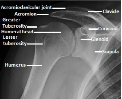
Bony Anatomy of the Shoulder
The shoulder joint is one of the most mobile and complex articulations in the human body. As a result the equilibrium between mobility and bony, ligamentous, and muscular support in this joint may be perturbed by even minor injury. The shoulder girdle consists of three bones and four major articulations. The three bones, the clavicle, scapula, and humerus, articulate with each other and the thoracic cage to form the sternoclavicular, acromioclavicular, scapulothoracic, and glenohumeral articulations (Figure 1)[20,21].
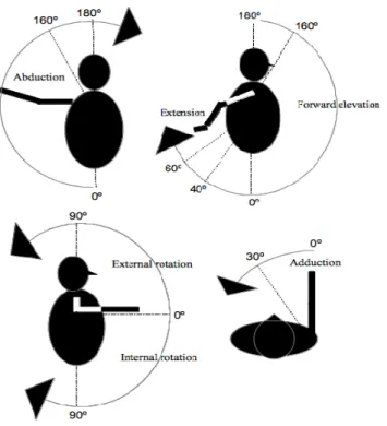
Range of Motion (ROM) around the Shoulder Joint
These four joints (sternoclavicular, acromioclavicular, scapulothoracic, glenohumeral) execute an intricate series of movements to allow a normal shoulder ROM of: 160 to 180 degrees abduction; 160 to 180 degrees forward flexion; 30 degrees adduction; and 40 to 60 degrees extension. Internal and external rotation is highly variable among individuals and may be tested at 0 or 90 degrees of shoulder abduction. Although the opposite limb is generally used as a control, external rotation at 90 degrees is normally about 20 degrees greater in the dominant extremity and may be up to 135 degrees in the throwing athlete (Figure 2)[22].
Figure 2: Range of Motion around the Shoulder Joint: Flexion/Extension, Abduction/Adduction, and External/Internal Rotation
The combination of translation, rotation, and angulation necessary for normal shoulder motion was first described by Codman in 1934 as “scapulohumeral rhythm”[23]. He discussed the vague descriptions of shoulder motion advanced by previous anatomists and physicians and although he recognized the difficulty of defining scapulohumeral rhythm, he was equally incapable of adequately illustrating it. Advances in the mathematic and mechanical understanding of scapulohumeral rhythm have been made in the past 80 years but we are far from fully understanding this sophisticated joint[23].
Muscular Anatomy of the Shoulder
The intricate movements arising in the shoulder are controlled and supported by a multitude of muscles and ligaments. A full review of the bony, muscular, and
ligamentous constraints of the entire shoulder girdle is beyond the scope of this study, so we will concentrate primarily on the glenohumeral joint. This “large ball-small socket” articulation offers very little intrinsic bony stability and is consequently the most frequently dislocated joint in the human body[20]. The surface of the glenoid is augmented radially by a dense fibrocartilagenous tissue called the labrum. This increases the depth of the shoulder socket by 50 percent and contributes to stability of the joint. The ligamentous constraints of the shoulder include the superior glenohumeral ligament, middle glenohumeral ligament, and inferior glenohumeral ligament. These ligaments stabilize the glenohumeral joint in the extremes of motion and resist inferior translation (in abduction), external rotation (in lower ranges of
abduction), and posterior or anterior translation (in greater than 45 degrees of abduction), respectively[20,24].
The muscles acting on the glenohumeral joint serve to create shoulder motion and dynamically stabilize the humeral head in the glenoid[25,26]. The rotator cuff envelops the humeral head and consists of the subscapularis, supraspinatus,
infraspinatus, and teres minor muscles. The subscapularis originates on the anterior surface of the scapula and inserts along the anterior aspect of the humeral head at the lesser tuberosity. This muscle acts as an internal rotator of the humerus and serves to dynamically inhibit antero-inferior humeral head displacement. The supraspinatus, infraspinatus, and teres minor all originate on the posterior surface of the scapula, wrap posteriorly around the humeral head and insert along the greater tuberosity. These muscles externally rotate the humerus, stabilise the shoulder posteriorly, and participate in a “force couple” with the subscapularis to stabilise the glenohumeral joint in abduction from 60 to 150 degrees. The supraspinatus muscle additionally initiates the shoulder abduction moment[24,25,27].
The long head of the biceps passes over the anterior aspect of the humerus through the bicipital groove between the greater and lesser tuberosities to insert in the supraglenoid tubercle. It enters the glenohumeral joint through the “rotator interval” (a triangular infolding of capsule between the supraspinatus and subscapularis muscles) and is stabilized in its groove by the transverse humeral, superior glenohumeral, and coracohumeral ligaments as well as the pectoralis major
muscle[28]. The biceps tendon creates a significant anterior stabilising force on the shoulder, particularly in abduction[29], but its location puts it at risk for injury.
Tendonitis, tears, and subluxation of the long head of the biceps tendon have been found in association with rotator cuff tears, osteoarthritis, and proximal humerus fractures[28,30].
Figure 3: Muscular Anatomy of the Glenohumeral Joint: Illustrations of (a) anterior and (b) posterior shoulder show supraspinatus (SS), infraspinatus (IS), subscapularis
(S), teres minor (Tm), and long head of the biceps brachii tendon (B). Subacromial-subdeltoid bursa is overlying the rotator cuff (light blue) [reproduced with
permission, © Carolyn Nowak, Ann Arbor 2011]
Other muscles acting around the glenohumeral joint include the deltoid, pectoralis major, teres major, and latissimus dorsi. These are the power movers of the shoulder and act as abductors, flexors, extensors, adductors, and internal or external rotators depending on the position of the upper limb.
1.2 – FRACTURES TO THE SHOULDER (PROXIMAL HUMERUS)
Fractures to the proximal humerus may have a substantial impact on motion, stability, and force of the glenohumeral joint. These generally occur along the physeal lines of the proximal humerus and split the humeral head into up to 4 parts: the greater tuberosity, lesser tuberosity, head and diaphysis[25,31]. The fragments then displace according to the pull of their relative musculature; the greater tuberosity is pulled posteriorly and superiorly by the supraspinatus and infraspinatus muscles; the lesser tuberosity is pulled medially by the subscapularis muscle; the head generally remains in close contact with the glenoid unless there is an associated glenohumeral
dislocation; and the diaphysis is pulled anteriorly, medially, and proximally by the pectoralis major and deltoid muscles[2].
Figure 4: Deforming Forces in 4-Part Fractures of the Proximal Humerus [reproduced with permission, Gruson 2008]
Numerous studies have demonstrated the negative impact of multiple fragments, increasing displacement, and associated tendon or ligamentous injury[8,30,32-38] on the functional outcome in proximal humerus fractures. As for greater tuberosity fractures, despite having garnered a significant amount of attention over the past 40 years, the appropriate evaluation and management of these fractures is still not clear. Even minimal displacement of greater tuberosity fractures can have a significant impact on post injury pain, strength and motion[39]. Unfortunately, the radiographic evaluation of greater tuberosity displacement is problematic, leading some authors to recommend computed tomography in this population[40].
Recommendations for surgical fixation of isolated greater tuberosity fractures have decreased over time from 1cm[8,35,41,42] to 5mm[9,40] to 3mm of displacement in overhead workers or athletes[39], yet no author has addressed the morphology of greater tuberosity fractures in their recommendations. Further complicating the issue, a wide variety of fixation techniques/strategies have been proposed for these
fractures. We believe this is due in part to the differing morphology of greater tuberosity fractures and leads to confusion when all types of greater tuberosity fractures are considered together.
1.3 – CLASSIFICATION OF SHOULDER FRACTURES
In 1934, Codman was the first to describe the four major fragments and deforming forces in proximal humerus fractures[31].
The first fragment, the humeral shaft, was created through fracture in the region between the surgical and anatomic humeral necks with the arm in abduction and elevation and with the acromion serving as the fulcrum for the long lever arm of the humerus. It would tend to displace medially due to the pull of the pectoralis major tendon[31].
The second fragment, the humeral head, was created when the tuberosities were sheared off at the transverse epiphyseal scar. With the arm in abduction, the superior edge of the glenoid would act as a wedge between the tuberosities and the articular humeral head, with the acromion as the fulcrum at the base of the greater tuberosity. The subsequently freed humeral head (no soft-tissue attachments) could rotate or dislocate depending on the magnitude of the trauma[31].
The third and fourth fragments, the tuberosities, would fracture apart lateral to the bicipital groove which follows the line of the vertical epiphyseal scar. The lesser tuberosity would displace medially due to the pull of the subscapularis muscle. The greater tuberosity would displace superiorly due to the pull of the supraspinatus muscle[31].
Interestingly, even in Codman’s original description of proximal humerus fractures, he warns readers that fractures of the greater tuberosity, and avulsions of the
supraspinatus facet in particular, are among the most serious types of humeral fractures. He recommended immediate surgical treatment[2].
In 1970, Neer published his four-part fracture classification of the proximal humerus. He used the same four fragments described by Codman but considered a fragment to be a “part” only if it was displaced more than 1cm or angulated more than 45 degrees. This classification was a major advancement in the understanding of proximal
humerus fractures because it considered the vascularity in the humeral head fragments as well as then tendency for patients with non-displaced fractures to do well[8,43].
Figure 5 = Neer’s 4-Part Classification of Proximal Humerus Fractures [reproduced with permission, Neer 1970]
The definition of displacement, however, was arbitrarily set and greater tuberosity fractures received no special attention in this classification. It has been the source of a fair amount of criticism for its poor intraobserver and interobserver reliability[44-49], particularly if based on simple radiography.
Neer responded to these criticisms in 2002 and noted that greater tuberosity fractures could often be missed[50]. He recommended the use of computed tomography to assist in the application of his classification, but the efficacy of this has also been questioned[51,52].
It has been noted by several authors that patients with greater tuberosity fractures displaced more than 5mm may do poorly with non-surgical management[9,39,40,53-55]. Both Codman[31] and Neer[8,43] observed that these injuries were difficult to treat and frequently resulted in inferior outcomes following non-surgical
management. They hypothesized that this was due to the deforming force of the rotator cuff but no clinically validated cutoff for surgical management was presented by either author[8,31]. Platzer et al. evaluated 135 patients with minimally displaced (1-5mm) greater tuberosity fractures at an average 3.7 years following injury[39]. Ninety-seven percent of these patients had good to excellent results with non-surgical management but patients with 3mm or more of displacement trended towards worse outcomes. The authors concluded that most patients with 1-5mm of greater tuberosity displacement could successfully be managed non-surgically but agreed with Park et al.[54] that surgical management could be considered in heavy labourers or overhead workers with greater tuberosity displacement of more than 3mm[39].
The current expert consensus is that patients with > 5mm of greater tuberosity
displacement would likely benefit from surgical management, in particular if they are young and physically active[9,40]. Displacement greater than this significantly negatively impacts the biomechanics of the shoulder[55]. Bono et al. developed a dynamic biomechanical model of greater tuberosity malunion and demonstrated that 5mm of superior greater tuberosity displacement increased the abduction force by 16%. Deltoid abduction force increased by 27% with 1cm of superior displacement and mechanical abutment on the undersurface of the acromion occurred. Although no bony abutment occurred with 5mm of displacement, they postulated that subacromial pressures would likely increase (through the decrease in volume) with abduction and noted this as a weakness in their model[55].
Various other authors have added contributions to the Neer Classification over the years and numerous other classifications have been proposed[56-62]. Since they have not been adapted into current use and, as a whole, contributed little to the
understanding or treatment of greater tuberosity fractures, they will not be described here.
The AO group developed a comprehensive classification of long bones with the aid of Mueller et al. in 1990[63]. It classifies fractures into A(extra-articular), B(partial-articular), or C(articular) groups with multiple sub-groups, leading to 27 possible fractures types in the proximal humerus alone[64]. In this classification, isolated fractures of the greater tuberosity are divided into 3 groups according to
Figure 6 = AO Classification of Greater Tuberosity Fractures: 11-A1.1 non-displaced GT fracture; 11-A1.2 displaced GT fracture; 11-A1.3 GT fracture with glenohumeral
dislocation [reproduced with permission, ©AO Foundation, Switzerland]
Similar to Neer, the AO Classification has also received numerous criticisms for poor intraobserver and interobserver reliability[46,47,51,60,65].
To date, Bahrs et al. are the only group to devote a significant amount of effort to the understanding and classification of isolated greater tuberosity fractures[66,67]. They also proposed a classification for proximal humerus fractures known as the Modular Topographic-Morphologic Classification[66]. However, in this classification they still fail to adequately discuss the variable morphology of greater tuberosity fractures.
In another paper, this same group later identified multiple possible mechanisms for greater tuberosity fracture including: avulsion through pull of the external rotators, a direct blow to the lateral shoulder, shearing by the glenoid rim during glenohumeral dislocation, and extreme rotation and abduction leading to impaction on the
fractures in the literature[53,68] and in their practice, and noted the contradiction this represented with the generally accepted avulsive mechanism of greater tuberosity fractures. They did not, however, translate this discussion of potential mechanisms into a viable classification using fracture morphology, nor were they able to demonstrate its clinical impact.
1.4 – RADIOLOGIC EVALUATION OF GREATER TUBEROSITY FRACTURES
Isolated greater tuberosity fractures of the proximal humerus are notoriously difficult to diagnose. In a study of 163 shoulders, Ogawa et al. noted that 60% of greater
tuberosity fractures were missed at the first consultation and this rate increased to 75% in minimally or non-displaced fractures[68]. Since the treatment of greater tuberosity fractures may change with as little as 3mm of displacement[39], complementary studies such as additional radiographic views, computed tomography, ultrasound, and MRI have been explored in the literature.
1.4.1 – PLAIN RADIOGRAPHY (XR)
In his original study, Neer recommended a minimum of two perpendicular
radiographic images for the evaluation of proximal humerus fractures[8]. These are the anteroposterior(AP) Grashley view and the scapular Y view(Neer view). The axillary view was later added to better evaluate glenohumeral dislocation and greater
tuberosity fracture[69]. In a comparative study by Sidor et al. of 50 radiographic series of the proximal humerus, the axillary view was shown to be more reliable than the scapular Y view and when combined with an AP, identified the final diagnosis in 99% of cases[70]. These three views (AP, Neer, Axillary) together comprise the standard trauma series[9,69].
The AP view is taken with the humerus in neutral rotation and the patient facing the XR source. The trunk is rotated 30 degrees to obtain a true AP of the glenoid.
The Neer view is taken with the injured shoulder against the plate and the XR source behind the patient. The trunk is rotated 30 degrees away from the source and the scapula forms a characteristic “Y” shape centered on the humeral head.
The Axillary view is taken with the patient supine with the plate at the superior aspect of the shoulder. The affected extremity is abducted 30 degrees and the image is taken through the axilla[64]. (This can be quite painful in the acute setting, so an alternative view, the Velpeau axillary view has been described.)
The Velpeau axillary view is taken with the shoulder sling in place. The patient stands in front of the radiographic table and the image is taken from above as the patient leans back 20 to 30 degrees over the XR plate[71].
Figure 7 = Common Radiographic Views of the Proximal Humerus: Anteroposterior(a,b), Neer(c,d), Axillary(e,f), and Velpeau [reproduced with
permission, ©AO Foundation, Switzerland and Bloom 1967]
This standard trauma series may be insufficient to adequately evaluate proximal humerus fractures due to bony overlap. In a radiographic study of 44 proximal humerus fractures, Bahrs et al. demonstrated a 72% overlap of the fractured regions on the transcapular Y view and 56% on the axillary view[72]. They concluded that computed tomography provided a better assessment of relevant structures regardless of fragment number or fracture severity.
The addition of internal and external rotation Grashley radiographs has been suggested[21,73] but Parsons et al. calculated up to 10mm of error in measuring
greater tuberosity displacement on these views[74]. In this cadaveric study, they did identify the AP view in external rotation as the most accurate for measuring greater tuberosity displacement of 2mm but this was surpassed in accuracy by the AP view with 15 degrees of caudal tilt for displacement of 5 to 10mm. They concluded by recommending multiple radiographic views for the assessment of greater tuberosity fractures.
1.4.2 – COMPUTED TOMOGRAPHY (CT)
Computed tomography has been suggested by some authors to improve the evaluation of proximal humerus fractures[40,75-77] and yet others have demonstrated that it is of little additional clinical utility[46,51,52,78].
Due to the aforementioned difficulties in measuring fracture displacement, CT has been recommended specifically for the evaluation of greater tuberosity fractures [39,40,50,52,75,79]. In the cases where plain radiographs are of poor quality, when there is a diagnostic or therapeutic uncertainty, or when physical conditions such as obesity make radiographic evaluation difficult, standard CT with coronal and sagittal plane reconstructions should be obtained[9]. Superior displacement of the greater tuberosity fracture is best obtained with the sagittal or coronal plane reconstructions while posterior displacement is best evaluated on the standard axillary view.
The increased exposure of the patient to radiation, however, cannot be ignored. A standard CT of the shoulder uses 2.06 mSv of radiation while a standard shoulder series uses 0.04 mSv[80]. This a 50-fold greater exposure to radiation and the
equivalent of one year exposure to background radiation in the average human[81]. Thus, while CT of the shoulder is a reasonable diagnostic step in the evaluation of greater tuberosity fractures and may help identify associated injuries such as glenoid fracture, it should be used only after a standard radiographic series has failed to provide diagnostic certainty.
1.4.3 – MAGNETIC RESONANCE IMAGING (MRI)
MRI is rarely, if ever, necessary for the diagnosis of proximal humerus fractures[82] but may provide some interesting information about associated soft tissue injury. Gallo et al. prospectively evaluated 30 patients with an MRI following proximal humerus fractures[83] and found that forty percent of patients under the age of 65 years had an associated complete rotator cuff tear or avulsion injury. This was positively correlated with an increasing number of fracture segments and displacement. Nanda et al. undertook a similar study with 85 proximal humerus fractures and confirmed the high incidence of rotator cuff tears in this population[84]. However, their population was slightly older and the presence of a full rotator cuff did not have an effect on functional outcome. They recommended against systematic screening for rotator cuff tears in proximal humerus fractures.
Isolated greater tuberosity fractures of the proximal humerus, however, tend to occur in a younger and more active population than proximal humerus fractures as a whole. These patients may be more negatively impacted by an associated rotator cuff tear and benefit from further imaging but the literature is silent on this subject. In the
same study above, Gallo noted that complete rotator cuff tears were more common in greater tuberosity fractures with more than 5mm of displacement. None of the
patients in Mason et al.’s series of 12 occult fractures of the greater tuberosity had significant associated rotator cuff pathology[85]. While they concluded that it was unlikely for the two pathologies to occur concurrently, it should be noted that all of the fractures were minimally or non-displaced. MRI is not currently recommended in the initial evaluation of isolated greater tuberosity fractures[9] but can be useful to evaluate for associated labral tears or rotator cuff injury in the patient with persistent shoulder pain following injury[79,86-88].
1.4.4 – ULTRASOUND (US)
Ultrasonography is a non-invasive and inexpensive test that shares many of the advantages and disadvantages of MRI with respect to proximal humerus fractures[9]. As with MRI, it is not currently recommended for the initial evaluation of these fractures and may, additionally, be too painful to perform in the acute setting.
Occult isolated greater tuberosity fractures have also been detected on ultrasound by looking for discontinuity and irregularity of the humeral cortex[89]. Associated rotator cuff tears were noted in 5% of the acute and 33% of the subacute trauma patients. Ultrasonography is a dynamic evaluation and affords the advantage of being able to assess for subacromial impingement[90], which has not yet been evaluated in greater tuberosity fractures.
Ultrasonography can additionally be used to evaluate for rotator cuff muscle atrophy. In the hands of an experienced radiologist, ultrasonography can reliably be used to calculate the “occupation ratios” of the rotator cuff muscles which range in value from 0.07(severe atrophy) to 0.81(normal). Khoury et al. calculated the occupation ratios using the “Y” view on both US and MRI and showed excellent correlation[91]. This atrophy has very important prognostic implications for rotator cuff repair[92,93] but, as is often the case with ultrasonography, is operator-dependent[94,95] and should be interpreted with caution.
Figure 8 = The Occupation Ratio on MRI (left) and Ultrasound (right) [reproduced with permission, Khoury 2008]
1.5 – ADDITIONAL CONSIDERATIONS FOR GREATER TUBEROSITY FRACTURES
Before evaluating or treating any fractures of the greater tuberosity, the anatomic particularities of this region must be taken into consideration. Due to the well-developed vascular network surrounding the proximal humeral head, ischemic complications following isolated greater tuberosity fractures are rarely a concern[9]. The proximity to neural structures, however, is an issue and isolated greater
tuberosity fractures may be associated with neurologic injury in up to 33% of cases[79]. This incidence increases with age and glenohumeral dislocation. The axillary nerve courses posteriorly in close proximity to the surgical humeral neck and gives off, among others, a motor branch to the deltoid muscle and a sensory branch to the lateral aspect of the shoulder[96]. This is the most commonly injured nerve in greater tuberosity fractures and should be tested prior to the initiation of any
treatment.
The most superior aspect of the greater tuberosity is situated 3 to 8mm inferior to the highest point of the humeral head[97-99]. This inferior offset allows for the
unobstructed passage of the greater tuberosity beneath the acromion through a full range of movement. Anatomic variations in the shape of the acromion,
coracoacromial arch and coracoid process may influence the incidence of
given to the position of the greater tuberosity and its role in the pathophysiology of subacromial impingement, particularly in the context of greater tuberosity fractures.
As previously mentioned, the greater tuberosity serves as the attachment site for three tendons of the external rotator cuff. The supraspinatus, infraspinatus and teres minor insert on distinct facets located at the superior-anterior, the posterior, and the posterior-inferior aspects of the greater tuberosity, respectively (this creates the rotator cuff “footprint”)[100]. These tendinous insertions may suffer differing
degrees of injury depending on the size and location of the greater tuberosity fracture.
Figure 9 = Insertion sites for the Muscles of the Rotator Cuff: Supraspinatus (green), Infraspinatus (red) and Teres Minor (black) on the Greater Tuberosity [reproduced with permission, Curtis 2006]
Superior and posterior displacement is the generally accepted rule for these fractures[9,40,79] due to the pull of the rotator cuff. However, a recent study by Edelson et al. using the 3D CT reconstructions of 248 proximal humerus fractures
demonstrated that posterior displacement of the greater tuberosity is often grossly underestimated[101]. The impact of superior displacement on rotator cuff mechanics and subacromial impingement is well described[33,40,54,74,79,102] but the impact of posterior displacement is much less clear and would benefit from further studies.
Bahrs et al.[67] also identified several greater tuberosity fractures with inferior displacement, which contradicted the commonly believed mechanism of bony
avulsion of the rotator cuff. These fractures may reflect an alternative mechanism for greater tuberosity fracture or may reflect an associated rotator cuff tear (due to the absence of posterior-superior pull of the rotator cuff).
Relative osteopenia of the greater tuberosity is also a consideration, particularly in the older patient and the patient with pre-existing rotator cuff pathology[100]. This may predispose patients to fracture through impaction of the greater tuberosity underneath the acromion although the exact pathobiomechanics are not well understood[67].
It is important not to confuse an impaction fracture of the greater tuberosity with the grooved defect of the humeral head described by Hill and Sachs in 1940[103]. While both may be associated with glenohumeral dislocation, the Hill-Sachs lesion does not involve the greater tuberosity. Also, a fracture involving the greater tuberosity would not be expected to engage with the glenoid since it would take approximately 120 degrees of external rotation to do so[104].
1.6 – TREATMENT OF GREATER TUBEROSITY FRACTURES
Isolated fractures of the greater tuberosity are rare, comprising approximately 1% of all fractures[1-3,9,10-12]. Well-designed prospective clinical studies for these injuries are therefore lacking.
1.6.1 – CONSERVATIVE TREATMENT
Over 95% of greater tuberosity fractures are non or minimally displaced[39]. While displacement was historically defined as fragments with more than 1cm of translation or 45 degrees of angulation[8,35,41,42], 5mm is currently the most widely accepted cut-off[9,40]. Of additional consideration in these fractures is the increased incidence of high-velocity trauma, glenohumeral dislocation, and young active patients when compared to the population of proximal humerus fractures in general[2,12-17]. Greater tuberosity fractures must therefore be considered injuries distinct from proximal humerus fractures and should be studied in isolation.
Neer traditionally treated greater tuberosity fractures displaced less than 1cm with “early functional exercises” and reported satisfactory results[8]. Very few details are provided as to the demographics of the greater tuberosity fracture population and patient outcomes are reported for two-part fractures as a whole rather than being considered in isolation. Other than to advise against closed manipulation in displaced greater tuberosity fractures, Neer lends no specific consideration to this injury group.
Jellad et al.[105] retrospectively evaluated the outcome of 22 isolated greater tuberosity fractures displaced less than 5mm following conservative management. The patients were majoritarily female and averaged 47 years of age. Physiotherapy was begun at one week post injury and consisted of cryotherapy, passive ROM for three weeks then active ROM with strengthening exercises delayed until normal ROM was achieved. Overall, patients did very well at one and three months post injury with 90% achieving “good” or better results. Return to work was not assessed.
Mattyasovszky et al.[106] similarly studied 14 patients with fractures displaced less than 5mm. Patients began oscillating movements of the arm after one week of immobilisation and were permitted active ROM three to four weeks post injury. Muscle strengthening was delayed for 6 to 8 weeks and the patients did well with 100% “good” to “excellent” results at an average of three years follow-up. These results should be interpreted with caution, however, as three patients underwent operative treatment and over half of the study population was lost to follow-up.
Obviously, there is a need for higher-quality studies in this area.
Platzer et al. are currently the only group that have adequately addressed the clinical impact of conservatively treated minimally displaced fractures of the greater
tuberosity[39]. They evaluated 135 patients at a mean follow-up time of 3.7 years, using radiographic measurement of greater tuberosity displacement and the Vienna Shoulder, Constant, and UCLA scores. Their conservative treatment protocol was 3 weeks of immobilisation followed by a rehabilitation regimen of physiotherapy, range of motion, and rotator cuff strengthening exercises.
Only cases with less than 6mm of displacement were included in the study and 97% had good to excellent clinical results. The average age was 56 years, more than half the patients were male, and 20% had an associated glenohumeral dislocation.
Younger patients demonstrated an increased tendency for further fragment displacement over time and patients with fractures displaced more than 3mm had worse results. This was not statistically significant. These results led the authors to conclude that greater tuberosity fractures displaced less than or equal to 5mm could be treated conservatively good to excellent clinical results 97% of the time. They agreed with Park et al.[54], however, that overhead athletes or workers may be considered for surgical fixation with displacement of 3mm or more.
1.6.2 – SURGICAL TREATMENT
The traditionally accepted indication for greater tuberosity fixation is fragment displacement of 1cm or more. The outcome for patients with 6 to 10mm of
displacement is still not clear but the current trend is towards surgical fixation in this group, particularly in young patients[9,39,40,53-55]. In active overhead athletes or workers, surgical intervention has been suggested for displacement as little as 3mm[39,54].
Unfortunately, any discussion of surgical outcomes following greater tuberosity fractures is necessarily confused by the multitude of fixation strategies that have been proposed. Given the wide variety of surgical strategies, clinical results from the literature cannot be correlated or compiled in a meaningful manner. I will therefore
provide a summary of the fixation methods proposed in the literature, as well as their reported results.
Suture Fixation
Suture fixation is the most frequent method reported in the literature for greater tuberosity fixation. The described techniques, however, vary and include direct parallel suture fixation[9,107], open 5-point transosseous suture fixation[108-110], open double row suture[111], and arthroscopic double-row or suture bridge
techniques[112-116].
Direct parallel suture fixation was assessed by Park et al. in their study involving 13 isolated greater tuberosity fractures[107]. All patients underwent open reduction and internal fixation of their fracture using a deltoid split approach under interscalene bloc anesthesia. Four to five polyester, number two sutures were passed from
superior (incorporating the rotator cuff) to inferior (through drilled bone tunnels) and tied off separately.
The average age of patients in this study was 64 years. They were followed post-operatively for an average of 4.4 years and no greater tuberosity fractures displaced following fixation. Overall, there were 89% of good to excellent results, as assessed by the pain scores (average 1), activities of daily living scores (average 25), and American Shoulder and Elbow Surgeons scores (average 87). The results were combined with some three-part fractures, however, so the results specific to isolated greater tuberosity fractures were difficult to isolate. One patient with greater
tuberosity lysis had an excellent result and the only three patients with unsatisfactory outcomes did not comply with their post-operative therapy.
Dimakopoulos et al. proposed another suture fixation technique for proximal humerus fractures[109]. They treated 34 patients with displaced greater tuberosity fractures following anterior shoulder dislocation with heavy, non-absorbable suture, using a 5-point transosseous technique. The average age was 53 years, average follow-up was 4.8 years, and over 90% of patients have a good or excellent result according to the Constant score. Partial lysis of the greater tuberosity was noted in 4 patients but this had no clinical impact.
Flatow et al. employed a very similar technique of suture fixation to Dimakopoulos. They evaluated 12 patients treated with this technique and reported similarly
excellent results[110].
Ishak et al. raised concerns for the use of suture fixation in greater tuberosity
fractures[117]. They evaluated suture fixation of greater tuberosity fractures using a figure-of-eight pattern in a biomechanical, cadaveric study and the results were abysmal. Fracture displacement of 6.5 to 8.5mm was recorded following initial loading regardless of suture type. The authors recommended against this technique for greater tuberosity fixation.
A number of other authors continue to use suture fixation for greater tuberosity fractures but add a double-row or a bridge technique for improved mechanical fixation[111-116]. This can be performed either open[111] or arthroscopically[112-116].
Bhatia et al. evaluated 21 patients following open, double-row, suture-anchor fixation for displaced and comminuted fractures of the greater tuberosity[112]. The patients were 51 years old on average and were followed for 3.5 years. Twenty of the greater tuberosity fractures healed without displacement and results were reported as good or excellent in 86% of patients. A reaction to fixation material resulted in severe, persistent pain in one patient.
While the arthroscopic methods of greater tuberosity fracture fixation are interesting, only technical notes and case reports are available for consultation. Arthroscopy allows for the concomitant treatment of associated labral pathology or rotator cuff tear[113,116], but the clinical results are not reported. Additionally, two of the technical notes described arthroscopic fixation of greater tuberosity fractures that were only minimally displaced[114,115]. The surgical indication in these cases is debatable.
Figure 10 = Suture Fixation of Greater Tuberosity Fractures: straight suture (A), 5 hole transosseous (B), open double row (C), arthroscopic double row
Tension Band and Screw Fixation
Tension band and/or screw fixation has been proposed by several authors to improve the biomechanic fixation of greater tuberosity fractures[102,118-121].
Braunstein et al. evaluated the biomechanic strength of greater tuberosity fixation in a cadaveric model[122]. They compared three methods: wire tension banding, two cancellous screws, and transosseous sutures, in 21 fractures and demonstrated the clear inferiority of the transosseous sutures. Of the three techniques, tension band wiring provided the most solid fixation but this was not statistically different from the two cancellous screws.
Platzer et al. used both of these techniques (tension band wiring and percutaneous screws) in their clinical study of 52 patients[102]. Their results were generally favourable (80% were good to excellent), but patients treated with closed reduction and percutaneous screw fixation had a tendency towards better clinical results. This may have been due to the lesser amount of soft tissue dissection required in this group. Overall, the works of Carrera, Xiang, Jiang and Taverna[118-121] support the clinical success of percutaneous screw fixation. Suggestions, such as the use of a washer[118] in osteoporotic bone and concurrent arthroscopic evaluation of the rotator cuff, were added.
In a study of 17 patients with isolated greater tuberosity fractures, Yin et al.
performed a variety of fixation techniques and evaluated the results as a whole. They determined that the presence of rotator cuff tears requiring repair, history of
Taverna, in contrast, recommended arthroscopic evaluation for all their cases of displaced greater tuberosity fractures[121].
Plate Fixation
Lastly, Schoffl et al reported their experience with the “Bamberg plate” in 10 patients with greater tuberosity fractures[124]. This is a low-profile plate that allows for the passage of multiple sutures through the rotator cuff, as well as solid fixation in bone. The patients in this series all had an excellent result.
Figure 11 = The Bamberg-type plate: comminuted GT fracture before (left) and following fixation with Bamberg-type plate (right) [reproduced with permission,
2.1 – SUMMARY OF THE PROBLEM
Fractures of the proximal part of the humerus are common[1] and represent 3-5 percent of all fractures[2,3]. They typically occur in an elderly, osteoporotic, female population[4-6] and may safely be treated non-operatively in a majority of cases[1,8].
In contrast, isolated greater tuberosity fractures occur in a younger, male population following more significant trauma[2]. They constitute one fifth of all proximal humerus fractures[1,9,10-12] and are associated with 15 to 30% of anterior glenohumeral dislocations[14-17]. Very few of these fractures are significantly displaced[8,18,19] but the higher physical demands in this particular patient population may require a more aggressive approach to surgical treatment[9].
Two major classification systems exist for greater tuberosity fractures: the Neer and the AO Classifications[8,43,63]. Both of these classifications use fragment
displacement as the basis for their different categories and have been highly criticised for poor interobserver and intraobserver reliability[44-49,51,52,60,64,65].
This poor reliability is due in part to the difficulty of measuring greater tuberosity displacement on plain radiography[68,69,72-74].
CT has been recommended by some authors for the evaluation of displacement in this population[40,75-77] while others have suggested that CT has no added clinical utility[46,51,52,78]. CT exposes the patient to 50 times the radiation dose of a standard shoulder series[81] and so should be used only in cases of diagnostic or therapeutic uncertainty[39,40,50,52,75,79].
MRI and ultrasound are rarely indicated in the acute evaluation of greater tuberosity fractures despite the high incidence of rotator cuff tears identified in this
population[83,84]. In the patient with persistent pain following greater tuberosity fracture, advanced imaging such as MRI or ultrasound is indicated[79,86-88,90].
Conservative treatment in non or minimally displaced isolated greater tuberosity fractures results in good to excellent results in the majority of cases[8,39,105]. However, active overhead workers or athletes may benefit from surgical reduction in fractures with as little as 3mm of displacement[39,54].
Five millimeters is generally accepted as a surgical indication in isolated greater tuberosity fractures [9,39,40,53-55] and multiple techniques have been described. These include suture fixation[9,107-117], tension band and/or screw
fixation[102,118-122], and suture-plate osteosynthesis[124].
The mechanism or morphology of greater tuberosity fractures is an interesting avenue of research that has been explored little in the literature. The typical injury
has been described with superior and posterior displacement due to avulsion by the rotator cuff. This is likely oversimplified[66,67] and Bahrs et al. identified other possible mechanisms for greater tuberosity fracture. These include impaction underneath the acromion, direct trauma, and shear on the rim of the glenoid[67]. However, the impact of greater tuberosity fracture morphology on clinical outcome has not been studied.
2.2 – GLOBAL OBJECTIVE
The overall objective of this study was to examine the relationship between patient demographic variables, fracture characteristics/displacement, and soft tissue injury of the shoulder girdle and the final clinical outcome following isolated fractures of the greater tuberosity of the proximal humerus. The purpose of this is to identify
demographic or fracture characteristics that would place the patients at risk for a poor clinical result.
In order to do this we needed to develop valid tools to accurately measure fracture characteristics such as greater tuberosity displacement and GT fracture morphology. The development of these two tools therefore became prerequisite objectives for the principle clinical goal.
2.3 – SPECIFIC OBJECTIVES
Objective 1: To develop and validate a reliable and accurate method of measuring greater tuberosity displacement on plain radiography. This will subsequently be referred to as the GT Ratio.
Objective 2: To develop and validate a simple and reliable classification for greater tuberosity fractures based on morphology. This will be called the Morphologic Classification.
Objective 3: To describe the incidence of rotator cuff pathology in patients following isolated fractures of the greater tuberosity and to evaluate its effect on shoulder function and quality of life.
Objective 4: To evaluate the association of patient demographic variables, greater tuberosity displacement, and glenohumeral dislocation with fracture morphology and, additionally, to evaluate the effect of these on patient prognosis.
2.4 – HYPOTHESES
Hypothesis 1: The GT Ratio on plain radiography is a reliable method of measuring superior/inferior greater tuberosity displacement and correlates well with
displacement measured on computed tomography.
Hypothesis 2: The Morphologic Classification is valid and performs at least as well as the Neer and AO classifications for intra- and inter-observer reliability.
Hypothesis 3: Isolated greater tuberosity fractures of the proximal humerus result in decreased strength and range of motion compared to the uninjured limb.
Hypothesis 4: The presence of a full-thickness rotator cuff tear is associated with a poor prognosis.
Hypothesis 5: Significant rotator cuff muscle atrophy is associated with a poor functional result.
Hypothesis 6: Avulsion type fractures are associated with a poor functional result.
2.5 – PRESENTATION OF THE ARTICLES
The first article (Chapter 4) addresses Objective 1 and Hypothesis 1. It deals with the development of the GT Ratio to measure greater tuberosity displacement on plain radiography and describes the interobserver and intraobserver reliability of this ratio for superior GT displacement on AP radiographs. A cohort of 40 radiologic records identified from the PACS system at our trauma hospital is used. The GT ratio is then correlated with GT displacement measured on CT for the entire cohort and its ability to differentiate displaced (≥5mm) versus non-displaced (<5mm) fractures is tested.
The second article (Chapter 5) addresses Objective 2 and Hypothesis 2. It presents the Morphologic Classification for isolated greater tuberosity fractures of the proximal humerus and discusses the likely pathophysiology, as well as association with age, sex, fracture displacement and glenohumeral dislocation. The interobserver and intraobserver reliability of the Morphologic Classification is tested using the Kappa statistic and compared with the AO and Neer Classifications.
The additional results section addresses Objectives 3 and 4 and Hypotheses 3 through 6. The clinical cohort of 65 patients is described in terms of demographic variables,
fracture displacement, fracture type, rotator cuff pathology, and clinical outcome. The effect of full rotator cuff tears, rotator muscle atrophy, biceps pathology and subacromial impingement on shoulder function is evaluated.
The association of fracture morphology type with age, trauma, glenohumeral dislocation, rotator cuff pathology and clinical outcome is also evaluated.
In this section a summary of the methods used for articles 1 (chapter 4) and 2 (chapter 5) will be presented. The materials and methods for the additional results section (chapter 6) will also be reviewed.
3.1 – RETROSPECTIVE REVIEW
Following ethics board approval, a retrospective review was performed of all shoulder radiographs ordered by the 13 practicing orthopaedic surgeons at Sacré-Coeur Hospital from July 2007 to April 2011. All radiographs were reviewed using the PACS (Picture Archive Computer/ Communication System) technology installed at this institution in July 2007.
Only radiographs ordered by orthopedic surgeons were included in the review to increase the percentage of radiographs with pathologic findings. Sacré-Coeur Hospital is a level 1 trauma center that serves the majority of the greater Montreal area and is a tertiary referral center for the eastern part of Quebec, Canada. As such, thousands of shoulder radiographs are performed monthly. However, all cases of shoulder fracture evaluated by a physician at Sacré-Coeur Hospital are subsequently referred to the Orthopedics Department with a follow-up radiograph 7-10 days post injury. Therefore, barring death or travel to another country or province before the
1st follow-up visit, all patients with a shoulder fracture diagnosed at Sacré-Coeur Hospital would be included in our review.
For the review, all cases of isolated fractures of the greater tuberosity of the proximal humerus (GT) were identified. At a minimum, adequate anteroposterior (AP) and lateral (Neer view) radiographs of the injured shoulder within 3 weeks of injury were required for inclusion in the study. All cases with isolated Hill-Sachs lesions, open physes, or evidence of previous bony injury to the proximal humerus were excluded. Cases with concurrent humerus fracture or evidence or glenohumeral arthritis were also excluded [table 1]. All cases of isolated GT fractures with glenohumeral dislocation were included if adequate post-reduction radiographs were available.
Inclusion criteria
Isolated fractures of the greater tuberosity Operative or conservative treatment Skeletal maturity
Minimum 1 year of follow-up
Good quality radiographs of the acute fracture with minimum AP and lateral views
Exclusion criteria
Local tumor, infection or significant glenohumeral arthritis
Previous injury of the same upper limb Presence of prior neurologic deficit of either upper limb
Patient or unable or unwilling to collaborate due to psychiatric illness or language barrier
Table 1: Inclusion and exclusion criteria
All radiographs were performed according to the standard protocol for the AP and Neer views of the shoulder[8] with the affected limb held in internal rotation (immobilised in a sling or a thoracobrachial brace).
Figure 12 = The AP(a,b) and Lateral or Neer(c,d) views of the Proximal Humerus [reproduced with permission, ©AO Foundation, Switzerland]
Basic demographic variables for all identified cases were obtained using information provided from the radiographs, official radiographic reports, and the computerised admission and discharge reports. These included age, sex, side of injury and the presence or absence of glenohumeral dislocation.
Additionally, cases identified using the search criteria above who also received a computer tomography (CT) scan of the shoulder within 24 hours of their initial shoulder radiographs, were set apart for further analysis (cf. section 3.2). All CT scans were performed with standard 0.625mm cuts and coronal and sagittal reconstructions. As with the radiographs, these images were obtained with the affected limb held in internal rotation.
3.2 – MEASUREMENT OF GREATER TUBEROSITY DISPLACEMENT
Due to the aforementioned unreliability of the current measurement methods for greater tuberosity displacement on plain radiography, we proposed and validated a displacement ratio (GT Ratio) on XR, using displacement measured on CT as the gold standard. We chose to use a ratio, as opposed to direct measurement, because shoulder radiographs performed in the trauma and standard follow-up settings often lack calibration markers. Additionally, the use of the patient’s own anatomy for calibration ensures identical calibration across serial radiographs and may represent a more clinically relevant measure of GT displacement. Numerous authors
[9,39,40,53-55] have suggested that loss of the lever arm for the rotator cuff, as well as impingement underneath the acromion are the main sources of pain and shoulder weakness with displaced GT fractures. If this is the case, patients with smaller bony anatomy or patients whose GT is normally positioned in a more superior position would be expected to be more adversely affected by superior GT displacement than their larger (or lower positioned GT) counterparts. This would be reflected in the GT Ratio.
Displacement Measured on Radiographs
The superior or inferior (SI) and anterior or posterior (AP) displacement of the GT fragment were calculated on XR. A brief review of the measurement techniques is
provided below. For full details on the methods applied, please refer to article 1 (Chapter 4).
Displacements recorded in the superior or anterior directions have positive values, while displacements recorded in the inferior or posterior directions have negative values.
Superior/Inferior Displacement
Figure 13 – Measurement of the GT Ratio on Plain Radiography for Superior/ Inferior Displacement
On the AP shoulder XR, the axis of the humeral diaphysis (AHD) and the HHT (humeral head tangent) are drawn as described in Chapter 4. Distance A (in mm) is the superior displacement of the GT fragment with respect to HHT. Distance B
represents the distance from the anatomic location of the GT (most lateral aspect of the humeral head) to the HHT.
The GT Ratio is then calculated using the formula: (A+B)/B.
ratios >1 represent GT fragments situated superior to HHT
ratios 0-1 have GT fragments displaced superior (but inferior to HHT) ratios <1 have GT fragments displaced inferiorly
Anterior/Posterior Displacement
Figure 14 – Measurement of the GT Ratio on Plain Radiography for Anterior/ Posterior Displacement
On the Neer shoulder XR the AHD is drawn as described in Chapter 4. Distance C represents the posterior displacement of the GT fragment with respect to AHD. Distance D is the width of the surgical humeral neck.

![Figure 4: Deforming Forces in 4-Part Fractures of the Proximal Humerus [reproduced with permission, Gruson 2008]](https://thumb-eu.123doks.com/thumbv2/123doknet/2069527.6520/23.918.374.630.645.979/figure-deforming-forces-fractures-proximal-humerus-reproduced-permission.webp)
![Figure 5 = Neer’s 4-Part Classification of Proximal Humerus Fractures [reproduced with permission, Neer 1970]](https://thumb-eu.123doks.com/thumbv2/123doknet/2069527.6520/27.918.208.747.179.899/figure-neer-classification-proximal-humerus-fractures-reproduced-permission.webp)


![Figure 8 = The Occupation Ratio on MRI (left) and Ultrasound (right) [reproduced with permission, Khoury 2008]](https://thumb-eu.123doks.com/thumbv2/123doknet/2069527.6520/37.918.246.759.453.704/figure-occupation-ratio-ultrasound-right-reproduced-permission-khoury.webp)
![Figure 9 = Insertion sites for the Muscles of the Rotator Cuff: Supraspinatus (green), Infraspinatus (red) and Teres Minor (black) on the Greater Tuberosity [reproduced with permission, Curtis 2006]](https://thumb-eu.123doks.com/thumbv2/123doknet/2069527.6520/39.918.247.756.468.717/insertion-rotator-supraspinatus-infraspinatus-greater-tuberosity-reproduced-permission.webp)
