Université de Montréal
Genetic studies on the role of type IA DNA topoisomerases
in DNA metabolism
and genome maintenance in Escherichia coli
par
Valentine Usongo
Département de microbiologie, infectiologie et immunologie Faculté de Médecine
Thèse présentée à la Faculté des études supérieures en vue de l’obtention du grade de
Philosophia Doctoris (Ph.D.) en Microbiologie et immunologie
Février 2014
Université de Montréal
Faculté des études supérieures et postdoctorales
Cette thèse intitulée
Genetic studies on the role of type IA DNA topoisomerases in DNA metabolism and genome maintenance in Escherichia coli
Présentée par: Valentine Usongo
a été évaluée par un jury composé des personnes suivantes:
Dr George Szatmari, Président-rapporteur Dr Marc Drolet, Directeur de recherche Dr Catherine Paradis-Bleau Membre du jury Dr Rodrigo Reyes-Lamothe Examinateur externe
Représentant du doyen de la FES Dr Dindial Ramotar
Résumé
Le surenroulement de l’ADN est important pour tous les processus cellulaires qui requièrent la séparation des brins de l’ADN. Il est régulé par l’activité enzymatique des topoisomérases. La gyrase (gyrA et gyrB) utilise l’ATP pour introduire des supertours négatifs dans l’ADN, alors que la topoisomérase I (topA) et la topoisomérase IV (parC et
parE) les éliminent. Les cellules déficientes pour la topoisomérase I sont viables si elles ont
des mutations compensatoires dans un des gènes codant pour une sous-unité de la gyrase. Ces mutations réduisent le niveau de surenroulement négatif du chromosome et permettent la croissance bactérienne. Une de ces mutations engendre la production d'une gyrase thermosensible. L’activité de surenroulement de la gyrase en absence de la topoisomérase I cause l’accumulation d’ADN hyper-surenroulé négativement à cause de la formation de loops. La surproduction de la RNase HI (rnhA), une enzyme qui dégrade l’ARN des R-loops, permet de prévenir l’accumulation d’un excès de surenroulement négatif. En absence de RNase HI, des R-loops sont aussi formés et peuvent être utilisés pour déclencher la réplication de l’ADN indépendamment du système normal oriC/DnaA, un phénomène connu sous le nom de « constitutive stable DNA replication » (cSDR).
Pour mieux comprendre le lien entre la formation de R-loops et l’excès de surenroulement négatif, nous avons construit un mutant conditionnel topA rnhA gyrB(Ts) avec l’expression inductible de la RNase HI à partir d’un plasmide. Nous avons trouvé que l’ADN des cellules de ce mutant était excessivement relâché au lieu d'être hypersurenroulé négativement en conditions de pénurie de RNase HI. La relaxation de l’ADN a été montrée comme étant indépendante de l'activité de la topoisomérase IV. Les cellules du triple mutant topA rnhA gyrB(Ts) forment de très longs filaments remplis d’ADN, montrant ainsi un défaut de ségrégation des chromosomes. La surproduction de la topoisomérase III (topB), une enzyme qui peut effectuer la décaténation de l’ADN, a corrigé les problèmes de ségrégation sans toutefois restaurer le niveau de surenroulement de l’ADN. Nous avons constaté que des extraits protéiques du mutant topA rnhA gyrB(Ts) pouvaient inhiber l’activité de surenroulement négatif de la gyrase dans des extraits d’une souche sauvage,
suggérant ainsi que la pénurie de RNase HI avait déclenché une réponse cellulaire d’inhibition de cette activité de la gyrase. De plus, des expériences in vivo et in vitro ont montré qu’en absence de RNase HI, l’activité ATP-dépendante de surenroulement négatif de la gyrase était inhibée, alors que l’activité ATP-indépendante de cette enzyme demeurait intacte. Des suppresseurs extragéniques du défaut de croissance du triple mutant topA rnhA
gyrB(Ts) qui corrigent également les problèmes de surenroulement et de ségrégation des
chromosomes ont pour la plupart été cartographiés dans des gènes impliqués dans la réplication de l’ADN, le métabolisme des R-loops, ou la formation de fimbriae.
La deuxième partie de ce projet avait pour but de comprendre les rôles des topoisomérases de type IA (topoisomérase I et topoisomérase III) dans la ségrégation et la stabilité du génome de Escherichia coli. Pour étudier ces rôles, nous avons utilisé des approches de génétique combinées avec la cytométrie en flux, l’analyse de type Western blot et la microscopie. Nous avons constaté que le phénotype Par- et les défauts de ségrégation des chromosomes d’un mutant gyrB(Ts) avaient été corrigés en inactivant topA, mais uniquement en présence du gène topB. En outre, nous avons démontré que la surproduction de la topoisomérase III pouvait corriger le phénotype Par- du mutant
gyrB(Ts) sans toutefois corriger les défauts de croissance de ce dernier. La surproduction
de topoisomérase IV, enzyme responsable de la décaténation des chromosomes chez E.
coli, ne pouvait pas remplacer la topoisomérase III. Nos résultats suggèrent que les
topoisomérases de type IA jouent un rôle important dans la ségrégation des chromosomes lorsque la gyrase est inefficace.
Pour étudier le rôle des topoisomérases de type IA dans la stabilité du génome, la troisième partie du projet, nous avons utilisé des approches génétiques combinées avec des tests de « spot » et la microscopie. Nous avons constaté que les cellules déficientes en topoisomérase I avaient des défauts de ségrégation de chromosomes et de croissance liés à un excès de surenroulement négatif, et que ces défauts pouvaient être corrigés en inactivant
recQ, recA ou par la surproduction de la topoisomérase III. Le suppresseur extragénique oriC15::aph isolé dans la première partie du projet pouvait également corriger ces
filaments remplis d’ADN d’apparence diffuse et réparti inégalement dans la cellule. Ces phénotypes pouvaient être partiellement corrigés par la surproduction de la RNase HI ou en inactivant recA, ou encore par des suppresseurs isolés dans la première partie du projet et impliques dans le cSDR (dnaT18::aph et rne59::aph). Donc, dans E. coli, les topoisomérases de type IA jouent un rôle dans la stabilité du génome en inhibant la réplication inappropriée à partir de oriC et de R-loops, et en empêchant les défauts de ségrégation liés à la recombinaison RecA-dépendante, par leur action avec RecQ.
Les travaux rapportés ici révèlent que la réplication inappropriée et dérégulée est une source majeure de l’instabilité génomique. Empêcher la réplication inappropriée permet la ségrégation des chromosomes et le maintien d’un génome stable. La RNase HI et les topoisomérases de type IA jouent un rôle majeur dans la prévention de la réplication inappropriée. La RNase HI réalise cette tâche en modulant l’activité de surenroulement ATP-dependante de la gyrase, et en empêchant la réplication à partir des R-loops. Les topoisomérases de type IA assurent le maintien de la stabilité du génome en empêchant la réplication inappropriée à partir de oriC et des R-loops et en agissant avec RecQ pour résoudre des intermédiaires de recombinaison RecA-dépendants afin de permettre la ségrégation des chromosomes.
Mots-clés : Surenroulement, RNase HI, R-loops, gyrase, ATP, topoisomérases de type IA, topoisomérase I, topoisomérase III, ségrégation des chromosomes, RecA, RecQ Abstract
Abstract
DNA supercoiling is important for all cellular processes that require strand separation and is regulated by the opposing enzymatic effects of DNA topoisomerases. Gyrase uses ATP to introduce negative supercoils while topoisomerase I (topA) and topoisomerase IV relax negative supercoils. Cells lacking topoisomerase I are only viable if they have compensatory mutations in gyrase genes that reduce the negative supercoiling level of the chromosome to allow bacterial growth. One such mutation leads to the production of a thermosensitive gyrase (gyrB(Ts)). Gyrase driven supercoiling during transcription in the absence of topoisomerase I causes the accumulation of hypernegatively supercoiled plasmid DNAs due to the formation of R-loops. Overproducing RNase HI (rnhA), an enzyme that degrades the RNA moiety of R-loops, prevents the accumulation of hypernegative supercoils. In the absence of RNase HI alone, R-loops are equally formed and can be used to prime DNA replication independently of oriC/DnaA, a phenomenon known as constitutive stable DNA replication (cSDR).
To better understand the link between R-loop formation and hypernegative supercoiling, we constructed a conditional topA rnhA gyrB(Ts) mutant with RNase HI being conditionally expressed from a plasmid borne gene. We found that the DNA of topA
rnhA gyrB(Ts) cells was extensively relaxed instead of being hypernegatively supercoiled
following the depletion of RNase HI. Relaxation was found to be unrelated to the activity of topoisomerase IV. Cells of topA rnhA gyrB(Ts) formed long filaments full of DNA, consistent with segregation defect. Overproducing topoisomerase III (topB), an enzyme that can perform decatenation, corrected the segregation problems without restoring supercoiling. We found that extracts of topA rnhA gyrB(Ts) cells inhibited gyrase supercoiling activity of wild type cells extracts in vitro, suggesting that the depletion of RNase HI triggered a cell response that inhibited the supercoiling activity of gyrase. Gyrase supercoiling assays in vivo as well as in crude cell extracts revealed that the ATP dependent supercoiling reaction of gyrase was inhibited while the ATP independent relaxation reaction was unaffected. Genetic suppressors of a triple topA rnhA gyrB(Ts) strain that
restored supercoiling and corrected the chromosome segregation defects mostly mapped to genes that affected DNA replication, R-loop metabolism and fimbriae formation.
The second part of this project aimed at understanding the roles of type IA DNA topoisomerases (topoisomerase I and topoisomerase III) in chromosome segregation and genome maintenance in E. coli. To investigate the role of type IA DNA topoisomerases in chromosome segregation we employed genetic approaches combined with flow cytometry, Western blot analysis and microscopy (for the examination of cell morphology). We found that the Par- phenotypes (formation of large unsegregated nucleoid in midcell) and chromosome segregation defects of a gyrB(Ts) mutant at the nonpermissive temperature were corrected by deleting topA only in the presence of topB. Moreover, overproducing topoisomerase III was shown to correct the Par- phenotype without correcting the growth defect, but overproducing topoisomerase IV, the major cellular decatenase, failed to correct the defects. Our results suggest that type IA topoisomerases play a role in chromosome segregation when gyrase is inefficient.
To investigate the role of type IA DNA topoisomerases in genome maintenance, in the third part of the project, we employed genetic approaches combined with suppressor screens, spot assays and microscopy. We found that cells lacking topoisomerase I suffered from supercoiling-dependent growth defects and chromosome segregation defects that could be corrected by deleting recQ, recA or overproducing topoisomerase III and by an
oriC15::aph suppressor mutation isolated in the first part of the project. Cells lacking both
type 1A topoisomerases formed very long filaments packed with diffuse and unsegregated DNA. Such phenotypes could be partially corrected by overproducing RNase HI or deleting
recA, or by suppressor mutations isolated in the first part of the project, that affected cSDR
(dnaT18::aph and rne59::aph). Thus, in E. coli, type IA DNA topoisomerases play a role in genome maintenance by inhibiting inappropriate replication from oriC and R-loops and by preventing RecA-dependent chromosome segregation defect through their action with RecQ.
The work reported here reveals that inappropriate and unregulated replication is a major source of genome instability. Preventing such replication will ensures proper chromosome segregation leading to a stable genome. RNase HI and type IA DNA topoisomerases play a leading role in preventing unregulated replication. RNase HI achieves this role by modulating ATP dependent gyrase activity and by preventing replication from R-loops (cSDR). Type IA DNA topoisomerases ensure the maintenance of a stable genome by preventing inappropriate replication from oriC and R-loops and by acting with RecQ to prevent RecA dependent-chromosome segregation defects.
Keywords : Supercoiling, RNase HI, R-loops, gyrase, ATP, type I A topoisomerases, topoisomerase I, topoisomerase III, chromosome segregation, genome maintenance, RecA, RecQ.
Table of contents
Résumé ... i
Abstract ... iv
Table of contents ... vii
List of tables ... x
List of figures ... xi
Abbreviations ... xv
Dedication ... xvii
Acknowledgements ... xviii
CHAPTER 1: Literature review ... 1
1.1. Historical perspective of the DNA entanglement problem ... 2
1.1.1. DNA supercoiling ... 4
1.1.2. Global supercoiling ... 6
1.1.3. Local supercoiling ... 10
1.1.4. Constrained versus unconstrained supercoiling ... 14
1.2. DNA topoisomerases ... 15
1.2.1. Classification of topoisomerases ... 18
1.2.2. Topoisomerase I (topo I) ... 24
1.2.3. Topoisomerase II (DNA gyrase) ... 26
1.2.4. Topoisomerase III (topo III) ... 28
1.2.5 Topoisomerase IV (topo IV) ... 31
1.3. DNA replication ... 32
1.3.1. The replication initiator DnaA ... 35
1.3.2. DnaB ... 39
1.3.3. DnaC ... 39
1.3.4. Helicase loading by DnaA ... 40
1.3.5. Dissociation of DnaB ... 41
1.3.7. Regulation of DNA replication initiation via origin sequestration ... 43
1.3.8. Regulation of replication initiation via DnaA availability ... 44
1.4. Role of DNA topoisomerases in DNA replication ... 46
1.4.1. Role of DNA topoisomerases and supercoiling in replication initiation ... 46
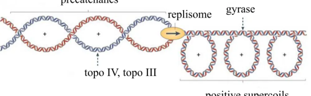
1.4.2. Role of DNA topoisomerases in the early stage of replication fork elongation 47 1.4.3. Role of DNA topoisomerases in the late stages of replication fork elongation . 50 1.4.4. Topoisomerases and replication termination ... 53
1.4.5. Role of DNA topoisomerases and supercoiling in chromosome segregation .... 54
1.5. Recombination dependent replication ... 56
1.5.1. Replication restart ... 63
1.6. Constitutive stable DNA replication (cSDR) ... 65
1.7. Topoisomerase inhibitors ... 68
1.8. Rationale, hypotheses and objectives ... 71
Preface to Chapter 2 ... 76
CHAPTER 2: Manuscript I ... 77
Depletion of RNase HI activity in Escherichia coli lacking DNA topoisomerase I leads to defects in DNA supercoiling and segregation ... 77
Abstract ... 78 Introduction ... 79 Results ... 81 Discussion ... 89 Experimental procedures ... 93 References ... 96 Acknowledgements: ... 102
TABLES AND FIGURES ... 103
Preface to Chapter 3 ... 120
CHAPTER 3: Manuscript II ... 121
Interplay between type IA topoisomerases and gyrase in chromosome segregation in Escherichia coli ... 121
Abstract ... 122
Introduction ... 123
Material and Methods ... 126
Results ... 128
Discussion ... 137
Acknowledgements ... 141
References ... 142
TABLES AND FIGURES ... 147
Preface to Chapter 4 ... 159
CHAPTER 4: Manuscript III ... 160
Roles of type IA topoisomerases in genome maintenance ... 160
Abstract ... 161
Author Summary ... 162
Introduction ... 163
Results ... 167
Discussion ... 179
Materials and Methods ... 185
Acknowledgments ... 187
References ... 188
TABLES AND FIGURES ... 194
Supplemental References ... 196
Supporting information ... 210
CHAPTER 5: Discussion ... 229
5.3 type IA topos and genome maintenance ... 238
CHAPTER 6: Conclusion and future directions ... 246
CHAPTER 7: References and Annexes ... 250
7.1. References ... 251
7.2 Annex I: Table showing suppressors of triple topA rnhA gyrB(Ts) mutant ... 297
List of tables
Chapter 2:
Table 1. Escherichia coli strains used in this study………103
Chapter 3:
Table 1. Escherichia coli strains used in this study………147
Chapter 4:
Table S1. E. coli strains and plasmids used
in this study……….……..194
Chapter 7:
Annex I: Table showing suppressors of the triple topA rnhA gyrB(Ts) mutant
List of figures
Chapter 1:
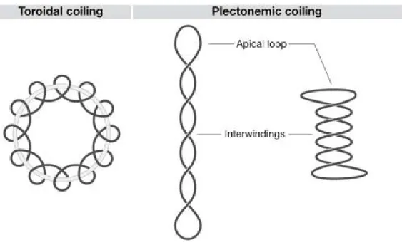
Figure 1. Illustration of the geometry of toroidal and plectonemic……….5
Supercoils Figure 2. Schematic representation of topological domain organization of the E. coli chromosome into independent loops………8
Figure 3. Twin supercoiled domain model………11
Figure 4. Constrained and unconstrained supercoils………...15
Figure 5. Catalysis of transient breakage of DNA by DNA topoisomerases ………17
Figure 6. Mechanism of action of type IA DNA topoisomerases ………...19
Figure 7. Mechanism of action of type IB DNA topoisomerases ………..20
Figure 8. Mechanism of the two gate model of type II DNA topoisomerases …………..23
Figure 9. A revised map of oriC depicting the model of pre-RC assembly ………...34
Figure 10. Domain organization of DnaA………...37
Figure 11. The functions of DNA topoisomerases during replication elongation………...48
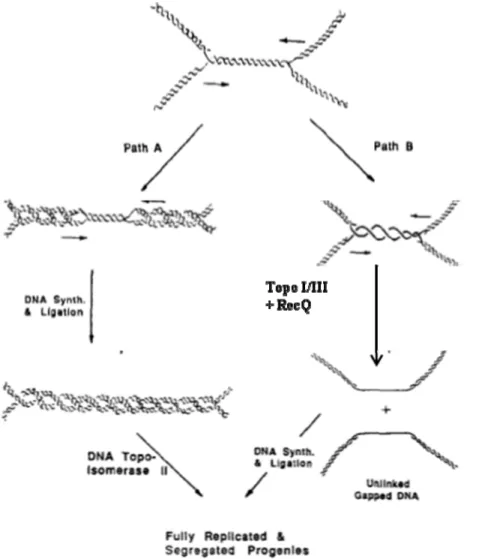
Figure 12. Two pathways showing the merging of a pair of converging replication forks.52 Figure 13. DNA repair by homologous recombination and subsequent replication restart………..59
Figure 14. Diagrammatic representation of two proposed models for the dissolution of dHJs………..62
Chapter 2:
Figure 1. Supercoiling in a topA rnhA null double mutant……….106 Figure 2. Supercoiling in a topA rnhA null double mutant after
transcription inhibition………..108 Figure 3. Western blot analysis to measure topoisomerase IV and gyrase levels
in topA rnhA mutants……… 109 Figure 4. Complementation of a topA rnhA mutant by topoisomerase III
Overproduction………..110 Figure 5. Topoisomerase IV inhibition does not promote hypernegative
supercoiling in a double topA rnhA mutant………111 Figure 6. GyrI, a gyrase inhibitor, play no role in supercoiling inhibition
in a topA rnhA null double mutant………..112 Figure 7. Supercoiling assays in crude cell extracts………...113 Figure 8. Topoisomerase III overproduction does not change supercoiling
in a topA rnhA null double mutant………..116 Figure 9. DAPI staining of topA rnhA null mutants reveals segregation defects
that are corrected by topoisomerase III overproduction………...117
Chapter 3:
Figure 1. Deleting topA complements the growth and Par- phenotype of
the gyrB(Ts) strain……….148 Figure 2. The topB gene is required for the growth of the topA gyrB(Ts)
strain at nonpermissive temperatures………150 Figure 3. The topB gene is required for chromosome segregation in
the topA gyrB(Ts) strain at nonpermissive temperatures………..151 Figure 4. The effect of overproducing a functional ParEC fusion protein
on the growth of RFM475 and CT170 strains………..152 Figure 5. Overproducing topo III at a very high level corrects the
Figure 6. Effects on DNA supercoiling of substituting the gyrB(Ts) allele
of strain SB383 (topA rnhA gyrB (Ts)) for a gyrB+ one……….156 Figure 7. Substituting the gyrB(Ts) allele of strain SB383 (topA rnhA gyrB (Ts))
for a gyrB+ one substantially corrects the chromosomal segregation defect………..157 Figure 8. Effects of the growth medium on unregulated replication
in topA and rnhA mutants………158
Chapter 4:
Figure 1. Growth and chromosome segregation defects in a topA gyrB(Ts) strain
at 30oC………198
Figure 2. Growth and chromosome segregation defects in topA gyrB(Ts) strains
at 24oC………199
Figure 3. recA and recQ deletions are epistatic to topB overexpression
in correcting the growth defect of a topA gyrB(Ts) strain………..200 Figure 4. Replication initiation asynchrony and reduced DNA/mass ratio conferred
by the oriC15::aph suppressor mutation isolated from a topA rnhA gyrB(Ts) strain…….201 Figure 5. The growth and chromosome segregation defects in cells lacking type 1A
topos are partially corrected by overproducing RNase HI or
by deleting recA but not recQ………....203 Figure 6. Both the dnaT18::aph and rne59::aph suppressor mutations isolated from
a topA rnhA gyrB(Ts) strain inhibit cSDR in an rnhA strain……….206 Figure 7. The growth and chromosome segregation defects in cells lacking type 1A topos are partially corrected by dnaT18::aph, rne59::aph and holC2::aph
suppressor mutations isolated from a topA rnhA gyrB(Ts) strain………..208
Supplementary information
Figure S1. Chromosome segregation defects in a topA gyrB(Ts) strain at 30oC………..211 Figure S2. Chromosome segregation defects in a topA gyrB(Ts) strain at 24oC………..213
Figure S3. Topo IV is not overproduced following the deletion of recQ
in strain RFM475………...215 Figure S4. Southern blot experiment showing the successful transfer of the oriC::Tn5 allele within topA null but not topA+ strains………...216 Figure S5. The oriC15::aph mutation complements the growth defect of a dnaAcos…..218
mutant at 30oC
Figure S6. Deleting recA or recQ, or overproducing RNase HI does not complement the growth and chromosome segregation defects of a topA topB gyrB(Ts)
strain at 40oC………..219
Figure S7. The growth and chromosome segregation defects of a topA topB
gyrB(Ts) strain are more severe
at 30 than at 37oC……….………....222
Figure S8. aph insertion sites for three suppressor mutants used in this study………...224 Figure S9. The effect of dnaT18::aph allele on the chromosome segregation
defects of a topA rnhA gyrB(Ts) strain………..226 Figure S10. The ori/ter ratio is similarly reduced in strains lacking either rnhA
or topA………227 Figure S11. Replication initiation asynchrony conferred by the holC2::aph mutation…228
Abbreviations
ATP: Adenosine triphosphate ADP: Adenosine diphosphateaph: Aminoglycoside phosphotransferase gene cassette bp: base pairs
cSDR: constitutive Stable DNA Replication DAPI: 4', 6-diamidino-2-phenylindole oC: Degrees Celsius
∆: Deletion
DNA: Deoxyribonucleic acid DUE: DNA unwinding element
E. coli: Escherichia coli
EOP: efficiency of plating Fig: Figure
h: Hours
iSDR: Inducible Stable DNA Replication IPTG: Isopropyl β-D-1-thiogalactopyranoside kanr: Kanamycin resistance
kb: kilo base LB: Luria-Bertani Lk: Linking number µg: Microgram µl: Microliters µM: Micromolar ml: Milliliters mM: Millimolar OD: Optical density
oriC: Origin of replication C oriK: Origin of replication K
Par-: Partitioning defective phenotype PCR: Polymerase chain reaction RPA: Replication protein A RNase: Ribonuclease Rif: Rifampicin
SSB: Single- Stranded DNA Binding protein NaCl: Sodium chloride
SDR: Stable DNA Replication σ: Supercoiling density
Ts: Temperature sensitive UV: ultraviolet
Dedication
This thesis is dedicated to the memory of my late father, Paa Usongo Isaac Akuta. His guidance and encouragement was crucial in my life. He initiated this process and I wish he was here to witness it. I miss him every day and I take solace in that he is resting in Gods kingdom. Thanks to mom, Ogwe Ruth Usongo for being there for me.
Acknowledgements
First of all, I want to express my sincere gratitude to my supervisor and mentor, Dr. Marc Drolet for giving me the opportunity to work in his laboratory. His vast knowledge, constructive comments, invaluable suggestions, guidance and corrections have been of great importance to me and from the bottom of my heart, I sincerely appreciate the chance he gave me. Without him, I could not have realized this project. His doors were open all the time and when confronted with a technical problem, I will call his phone for suggestions/advice and he will happily help out.
I will also like to thank members of the Drolet’s lab, past and present for their support. Special thanks to Flora Nolent, Patrick Sanscartier, Jill Egbe and Christy Manuella.
I will also like to thank the professors, staff and fellow students of the department especially Dr. George Szartmari for his support and advice, Martin Clement for his input and suggestions, and Serge Sénéchal for his invaluable assistance in flow cytometry. In the same light, I would also like to extend my gratitude to Christian Charbonneau at l’Institut de recherche en immunologie et en cancérologie.
I am grateful to the members of my thesis evaluation committee Dr. Reyes-Lamothe Rodrigo, Dr. Catherine Paradis-Bleau, Dr. Dindial Ramotar and Dr. George Szatmari for accepting to evaluate my thesis. Your comments/suggestions were very useful in the realization of this work.
My sincere gratitude and appreciation to my dear mom Ogwe Ruth Usongo, my beloved brothers and sisters for their encouragement, moral and spiritual support. To all my friends I sincerely thank you all for always being there for me.
Finally I will like to thank my beloved wife Karen, my daughter Karissa and my son Kaelen-Isaac for all their love, support and encouragement.
1.1. Historical perspective of the DNA entanglement problem
The double helical structure of DNA described by Watson and Crick took the scientific community by storm and projected them to instant fame. In their model termed the double helix model, the two strands are interwound and follow a right hand helical path around a central axis and run in opposite directions with a helical repeat of 10 bp per turn. The two strands are held together by base pairing through the formation of hydrogen bonds with thymine pairing with adenine and cytosine pairing with guanine (Watson & Crick, 1953b). In regards to the base pairing in the double helix, Watson and Crick stated that "It has not escaped our notice that the specific pairing we have postulated immediately suggests a possible copying mechanism for the genetic material" (Watson & Crick, 1953b). The concept of complementarity that they predicted in their model suggested that a parent DNA molecule could be duplicated by copying each strand in tandem with the Watson and Crick base pairing (Watson & Crick, 1953a).The interwound nature of the helix imposes a strict requirement on the mechanism of replication. The parental strands need to be unlinked for the separation of daughter chromosomes to occur. Watson and Crick acknowledged the challenge but stated that it was "not insuperable" (Watson & Crick, 1953b). This admission by Watson and Crick was not enough to dissuade criticism of their model. Among them Max Delbruck raised the concern on how the semi-conservative scheme of DNA replication (an appellation designated since it was envisioned that one half of the progeny duplex was inherited and the other half newly synthesized) could be successfully executed without tangling of the progeny DNA, with the parental DNA spelling disastrous consequences for the cell following division. He went ahead to propose solutions involving breakage every 1/2 turn of the helix during replication and suggested an alternate model of discontinuous DNA replication to avoid the linking problem (Delbruck, 1954). The confirmation that DNA replicated semi-conservatively (Meselson & Stahl, 1958) implied that the entanglement problem was real.
In addition, the famous autoradiographic images (Cairns, 1963b) of E. coli showing a circular chromosome that replicates semi-conservatively from a unique region of the parent DNA ring (Cairns, 1963a) equally proved the entanglement problem.
Further solidifying the entanglement problem was the discovery that the DNA of the polyomavirus that infects animals was a double stranded ring with intact strands (Weil & Vinograd, 1963). This observation further complicated the issue of separating the DNA strands which became a topological problem. Another observation that added a piece to the puzzle was the discovery that polyoma DNA existed in varying forms namely "extended cyclic form" and "tightly coiled cyclic form" or supercoiled DNA (Vinograd et al., 1965). The observation that DNA from polyomavirus was negatively supercoiled triggered more structural studies (Vinograd et al., 1965) which explained the varying forms of DNA observed through a quantity called the linking number (Lk). Lk is the number of times one strand crosses the other and it is the sum of two terms twist (Tw) which is the local winding of the two strands around each other, and writhe (Wr) a measure of DNA supercoiling (White, 1969). Tw and Wr are related to Lk by the simple equation: Lk = Tw +Wr (White, 1969).
It was soon deduced that a DNA ring from a natural source would display a value of LK lower than that of the same ring in its most stable structure under typical experimental conditions used in the laboratory. This quantity termed the linking number of a relaxed DNA ring has a symbol LK0. A DNA ring with a linking number Lk is said to be negatively supercoiled if Lk < LK0, or (Lk- LK0) < 0 or positively supercoiled if Lk > LK0 or (Lk- LK0) > 0 (J. C. Wang, 1974). In this regard, the polyoma DNA rings from natural sources were thus defined to be negatively supercoiled and the supercoiled state of the DNA was viewed as a compensation for the torsional stress produced by a reduction in Lk (Vinograd et al., 1965).
Proper duplication and transmission of genetic information from one generation to another warrant that the linking number must be reduced to zero. It is in this light that a prominent topologist named William Pohl rejected the plectonemic Watson-Crick model for a paranemic (side-by-side) structure of DNA, for he could not imagine how a global property like the Lk could be nulled enzymatically (Pohl & Roberts, 1978). The quest to know why DNA from natural sources was negatively supercoiled lead to the discovery of
DNA topoisomerases as nature's solution for DNA strand unlinking and modulation of the torsional state of the duplex (Wang, 1971, 1985).
1.1.1. DNA supercoiling
As mentioned earlier, supercoiling in a DNA molecule can either be positive or negative. Literally speaking, DNA is said to be negatively supercoiled when it has a deficiency in the linking number compared with the relaxed DNA. Because negatively supercoiled DNA has fewer helical turns than the molecule would normally contain as a linear or relaxed molecule, it is said to be underwound (Sinden, 1994). The consequence of this underwinding in the number of helical turns is that there are more base pairs per helical turn, which leads to a decrease in the angle of twist between the adjacent base pairs. Underwinding thus creates torsional tension following the winding of the double helix. This torsional tension drives the supertwisting of the molecule forming a right-handed (or clockwise) supercoil (Sinden, 1994). On the contrary, positively supercoiled DNA is overwound in terms of the number of helical turns, leading to fewer bases per helical turn and thus an increase in the winding angle between the adjacent base pairs, which equally create torsional tension in the winding of the helix. The tension in the winding helix is relieved by the positive supercoiling of the DNA forming a left-handed (counterclockwise) supercoil (Sinden, 1994).
DNA isolated from natural sources is negatively supercoiled (Vinograd et al., 1965). In fact DNA supercoiling has been shown to be a target of evolutionary selection for mutations that lead to an overall increase in negative supercoiling have been identified in evolutionary experiments. Mutations in genes such as topA encoding topoisomerase I (topo I) and dusB which regulates the expression of fis (encoding the histon-like protein Fis ) have been identified in long term evolutionary experiments (Crozat et al., 2010). Several lines of experimental evidence have confirmed that negative supercoiling causes the double helix to adopt a branched and plectonemic or interwound structure. Non-viscous nucleoids obtained by treating cells with lysozyme (enzymes that hydrolyses bacterial cell walls) and mild ionic detergent (for cell lysis) and analyzed by sedimentation and electron
microscopy reveals an interwound DNA structure (Giorno, Hecht, & Pettijohn, 1975; Kavenoff & Bowen, 1976; Worcel & Burgi, 1972). Plasmid DNA structure produced by recombination with the lambda integrase and analyzed with sensitive and non-disruptive topological tests have confirmed the presence of interwound DNA in vivo (Bliska & Cozzarelli, 1987).
Supercoils do not only exist as interwound supercoils. Negative supercoils can also physically exist as left-handed toroidal coils whereby the DNA is wrapped around proteins.
Figure 1. Illustration of the geometry of toroidal and plectonemic supercoils. DNA depicting a single form of toroidal DNA and two geometries of plectonemes which have the same superhelical density. Conversion of the long plectoneme to the shorter one is achieved by DNA bending and untwisting of the DNA duplex (Travers & Muskhelishvili, 2007).
Figure used with permission from Nature publishing group.
In solution, supercoils are manifested as a mixture of interwound and toroidal coils. Studies have shown that for DNA in bacteria about 70 % of the linking number deficiency is distributed as writhe change and about 30 % is distributed as a change in twist (Boles, White, & Cozzarelli, 1990). Toroidal supercoils are very important for the biological organization of the nucleosome core in eukaryotes that involves the toroidal coiling of the DNA around proteins. In this organization, the supercoiling energy is restrained by this
wrapping. In bacteria, studies have shown that the dominant form of supercoiled DNA is a plectoneme (Crisona et al., 1999). In fact, measurements using the cross-linking agent trimethyl-psoralen which forms interstrand abducts on DNA have revealed that most of the DNA in bacteria exists as unconstrained plectonemic supercoils in vivo, whereas little if any of the DNA in eukaryotic nucleus is in this form (Sinden, Carlson, & Pettijohn, 1980). More importantly it has been found that in the nucleosome, DNA is marginally overtwisted (White, Cozzarelli, & Bauer, 1988; Zivanovic et al., 1988) whereas in the plectonemic form when free in solution, negatively supercoiled DNA is undertwisted significantly (Boles et al., 1990).
Undertwisting facilitates processes that require strand separation and this explains the importance of negative supercoiling in cellular processes such as initiation of DNA replication (Alexandrov et al., 1999; Pruss & Drlica, 1989), positive or negative modulation of transcription of numerous bacterial genes (Lim et al., 2003; Peter et al., 2004; Travers & Muskhelishvili, 2005b), chromosome segregation (Holmes & Cozzarelli, 2000; Sawitzke & Austin, 2000; Usongo et al., 2008), and recombination (Alexandrov et al., 1999; Crisona et al., 1994; Hatfield & Benham, 2002). Even though cellular processes, especially replication and transcription, generate strong torsional forces along the DNA axis leading to the buildup of transient supercoiling in both the positive and negative directions (Liu & Wang, 1987; Lockshon & Morris, 1983), homeostatic regulation by DNA topoisomerases (to be discussed ahead) resets the topological status into an underwound state (Travers & Muskhelishvili, 2007; Zechiedrich et al., 2000).
1.1.2. Global supercoiling
The importance of DNA supercoiling in E. coli has no yard stick measurement as it is implicated in numerous cellular transactions as stated below. Because of its importance, a modest reduction of negative supercoiling in bacteria is lethal (Zechiedrich, Khodursky, & Cozzarelli, 1997). In addition, supercoiling is equally fragile as just a single break in DNA can relax the entire chromosome killing the cell since free negative supercoiling is necessary for viability (Gellert et al., 1976). The process of DNA replication also causes
relaxation because of the presence of gaps in the lagging strand. Fortunately for the cell, these doom day scenarios are avoided thanks to the intuitive organization of the chromosome. Actively replicating parts of the chromosome and any insults be it physical, chemical or enzymatical to the DNA backbone are confined to isolated regions of the chromosomes that are topologically independent and protect the bulk of the chromosome from the dangers of relaxation. These regions are called domains (Postow et al., 2004). Topological domains are defined as regions of the DNA that are topologically constrained at their ends and are thus independent with regards to the entire chromosome. Global supercoiling is defined as the average superhelical density of all supercoiling domains. The superhelical density (σ) is controlled in a very narrow range and deviations from this range in either direction are growth inhibitory (DiNardo et al., 1982). Mathematically, σ can be defined as ∆LK/LK0 (Vologodskii & Cozzarelli, 1994). LK0 = N/γ where N is the number of base pairs in the molecule and γ is the mean number of base pairs per turn in the double helix under a given set of conditions, ∆LK = LK- LK0 (Vologodskii & Cozzarelli, 1994). DNA extracted from cells has a σ between -0.03 and -0.09 (Bauer, 1978). In E. coli, genetic studies using DNA topoisomerase mutants have established that for vigorous growth to occur, global supercoiling must lie within a +15% range of supercoiling (Drlica, 1992).
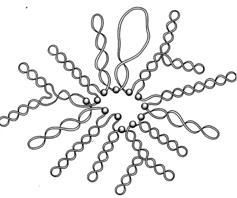
Figure 2. Schematic representation of topological domain organization of the E. coli chromosome. The E. coli chromosome is organized into independent topological domains
in vivo. The shaded spheres represent the domain boundaries (Sinden, 1994). Figure used
with permission from Elsevier.
Evidence of the existence of domains has been documented. Chromosomes isolated from E. coli are found to be folded by unconstrained negative supercoiling and several nicks are required to relax the entire chromosome (Delius & Worcel, 1974; Worcel & Burgi, 1972). The conclusion from these papers is that the E. coli chromosome is divided into no more than 50 topological domains with an average length of about 100 kb. By counting the number of supercoiled loops visible by electron microscopy, domains were estimated to be between 65 and 200 per nucleoid (Kavenoff & Bowen, 1976; Kavenoff & Ryder, 1976). Another method that is used to estimate the number of domains per nucleoid is psoralen photobinding to bacterial chromosomes partially relaxed by DNA breaks induced with X-rays in vivo. This method estimates the number of domains to be around 50 (Sinden & Pettijohn, 1981). More recent experiments employing less invasive approaches have shown that domain sizes are much smaller. A study in S. enterica examining the
topological requirements of site specific recombinases concluded that topological domains average 25 kb in length (Higgins et al., 1996).
A recent less invasive method to determine the size of domains has been developed (Postow et al., 2004) and it is based on the fact that negative supercoiling can modulate the activity of specific promoters by either increasing or decreasing their output (McGovern et al., 1994; Travers & Muskhelishvili, 2005b; Willenbrock & Ussery, 2004). This method takes advantage of the 306 supercoiling sensitive genes that are distributed widely on the genome and respond rapidly and reliably to supercoil relaxation. After DNA relaxation, 106 of the supercoiling sensitive genes (SSGs) become induced for transcription while 200 genes are repressed (Peter et al., 2004). In this method, double strand breaks were introduced onto specific locations on the chromosome by controlling the in vivo expression of restriction enzymes. The measure of the distance from a SwaI site to the promoter of a supercoiling sensitive gene (SSG) was combined with microarray expression patterns before and after cleavage with restriction enzymes. Following the analysis of the SSG transcription data, a model was proposed which predicts that the E. coli chromosome consists of variable loops and random distribution of domain barriers with an average domain size of 10 kb, significantly smaller than the previously estimated domain sizes (Postow et al., 2004).
Small domains confer several advantages to the cell. Small domains make life easy for decatenating enzymes by concentrating catenane links, making global processes more local (Espeli et al., 2003). By concentrating catenane links, the free energy of catenation will also increase and this will help drive the decatenation reaction to completion (Vologodskii & Cozzarelli, 1993). Catenanes and precatenanes will easily be resolved following replication in small domains (Schvartzman & Stasiak, 2004). Domain independence implies that each domain is shielded from the other by a barrier. Despite the importance of domain barriers little is known about the cellular components of these barriers in vivo. For bacteria, in vitro studies with proteins such as FtsK and SpoIIIE found that they are able to constrain DNA loops (Aussel et al., 2002; Bath et al., 2000; Pease et al., 2005). Moreover, these proteins are anchored in the bacterial inner membrane, making
them likely to form topological barriers on chromosomes that they act upon. Stable RNAs or transcriptional complexes can also stabilize chromosomes. RNases can decondense isolated chromosomes and these decondensed chromosomes contain a high fraction of nascent mRNA and DNA-bound RNA polymerase (Pettijohn et al., 1970; Stonington & Pettijohn, 1971; Worcel & Burgi, 1972).
The role of transcription in domain barrier formation has also been tested with the transcriptional inhibitor rifampicin. Although rifampicin causes chromosomes to decondense upon isolation (Dworsky & Schaechter, 1973; Pettijohn & Hecht, 1974), there is no change in domain numbers in vivo (Sinden & Pettijohn, 1981). In bacteria, since transcription and translation are coupled, insertion of the nascent polypeptide into the membrane in the case of mRNA encoding membrane bound proteins can also act as a domain barrier and this has been shown using plasmid DNA (Lynch & Wang, 1993). The role of transcription-translation coupling in domain barrier formation has also been demonstrated in the Salmonella typhimurium chromosome (Deng, Stein, & Higgins, 2004). A more recent in vivo study identified H-NS, Fis and transketolase (TktA) as domain barrier proteins and these proteins play a role in the supercoiling of domains by forming topological barriers in the chromosomes (Hardy & Cozzarelli, 2005).
1.1.3. Local supercoiling
Cellular processes such as DNA replication and transcription that involve tracking of huge protein complexes along double stranded DNA can transiently cause local perturbations in DNA topology. Evidence implicating transcriptional activity with DNA supercoiling has been documented. Highly positively supercoiled pBR322 DNA has been extracted from E. coli treated with gyrase inhibitors (Lockshon & Morris, 1983). pBR322 DNA extracted from topA mutants of E. coli and S. typhimurium harbors a high degree of negative supercoiling and this depends on the transcription of the tetA gene (Pruss & Drlica, 1986).
These observations led Liu and Wang (Liu & Wang, 1987) to propose the twin supercoiled domain model of transcription. The basis of this model is that elongating RNA polymerase molecules cannot rotate freely around the double-helical DNA because of the bulk of the polymerase with associated nascent transcripts as well as attached ribosomes. As a result, the DNA is forced to rotate upon itself. If the end of the DNA molecules are constrained in some way either by being very long or attached to cellular structures, polymerase tracking will cause the DNA ahead of the transcription complex to be overwound and the DNA behind to be underwound. Thus, polymerase movement will generate domains of positive supercoiling ahead of its passage and domain of negative supercoiling behind it. This model has been supported experimentally both in vitro and in
vivo (Drolet, Bi, & Liu, 1994; Leng, Amado, & McMacken, 2004; Leng & McMacken,
2002; Rovinskiy et al., 2012).
Figure 3. Twin supercoiled domain model: Transcription generates domains of positive and negative supercoils respectively in front and behind of the transcription complex represented by RNAP. E. depicts the frictional barrier against the rotation of the duplex around it helical axis (Wu et al., 1988). Figure adapted with authorization from Elsevier.
Even though it has been shown that topoisomerases are able to relax transcription induced supercoiling (Cook et al., 1992; Drolet, 2006; Massé & Drolet, 1999b; Rovinskiy et al., 2012), some studies have shown that irrespective of the presence of topoisomerases, localized supercoiling exists and can be exploited to trigger a variety of important DNA transactions (Bowater, Chen, & Lilley, 1994; Dunaway & Ostrander, 1993; Figueroa & Bossi, 1988; Kouzine & Levens, 2007; Kouzine et al., 2004; Kouzine et al., 2008; Ljungman & Hanawalt, 1992). It has also been found that depending on transcription
intensity and the disposition of topoisomerases, local supercoiling may exceed the relaxation activity of topoisomerases, forcing the residual DNA torsional stress to propagate through the surrounding DNA (referred to as dynamic supercoiling) (Kouzine et al., 2008).
The double helix which is in the predominant B-form (the most common form of DNA inside the cell and the one described by Watson and Crick) could adopt, depending on the sequence composition, a variety of alternative structures. In order to form these structures, the DNA duplex must be melted and this can be achieved by a high level of negative supercoiling (Bloomfield, Crothers, & Tinoco, 1974). In humans cells, dynamic supercoiling has been measured in vivo and in vitro by identifying chromosomal regions that have sequences susceptible to the formation of non-B DNA structures, and these regions are found to be mostly located upstream of active promoters (Kouzine et al., 2004; Kouzine et al., 2008). Dynamic supercoiling has been shown to persist for some time irrespective of the presence of normal concentrations of functional topoisomerases in the cell (Kouzine et al., 2008) and this suggests that topoisomerases are unable to immediately keep up or prevent the build-up of transient torsional stress induced by transcription. The non-B DNAs produced as a result of dynamic supercoiling have been shown to be bound by a variety of proteins that can change DNA conformation, implying that these non-B DNAs structure are important to the cell (Brooks, Kendrick, & Hurley, 2010; Kouzine & Levens, 2007).
The importance of non-B-DNA in gene regulation is illustrated by the human c-myc proto-oncogene. The protein c-Myc is a crucial regulator of up to 15 % of human genes, and this protein is essential for cell homeostasis, differentiation and growth (Liu et al., 2006). If not properly regulated, c-myc becomes a lethal oncogene that plays a role in many cancers (Hanahan & Weinberg, 2011). Because both the c-Myc mRNA and c-Myc protein are too short-lived to provide an effective feedback mechanism, the cell has evolved an alternative feedback mechanism that uses DNA dynamics to ensure regulation of c-myc transcription. This is provided by a 90 bp far upstream element (FUSE) of the human c-myc gene. The transcriptional activity of the c-myc promoter is enhanced by the supercoiling sensitive FUSE sequence. This sequence is sensitive to negative supercoiling. Elevated
levels of negative supercoiling melts this sequence. This in turn enables this sequence to bind to the transcription activator FUSE- binding protein (FBP). FBP increases promoter activity by interacting with the general transcription factor TFIIH to drive c-myc transcription. Another protein called the FBP interacting repressor (FIR) binds to FBP and FUSE and represses c-myc transcription (Liu et al., 2006). Therefore FUSE melting acts as a sensor for transcription to provide either positive feedback (via FBP) or negative feedback (via FIR) in the regulation of c-myc transcription.
Another important conformational sequence involved in c-myc regulation is the CT-element located 250 bp upstream of the main promoter (Brooks & Hurley, 2009; Siddiqui-Jain et al., 2002). This element has been shown to adopt non-B structures in supercoiled DNA, in vitro as well as in vivo (Kohwi & Kohwi-Shigematsu, 1991; Michelotti et al., 1996). The transcriptional activator Sp1 binds the CT element in its normal B-DNA structure to activate transcription. It has also been suggested that this sequence can also adopt a single-stranded conformation due to supercoiling accumulation generated by transcription. It is in this single-stranded form that the transcription factors hnRNPK and CNBP bind to maintain the active state of transcription (Brooks & Hurley, 2009; Michelotti et al., 1995; Tomonaga & Levens, 1996). The CT element can also adopt a non-B DNA structure conformation and in this conformation, the binding sites of the transcription factors are sequestered, leading to transcriptional silencing (Sun & Hurley, 2009).
Transcription induced local supercoiling also plays a role in the regulation of gene expression. A well-documented example is the activation of the S. typhimurium leu-500 promoter. It has been found that a point mutation in this promoter that confers leucine auxotrophy is phenotypically suppressed by a mutation in the gene coding for topoisomerase I (Lilley & Higgins, 1991). This point mutation is located in the -10 region, making open complex formation energetically expensive, and the absence of topo I would energetically favor open complex formation at the mutant promoter. However, placing this mutant promoter on an extra chromosomal plasmid even in the absence of topo I (Richardson, Higgins, & Lilley, 1988) does not reproduce the same effect, thereby implicating local rather than global change in template topology. In addition, the leu-500 promoter can be activated in a topo I negative background when it is placed divergent to the
tetracycline gene (tetA) on a plasmid (Chen et al., 1992). Activation only occurs upon transcription and translation of the tetA gene. Thus, the level of supercoiling that is generated is sufficient to induce open complex formation on the leu-500 promoter located upstream.
1.1.4. Constrained versus unconstrained supercoiling
A topological domain can harbor two sorts of supercoils: constrained and unconstrained supercoils (Cozzarelli & Wang, 1990). Though hidden, constrained supercoils are not lost when DNA is nicked (Pettijohn & Pfenninger, 1980). The portion of the overall supercoiling that is lost when DNA is nicked is called unconstrained supercoiling or superhelical tension. In E. coli, about 50% of the DNA supercoils are free (Bliska & Cozzarelli, 1987). Thus, about 50 % of supercoils are constrained by proteins binding to DNA (Bensaid et al., 1996; Bliska & Cozzarelli, 1987; Pettijohn & Pfenninger, 1980; Sinden & Kochel, 1987). Although a major source of this restraining comes from proteins that separate DNA strands as well as proteins of the replication apparatus and RNA polymerases, architectural proteins such as heat-stable-nucleoid-structuring protein (H-NS), integration host factor (IHF), the histone-like HU proteins, and the factor for inversion stimulation (FIS) have also been implicated (Drlica & Rouviere-Yaniv, 1987; Travers & Muskhelishvili, 2005a). Even though the exact role and mode of DNA interactions of these proteins are not entirely clear, some clues are beginning to emerge. The HU heterodimer not only constrains negatively supercoiled DNA by stabilizing plectoneme folds, it may also wrap DNA into a left-handed nucleosome-like structure (Guo & Adhya, 2007). HU deficient mutant strains have been shown to exhibit levels of supercoiling 15 % lower than those of wild-type strains (Hsieh, Rouviere-Yaniv, & Drlica, 1991). The stabilization of negative supercoils by HNS is likely achieved by interacting with DNA crossings. H-NS and IHF mutants show a high reduction in negative supercoiling compared to the wild-type (Higgins et al., 1988; Parekh, Sheridan, & Hatfield, 1996). The factor for inversion stimulation (FIS) binds at crossovers in plectonemes (Schneider et al., 2001) but it can also locally stabilize a DNA toroid (Maurer et al., 2006). In eukaryotes, the majority of
supercoils are restrained by nucleosomal organization following the toriodal wrapping of DNA around histones (Holmes & Cozzarelli, 2000).
Figure 4.Constrained and unconstrained supercoils. In the unrestrained supercoil upper figure, any insult on the DNA backbone leading to chromosome breakage will lead to complete DNA relaxation. In the constrained supercoil represented below, supercoils are constrained by proteins and, in the event of strand breakage, supercoils are not lost since they are constrained. Figure obtained from (Sinden, 1994). Figure used with permission from Elsevier.
1.2. DNA topoisomerases
The choice was made by nature early in evolution to have the plectonemic DNA double helix as the carrier of genetic information. From this initial choice, the challenge to duplicate DNA became apparent and this was deeply rooted in the plectonemic nature of the DNA helix. However, this challenge was no match to nature's remedy, DNA topoisomerases. They evolved alongside DNA to solve the topological problems associated with it. Their universal partnership with DNA was blessed by nature from the beginning. Equipped with the ability to cut, shuffle and religate DNA strands, topoisomerases can add or remove supercoils, untangle interlocked double stranded DNA segments (catenanes)
and introduce or remove knots from DNA rings (Hartman et al., 2013; Wang, 2002). The dexterity of these enzymes at solving topological puzzles is achieved via the simple and elegant chemistry of transesterification (Wang, 2002). It is through this mechanism that the strand breakage reaction of topoisomerases is achieved. The first step is the attack of the DNA phosphorus by tyrosyl oxygen of the enzyme, thus forming a covalent phosphotyrosine link and at the same time breaking a DNA phosphodiester bond. Through a second transesterification reaction, which is the reverse of the first, the DNA strands are rejoined. In the reverse reaction, basically the oxygen from the DNA hydroxyl group that is generated from the first reaction attacks the phosphorous of the phosphotyrosine link and as a result, the covalent bond between the protein and DNA is broken and the DNA backbone bond is reformed (Wang, 2002).
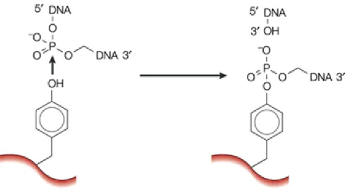
Figure 5. Catalysis of transient breakage of DNA by DNA topoisomerases. In the transesterification reaction, the tyrosyl oxygen of the enzyme attacks the DNA phosphorus leading to the breakage of the DNA backbone bond and the formation of a covalent enzyme -DNA intermediate. Rejoining the backbone bond occurs by the reversal of the reaction shown above. In the reverse or second transesterification reaction, the oxygen of the DNA hydroxyl group generated in the first reaction, attacks the phosphorus of the phosphotyrosine link breaking the covalent bond between the protein and DNA and reforms the DNA backbone bond. In the reaction catalyzed by a type IA or type II enzyme, a 3'-OH is the leaving group and the active tyrosyl becomes covalently linked to a 5'-phosphoryl group, as shown. In the reaction catalyzed by a type IB enzyme (not shown) a 5'-OH is the leaving group and the active-site tyrosyl becomes covalently linked to a 3'-phosphoryl group (Wang, 2002). Figure used with permission from Nature Publishing Group.
The end result of this reaction is the creation of an enzyme mediated transient DNA gate through which another DNA strand or a double helix can pass, a phenomenon termed enzyme-bridging mechanism (Wang, 2002). This mechanism is illustrated in Figure 8. This mechanism has been exploited for clinical purposes to develop drugs that act by trapping the covalent enzyme-DNA complex and, this has made these enzymes targets of the pharmaceutical industry, as a lot of quinolone antibiotics (Drlica & Zhao, 1997) and
anticancer drugs (Pommier, 2013; Staker et al., 2005; Staker et al., 2002) wreak havoc by exploiting this route.
1.2.1. Classification of topoisomerases
Using DNA strand cleavage as a discriminatory factor, topoisomerases are classified into two broad categories: type I, those that cleave only one DNA strand, and type II, those that cleave both DNA strands to generate staggered double strand breaks (Hartman et al., 2013; Wang, 2002). Further discrimination of the type I topoisomerases in to the subfamily types IA, IB and IC is based on structure and/or mechanistic properties (Hartman et al., 2013). Mechanistic discrimination is based on the point of linkage of the enzyme to the phosphate in the DNA. If the enzyme is linked to a 5' phosphate, they are classified as type IA and if the enzyme is attached to the 3' phosphate, they are classified as types IB (Champoux, 2001; Hartman et al., 2013; Schoeffler & Berger, 2008; Vos et al., 2011; Wang, 2002) and IC (Forterre, 2006; Schoeffler & Berger, 2008; Vos et al., 2011). Prompted by the discovery of a novel type II enzyme from the hyperthermophilic archaeon
Sulfolobus shibatae (Bergerat et al., 1997; Buhler et al., 1998) the type II topoisomerases
were further divided into the subfamilies type IIA and type IIB based on structural differences (Nichols et al., 1999). Type IA topoisomerases are found in all three domains of life namely bacteria, archaea and eukarya (Hartman Chen et al., 2013). The relaxation of negatively supercoiled DNA is their primary activity (Hiasa, DiGate, & Marians, 1994; Wang, 1971) and this requires an exposed single-stranded region within the DNA substrate for activity (Kirkegaard & Wang, 1985). Overwound or positively supercoiled DNA is refractive to type IA enzymes and relaxation of positively supercoiled DNA can only be realized if a pre-existing single-stranded region is present (Kirkegaard & Wang, 1985). Based on the crystal structure of the N-terminal fragment of type IA enzymes, a mechanism of DNA relaxation or catenation-decatenation by type IA topoisomerases has been proposed and confirmed subsequently in biochemical and biophysical reactions (Dekker et al., 2002; Dekker et al., 2003; Li, Mondragon, & DiGate, 2001). In this mechanism, the type IA enzyme, cleaves one strand to generate a single-strand break which is bridged by
the formation of a phosphotyrosine linkage between the enzyme and the 5' end of the broken DNA strand, while holding the 3'-end hydroxyl group non-covalently (Brown & Cozzarelli, 1981; Champoux, 1981; Depew, Liu, & Wang, 1978; Tse, Kirkegaard, & Wang, 1980; Zhang, Cheng, & Tse-Dinh, 2011). Magnesium ions are required for relaxation by type IA topoisomerases, as they are essential for catalysis by helping to keep the 3' end of the cleaved strand in a proper position in the catalytic site (Schmidt et al., 2010; Zhang et al., 2011).
Figure 6. Mechanism of action of type IA DNA topoisomerases. Following the transient breakage of a DNA strand ( blue line), the 5' end of the broken DNA is attached covalently to the active-site tyrosyl group (red circle) in the lid of the enzyme, while the 3' end is non covalently bound to the base of the enzyme. The passage of another strand (blue circle) is achieved by lifting the lid away from the base which opens up the gate (Wang, 2002). Figure adapted with permission from Macmillan publishers Ltd.
In addition to negative supercoils relaxation, these enzymes can also catalyze knotting, unknotting and interlinking of single stranded circles as well as knotting, unknotting catenation and decatenation of double stranded DNA molecules provided there is a gap in one of them (Dean & Cozzarelli, 1985). Members of this family include bacterial DNA topoisomerase III and I (Srivenugopal, Lockshon, & Morris, 1984; Wang, 1971), eukaryotic DNA topoisomerase III (Wallis et al., 1989) and reverse gyrase (Kikuchi & Asai, 1984). Recent single-molecule techniques have confirmed the general features of the strand passage mechanism in type IA topoisomerases as well as the reduction of linking number in steps of one per catalytic event (Dekker et al., 2002). The energy required to
power reactions catalyzed by type IA topoisomerases is provided by the mechanical tension of supercoiled DNA as reactions catalyzed by type IA topoisomerases proceeds without ATP. One exception is reverse gyrase which uses ATP to introduce positive supercoils and, as its name reflects, its positive supercoiling activity is the opposite of the negative supercoiling activity of DNA gyrase (Kikuchi & Asai, 1984).
Unlike the type IA enzymes which rely on strand passage, type IB enzymes are thought to effectuate supercoil relaxation by swiveling the DNA opposite its nicking point and this mechanism of supercoil relaxation is supported by structural (Stewart et al., 1998) and kinetic (Stivers, Harris, & Mildvan, 1997) data. In this mechanism, a type IB enzyme cleaves a single-strand of the duplex DNA and allows one duplex end to rotate with respect to the other around the intact phosphodiester bond on the opposing strand. In this scheme, only the 3'-OH end of the broken strand is tightly bound to the enzyme through covalent binding with the active site tyrosine residue. Because the 5' end of the DNA strand is only bound to the enzyme via nonspecific interactions, it can rotate freely (Stivers et al., 1997). However, free rotation is hindered by friction between the DNA and the enzyme and this helps to align the broken ends prior to resealing, a mechanism termed "controlled rotation" (Koster et al., 2005).
Figure 7. Mechanism of action of type IB DNA topoisomerases. In the case of type IB enzymes, the 3' end of the broken DNA is covalently linked to the active-site tyrosyl group (Y) of the enzyme (red circle) (Wang, 2002). Figure used with permission from Macmillan
publishers Ltd.
The efficiency of relaxation by type IB enzymes is also affected by the extent of supercoiling. It has been shown that higher supercoiling levels lead to an increase in the
5ꞌ
3ꞌ
mean number of supercoils removed by the enzyme per cleavage/religation cycle, thus indicating that type IB topoisomerases are sensitive to the torque stored in under-or overwound DNA (Koster et al., 2005). The number of supercoils decreases by one per DNA rotation. During one catalytic event, several rotations may occur between the strand cleavage and ligation events, and hence the DNA linking number changes at random by several units, unlike the type IA topoisomerases whereby the linking number changes in steps of one (Koster et al., 2005). Type IB enzymes efficiently relax positively and negatively supercoiled DNA (Madden, Stewart, & Champoux, 1995). The catalytic domains of type IB enzymes and tyrosine recombinases are evolutionarily related (Cheng et al., 1998). Members of this family include eukaryotic DNA topoisomerase I, poxvirus topoisomerase I as well as homologues found in certain bacteria (Krogh & Shuman, 2002) and in Mimivirus (Benarroch et al., 2006).
Topoisomerase V (Slesarev et al., 1993) the only known member of the Type IC topoisomerases has so far been found only in the archaea Methanopyrus kandleri which is one of the most hyperthermophilic organisms known (Forterre, 2006). Like the type IB enzymes, type IC topoisomerases also relax positively and negatively supercoiled DNA via a nicking and rotation mechanism and requires neither magnesium ions nor ATP as a cofactor (Slesarev et al., 1993; Taneja et al., 2007). Structurally, the active site of type IC enzymes show little similarity to that of type IB and appear to be evolutionarily distinct (Forterre, 2006; Taneja et al., 2006).
As is the case with type IA topoisomerases, type IIA enzymes effect topological changes on DNA by creating an enzyme bridge gap in DNA and passing a second DNA segment through the break (Brown & Cozzarelli, 1979; Mizuuchi et al., 1980). Type IIA topoisomerases however differ in that they cleave both strands of DNA generating a double strand break through which a second duplex is passed (Brown & Cozzarelli, 1981; Liu, Liu, & Alberts, 1980; Mizuuchi et al., 1980; Sander & Hsieh, 1983). To achieve this feat, the enzymes bind and open up a gate in the duplex termed the G segment, and via the transesterification mechanism earlier described, a second DNA piece termed the transfer or T segment is captured and transported through the gate or G segment. Through a second transesterification reaction which is basically the reverse of the first reaction, the gate is
closed and the active site tyrosine is reset for the next catalytic event (Hartman Chen et al., 2013; Wang, 2002). This transient double strand break reaction inverts double-stranded DNA crossovers, changing the LK in steps of two (Brown & Cozzarelli, 1979). Unlike the type IA topoisomerases, type IIA topoisomerases uses ATP as the driving force behind strand passage reactions (Brown & Cozzarelli, 1979; Gellert et al., 1976; Goto & Wang, 1982). This ATP dependent ability to transport one double helix through another equally endows these topoisomerases with the ability to resolve catenanes, the decatenation of double stranded rings, as well as the relaxation of positively or negatively supercoiled DNA (Champoux, 2001; Mizuuchi et al., 1980; Wang, 1996, 1998).
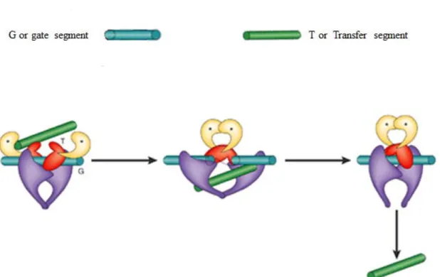
Figure 8. Mechanism of the two gate model of type II DNA topoisomerases. The G segment represented by the blue rod is the double stranded DNA segment that contains the enzyme-mediated DNA gate through which the T or transfer segment represented by the green rod is passed through. The transport and subsequent exit of the T-segment through the gate is mediated by ATP binding and hydrolysis respectively. ATP binding sites are represented by asterisks (Wang, 2002). Figure adapted with permission from Macmillan
publishers Ltd.
Type IIA topoisomerases are widely distributed in all cellular organisms and they include: eukaryotic topo II (Baldi et al., 1980; Hsieh & Brutlag, 1980; Miller, Liu, & Englund, 1981), viral and bacteriophage topo II (Lavrukhin et al., 2000; Liu, Liu, & Alberts, 1979; Raoult et al., 2004; Stetler, King, & Huang, 1979), bacterial, chloroplast, and archael DNA gyrase (Gellert et al., 1976; Sioud et al., 1988; Thompson & Mosig, 1985), and bacterial topo IV (Kato et al., 1990). Bacterial DNA gyrase is the only known type II A topoisomerase that is capable of introducing negative supercoils into DNA (Gellert et al., 1976).
Type IIB topoisomerases like their type IIA counterparts, can relax both positive and negative supercoils by strand passage utilizing ATP in the process (Bergerat, Gadelle, & Forterre, 1994). Type IIB topoisomerases are found in archaea, plants, and some bacterial, protists and algal lineages (Bergerat et al., 1997; Malik et al., 2007). So far this family is represented by topo VI (Bergerat et al., 1997).
In E. coli four topoisomerases have been identified with two belonging to the type I A family (topoisomerase I and topoisomerase III) and two belonging to the type IIA family (DNA gyrase and topoisomerase IV).