Clinical Utility of Amyloid PET Imaging in the Differential
Diagnosis of Atypical Dementias and Its Impact on Caregivers
Mémoire
Mohamed Réda Bensaïdane
Maîtrise en médecine expérimentale
Maître ès sciences (M. Sc.)
Québec, Canada
Clinical Utility of Amyloid PET Imaging in the Differential
Diagnosis of Atypical Dementias and Its Impact on Caregivers
Mémoire
Mohamed Réda Bensaïdane
Sous la direction de :
III
RÉSUMÉ
La maladie d'Alzheimer (MA) se caractérise pathologiquement par l'accumulation de plaques amyloïde dans le cerveau. La tomographie par émission de positrons (TEP) permet d'imager les plaques amyloïde in vivo. Le but de ce projet est d’évaluer le rôle de la TEP amyloïde dans le processus diagnostique de la MA dans des cas de démences atypiques. Le deuxième but de ce projet est de déterminer l'impact de la
révélation d’un diagnostic plus certain chez les proches aidants. 28 patients sans diagnostic malgré une investigation exhaustive ont été sélectionnées et imagées avec le traceur amyloïde 18F-NAV4694 (âge 59,3
ans, é-t. 5,8; MMSE 21.4, é-t 6.0). Les neurologues référents documentaient par la suite tout changement de niveau de certitude, de diagnostic, de traitement et/ou de prise en charge. Les proches aidants
consentants ont été rencontrés subséquemment, et un questionnaire avec une échelle de Likert a été utilisé afin de documenter l’impact de l’imagerie leur perception de la maladie. Notre cohorte a été également divisée entre amyloïde positifs (14/28) et négatifs (14/28). Un changement de diagnostic a lieu dans 9/28 cas (32,1% :17.8% ont changé de MA à non-MA, 14,3% de non-MA à MA). Il y avait une augmentation significative (p<0,05) de 44% dans la certitude du neurologue suite à cet examen. Un changement de prise en charge a été obtenu dans 20/28 (71,4%) des cas. Bien que non significatifs statistiquement, un impact favorable sur les proches-aidants a été noté. Cette étude suggère que l’imagerie amyloïde a un rôle bénéfique dans les cas de démences atypiques n’ayant pu être élucidés avec les techniques
d’investigations actuellement recommandées. De plus, le processus a été perçu positivement par les proches aidants, notamment en encourageant du temps de qualité avec leurs personnes chères. Ceci illustre un rôle prometteur des biomarqueurs, qui sont de plus en plus explorés.
IV
ABSTRACT
Recent studies have supported a role for amyloid PET imaging in distinguishing Alzheimer’s disease (AD) pathology from other pathological protein accumulations leading to dementia. We investigated the clinical utility of amyloid PET in the differential diagnosis of atypical dementia cases and its impact on caregivers. Using the amyloid tracer 18F-NAV4694, we prospectively scanned 28 patients (mean age 59.3
y, s.d. 5.8; mean MMSE 21.4, s.d. 6.0) with an atypical dementia syndrome. Following a comprehensive diagnostic workup (i.e., history taking, neurological examination, blood tests, neuropsychological evaluation, MRI and FDG-PET), no certain diagnosis could be arrived at. Amyloid PET was then conducted and classified as positive or negative. Attending physicians were asked to evaluate whether this result led to a change in diagnosis or altered management. They also reported their degree of confidence in the diagnosis. Caregivers were met after disclosure of amyloid PET results and completed a questionnaire/interview to assess the impact of the scan. Our cohort was evenly divided between positive (14/28) and negative (14/28) 18F-NAV4694 cases. Amyloid PET resulted in a diagnostic change in 9/28
cases (32.1%: 17.8% changed from AD to non-AD, 14.3% from non-AD to AD). There was a 44% increase in diagnostic confidence. Altered management occurred in 71.4% (20/28) of cases. Knowledge of amyloid status improved caregivers’ outcomes in all domains (anxiety, depression, disease perception, future anticipation, and quality of life). This study suggests a useful additive role for amyloid PET in atypical cases with an unclear diagnosis beyond the extensive workup of a tertiary memory clinic. Amyloid PET increased diagnostic confidence and led to clinically significant alterations in management. The information gained from that test was well received by caregivers and encouraged spending quality time with their loved ones. This study underscores the promising role of biomarkers in the study of dementias.
V
TABLE DES MATIÈRES
RÉSUMÉ ... III
ABSTRACT ... IV
TABLE DES MATIÈRES ... V
DÉDICACE ... VII
REMERCIEMENTS ...VIII
AVANT-PROPOS ... IX
INTRODUCTION ... 1
Background Theory ... 1 Objectives ... 3METHODS ... 5
Patients and diagnostic procedures ... 5
Amyloid PET imaging and interpretation ... 6
Assessment of the impact of amyloid PET on the diagnostic process ... 7
Assessment of the impact on caregivers’ life ... 7
Statistical analysis ... 8
RESULTS ... 9
Subjects ... 9
Illustrative case example... 9
Amyloid status ... 10
Change in diagnosis and diagnostic confidence following amyloid PET ... 10
Effect on treatment and patient management ... 11
Impact on caregivers ... 12
DISCUSSION ... 14
Impact on caregivers ... 15 Limits ... 16CONCLUSION ... 18
REFERENCES ... 20
ANNEXES ... 24
TABLES ... 24Table 1. Patient characteristics. ... 24
Table 2. Diagnostic change following amyloid PET scan. ... 25
Table 3. Effect of amyloid scan on treatment and patient management. ... 26
Table 4. Most polarized answers on the caregiver questionnaire. ... 27
FIGURES ... 28
VI
Figure 2. Assessment of the impact of amyloid imaging on the diagnostic process and main
caregiver. ... 29
Figure 3. Positive amyloid-PET study using NAV4694. ... 30
Figure 4. Global Aβ burden with NAV4694 in our 28 participants. ... 31
Figure 5. Change in diagnostic confidence following amyloid scan. ... 32
Figure 6. Impact of amyloid imaging on caregivers. ... 33
LIST OF ABBREVIATIONS ... 34
SUPPLEMENTARY MATERIAL ... 34
Caregiver Questionnaire ... 34
* Les parties en gras sont les sections qui proviennent de manière quasi-intégrale de l’article que nous avons publié dans le « Journal of Alzheimer’s Disease » (voir section Avant-Propos)
VII
DÉDICACE
À mes parents, ma sœur, mes grands-parents, mes oncles et ma tante.
VIII
REMERCIEMENTS
J’aimerais tout d’abord remercier Dr. Robert Laforce, pour tout l’enseignement qu’il m’a offert et pour m’avoir transmis sa passion pour les sciences neurologiques.
Mes remerciements vont également à Mme. Audrey Paradis et M. André Boucher pour leur inestimable aide à la coordination tout au long de l’étude.
Tous mes remerciements à Dr Jean-Mathieu Beauregard, Dr Stéphane Poulin, Dr François-Alexandre Buteau, Dr Jean Guimond, David Bergeron, Dr Louis Verrt, Dre Marie-Pierre Fortin, Dre Michèle Houde, Dr Rémi W. Bouchard et Dr Jean-Paul Soucy pour leurs conseils, leurs encouragements, et leur contribution à la réalisation du projet.
Je remercie aussi Navidea pour avoir facilité notre projet, notamment M. Stephen Haber, ainsi que Dr. Gassan Massaraweh et l’équipe au McConnell Brain Imaging Center pour la synthèse du ligand amyloïde. Également, remerciements à M. Benoît Galarneau et M. René Rebeaud de Hermes Medical Solutions, pour nous avoir donné accès au logiciel d’analyse d’images. Finalement, je remercie La Société Alzheimer de Québec, La fondation du CHUL de Québec, Les Fonds sur la Maladie d’Alzheimer de l’Université Laval, et Le Réseau de Bio-Imagerie du Québec, pour leurs supports financiers.
IX
AVANT-PROPOS
Ce mémoire de maîtrise est fait sur un projet effectué à la Clinique Interdisciplinaire de Mémoire, du Centre Hospitalier Universitaire (CHU) de Québec. Le projet a ultimement a été soumis au « Journal of Alzheimer’s Disease » et a été officiellement publié le 18 avril 2016 (PUBMED ID : 27104896).
Une large partie de ce mémoire est basé sur l’article. Plus précisément, les sections MÉTHODS, RESULTS, DISCUSSION sont identiques à celle du mémoire. L’introduction et la conclusion sont différentes, et ont été faites afin de fournir une vision encore plus globale que celle publiée ; publiée dans un journal que lisent déjà des experts du domaine. Afin d’avoir un texte continu, tout le mémoire a été rédigé en anglais, langue originale de l’article.
Les auteurs de l’article sont les suivants : Dr. Robert Laforce (neurologue), Dre Marie-Pierre Fortin (Gériatre), Dr Jean-Mathieu Beauregard (radiologiste nuclésite), Dr. Stéphane Poulin (psychiatre), Dr. François-Alexandre Buteau (radiologiste nucléiste), Dr. Jean Guimond (radiologiste nucléiste), Dr Louis Verret (neurologue), Dre Michèle Houde (gérontopsychiatre), Dr. Rémi W Bouchard (neurologue), Dr Jean-Paul Soucy (radiologiste nucléiste), David Bergeron (étudiant MD-Ph.D) et moi-même. Ces personnes ont tous été nécessaires à la réalisation du projet ; soit en référant des patients, en interprétant des images radiologiques, ou en donnant des conseils qui ont bénéficié à la bonne réalisation du projet. Dans cet article, j’ai été premier auteur et j’ai entrepris, sous les corrections et la supervision du Dr Laforce, la rédaction de l’article. Les co-auteurs ont tous approuvé l’article avant sa publication et ont permis d’apporter des commentaires qui ont augmenté la qualité du rapport. Évidemment, chaque auteur a accepté à ce qu’une partie majeure de l’article soit inséré dans ce mémoire.
En espérant le tout à votre convenance, je vous invite à vous plonger dans le monde fascinant des démences atypiques par la lecture de ce mémoire.
1
INTRODUCTION
Background Theory
Dementias are a group of neurodegenerative diseases characterized by a significant dysfunction of cognitive function associated with impairment in functionality (McKhann, 2011). Since age is the most important risk factor for dementia (van der Flier, 2005), it is important to acknowledge the increasing prevalence of the disease in today’s ageing society (Lobo, 2000; Fratiglioni, 2000). In fact, over 750 000 Canadians are living with a dementing disorder, and this number is expected to double to 1.4 million by 2031 (Alzheimer Society of Canada, 2012). On a more global scale, over 40 million individuals are living with dementia and this has humongous economic impacts, as over 1% of the global gross domestic product (GDP) is dedicated to health-related costs of such conditions (Prince, 2013). While Alzheimer’s disease (AD) is the most common cause of dementia, accounting for about 40-60% (World Health Organization, 2012), other causes include frontotemporal lobar degeneration (FTLD), vascular dementia, Lewy Body dementia, and Parkinson disease with dementia (Caselli, 2003).
AD was first described by German psychiatrist, Alois Alzheimer, who examined the brain of a cognitively impaired patient that fascinated him (Cipriani, 2011). He showed the histological features that are today associated with AD: amyloid beta senile plaques and neurofibrillary tangles (NFTs). The former is an accumulation of a constitutively generated peptide, named amyloid beta, formed from the cleavage of amyloid precursor protein. The extra and intracellular accumulation of this peptide has been shown to be toxic to neurons. However, several studies have showed, through its complex uptake and breakdown pathways, that amyloid beta is not just a toxic protein but that it may have a physiological role that is yet to be fully understood (Pearson, 2006). NFTs are intracellular microtubule-associated aggregations of hyperphosphorilated tau protein. In addition to being toxic to synaptic function (Spires-Jones, 2014), they are highly correlated in their spatial distribution with the cortical atrophy observed in AD (Braak, 1991) and are correlated in an inversely proportional manner to cognitive performance (Nelson, 2012). Whether these two pathological entities are related or independent is still a matter of debate, and their review exceeds the objectives of this introduction. However, let us underscore an important study, conducted by Jack and colleagues, which had major impacts on the field and that allows us to see the big picture (Jack, 2010; Jack, 2013). Indeed, they suggested a model in which there is a temporal ordering of
2
AD biomarkers: first appear amyloid plaques, several years before symptom onset; then appear tau protein abnormalities causing neuronal injury. It is not until after brain structure begins to significantly alter, as objectified with magnetic resonance imaging (MRI), that early AD symptoms begin.
Until recently, AD was considered to be a homogenous entity that required distinctive memory impairment, as it was indicated in the 1984 criteria (McKhann, 1984). It is now clear that AD includes several distinct cognitive profiles, ranging from the typical late-onset amnestic AD to more focal syndromes (Galton, 2000; Alladi, 2007; Lam, 2013; Warren, 2012; Bergeron, 2015). Briefly, these include language variants of AD, a condition with different variants called primary progressive aphasia (PPA) (Mesulam, 1982; Gorno-Tempini, 2011), behavioural-executive variant (Taylor, 2008; Ossenkoppele, 2015), and visuospatial variant, also known as posterior cortical atrophy (Crutch, 2012). For this, new criteria have been developed, which not only take in account nonamnestic presentations, but can also incorporate biomarkers (McKhann, 2011). Despite such definitions, AD diagnosis remains challenging especially in younger patients with atypical symptoms or comorbid conditions (Vandenberghe R, 2013). Indeed, trained physicians can diagnose “probable AD” with a sensitivity and specificity of only 71% when compared to pathology, and up to 39% of patients diagnosed with non-AD dementia show postmortem AD-histopathological features (Beach, 2010). In addition, misdiagnosis rates are higher in complex, atypical patients with an uncertain diagnosis, approaching 30% (Grundman, 2013; Wolk, 2012). In Canada, dementia diagnosis follows the recommendations of 4th Canadian Consensus
Conferences on the Diagnosis and Treatment of Dementia (CCCDTD4) (Gauthier, 2012). In these, structural imaging, using MRI, is reserved for young patients (< 60 years) or for those with unexplained symptomatology despite a thorough history, physical examination and basic laboratory tests. Nuclear imaging, using fluorodeoxyglucose (FDG) positron emission tomography (PET), is reserved for patients who have undergone structural brain imaging and who have been evaluated by a dementia specialist but whose diagnosis remains unclear. FDG-PET can show distinctive patterns of hypometabolism, all depending on the different underlying pathologies (Schöll, 2014). It can significantly aid in the differential diagnosis of complex/atypical dementing disorders with accuracies varying between 79 and 93% (Jagust, 2007; Laforce, 2011; Soucy, 2013).
Because of its high sensitivity and specificity for the detection of neuritic plaque densities associated with AD (Klunk, 2004; Clark, 2012; Curtis, 2015; Sabri, 2015), amyloid PET imaging
3
(or amyloid PET) has recently been shown by several studies to be useful in the differential diagnosis of atypical dementias (Wolk, 2012; Ossenkoppele, 2013; Bensaïdane, 2014; Sanchez-Juan, 2014; Zannas, 2014; Mitsis, 2014). When compared to FDG-PET in discriminating AD (n=62) from FTLD (n=45), it was shown to have higher sensistivity and inter-rater agreement in the diangosis of FTLD (90%, k=0.96, vs. 78% and k=0.72, p<0.05) (Rabinovici, 2011). In another effort where amyloid accuracy was assessed against pathologically confirmed cases (n=37), negative predictive value was 100%, along with excellent sensitvity and specificy, being 90% and 100% respectively (Rabinovici, 2014). Furthermore, in a major study in which authors recruited 229 patients from 19 clinical sites (Grundman, 2013), amyloid PET was found to change diagnosis in 54.6% of cases, increase diagnostic confidence by 21.6%, and to significantly alter care management. Intended acetylcholinesterase inhibitors (ACheI) or memantine treatment increased by 17.7% with positive scans and decreased by 23.3% with negative scans. Similar findings were reported by another group (Zwan, 2015), which studied 211 early-onset dementia patients (aged ≤70 years) with an MMSE ≥18, and in whom diagnostic confidence was <90% after routine diagnostic work-up. After disclosure of PET results, change in diagnosis occurred in 19% of cases, diagnostic confidence increased from 69% to 88%, and here again PET led to a significant change in patient management. Altogether, this has major implications for a cohort of individuals who are often younger than 65 years old and still active in the workforce. In those patients, an accurate diagnosis can help direct therapy, determine a better care plan, and enable patients to participate in legal and financial planning that will directly affect them.
Objectives
Beyond converging evidence for the clinical utility of amyloid imaging in complex cases, two particular issues remain poorly studied. First, definitions of what constitutes a “complex atypical/unclear dementia case” vary among authors. For some, this would be a case where age, symptoms and/or standard diagnostic evaluation do not fit a recognizable pattern while for others it would be a case where diagnosis is confounded by comorbid psychiatric issues and/or polypharmacy. Moreover, the sequence of investigations which leads to amyloid imaging can be very different from one center to another, some having access to hippocampal volumetry and FDG-PET, while others must rely on more limited techniques. Second, to our knowledge, none of these studies have addressed the impact of a more definitive pre-mortem diagnosis on caregivers. Caregivers constitute an essential part of the care of patients with early-onset atypical dementias, yet no authors have explored their emotional reaction to the results from an amyloid imaging study.
4
Clinicians find it to be beneficial because it helps them arrive at a more definite diagnosis, but how is this viewed from the caregiver perspective? Would they do it again? Does it impact quality of life, and how so?
The main goal of this study was therefore to investigate the clinical utility of amyloid PET imaging in the differential diagnosis of early-onset atypical cases. It is different from previous work in two distinct ways. First, we followed appropriate use criteria for amyloid imaging (Johnson, 2013) which recommend using it only in cases where three prerequisites are met: (1) there is an objectively confirmed cognitive impairment, (2) AD is a possible diagnosis but it remains unconfirmed after a comprehensive assessment, and (3) evidence of increased levels of amyloid in the brain is expected to increase diagnostic certainty and alter management. We defined “comprehensive assessment” as the combination of history taking, neurological examination, basic blood tests, detailed neuropsychological examination, magnetic resonance imaging (MRI) – dementia protocol (see below) – and FDG-PET. The important nuance here is that we recruited highly atypical cases, assuming from previous literature and clinical experience that the sequence of testing described above should have a sensitivity and specificity well above 90% (Jagust, 2007; Laforce, 2010; Rabinovici, 2011). Yet, none of the patients included in this study had a clear diagnosis, and therefore were considered to be highly complex/atypical cases. The second way in which our study differed from previous work is that we specifically addressed impact on caregiver by exposing them to the results of the amyloid PET and measuring a number of key variables using a 21-item Likert scale questionnaire along with a 1-hour interview. Based on recent literature we hypothesized that amyloid imaging would provide important added clinical information in atypical cases with an unclear diagnosis beyond the detailed workup of a tertiary memory clinic, and that the overall process would have a positive impact on caregivers’ life.
5
METHODS
Patients and diagnostic procedures
Patients were recruited from La Clinique Interdisciplinaire de Mémoire du CHU de Québec (www.cliniquedememoire.ca), one of the oldest tertiary memory clinics in Canada, which relies on the expertise of six dementia specialists (3 behavioral neurologists [RWB, LV, RL], 1 geriatrician [MPF], 1 geriatric psychiatrist [MH], and 1 neuropsychiatrist [SP]). This clinic is visited by an average of 1,000 patients per year, referred from Eastern Quebec, generally for further evaluation after an inconclusive initial assessment.
Compliance with guidelines on human experimentation was enforced at all phases of the study and protocol approval by our local Institutional Review Board was obtained. Each patient and caregiver provided written consent.
Prospective recruitment took place between January 1st 2013 and September 1st 2015. During that
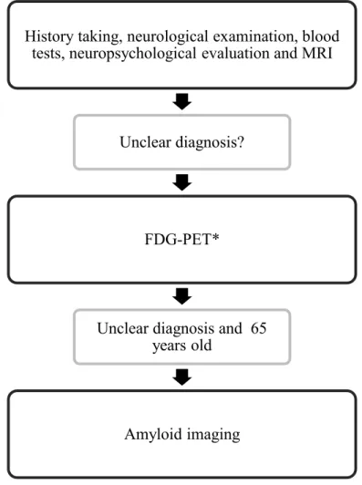
period, a total of 3,542 patients were seen at our clinic. All patients were assessed according to the “Recommendations of the 4th Canadian Consensus Conference on the Diagnosis and Treatment of
Dementia” (Gauthier, 2012). Following history taking, detailed neurological examination, basic blood tests (complete blood count, ions, TSH, B12, Ca/Mg/Ph, Rapid Plasma Reagin for screening of syphilis), comprehensive neuropsychological evaluation (Trail Making Test A and B, Stroop Color-Word Test, Consonant Trigrams, IVA Continuous Performance Test, Wechsler Adult Intelligence Scale-III, Controlled Oral Word Association Test, Boston Naming Test, Clock Drawing test, Rey Complex Figure [copy], Hooper Visual Organizational Test, Wisconsin Card-Sorting Test, Wechsler Memory Scale-III, California Verbal Learning Test, ABC 15 items, Beck Depression Inventory), and MRI (dementia protocol – includes axial and coronal T2/FLAIR, axial SWI and diffusion, and 3-D T1 sequences with detailed atrophy measures using Scheltens scale and grading of white matter changes using Fazekas scale interpreted by a neuroradiologist with expertise in dementia [MB]), only a limited number of cases remained unclear (n=270). In such a situation, our investigative sequence is to request an FDG-PET scan, which is covered by the Quebec public health insurances. Cerebrospinal fluid (CSF) analyses were not covered at the time of the study and therefore were only very rarely used. Among the 270 FDG-PET scans that were requested during that period (and interpreted by nuclear medicine specialists with expertise in dementia [FAB, JMB, JPS, JG]), 30 cases corresponded to our inclusion and exclusion criteria, which are: inclusion
6
criteria: 1) ≤ 65 years old, 2) an atypical/unclear case with no specific diagnosis despite history taking, neurological examination, neuropsychological assessment, MRI, and FDG-PET; exclusion criteria: 1) typical dementia syndrome (see Figure 1), and 2) absence of FDG-PET. Of note, one patient was cancelled twice because of unavailability of the tracer and decided to withdraw. Another patient was deemed typical after blinded review of the FDG-PET by one of the co-authors (JPS). Consequently, a total of 28 patients underwent an amyloid scan.
--- Insert Figure 1 here
---
Amyloid PET imaging and interpretation
Patients underwent amyloid imaging using 18F-NAV4694 (NAV4694; Navidea
Biopharmaceuticals, Dublin, OH, USA). Authorization was obtained from Health Canada to use this non-approved tracer in a research protocol. This fluorine-based ligand was shown to be equivalent to the reference standard carbon-11 (11C)-labeled Pittsburgh Compound B (PiB), but with a 110
minutes half-life (Jereus, 2010; Cselenyi, 2012; Rowe, 2013), it has the added advantage of much more flexible scanning scheduling. NAV4694 was radiolabeled at the PET Unit, McConnell Brain Imaging Centre (McGill University, Montreal [QC], Canada). Briefly, it was produced by radiofluorination of the corresponding N-Boc–protected nitro precursor, followed by acidic deprotection with hydrochloric acid. Average radiochemical yield is 16% after a synthesis time of 65 min, with radiochemical purity greater than 98% and an average specific activity of 555 ± 230 GBq/μmol.
PET scans were acquired in 3-D mode on a Siemens Biograph 6 PET/CT scanner (Siemens Medical Solutions, Erlangen, Germany) at our PET unit, CHU de Québec, Québec City (QC), Canada. After the intravenous injection of 200 to 370 MBq of NAV4694, an uptake period of 40 minutes was observed. A low-dose, non-contrast CT of the head was acquired for attenuation correction and anatomical correlation, followed by a 30 minutes dynamic PET acquisition. Three 10-minute frames were reconstructed using 3-D ordered subset expectation maximization with a point-spread function (Siemens HD-PET) with a 256 x 256 matrix. Analysis was performed with automated Brass software (Hermes Medical Solutions, Stockholm, Sweden), which co-register the PET images to an atlas and segment the brain in standard regions of interest (ROIs) to generate standardized uptake
7
value ratios (SUVRs) referenced to the cerebellar cortex, a region relatively unaffected by dense Aβ plaques in sporadic AD, for NAV4694. Global Aβ burden was expressed as the average SUVR of the mean of the following cortical ROIs: frontal (consisting of dorsolateral prefrontal, ventrolateral prefrontal, and orbitofrontal regions), superior parietal, lateral temporal, lateral occipital, and anterior/posterior cingulate and precuneus. As in previous studies using NAV4694 (Jereus, 2010; Cselenyi, 2012; Rowe, 2013), SUVR cutoff was set at 1.50. In addition to quantitative interpretation using SUVRs, images were qualitatively interpreted by two highly trained nuclear medicine physicians (JPS, JMB). These clinicians were blinded to clinical information and performed their readings independently. In the end, each amyloid PET scan was judged as either positive or negative based on qualitative and quantitative reads.
Assessment of the impact of amyloid PET on the diagnostic process
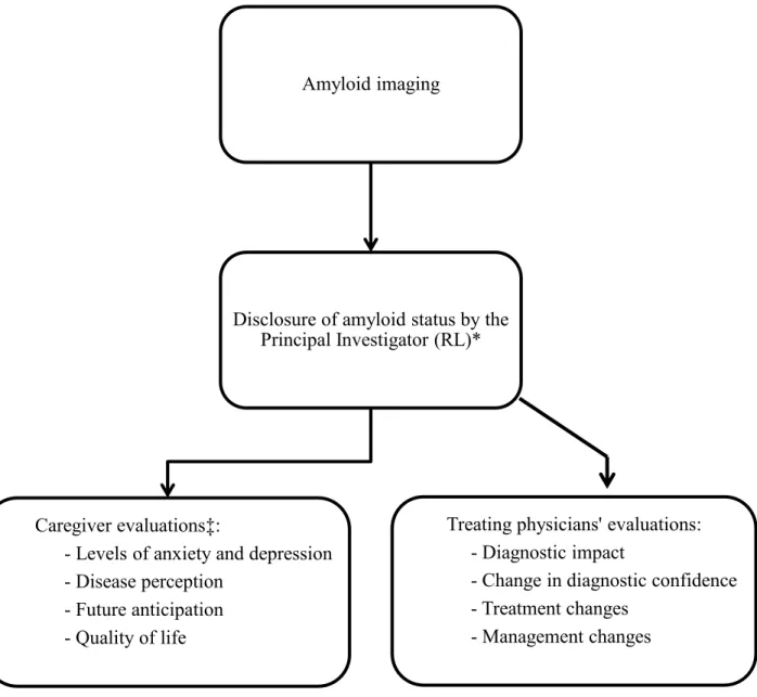
Amyloid PET was presented to both the patient and the caregiver as an additional test that was part of a research protocol and that could help clarify the diagnosis. Following each amyloid PET, the principal investigator (RL) of the study met with the patient and family to disclose amyloid status (either positive or negative) and further inquire about any side effects or discomfort associated with the process. The amyloid PET image was shown to each case. Amyloid status was then disclosed to the patient’s treating dementia expert who scheduled another appointment to discuss the most recent diagnostic opinion. Each physician filled a questionnaire detailing pre-scan and post-scan diagnoses with their corresponding confidence levels. Confidence level was assessed on a scale of 5 where 1 = very weak, 2 = weak, 3 = average, 4 = high, and 5 = very high. Treatment changes and altered management following scan were also documented in this questionnaire. Physicians were asked to explain in what way the amyloid scan had contributed to the overall assessment process.
Assessment of the impact on caregivers’ life
All participating caregivers were very close to the patients and aware of each step of the diagnostic process. They were present at each appointment with their treating physician, and for this reason had access to the same information as the patient prior to the amyloid scan. Amyloid PET was presented as an additional test that could help clarify the diagnosis (the sequence of events that led to disclosure of amyloid status is detailed in the section above).
Consenting caregivers were met at least 30 days after disclosure of amyloid status by their treating dementia expert. A 21-item Likert scale questionnaire along with a 1-hour interview designed to assess the impact of the amyloid scan results was then administered (see Supplementary Material –
8
Caregiver Questionnaire). More specifically, the purpose of this interview was to go over each question individually, and, by taking in account the caregivers’ psychosocial contexts, try to understand the reasons that motivated his or her answers. This was done in order to try and correct for the different interpretations that may have the caregivers of the different statements. The questionnaire and interview covered the following domains: anxiety, depression, disease perception, future anticipation, and quality of life. In fact, 4 questions assessed “Anxiety” (e.g. I am more afraid of developing a neurodegenerative disease since the diagnosis has become more certain), 4 assessed “Depression” (e.g. I sleep better since my loved one’s condition is known), 6 assessed “Disease perception” (e.g. The family atmosphere is better since the precise diagnosis of my loved one has become clearer), 4 assessed “Future anticipation” (e.g. Better assessment has allowed us to better plan for the future) and 3 assessed general “Quality of life” (e.g. I appreciate more every moment with my loved one since clarification of the diagnosis). In some cases, more than one caregiver was interviewed (e.g. both parents, or spouse and daughter). Figure 2 recapitulates the sequence of events that occurred following amyloid PET scan.
--- Insert Figure 2 here
---
Statistical analysis
Independent sample t-tests were used to assess group differences. Paired-sample t-tests were used to assess change in diagnostic certainty before and after amyloid PET scan. Pearson χ2 tests were used to assess differences in patient management plan.
9
RESULTS
Subjects
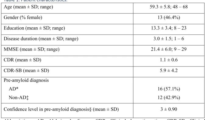
Patient characteristics are shown in Table 1. Treating physicians’ hypotheses (AD vs. non-AD) of the 28 participants with complex/atypical dementias are listed in Table 1. There were a total of 16 AD diagnoses; 8 amnestic AD, 3 behavioral/dysexecutive AD, 3 language variant AD/logopenic variant of primary progressive aphasia, 1 visual variant AD/posterior cortical atrophy and 1 corticobasal syndrome. A total of 12 cases were judged non-AD; 3 behavioral variant frontotemporal dementia, 2 corticobasal degeneration, 1 non-fluent/agrammatic variant primary progressive aphasia, and 6 of other causes (psychiatric condition, intellectual deficiency, etc.). Diagnostic confidence of attending physicians in their hypotheses before amyloid scanning (scale 1-5) is also listed in Table 1.
--- Insert Table 1 here
---
Illustrative case example
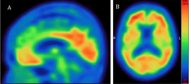
A 65-year-old woman was referred to our Memory Clinic for complaints of progressive memory difficulties. No changes in behavior were noted. Memory problems were combined with increasing functional difficulties. Neurological examination documented the absence of eye movement irregularities as well as of extrapyramidal signs. Initial blood tests were non-contributory. Neuropsychological evaluation showed spared verbal episodic memory but impaired visual memory along with visuospatial deficits. There were impairments in lexical verbal fluency as well. MRI did not show any specific pattern of focal atrophy or significant vascular burden. Given the atypical nature of her deficits, her young age and the functional impact of her symptoms, FDG-PET was ordered. This test showed diffuse fronto-parieto-temporal hypometabolism with a frontal predominance and sparing of the posterior portions of the cingulate gyri, suggestive of frontotemporal lobar degeneration (FTLD). The treating behavioral neurologist (LV) explained to the family that there was a discrepancy between the clinical presentation and the results of the investigation, and suggested further imaging. Amyloid imaging revealed high deposition of amyloid
10
plaques with an average SUVR of 2.15 (figure 3) with a positive read from two different nuclear medicine specialists blinded to clinical data. A diagnosis of Alzheimer’s disease was announced with greater confidence. The full one-hour interview with the primary caregiver revealed that family members were glad to be finally clear on diagnosis, as they had been seeking answers for over a year at that time.
--- Insert Figure 3 here
---
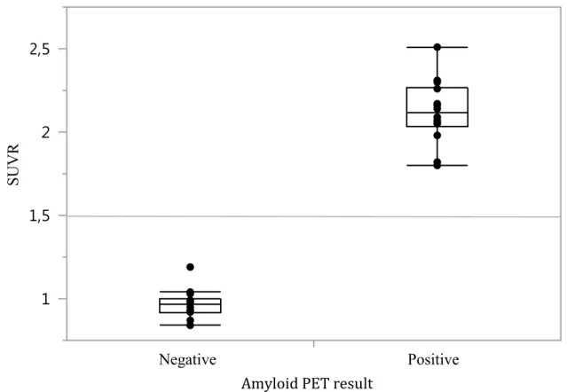
Amyloid status
Based on qualitative (visual interpretation) and quantitative (SUVR > 1.5 means positive) blinded reads by 2 independent expert nuclear medicine specialists, half of our patients (n=14) had a positive amyloid scan (see Figure 4). No significant differences were found between amyloid-positive and amyloid-negative groups in terms of age, gender, education, disease duration and MMSE scores. The average SUVRs were 2.12 (± 0.19 ; 1.80 – 2.51) and 0.97 (± 0.85; 0.84 – 1.19) in the amyloid-positive and amyloid-negative groups, respectively. Therefore, no overlap was found, and the difference between the highest SUVR value of the negative group and the lowest SUVR value of the positive group was quite large. The concordance rate between our two nuclear medicine specialists was 100%. Of interest, 3 patients (2 amyloid-positive and 1 amyloid-negative) also had lumbar punctures with analysis of Aβ1-42, total tau and p-tau, and their CSF levels were
consistent with amyloid PET scan results.
--- Insert Figure 4 here
---
Change in diagnosis and diagnostic confidence following amyloid PET
Table 2 indicates the changes in diagnosis following the amyloid scan. Overall, diagnostic changes occurred in 9 (32.1%) cases and were found slightly more often in the direction from AD to non-AD (17.8%) then the opposite (14.3%). Diagnostic change occurred in 28.6% and 35.7% in amyloid positive and amyloid negative groups respectively. There were no statistical differences between
11
these 2 groups in terms of diagnostic change.--- Insert Table 2 here
---
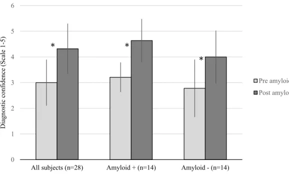
Amyloid imaging generated a significant increase (p<0.05) of 44% in diagnostic confidence from pre- to post-amyloid scanning (see Figure 5). This did not significantly differ between the amyloid-positive and amyloid-negative groups (p=0.169).
--- Insert Figure 5 here
---
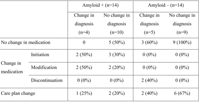
Effect on treatment and patient management
A total of 11/28 patients saw their medication changed (initiation of an ACheI, change in dosage or molecule, or discontinuation of the treatment) following amyloid PET (see Table 3). All amyloid-positive patients who saw their diagnosis changed also had a change in medication (initiation or modification, but no discontinuation). Amongst the patients who were amyloid-negative and saw their diagnosis changed from AD to non-AD, 3 did not have medication change because they were not previously on an ACheI. The other two patients were previously on such medication, which was discontinued.
Changes in patient management (or care plan) are also shown in Table 3. This excludes medication changes and includes, but is not limited to, registration to a vaccine trial, lumbar puncture for CSF analysis, referral to a speech therapist and/or referral to a psychologist. The amyloid-negative group showed the most care plan changes (57% vs. 21%; p=0.120 by chi-square test). When there was a negative amyloid scan and no diagnostic change, the care plan changed most often (67%). This proportion approached significance when compared to the amyloid-positive group without change in diagnosis (67% vs. 20%; p=0.07 by 2-sided chi-square test).
--- Insert Table 3 here
12
---
Impact on caregivers
A total of 23 caregivers were met. All participating caregivers completed the 21-item Likert scale questionnaire as well as the 1-hour interview. Figure 6 illustrates the impact of amyloid imaging on various variables. There were no statistically significant differences between the amyloid-positive and amyloid-negative groups.
--- Insert Figure 6 here
---
An in-depth and purely qualitative exploration of the most polarized answers from the questionnaire revealed interesting characteristics (see Table 4). Most importantly, and not surprisingly, different questions were understood differently by caregivers depending on their standpoint and life situation (for example, in one instance the caregiver was the 82-year-old father of the patient).
--- Insert Table 4 here
---
The 1-hour interview with caregivers provided a different perspective on things. Caregivers expressed in their own words the impact of amyloid PET results on their overall appreciation of life. First and foremost, caregivers reported a better understanding of their loved one’s condition. They consistently described their state of mind prior to amyloid imaging, all of them reporting many periods of confusion and frustration towards unexplainable behavioral and cognitive changes seen in their loved ones. They expressed frustration towards the difficulty physicians showed in providing appropriate answers to their questions. Amyloid imaging was correctly perceived as the final test that was meant to indicate if Alzheimer’s disease was the causal pathology. Generally, after amyloid imaging, caregivers felt more confident as to the next steps ahead. For example, in question #10 of the questionnaire “I appreciate every instant with my beloved one even more since we know the precise diagnosis”, some caregivers described how they stopped working in order to spend more time with their loved one now that the diagnosis was clear and that the reserved
13
prognosis of the disease was clarified. Examples of future plans were given as well (buying plane tickets for vacations and making sure that wills and testaments were completed). For such reasons, despite remaining saddened by the incurable nature of neurodegenerative diseases, they all saw themselves as having a better acceptance of the disease.
14
DISCUSSION
This study investigated the clinical utility of amyloid PET imaging in the differential diagnosis of early-onset atypical cases of dementia. It demarcated itself from previous work in two ways. First, by defining ‘uncertainty after a comprehensive assessment’ (see AUC criteria (Johnson, 2013)) as an “uncertain diagnosis after history taking, neurological examination, blood tests, comprehensive neuropsychological examination, MRI – dementia protocol and FDG-PET”, therefore recruiting the most challenging atypical cases. Second, it addressed impact on caregivers using a 21-item Likert scale questionnaire and a 1-hour interview specifically designed for this study. Amyloid PET was associated with a diagnostic change in 9/28 cases (32.1%). There was a 44% increase in diagnostic confidence. Altered management occurred in 71.4% (20/28) of cases. Knowledge of amyloid status improved caregivers’ outcomes in all domains (anxiety, depression, disease perception, future anticipation, and quality of life). Altogether, our results suggested a useful and additive role for amyloid PET in atypical cases with an unclear diagnosis beyond the detailed workup of a tertiary memory clinic. Amyloid PET increased diagnostic confidence and generated clinically significant alterations in management. The overall process was positive for caregivers notably by making it easier to spend quality time with their loved ones.
Usefulness of amyloid imaging in cases where diagnostic confidence is low has been reported in several studies (Grundman, 2010; Ossenkoppele, 2013; Zannas, 2013; Wolk, 2013), and our results are consistent with those findings. Indeed, a total of 32.1% of the subjects had a diagnosis change following amyloid PET scan. Despite this, amyloid imaging did not replace clinical judgment. In some instances, ACheI treatment was maintained in the presence of a negative amyloid scan because of a lack of negative impact on the patient. Most often, however, acetylcholinesterase treatment was initiated in patients thought to have AD pathology and stopped in FTLD cases. This is relevant because FTLD patients were shown to have worsening of their symptoms following acetylcholinesterase treatment (Mendez, 2007; Boxer, 2013; Arciniegas, 2013).
Clinically significant alterations in management have been defined in various ways by authors. The most obvious problem regarding this claim is what is significant exactly? In all cases, the pre-test diagnosis was an incurable dementia and neurodegenerative disease, as was the post-test diagnosis. Does a change in cholinesterase inhibitor use really indicate a significant alteration in therapy?
15
Nonetheless, it is important to try and quantify this concept. The definition of altered management used in our study excluded medication changes and included registration to a vaccine trial, lumbar puncture for CSF analysis, referral to a speech therapist and/or referral to a psychologist, as well as other similar interventions. Although it failed to reach statistical significance, we found a change in careplan (or management) in up to 11/28 (39.2%) of our cohort.
Interestingly, the group that had the most careplan/management changes (excluding medication change according to the definition used in the current study) was the group with a negative amyloid PET and no change in diagnosis. In that group, 2 patients were referred to speech-therapy, 1 for psychotherapy, 1 was encouraged to focus on a number of compensatory strategies, and 2 had lumbar puncture for further investigations. This opposes our amyloid positive group without diagnosis change where out of all patients in this group, the only management change included 2 patients referred for vaccine protocol trials. The most likely explanation for this could be that non-AD entities appear to have more components where clinicians can intervene. For example, non-non-AD cases can benefit from speech therapy, when appropriate. Our pseudo-dementia case could benefit from psychotherapy but no amyloid positive AD got such referral because of lack of expected benefit from psychotherapy. Another explanation could be that once clinicians eliminate the possibility of prescribing acetylcholinesterase inhibitors; they take an extra step in attempting to help their patients. This could explain why 2 patients had lumbar puncture for more advanced analyses.
There was a 100% inter-rater agreement in the readings of amyloid scans in this study. The experts’ interpretations also matched with quantitative (SUVR) results 100% of the time. This has also been reported previously in the literature. For instance, a major multicenter study identified an overall agreement of 93% between 3 independent readers (Barthel, 2011). The higher agreement in our study may reflect the quality of the ligand, as NAV4694 has less white matter binding than other fluorine-based compounds commonly used (Rowe, 2013). Consequently, images are easier to interpret. SUVRs in our study followed a bimodal distribution with no borderline cases approaching recommended threshold. This certainly made the cases easier for the nuclear medicine physicians to interpret.
Impact on caregivers
Caregivers constitute an essential part of the care of patients with early-onset atypical dementias, yet no authors have explored their emotional reaction to the results of an amyloid scan in the
16
context of a tertiary memory clinic investigation. Clinicians believe it is beneficial because it helps them establish a diagnosis but we wished to explore how this is viewed from the caregiver’s perspective. In a study where participants were asked whether they wanted or not to know if they had AD pathology, older adults with subjective memory complaints had two kinds of reactions: there were those who did not want to know, who stated it would create anxiety, whereas those who wanted to know reported several potential benefits (Elson, 2006). Amongst those were developing plans for the future, considering early treatment, facilitating psychological adjustments, enabling family members to understand behaviours, and the desire to be kept informed. Another study suggests that disclosure of AD status could help patients deal with anxiety (Mormont, 2014). Overall, these results suggest a benefit in the disclosure of a correct diagnosis. However, no study had investigated the impact of a more confident AD diagnosis on caregivers, particularly when their loved ones present with an atypical syndrome. Since no disease-modifying treatments are available yet for AD, we believe that it is of the uttermost importance to assess the impact of a correct diagnosis on caregivers. Hence, the second objective of this study was to see the impact on caregivers of disclosure of diagnosis following amyloid imaging.
Overall, we found a clear qualitative improvement in the variables assessed namely anxiety, depression, disease perception, anticipation of the future, and quality of life following disclosure of the amyloid scan findings. However, none of these trends reached statistical significance. This could be biased by the fact that, at the end of the day, one can be informed with greater confidence about a diagnosis but the fact remains that a young individual is faced with a progressive and irreversible neurodegenerative disease. This can never be a source of joy. Following amyloid PET, caregivers appreciated more each moment with their loved ones and were better able to make plans for the future, as shown in Table 4. Most reported they did not regret going through all of the investigations. Furthermore, there were no differences between positive and amyloid-negative groups with respect to caregiver impact. Nevertheless, those with a amyloid-negative scan often had a more nebulous understanding of the disease and of its natural history.
Limits
This study has limitations. First, it was conducted on a small sample of 28 patients without pathological confirmation. However, this sample was carefully selected by experienced experts in the field of behavioral neurology. It is also unique in that contrary to previous reports, it addressed the value of amyloid imaging in the most atypicals among atypicals. Second, the questionnaire used
17
to assess impact on caregivers was not validated and only reflected the general feelings of the caregivers. A difficulty that was encountered with our questionnaire is variable interpretations of the same statement. Caregivers sometimes showed problems relating to how they actually felt at the moment when the diagnosis was announced. In other words, caregivers tended sometimes to answer the questions relating to how they lived through their loved one’s disease in general, instead of concentrating on what happened at the point in time where they obtained a more accurate diagnosis. A possible way to prevent this type of confusion would have been to perform the questionnaire before the amyloid PET, and to re-perform it after the amyloid PET. Third, those with a negative scan often had a more nebulous understanding of the disease and of its natural history. Fourth, this study was conducted in young dementia patients in whom the prevalence of “age-related” amyloid pathology is less likely to be present compared to older patients in whom (mixed) amyloid pathologies are more frequently found. Finally, all were recruited from a tertiary memory clinic, which may not reflect the impact of amyloid PET in a general, often older aged population. However, it supports the usefulness of appropriate use criteria for amyloid PET imaging, which describe a potential added value in patients with early-onset dementia with unclear clinical presentations (Johnson, 2013; Dubois, 2015; Laforce, 2016)
18
CONCLUSION
Our findings support an additive value to amyloid PET imaging in the context of a comprehensive tertiary memory clinic work-up in patients suspected of early-onset atypical dementia. This has an impact on clinical diagnosis, it increases overall diagnostic confidence and alters patient management plan. In these patients, an accurate diagnosis can help direct therapy (i.e. avoid unnecessary or undesired ACheI or memantine prescriptions), determine a better care plan (which considers patient safety and minimizes the risk of preventable complications), and enable patients to participate in legal and financial planning. The information gained from this technique is well received by caregivers and encouraged spending quality time with their loved ones.
Since our main inclusion criteria was that our patients did not have a clear diagnosis despite a thorough investigation, our results provide additional evidence for the recommendations put forward in the appropriate use criteria for amyloid PET in clinical practice (Johnson, 2013; Laforce, 2016). As previously mentioned in the introduction, these criteria are: (1) there is an objectively confirmed cognitive impairment, (2) AD is a possible diagnosis but it remains unconfirmed after a comprehensive assessment, and (3) evidence of increased levels of amyloid in the brain is expected to increase diagnostic certainty and alter management. Such evidence-based criteria are important because amyloid imaging can be harmful if not used properly. A recent cross-sectional study which measured amyloidosis and neurodgeneration in 985 cognitively normal participants aged 50-89 years old, it was demonstrated that amyloid inveitably acumulates with age. Yet, many people retain normal cognivitve function despite this substantial amyloid burden (Jack, 2014). Despite some evidence suggesting that participants who are APOE e4 positive (an allele known to be a risk factor for AD) and have high amyloid beta burden on amyloid PET show the highest cognitive decline on MMSE, these are merely risk factors for developping the disease (Mormino, 2014). Thus, inappropriate amyloid imaging can lead to disclosure of results that can create distress and be harmful.
Our study can be continued as part of doctoral studies and could investigate the overall use of biomarkers in atypical dementias. Recent advances in neurosciences have led to a better understanding of neurodegenerative diseases’ pathophysiological processes, and have resulted in the incorporation of biomarker evidence as part of some disorders’ criteria (see updated McKhann criteria for AD for example). However, the combined use of these biomarkers in a large (>100)
19
cohort of atypical dementias has never been studied. Such biomarkers not only include amyloid imaging, but also CSF analyses, and tau imaging, whose clinical role is still ambiguous. Subgroups analyses can then be performed to compare each biomarker and better define their roles and limitations within a complete clinical setting that assesses every variable, from the diagnosis to the management of patients.
Furthermore, much is expected from the recently launched Imaging Dementia – Evidence for Amyloid Scanning (IDEAS) Study, a $100M open-label longitudinal cohort effort on approximately 18,500 US Medicare beneficiaries. In this venture, diagnostically uncertain cases of mild cognitive impairment and atypical dementia will be referred by physicians of all specialities, including those in primary care, to be scanned to determine whether knowledge of amyloid status leads to significant changes in patient management and if this translates into improved medical outcomes. In addition to enhancing the diagnostic process of atypical dementing syndromes and allowing earlier treatment, we can expect amyloid imaging to be a major contributor to future advancements and discoveries in the field of Alzheimer disease and the natural evolution of dementias. For example, amyloid imaging has served as a secondary outcome measure in Alzheimer disease clinical trials with disease-modifying agents such as the anti-amyloid monoclonal antibodies bapineuzemab and solanezumab. Recently, a study has shown brain amyloid reduction and slowing of cognitive decline after one year of treatment with aducanumab, a human IgG1 monoclonal antibody against a conformational epitope found on Aβ (Keller, 2015). Hence, confirming the amyloid-positive status of patients in clinical trials (e.g., the Anti-Amyloid Treatment in Asymptomatic Alzheimer’s Disease (A4) Study) will be of the uttermost importance for validation of therapies.
20
REFERENCES
Alladi S, Xuereb J, Bak T, Nestor P, Knibb J, Patterson K, Hodges JR (2007) Focal cortical presentations of Alzheimer's disease. Brain 130, 2636-2645.
Alzheimer Society of Canada (2012) A new way of looking at the impact of Dementia. Web. Retrieved January 14, 2016. http://www.alzheimer.ca/en/About-dementia/What-is-dementia/Dementia-numbers
Arciniegas DB, Anderson CA (2013) Donepezil-induced confusional state in a patient with autopsy-proven behavioral-variant frontotemporal dementia. J Neuropsychiatry Clin Neurosci 25, E25-26.
Barthel H, Gertz H-J, Dresel S, Peters O, Bartenstein P, Buerger K, Hiemeyer F, Wittemer-Rump SM, Seibyl J, Reininger C (2011) Cerebral amyloid-β PET with florbetaben (18 F) in patients with Alzheimer's disease and healthy controls: a multicentre phase 2 diagnostic study. Lancet Neurol 10, 424-435.
Beach TG, Monsell SE, Phillips LE, Kukull W (2012) Accuracy of the clinical diagnosis of Alzheimer disease at National Institute on Aging Alzheimer Disease Centers, 2005-2010. J Neuropathol Exp Neurol 71, 266-273.
Bensaïdane M, Fortin M, Damasse G, Chenard M, Dionne C (2014) Clinical Utility of Amyloid Imaging in a Complex Case of Corticobasal Syndrome Presenting with Psychiatric Symptoms. J Neurol Disord 2, 2.
Bergeron D, Bensaïdane M, Laforce RJ (2016) Untangling Alzheimer’s disease clinicoanatomical heterogeneity through selective network vulnerability – an effort to understand a complex disease. Current Alzheimer Research 13, 589-596. Boxer AL, Knopman DS, Kaufer DI, Grossman M, Onyike C, Graf-Radford N, Mendez M, Kerwin D, Lerner A, Wu CK, Koestler M, Shapira J, Sullivan K, Klepac K, Lipowski K, Ullah J, Fields S, Kramer JH, Merrilees J, Neuhaus J, Mesulam MM, Miller BL (2013) Memantine in patients with frontotemporal lobar degeneration: a multicentre, randomised, double-blind, placebo-controlled trial. Lancet Neurol 12, 149-156.
Braak H, Braak E (1991) Neuropathological stageing of Alzheimer-related changes. Acta Neuropathologica 82, 239-259. Caselli RJ (2003) Current issues in the diagnosis and management of dementia. Semin Neurol 23, 231-240.
Cipriani G, Dolciotti C, Picchi L, Bonuccelli U (2011) Alzheimer and his disease: a brief history. Neurol Sci 32, 275-279. Clark C, Pontecorvo M, Beach T, Bedell B, Coleman R, Doraiswamy P, Fleisher A, Reiman E, Sabbagh M, Sadowsky C, Schneider J, Arora A, Carpenter A, Flitter M, Joshi A, Krautkramer M, Lu M, Mintun M, Skovronsky D (2012) Cerebral PET with florbetapir compared with neuropathology at autopsy for detection of neuritic amyloid-beta plaques: a prospective cohort study. Lancet Neurol 11, 669-678.
Crutch SJ, Lehmann M, Schott JM, Rabinovici GD, Rossor MN, Fox NC (2012) Posterior cortical atrophy. Lancet Neurol 11(2), 170-8.
Cselenyi Z, Jonhagen ME, Forsberg A, Halldin C, Julin P, Schou M, Johnstrom P, Varnas K, Svensson S, Farde L (2012) Clinical validation of 18F-AZD4694, an amyloid-beta-specific PET radioligand. J Nucl Med 53, 415-424.
Curtis C, Gamez JE, Singh U, Sadowsky CH, Villena T, Sabbagh MN, Beach TG, Duara R, Fleisher AS, Frey KA, Walker Z, Hunjan A, Holmes C, Escovar YM, Vera CX, Agronin ME, Ross J, Bozoki A, Akinola M, Shi J,
Vandenberghe R, Ikonomovic MD, Sherwin PF, Grachev ID, Farrar G, Smith AP, Buckley CJ, McLain R, Salloway S (2015) Phase 3 trial of flutemetamol labeled with radioactive fluorine 18 imaging and neuritic plaque density. JAMA
Neurol 72, 287-294.
Dubois B, Padovani A, Scheltens P, Rossi A, Dell'Agnello G. (2015) Timely Diagnosis for Alzheimer's Disease: A Literature Review on Benefits and Challenges. J Alzheimers Dis 49, 617-31.
21
Elson P (2006) Do older adults presenting with memory complaints wish to be told if later diagnosed with Alzheimer's disease? Int J Geriatr Psychiatry 21, 419-425.
Fratiglioni L, Launer LJ, Andersen K, Breteler MM, Copeland JR, Dartigues JF, Lobo A, Martinez-Lage J, Soininen H, Hofman A (2000) Incidence of dementia and major subtypes in Europe: A collaborative study of population-based cohorts. Neurologic Diseases in the Elderly Research Group. Neurology 54 (11 Suppl 5), S10-15.
Galton CJ, Patterson K, Xuereb JH, Hodges JR (2000) Atypical and typical presentations of Alzheimer's disease: a clinical, neuropsychological, neuroimaging and pathological study of 13 cases. Brain 123, 484-498.
Gauthier S, Patterson C, Chertkow H, Gordon M, Herrmann N, Rockwood K, Rosa-Neto P, Soucy JP (2012)
Recommendations of the 4th Canadian Consensus Conference on the Diagnosis and Treatment of Dementia (CCCDTD4).
Can Geriatr J 15, 120-126.
Gorno-Tempini ML, Hillis AE, Weintraub S, Kertesz A, Mendez M, Cappa SF, Ogar JM, Rohrer JD, Black S, Boeve BF, Manes F, Dronkers NF, Vandenberghe R, Rascovsky K, Patterson K, Miller BL, Knopman DS, Hodges JR, Mesulam MM, Grossman M (2011) Classification of primary progressive aphasia and its variants. Neurology 15, 1006-1014. Grundman M, Pontecorvo MJ, Salloway SP, Doraiswamy PM, Fleisher AS, Sadowsky CH, Nair AK, Siderowf A, Lu M, Arora AK (2013) Potential impact of amyloid imaging on diagnosis and intended management in patients with
progressive cognitive decline. Alzheimer Dis Assoc Disord 27, 4-15.
Jack CR, Jr, Knopman DS, Jagust WJ, , Petersen RC, Weiner MW, Aisen PS, Shaw LM, Vemuri P, Wiste H, Weigand SD, Lesnick TG, Pankratz VS, Donohue MC, Trojanowski JQ (2013) Update on hypothetical model of Alzheimer’s disease biomarkers. Lancet Neurol 12, 207-216.
Jack CR, Jr, Knopman DS, Jagust WJ, Shaw LM, Aisen PS, Weiner MW, Petersen RC, Trojanowski JQ (2010) Hypothetical model of dynamic biomarkers of the Alzheimer’s pathological cascade. Lancet Neurol 9, 119.
Jack CR, Wiste HJ, Weigand SD, Rocca WA, Knopman DS, Mielke NM, Lowe VJ, Senjem ML, Gunter JL, Preboske GM, Pankratz VS, Vemuri P, Petersen RC (2014) Age-specific population frequencies of cerebral beta-amyloidosis and neurodegeneration among people with normal cognitive function aged 50-89 years: a cross-sectional study. Lancet Neurol 13, 997-1005.
Jagust W, Reed B, Mungas D, Ellis W, DeCarli C (2007) What does fluorodeoxyglucose PET imaging add to a clinical diagnosis of dementia? Neurology 69, 871-877.
Johnson K, Minoshima S, Bohnen N, Donohoe K, Foster N, Herscovitch P, Karlawish J, Rowe C, Carrillo M, Hartley D, Hedrick S, Pappas V, Thies W (2013) Appropriate use criteria for amyloid PET: a report of the Amyloid Imaging Task Force, the Society of Nuclear Medicine and Molecular Imaging, and the Alzheimer's Association. Alzheimers Dement 9, 1-15.
Johnson KA, Minoshima S, Bohnen NI, Donohoe KJ, Foster NL, Herscovitch P, Karlawish JH, Rowe CC, Carrillo MC, Hartley DM (2013) Appropriate use criteria for amyloid PET: a report of the Amyloid Imaging Task Force, the Society of Nuclear Medicine and Molecular Imaging, and the Alzheimer’s Association. J Nucl Med 54, 476-490.
Johnson KA, Minoshima S, Bohnen NI, Donohoe KJ, Foster NL, Herscovitch P, Karlawish JH, Rowe CC, Hedrick S, Pappas V, Carrillo MC, Hartley DM (2013) Update on appropriate use criteria for amyloid PET imaging: dementia experts, mild cognitive impairment, and education. J Nucl Med 54, 1011-1013.
Jureus A, Swahn BM, Sandell J, Jeppsson F, Johnson AE, Johnstrom P, Neelissen JA, Sunnemark D, Farde L, Svensson SP (2010) Characterization of AZD4694, a novel fluorinated Abeta plaque neuroimaging PET radioligand. J Neurochem 114, 784-794.
Keller D (2015) Finally, a Big Win for a Monoclonal in Alzheimer's: International Conference on Alzheimer's and Parkinson's Diseases. Web. Retrieved January 14th 2016. http://www.medscape.com/viewarticle/841856
Klunk WE, Engler H, Nordberg A, Wang Y, Blomqvist G, Holt DP, Bergström M, Savitcheva I, Huang GF, Estrada S, Ausén B, Debnath ML, Barletta J, Price JC, Sandell J, Lopresti BJ, Wall A, Koivisto P, Antoni G, Mathis CA, Långström B (2004) Imaging brain amyloid in Alzheimer's disease with Pittsburgh Compound-B. Ann neurol 55, 306 - 319. Laforce R, Buteau J, Paquet N, Verret L, Houde M, Bouchard R (2010) The value of PET in mild cognitive impairment, typical and atypical/unclear dementias: a retrospective memory clinic study. Am J Alzheimers Dis Other Dement 25,
324-22
332.
Laforce RJ, Rosa-Neto P, Soucy JP, Rabinovici GD, Dubois B, Gauthier S, participants. obotcm (2016) Canadian Consensus Guidelines on Use of Amyloid Imaging in Canada: Update and Future Directions from the Specialized Task force on Amyloid imaging in Canada (STAC). Can J Neurol Sci Epub ahead of print.
Lam B, Masellis M, Freedman M, Stuss DT, Black SE (2013) Clinical, imaging, and pathological heterogeneity of the Alzheimer's disease syndrome. Alzheimers Res Ther 9, 1.
Lobo A, Launer LJ, Fratiglioni L, Andersen K, Di Carlo A, Breteler MM, Copeland JR, Dartigues JF, Jagger C, Martinez-Lage J, Soininen H, Hofman A (2000) Prevalence of dementia and major subtypes in Europe: A collaborative study of population-based cohorts. Neurologic Diseases in the Elderly Research Group. Neurology 54 (11 Suppl 5), S4-9. McKhann G, Drachman D, Folstein M, Katzman R, Price D, Stadlan EM (1984) Clinical diagnosis of Alzheimer’s disease: report of the NINCDS-ADRDA Work Group under the auspices of Department of Health and Human Services Task Force on Alzheimer’s Disease. Neurology 34, 939-944.
McKhann G, Knopman D, Chertkow H, Hyman B, Jack C, Kawas C, Klunk W, Koroshetz W, Manly J, Mayeux R, Mohs R, Morris J, Rossor M, Scheltens P, Carrillo M, Thies B, Weintraub S, Phelps C (2011) The diagnosis of dementia due to Alzheimer's disease: recommendations from the National Institute on Aging and the Alzheimer's Association workgroup.
Alzheimers Dement 7, 263 - 269.
Mendez M, Shapira J, McMurtray A, Licht E (2007) Preliminary findings: behavioral worsening on donepezil inpatients with frontotemporal dementia. Am J Geriatr Psychiatry 15, 84 - 87.
Mesulam MM (1982) Slowly progressive aphasia without generalized dementia. Ann Neurol 11, 592-598.
Mitsis E, Bender H, Kostakoglu L, Machac J, Martin J, Woehr J, Sewell M, Aloysi A, Goldstein M, Li C, Sano M, Gandy S (2014) A consecutive case series experience with [18F] florbetapir PET imaging in an urban dementia center: impact on quality of life, decision making, and disposition. Mol Neurodegener 9, 10.
Mormino EC1, Betensky RA, Hedden T, Schultz AP, Ward A, Huijbers W, Rentz DM, Johnson KA, Sperling RA; Alzheimer's Disease Neuroimaging Initiative; Australian Imaging Biomarkers and Lifestyle Flagship Study of Ageing; Harvard Aging Brain Study (2014) Amyloid and APOE epsilon4 interact to influence short-term decline in preclinical Alzheimer disease. Neurology 82, 1760-1767.
Mormont E, Jamart J, Jacques D (2014) Symptoms of depression and anxiety after the disclosure of the diagnosis of Alzheimer disease. J Geriatr Psychiatry Neurol 27, 231-236.
Nelson PT, Alafuzoff I, Bigio EH, Bouras C, Braak H, Cairns NJ, Castellani RJ, Crain BJ, Davies P, Del Tredici K, Duyckaerts C, Frosch MP, Haroutunian V, Hof PR, Hulette CM, Hyman BT, Iwatsubo T, Jellinger KA, Jicha GA, Kövari E, Kukull WA, Leverenz JB, Love S, Mackenzie IR, Mann DM, Masliah E, McKee AC, Montine TJ, Morris JC, Schneider JA, Sonnen JA, Thal DR, Trojanowski JQ, Troncoso JC, Wisniewski T, Woltjer RL, Beach TG (2012) Correlation of Alzheimer disease neuropathologic changes with cognitive status: a review of the literature. J Neuropathol
Exp Neurol 71, 362- 381.
Ossenkoppele R, Pijnenburg YA, Perry DC, Cohn-Sheehy BI, Scheltens NM, Vogel JW, Kramer JH, van der Vlies AE, Joie RL, Rosen HJ, van der Flier WM, Grinberg LT, Rozemuller AJ, Huang EJ, van Berckel BN, Miller BL, Barkhof F, Jagust WJ, Scheltens P, Seeley WW, Rabinovici GD (2015) The behavioural/dysexecutive variant of Alzheimer's disease: clinical, neuroimaging and pathological features. Brain 138, 2732-2749.
Ossenkoppele R, Prins ND, Pijnenburg YA, Lemstra AW, van der Flier WM, Adriaanse SF, Windhorst AD, Handels RL, Wolfs CA, Aalten P, Verhey FR, Verbeek MM, van Buchem MA, Hoekstra OS, Lammertsma AA, Scheltens P, van Berckel BN (2013) Impact of molecular imaging on the diagnostic process in a memory clinic. Alzheimers Dement 9, 414-421.
Pearson HA, Peers C (2006) Physiological roles for amyloid beta peptides. J Physiol 575, 5-10
Prince M, Bryce R, Albanese E, Wimo A, Ribeiro W, Ferri CP (2013) The global prevalence of dementia: a systematic review and metaanalysis. Alzheimers Dement 9, 63-75.
Rabinovici G, Rosen H, Alkalay A, Kornak J, Furst A, Agarwal N, Mormino E, O'Neil J, Janabi M, Karydas A, Growdon M, Jang J, Huang E, Dearmond S, Trojanowski J, Grinberg L, Gorno-Tempini M, Seeley W, Miller B, Jagust W (2011)
23
Amyloid versus FDG PET in the differential diagnosis of AD and FTLD. Neurology 77, 2034-2042
Rowe CC, Pejoska S, Mulligan RS, Jones G, Chan JG, Svensson S, Cselenyi Z, Masters CL, Villemagne VL (2013) Head-to-head comparison of 11C-PiB and 18F-AZD4694 (NAV4694) for beta-amyloid imaging in aging and dementia. J
Nucl Med 54, 880-886.
Sabri O, Sabbagh MN, Seibyl J, Barthel H, Akatsu H, Ouchi Y, Senda K, Murayama S, Ishii K, Takao M, Beach TG, Rowe CC, Leverenz JB, Ghetti B, Ironside JW, Catafau AM, Stephens AW, Mueller A, Koglin N, Hoffmann A, Roth K, Reininger C, Schulz-Schaeffer WJ (2015) Florbetaben PET imaging to detect amyloid beta plaques in Alzheimer's disease: Phase 3 study. Alzheimers Dement 11, 964-974.
Sanchez-Juan P, Ghosh PM, Hagen J, Gesierich B, Henry M, Grinberg LT, O'Neil JP, Janabi M, Huang EJ, Trojanowski JQ, Vinters HV, Gorno-Tempini M, Seeley WW, Boxer AL, Rosen HJ, Kramer JH, Miller BL, Jagust WJ, Rabinovici GD (2014) Practical utility of amyloid and FDG-PET in an academic dementia center. Neurology 82, 230-238.
Schöll M, Damián A, Engler H (2014) Fluorodeoxyglucose PET in Neurology and Psychiatry. PET Clin 9, 371-390 Soucy JP, Bartha R, Bocti C, Borrie M, Burhan AM, Laforce R, Rosa-Neto P (2013) Clinical applications of neuroimaging in patients with Alzheimer's disease: a review from the Fourth Canadian Consensus Conference on the Diagnosis and Treatment of Dementia 2012. Alzheimers Res Ther 5, S3.
Spires-Jones TL, Hyman BT (2014) The intersection of amyloid beta and tau at synapses in Alzheimer's disease. Neuron 82, 756-771.
Taylor KI, Probst A, Miserez AR, Monsch AU, Tolnay M (2008) Clinical course of neuropathologically confirmed frontal-variant Alzheimer's disease. Nat Clin Pract Neurol 4, 226-232.
van der Flier WM, Scheltens P (2005) Epidemiology and risk factors of dementia. J Neurol Neurosurg Psychiatry 76 (Suppl 5), 2-7.
Vandenberghe R, Adamczuk K, Dupont P, Laere KV, Chetelat G (2013) Amyloid PET in clinical practice: Its place in the multidimensional space of Alzheimer's disease. Neuroimage Clin 2, 497-511.
Warren JD, Fletcher PD, Golden HL (2012) The paradox of syndromic diversity in Alzheimer disease. Nat Rev Neurol 8, 451-464.
Wolk DA (2013) Amyloid imaging in atypical presentations of Alzheimer's disease. Curr Neurol Neurosci Rep 13, 412. Wolk DA, Price JC, Madeira C, Saxton JA, Snitz BE, Lopez OL, Mathis CA, Klunk WE, DeKosky ST (2012) Amyloid imaging in dementias with atypical presentation. Alzheimers Dement 8, 389-398.
World Health Organization, Alzheimer's Disease International (2012) Dementia: a public health priority. World Health
Organization.
Zannas AS, Doraiswamy PM, Shpanskaya KS, Murphy KR, Petrella JR, Burke JR, Wong TZ (2014) Impact of 18F-florbetapir PET imaging of beta-amyloid neuritic plaque density on clinical decision-making. Neurocase 20, 466-473. Zwan MD, Bouwman F, Konijnenburg E, van der Flier WM, Lammertsma AA, Van Berckel BNM, Scheltens P (In press) in Alzheimer's and Dementia Alzheimer's Association International Conference, Washington.
24
ANNEXES
TABLES
Table 1. Patient characteristics.
Abbreviations: AD: Alzheimer’s disease; CDR: Clinical dementia rating; CDR-SB: Clinical dementia rating – Sum of boxes; MMSE: Mini-Mental State Examination; SD: Standard deviation. *AD means: 8 amnestic AD, 3 behavioral/dysexecutive AD, 3 language variant AD (or logopenic variant of primary progressive aphasia), 1 visual variant AD (or posterior cortical atrophy) and 1 corticobasal syndrome.
‡Non-AD means: 3 behavioral variant frontotemporal dementia, 2 corticobasal degeneration, 1 non-fluent/agrammatic variant primary progressive aphasia, and 6 of other causes (psychiatric condition, intellectual deficiency, etc.).
§Confidence level was assessed on a scale of 1-5.
Age (mean ± SD; range) 59.3 ± 5.8; 48 – 68
Gender (% female) 13 (46.4%)
Education (mean ± SD; range) 13.3 ± 3.4; 8 – 23
Disease duration (mean ± SD; range) 3.0 ± 1.5; 1 – 6
MMSE (mean ± SD; range) 21.4 ± 6.0; 9 – 29
CDR (mean ± SD) 1.1 ± 0.6 CDR-SB (mean ± SD) 5.9 ± 4.2 Pre-amyloid diagnosis AD* Non-AD‡ 16 (57.1%) 12 (42.9%) Confidence level in pre-amyloid diagnosis§ (mean ± SD) 3 ± 0.90