Open Archive Toulouse Archive Ouverte (OATAO)
OATAO is an open access repository that collects the work of some Toulouse
researchers and makes it freely available over the web where possible.
This is
an author'sversion published in:
https://oatao.univ-toulouse.fr/23118Official URL :
https://doi.org/10.1177/0363546517733472
To cite this version :
Any correspondence concerning this service should be sent to the repository administrator: [email protected]
Cavaignac, Etienne and Marot, Vincent and Faruch, Marie and Reina, Nicolas and Murgier,
Jérôme and Accadbled, Franck and Berard, Emilie and Chiron, Philippe Hamstring graft
incorporation according to the length of the graft inside tunnels. (2018) The American Journal
of Sports Medicine, 46 (2). 348-356. ISSN 0363-5465
OATAO
Hamstring Graft Incorporation According
to the Length of the Graft Inside Tunnels
Et
i
enne Cava
i
gnac,*
t
MD,
V
i
ncent Marot,
t
MD, Mar
i
e
Faruch,
*
MD, PhD,
N
i
co
l
as
Re
i
na,
t
MD, PhD, Jerome Murg
i
er,
t
MD, Franck Accadb
l
ed,§
MD, PhD,
Em
ili
e
Berard,11 MD,
and
Ph
ili
ppe
Ch
i
ron,
t
MD, PhD
Investigation performed at
Department
of Orthopedic Surgery and Trauma,
H6pital Pierre-Paul
Riquet, Toulouse,
France
Background: Anterior cruciate ligament (ACL) reconstruction with a quadrupled semitendinosus (ST4) graft is an evolution of the standard technique with 2 hamstring tendons (semitendinosus + gracilis [STG]). However, there is no published comparison of how well these 2 types of hamstring grafts are incorporated into the bone tunnels. Because the ST4 graft is shorter, there is less graft material inside the tunnels.
Purpose: To use magnetic resonance imaging (MRI) to compare graft incorporation in the tibial bone tunnels 1 year after ACL reconstruction with either an STG graft or ST4 graft.
Study Design: Cohort study; Level of evidence, 2.
Methods: Sixty-two patients who underwent ACL reconstruction were enrolled prospectively: 31 with an ST4 graft and 31 with an STG graft. The same surgical technique, fixation method, and postoperative protocol were used in both groups. Graft i ncorpo-ration and ligamentization were evaluated with MRI after 1 year of follow-up. The following parameters were evaluated: signal -to-noise quotient (SNQ), tibial tunnel enlargement, signal intensity at the bone-graft interface, and graft signal according to the Howell scale. The number of participants needed to show that the mean SNQ did not differ between the 2 techniques was 31
in each group (with a 1-sided alpha of 2.5% and a 1-sided beta of 10.0%). The Student ttest was used to compare the distribution of continuous secondary endpoints.
Results: The mean SNQ was 5.2 ::!: 4.5 for the STG group and 5.9 ::!: 3.7 for the ST4 group (P = .5100). The mean tibial tunnel
widening was 93. 7% ::!: 51. 7% for the STG group versus 80.0% ::!: 42.9% for the ST4 group (P = .2605). The groups did not differ
in signal intensity at the bone-graft interface (P
=
.7502) or in graft signal according to the Howell scale (P=
.4544).Conclusion: At the 1-year postoperative follow-up, incorporation and ligamentization of the STG and ST4 grafts were the same based on MRI analysis. The results were at least as good with the ST4 technique as with the standard STG technique in terms of
incorporation and ligamentization.
Keywords: ACL reconstruction; ST4; ligamentization; graft incorporation
The hamstring tendons are the most commonly used type of graft for anterior cruciate ligament (ACL) reconstruction.43
•Address correspondence to Etienne Cavaignac, MD, Department of
Orthopedic Surgery and Trauma, Hopital Pierre Paul Riquet, Rue Jean Dausset, 3105 Toulouse, France (email: [email protected]).
tDepartment of Orthopedic Surgery and Trauma, H6pital Pierre Paul Riquet, Toulouse, France.
*Department of Radiology, Centre Hospitalier Universitaire de Tou louse, Toulouse, France.
§Department of Orthopedic Surgery, Hopital des Enfants, Toulouse, France.
11Department of Epidemiology, Health Economics and Public Health, UMR1027 INSERM Universite de Toulouse 111, Centre Hospitalier Univer sitaire de Toulouse, Toulouse, France.
The authors declared that they have no conflicts of interest in the authorship and publication of this contribution.
The graft configuration typically consists of the semitendi-nosus and gracilis (STG) tendons. Some authors have pro-posed using the semitendinosus only (folded in 4) because this graft has a larger diameter and greater mechanical strength6; this would preserve the gracilis tendon and potentially improve functional out.comes.3•18•27•49 The
qua-drupled semitendinosus (ST4) graft has also been called a short graft12 and an all-inside graft.32
Because the ST4 graft is shorter than the STG graft, it is justified to wonder whether the ST4 graft is too short to become integrated and heal inside the tibial and femoral tunnels in which the graft is secured. Histological animal studies have shown that graft length must be at least
5 mm inside the tunnel because the graft can only attach itself in the zone nearest the joint surface with a specific type of collagen fiber, called Sharpey fibers.10.33 Yamazaki
et al54 have reported that placing a longer portion of the graft in the tibial tunnel does not improve anchoring because it occurs only near the joint. In an animal study,
Zantop et al56 showed that only 15 mm of tissue was
needed inside the tunnels for good incorporation of the graft. Yet, because all those studies were performed in a con-trolled laboratory setting, our knowledge of this topic is based on experimental studies that do not replicate real life. Magnetic resonance imaging (MRI) can be used to eval-uate the incorporation and healing of grafts inside bone
tunnels with specific slices and measurement tools.17•22
Weiler et al52 compared the MRI signal intensity of the
graft with its biomechanical and histological properties in animals to create the signal-to-noise quotient (SNQ).
They demonstrated that the SNQ was inversely
propor-tional to the graft's tensile strength. Many studies have
compared the clinical outcomes and residual knee laxity between the STG and ST4 techniques.1 To the best of our
knowledge, no study up to now has compared the incorpo-ration of these 2 types of grafts.
We hypothesized that the ST4 graft can be integrated into bone tunnels and undergo ligamentization as well as
the standard STG graft. The primary objective of this study was to compare STG and ST4 graft incorporation in the
tib-ial bone tunnels 1 year after ACL reconstruction based on
MRI analysis. The secondary objective was to compare ante-rior knee laxity, functional outcomes, clinical outcomes, and patient satisfaction between these 2 techniques.
METHODS
This was a prospective, single-center, blinded, noninferior-ity study performed according to the recommendations of
Piaggio et al41 for noninferiority studies. A noninferiority
study aims to determine whether a new treatment is no
worse than a reference treatment. In this study, STG
was the reference technique, and ST4 was the "new'' tech-nique being evaluated. The study was approved by our
institutional review board.
Patients
Between January and November 2015, 169 patients
under-went ACL reconstruction at our facility. The following
inclusion criteria were used: (1) male sex,28 (2) closed
growth plates and less than 50 years of age at the time of surgery, (3) symptoms and clinical examination and MRI
findings showing an ACL rupture, (4) healthy
contralat-eral knee, (5) no prior injuries in the knee undergoing
sur-gical repair, (6) no patellofemoral pain, and (7) agreement
to return for 1-year follow-up.
The following exclusion criteria were used during the
preoperative phase: (1) posterior cruciate ligament (PCL),
lateral collateral ligament, or medial collateral ligament injuries superior to grade 2; and (2) cartilage damage or signs of osteoarthritis stage >2 according to the Outer-bridge classification. One additional exclusion criterion was applied during the analysis of results: (3) wrong
tun-nel position, defined by Ayala-Mejias et al4 as an overly vertical tibial tunnel that leads to excessive widening.
,References 1, 3, 5, 18, 23, 27, 38, 45, 49, 55. 169 patients with ACL reconstruction 82 patients did not meet inclusion criteria 11 women excluded 8 lost to follow-up
68 patients who agreed to 1-year follow-up
65 patients analyzed 62 patients included (31 STG + 31 ST4) 3 retears (2 STG + 1 ST4) 3 patients with incorrect tunnel positioning (2 STG + 1 ST4)
Figure 1. Flow chart. ACL, anterior cruciate ligament; ST4, quadrupled semitendinosus graft; STG, semitendinosus
+
gracilis tendon graft.
Tunnel placement was evaluated on MRI using
3-dimen-sional proton density weighted turbo spin echo (3D-PD -TSE) sequences based on the following criteria51:
• For the tibial tunnel: on sagittal slices, tunnel positioned behind the Blumensaat line (defined as a tangent line to the cortex of the bottom of the intercondylar notch) with
the knee extended.
• For the femoral tunnel: on sagittal slices, tunnel
posi-tioned at the intersection between the Blumensaat line and the posterior femoral cortex, and on frontal slices, tunnel positioned at the 11-o'clock and 1-o'clock lines
for right and left knees, respectively.
Of the initial 169 patients, 82 patients did not meet the
inclusion criteria. Eleven operated women were excluded
(5 STG and 6 ST4). Of the remaining 76 patients, 68 agreed
to return for 1-year follow-up. Three patients were excluded after enrollment because of an ACL retear (2 STG, 1 ST4). Three patients were excluded after enrollment because of
incorrect tunnel positioning (2 STG, 1 ST4). Finally, 62 patients were included: 31 STG and 31 ST4 (Figure 1).
The 2 groups were comparable based on all the criteria
shown in Table 1, except for age and graft diameter. The analysis of the primary endpoint (SNQ) was adjusted for these differences between the groups.
Surgical Procedure
The patients underwent ACL reconstruction using an STG or ST4 graft, depending on which surgeon was treating them. In their daily practice, one surgeon always per-formed ACL reconstruction with STG grafts, and the other one always used ST4 grafts. This was the only factor con-sidered when selecting one graft over the other.
The type of anesthesia was determined based on patient preference. The patient was placed supine on the operating table with the operated knee in 90° of flexion. A tourniquet was applied in all cases (250 mm Hg). The knee was cleaned with povidone-iodine solution and covered with disposable draping.
For hamstring graft harvesting, a vertical skin incision was made on the anteromedial aspect of the proximal tibia over the pes anserinus. Tendons were harvested using an open stripper (Zimmer Biomet). Both the gracilis and semi-tendinosus were harvested in the STG group and only the semitendinosus in the ST4 group. In the STG group, ten-dons were then folded to form a 4-strand hamstring graft using a cortical device to prepare the graft (ToggleLoc Device with ZipLoop Technology; Zimmer Biomet); the free ends were secured with a whipstitch using No. 2 Vicryl suture (Ethicon). In the ST4 group, the semitendinosus was folded in 4 using the same cortical device, and the ends were secured using FiberWire (Arthrex).
The intra-articular surgical technique was identical: a sin-gle femoral tunnel was defined using the inside-out method by the anteromedial portal closest to the anteromedial footprint. The femoral aiming device used rests on the posterior cortex; the distance between the support on the posterior cortex and the guide wire varied according to graft size. A 4.5 mm diam-eter tunnel was drilled to allow passage of the cortical device. Then, a cannulated reamer was used to create the tunnel. The tunnel length was 20 mm in the STG group and 10 mm in the ST4 group. The tibial tunnel was created with a tibial jig set at 55° and then drilled with a cannulated reamer; the aiming point was the center of the footprint. The same tibial and fem-oral aiming devices were used in both patient groups.
In both methods, the graft was then passed through the tib-ial tunnel, across the joint, and into the femoral tunnel. The femoral part of the graft was fixed first using a cortical device (ToggleLoc Device with ZipLoop Technology). After the graft was tensioned several times, the tibial part of the graft was
then fixed using an interference screw (Lacto Sorb; Zimmer Biomet) (Figure 2). This screw is made of 82% polylactic acid and 18% polyglycolic acid. The screw size was the same size as the graft diameter and the bone tunnel; the screw length was always 23 mm. Hence, for an 8-mm graft, an 8 mm diam-eter tunnel was drilled, and an 8 mm diamdiam-eter screw was used. The screw was inserted as proximal as possible, making sure that it did not protrude inside the joint. By raising the screw as proximal as possible, contact between the graft (even the short graft) and the screw was maximized (Figure 2). All patients participated in the same postoperative rehabilitation protocol. Full weightbearing was allowed immediately after surgery.
To summarize, the patients in this study came from the same population pool and were operated by surgeons with similar training using the same instrumentation and the same technique. The fixation method and rehabilitation protocol were identical for both groups. Only the type of graft and surgeon differed between the 2 groups.
Endpoints
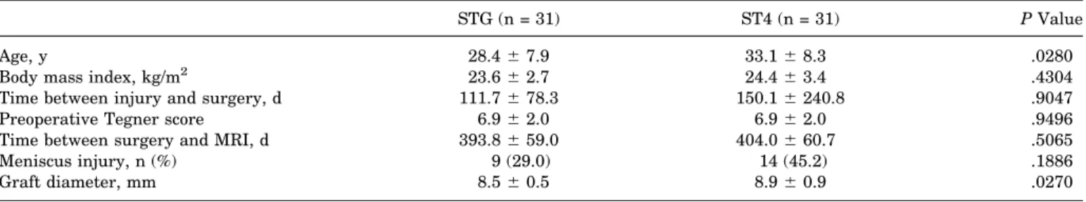
According to Claes et al,11ligamentization is the histologi-cal evolution of the graft. As histologihistologi-cal sections are TABLE 1
Characteristics for STG and ST4 Groupsa
STG (n = 31) ST4 (n = 31) P Value
Age, y 28.4 6 7.9 33.1 6 8.3 .0280
Body mass index, kg/m2 23.6 6 2.7 24.4 6 3.4 .4304
Time between injury and surgery, d 111.7 6 78.3 150.1 6 240.8 .9047
Preoperative Tegner score 6.9 6 2.0 6.9 6 2.0 .9496
Time between surgery and MRI, d 393.8 6 59.0 404.0 6 60.7 .5065
Meniscus injury, n (%) 9 (29.0) 14 (45.2) .1886
Graft diameter, mm 8.5 6 0.5 8.9 6 0.9 .0270
aValues are shown as mean 6 SD unless otherwise indicated. MRI, magnetic resonance imaging; ST4, quadrupled semitendinosus; STG, semitendinosus 1 gracilis.
Figure 2. Tunnels of the 2 types of graft: (A) semitendinosus 1gracilis (STG) and (B) quadrupled semitendinosus (ST4).
impossible in humans, the best way to evaluate ligamenti-zation is with MRI.
Several MRI criteria have been validated for evaluating graft ligamentization: (1) SNQ,19,22,37,52 (2) tibial tunnel widening,17,21,24 (3) graft healing (signal intensity at the
bone-graft interface),17and (4) graft maturity (water con-tent of the graft based on the Howell scale).22
The knee MRI examination was conducted after the patient rested for 1 hour; a 3-T MRI unit (Magnetom Skyra; Siemens Medical Solutions) with a 15-channel vol-ume array coil was used. The following sequences were obtained: 3D-PD-TSE and sagittal proton density weighted fat suppressed (PD-FS).
The SNQ for each graft was calculated with the follow-ing formula:
SNQ¼graft signal PCL signal background signal :
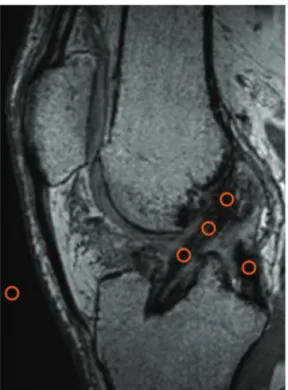
The graft signal values were averaged as described by Weiler et al.52For the MRI analysis, signal intensity was measured in 0.05-cm2circular regions of interest on
obli-que sagittal PD-FS images. The graft signal was measured in its intra-articular portion at 3 sites (superior, middle, and inferior), and the average was calculated. The back-ground signal was measured 2 cm anterior to the patellar tendon (Figure 3). SNQ reflects the graft’s mechanical strength.19,22,37,52
To determine tunnel widening,16 we measured the mean area at the entrance of each tibial tunnel on oblique MRI perpendicular to the tunnel’s section. The cross-sectional area (CSA; in cm2) of the superior portion of the tibial bone tunnel was measured using image postprocess-ing software (TeraRecon) on 3D-PD-TSE sequences (Figure 4). Three-dimensional reconstruction was used to define a perpendicular axis to the graft axis. Tunnel widening was calculated with the following formula:
CSA increase %ð Þ5Measured CSA Drilled CSA Drilled CSA 3100:
We used the protocol described by Ge et al17to measure graft healing (signal intensity at the bone-graft interface). Healing was evaluated on sagittal oblique images from PD-FS sequences. Based on this information, the patients were assigned 1 of 3 grades (Figure 5):
1. Low intensity, no fibrosis at the bone-graft interface, full attachment.
2. High intensity over a portion of the interface.
3. High intensity over the entire bone-graft interface, poor attachment.
To determine graft maturity, we used a 4-grade system based on the graft’s MRI signal inside the tibial tunnel according to Howell et al22(Figure 6):
1. Homogeneous, low-intensity signal indistinguishable from the PCL and patellar tendon.
Figure 3. Placement of regions of interest (ROIs) used to cal-culate the signal-to-noise quotient (ROI = 0.05 cm2). Three ROIs were placed on the graft (superior, middle, inferior), 1 ROI on the posterior cruciate ligament, and 1 ROI on an empty area 2 cm anterior to the patellar tendon.
Figure 4. Measurement of the cross-sectional area of the tibial bone tunnel with TeraRecon software using 3-dimensional reconstruction.
2. Normal ligament signal over at least 50% of its volume, intermingled with portions that have increased signal intensity.
3. Increased signal intensity over at least 50% of its vol-ume, intermingled with portions that have a normal lig-ament signal.
4. Diffuse increase in signal intensity without strands with a normal ligament appearance.
Knee stability was measured at the last follow-up using the Lachman test, the pivot-shift test, and Telos at 150 N.25,26The Lachman test results were graded as either 0 (\3 mm), 1 (3-6 mm), 2 (7-10 mm), or 3(.10 mm).20The
pivot shift was graded as 0 (absent), 1 (subluxation), 2 (jump), or 3 (transient lock).20The clinical examination was performed by a trained orthopaedic fellow blinded to the graft choice.
The MRI and Telos images were analyzed by 2 raters (a radiologist and an orthopaedic surgeon) in a double-blind manner. Neither rater knew the grade assigned by the other rater on the same examination, nor the result of the other examination (MRI, Telos). Similarly, the raters did not know which group the patient belonged to when they ana-lyzed the images. The analysis was performed using a PACS workstation (Horizon Rad Station; McKesson).
Functional outcomes consisted of Lysholm,8 Tegner,50 and International Knee Documentation Committee (IKDC) subjective20 scores at 1 year of follow-up. Patient satisfac-tion was evaluated with a simple 3-level quessatisfac-tionnaire: very satisfied, satisfied, and dissatisfied.
Statistical Analysis
We assumed that patients in the STG group had a mean SNQ at 1 year of 2.4 6 0.6 and that patients in the ST4 group had a mean SNQ at 1 year of 2.5.34We wanted to show that, at worst, the mean standardized difference in the SNQ between the ST4 and STG groups was less than 1 SD. With a sided alpha of 2.5% (ie, sided 97.5% CI) and a 1-sided beta of 10.0%, 31 patients were needed in each group.35
Before the analyses, we checked for missing, unusual, or inconsistent data. After corrections, the database was
locked. The analysis was performed on the locked database. The baseline characteristics of the patients in each group were expressed using the appropriate descriptive statistics for the type of variables. Descriptive statistics included the number of nonmissing observations, mean with SD, or median with interquartile range, as appropriate, for contin-uous variables and the number of nonmissing observations with frequency (%) for categorical variables. The intraclass correlation coefficient (ICC) with 95% CIs was calculated to assess interobserver reproducibility for the SNQ values and for the other MRI endpoints. The primary endpoint was analyzed in each group by comparing the 1-sided 97.5% CI of the mean SNQ at 1 year to the limit of noninfer-iority. To take into account imbalanced baseline character-istics between the groups, the adjusted mean SNQ (with 1-sided 97.5% CI) was assessed in each group using a linear regression model. Categorical secondary endpoints were compared between the groups using the chi-square test (or the Fisher exact test when necessary). The Student t test was used to compare the distribution of continuous second-ary endpoints (or the Mann-Whitney test when the data were not normally distributed or when the homoscedasticity assumption was rejected). All reported P values were 2-sided, with a significance threshold of \.05. Statistical anal-yses were performed using STATA software (version 14.1; StataCorp).
RESULTS
Signal-to-Noise Quotient
The mean SNQ was 5.2 6 4.5 in the STG group and 5.9 6 3.7 in the ST4 group (P = .5100). The unilateral 97.5% CI of the mean SNQ was 5.2 ( to 6.9) for the STG group and 5.9 ( to 7.3) (\9.7 at the limit of noninferiority) for the ST4 group (P = .5100). After adjusting for differences in age, smoking, and graft diameter, the unilateral 97.5% CI of the mean SNQ was 5.2 ( to 6.5) for the STG group and 5.9 ( to 7.1) (\9.7 at the limit of noninferiority) for the ST4 group (P = .1300). Hence, the ST4 graft is not statisti-cally different to the STG graft in terms of the SNQ. Figure 5. Examples of the 3 grades assigned to the bone-graft interface: (A) grade 1, arrow indicates low intensity signal, no fibrosis at bone-graft interface, full attachment; (B); grade 2, arrow indicates high intensity signal over a portion of the interface; and (C) grade 3, arrow indicates high intensity signal over the entire bone-graft interface, poor attachment.
Secondary Endpoints
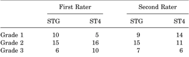
The mean tibial tunnel widening was 93.7% 6 51.7% for the STG group versus 80.0% 6 42.9% for the ST4 group (P = .2605). In terms of graft healing (signal intensity at the bone-graft interface), 10 patients were assigned grade 1, 15 patients grade 2, and 6 patients grade 3 in the STG group versus 5 patients assigned grade 1, 16 grade 2, and 10 grade 3 in the ST4 group by the first rater. There were 9 patients assigned grade 1, 15 patients grade 2, and 7 patients grade 3 in the STG group versus 14 patients assigned grade 1, 11 grade 2, and 6 grade 3 in the ST4 group by the second rater. There was no significant difference in
the population distribution based on the signal intensity between the 2 groups (P = .7502) (Table 2).
For graft maturity (Howell scale), 6 patients were assigned grade 1, 12 patients grade 2, 9 patients grade 3, and 4 patients grade 4 in the STG group versus 4 patients assigned grade 1, 14 grade 2, 12 grade 3, and 1 grade 4 in the ST4 group (Figure 5). There were no significant differ-ences between the 2 groups (P = .4544).
The mean side-to-side difference in anterior laxity was 0.8 6 4.1 for the STG group and 1.2 6 2.2 for the ST4 group (P = .7353) (Table 3). The ST4 group was noninferior to the STG group for all the functional outcome scores: Lysholm (P = .1890), Tegner (P = .8936), and subjective IKDC (P = .2350).
On the clinical examination, there was no significant dif-ference between the 2 groups in any of the clinical tests: Lachman (P = .6123), anterior drawer (P . .9999), and pivot shift (P = .6123). For the Lachman test, 28 patients were stage 0 (\3 mm), 3 patients were stage I (3-6 mm), and 0 patients were stage II (7-10 mm) or III (.10 mm) in the STG group; 30 patients were stage 0 (\3 mm), 1 patient was stage I (3-6 mm), and 0 patients were stage II (7-10 mm) or III (.10 mm) in the ST4 group.
In terms of satisfaction, 16 patients were very satisfied, 13 were satisfied, and 2 were dissatisfied in the STG group. In the ST4 group, 22 patients were very satisfied, 9 were satisfied, and 0 were dissatisfied (P = .1703).
Interobserver Reproducibility
For the SNQ measurement, the interobserver reproducibil-ity relative to the mean was 72% (95% CI, 54%-83%). The
TABLE 2
Grade of Signal Intensity at Bone-Graft Interfacea First Rater Second Rater
STG ST4 STG ST4
Grade 1 10 5 9 14
Grade 2 15 16 15 11
Grade 3 6 10 7 6
aValues are shown as No. of patients. ST4, quadrupled semiten dinosus; STG, semitendinosus 1 gracilis.
Figure 6. Examples of the 4 grades assigned to the water content of the graft according to the Howell scale: (A) grade 1, (B) grade 2, (C) grade 3, and (D) grade 4.
TABLE 3
Functional Outcomes in STG and ST4 Groupsa
STG ST4 P Value
Lysholm 80.9 6 17.7 92.0 6 5.6 .1890 Tegner 5.8 6 1.8 5.7 6 2.0 .8936 IKDC subjective 79.6 6 14.4 89.1 6 9.7 .2350 Anterior laxity (Telos) 0.8 6 4.1 1.2 6 2.2 .7353
a
Values are shown as mean 6 SD. IKDC, International Knee Documentation Committee; ST4, quadrupled semitendinosus; STG, semitendinosus 1 gracilis.
MRI endpoints are the mean of both raters. Hence, the ICC
corresponds to the reliability of the mean. There was good
interobserver reproducibility for the evaluation of the
sig-nal intensity at the bone-graft interface (ICC [3,2] = 0. 739; 95% Cl, 0.567-0.843), tibial tunnel widening (ICC
[3,2] = 0.820; 95% Cl, 0. 702-0.892), and knee laxity (ICC
[3,2] = 0.893; 95% Cl, 0.822-0.935).
DISCUSSION
Our hypothesis was confirmed: the ST4 technique was not inferior to the STG technique in terms of graft
incorpora-tion. There was also no inferiority in terms of healing, graft maturity, or tibial tunnel widening. This is the first study to compare the incorporation of STG and ST4 grafts.
Based on the assumptions made when calculating the sample size for the study, the noninferiority limit was equal to 1 SD (STG group), or 4.5. To demonstrate the non-inferiority of the ST4 graft relative to the STG graft, the
upper limit of the unilateral 97.5% Cl of the mean SNQ
of the ST4 group had to be less than 9.7 (ie, 5.2
+
4.5 = 9.7). The unilateral 97.5% Cl of the mean SNQ was 5.2( to 6.9) for the STG group and 5.9 ( to 7.3) (<9.7 at
the limit ofnoninferiority) for the ST4 group. After adjust-ing for differences in age, smoking, and graft diameter, the
unilateral 97.5% Cl of the mean SNQ was 5.2 ( to 6.5) for the STG group and 5.9 ( to 7 .1) ( <9. 7 at the limit of non-inferiority) for the ST4 group. Hence, the ST4 graft was not inferior to the STG graft in terms of the SNQ.
The Sharpey fibers that help a graft attach to bone develop over the 5 mm closest to the joint surface.10·33
The ST4 graft has the same outcome as the STG graft in terms of its incorporation into bone tunnels. By preserving
the gracilis, this tendon can be used for lateral tenodesis.53 The gracilis is an interesting option for reinforcing the
anterolateral aspect of the knee.53 Sonnery-Cottet et al47
showed that the STG technique can be used to perform intra-articular ACL reconstruction and lateral tenodesis with good clinical outcomes.
Pailhe et al40 showed that the ST4 graft is stronger than
the STG graft. In that cadaveric study, the tests were
per-formed on tissues in their graft configuration. Cavaignac et al9 showed that the diameter of an SI'4 graft is, on average,
20% greater than that of the SI'G graft in the same person. The mean graft diameter was 8.45 ::!: 0.51 mm for the STG group and 8.90 ::!: 0.94 mm for the ST4 group in this study.
Weiler et al52 observed that a higher signal intensity on contrast-enhanced MRI corresponded to lower mechanical
strength of the graft during the early remodeling phase. Hence, the SNQ is inversely proportional to the graft's
ten-sile strength. Several variations of the SNQ have been described, many of which do not require gadolinium
injec-tions.19·22.37 Other authors have compared the graft signal to that of the quadriceps tendon2·17.37 instead of that of the
PCL as did Weiler et al.13·34•52 We decided to use the same methodology as Weiler et al,52 who developed the SNQ measurements on MRI by comparing it to histological eval-uations. We did not perform a gadolinium injection because Weiler and colleagues52 had shown that it does not alter
the signal in the graft at 1 year. The SNQ values found
in the literature range from 0.078 ::!: 0.62 for an ST4 graft at 6 months13 to 5.49 ::!: 3. 71 for an allograft after 2 years.17
We found mean SNQ values of 5.2 ::!: 4.5 (STG) and 5.9 ::!: 3. 7 (ST4) at 1 year.
The tibial tunnel widens during the first few months after ACL reconstruction surgery. Fules et al16 showed that MRI was a good examination modality for evaluating
tunnel widening on transverse slices. Published tibial
tun-nel widening values range from 33% for the STG technique at 6.5 months16 to 80% for the ST4 technique at 10 years. 48
We found mean values of93.7%(STG) and 80.0% (ST4) at 1
year. We believe that these values are higher than in other studies because we used a screw of the same diameter as
the graft, which further widened the tunnel.
Women were excluded from this study because hor-monal changes can affect the graft's ligamentization dur-ing the menstrual cycle.7·14·17·31·39 In animal studies,
Kiapour et al28 have shown that female animals had worse results than male animals in terms of graft structural
properties and knee laxity.
We verified the plausibility of our results by comparing the values found in our study with those found in pub-lished studies. The side-to-side difference in anterior
trans-lation found in the literature ranges from 0.62 ::!: 2.13 for the ST4 graft at 6 months13 to 2.7 mm (range, 4.5 to
9.5) for the ST4 graft at 7 years.15 Our mean values on the Telos knee laximeter were 0.8 (STG) and 1.2 (ST4) at 1 year. The number of retears was consistent with pub-lished results (>3% retear rate at 2 years after ACL
recon-struction with the STG graft according to Mohtadi et al's36 meta-analysis).
In terms of clinical scores, we did not find any studies that have shown a significant difference between the 2 techniques. For both the IKDC and Lysholm scores, every
study has found the STG and ST4 grafts to be equal.3·5·18.23,27·29•45 In isokinetic testing, Gobbi et al18
showed that the STG technique resulted in a greater inter-nal rotation deficit in the knee than the ST4 technique. Other studies3·49 have shown that the ST4 technique resulted in less loss of active flexion range and flexion
strength than the STG technique. Karimi-Mobarakeh et al27 reported the same observation in terms of knee flex-ion strength loss. No study has found a significant differ-ence in knee stability between the 2 techniques. This was confirmed in a meta-analysis conducted by Sharma et al46 that included 8 studies.
Our study had several limitations. The SNQ is a variable
parameter that peaks at 6 months and then decreases until 60 months postoperatively.37 This means that we may have evaluated our patients too early in the follow-up
period. This is consistent with studies42•44 showing that
remodeling persists for up to 24 to 36 months, at which
point the graft becomes quiescent. However, the meta-analysis performed by Claes et al11 found that there was no agreement on the duration of the various stages
ofliga-mentization. Also, according to Li et al,30 MRI-based graft maturity did not predict the clinical and functional out-comes in patients at 1-year follow-up. The length of follow-up was too short for a clinical follow-up but chosen
for the imaging follow-up as the primary endpoint. In addi-tion, our 2 groups were not comparable in terms of age, smoking, and graft diameter; however, the data were adjusted for those parameters. The patients in the 2 groups were not operated by the same surgeon, which can in tro-duce a performance bias; however, the surgeons had
simi-lar training and used the same surgical technique, except
for the graft. The length of the graft inside the tunnels was not measured intraoperatively or on MRI. Because the primary objective of this study was to compare 2 types of grafts (ST4 vs STG), we cannot draw any conclusions about the length of graft needed for optimal incorporation.
We chose a hybrid suspension technique because an adjustable cortical device helps us to fix the femoral side into the socket and maintain a reserve length that allowed enough graft length in the tibia! tunnel.
CONCLUSION
At 1 year postoperatively, there were no differences between the incorporation and ligamentization of STG and ST4 grafts based on MRI analysis. The functional out-comes and residual laxity were equal in the 2 groups.
REFEREN
C
ES
1. Adachi N, Ochi M, Uchio Y, Sakai Y, KuriwakaM, Fujihara A. Harvest
ing hamstring tendons for ACL reconstruction influences postopera tive hamstring muscle performance. Arch Orthop Trauma Surg. 2003;123(9):460 465.
2. Ahn JH, Lee SH, Choi SH, Lim TK Magnetic resonance imaging eval
uatbn of anterbr cruciate ligament reconstruction using quadrupled hamstring tendon autografts: comparison of remnant bundle preserva tion and standard technque. Am J Sports Mad. 2010;38(9):1768 1777. 3. Ardern CL, Webster KE, Taylor NF, Feller JA. Hamstring strength recovery after hamstring tendon harvest for anterior cruciate ligament reconstruction: a comparison between graft types. Arthroscopy.
2010;26(4):462 469.
4. Ayala Mejias JO, Garcia Gonzalez B, Alcocer Perez Espana L, Villafane JH, Berjano P. Relationship between widening and position of the tunnels and clinical results of anterior cruciate ligament recon struction to knee osteoarthritis: 30 patients at a minimum follow up of 10 years. J Knee Surg. 2017;30(6):501 508.
5. Barenius B, Webster WK, McClelland J, Feller J. Hamstring tendon anterior cruciate ligament reconstruction: does gracilis tendon har vest matter? lnt Orthop. 2013;37(2):207 212.
6. Biuk E, Zelic: Z, Rapan S, Curie G, Biuk D, Radie: R. Analysis of bio mechanical properties of patellar ligament graft and quadruple ham string tendon graft Injury. 2015;46 Suppl 6:S14 S17.
7. Blacher AM, Richmond JC. Transient laxity of an anterior cruciate lig ament reconstructed knee related to pregnancy. Arthroscopy.
1998;14(1):77 79.
8. Briggs KK, Lysholm J, Tegner Y, Rodkey WG, Kocher MS, Steadman JR. The reliability, validity, and responsiveness of the Lysholm score and Tegner activity scale for anterior cruciate ligament injuries of the knee: 25 years later. Am J Sports Mad. 2009;37(5):890 897. 9. Cavaignac E, Pailhe R, Murgier J, Reina N, Lauwers F, Chiron P. Can
the gracilis be used to replace the anterior cruciate ligament in the knee? A cadaver study. Knee. 2014;21 (6):1014 1017.
10. Chen C H. Graft healing in anterior cruciate ligament reconstruction.
Sports Mad Arthrosc Rehabil 7her Techno/. 2009;1 (1 ):21.
11. Claes S, Verdonk P, Forsyth R, Bellemans J. The "ligamentization" process in anterior cruciate ligament reconstruction: what happens
to the human graft? A systematic review of the literature. Am J Sports
Mad. 2011 ;39(11):2476 2483.
12. Collette M, Cassard X. The Tape Locking Screw technique (TLS): a new ACL reconstruction method using a short hamstring graft
Orthop Traumato/ Surg Res. 2011 ;97(5):555 559.
13. Colombet P, Graveleau N, Jambou S. Incorporation of hamstring grafts within the tibial tunnel after anterior cruciate ligament recon struction: magnetic resonance imaging of suspensory fixation versus
interference screws. Am J Sports Mad. 2016;44(11):2838 2845.
14. Dragoo JL, Lee RS, Benhaim P, Finerman GAM, Harne SL. Relaxin receptors in the human female anterior cruciate ligament Am J
Sports Mad. 2003;31(4):577 584.
15. Ejerhed L, Kartus J, Sernert N, Kohler K, Karlsson J. Patellar tendon or semitendinosus tendon autografts for anterior cruciate ligament reconstruction? A prospective randomized study with a two year follow up. Am J Sports Mad. 2003;31(1):19 25.
16. Fules PJ, Madhav RT, Goddard RK, Newman Sanders A, Mowbray MAS. Evaluation of tibial bone tunnel enlargement using MRI scan cross sectional area measurement after autologous hamstring ten don ACL replacement. Knee. 2003;10(1):87 91.
17. Ge Y, Li H, Tao H, Hua Y, Chen J, Chen S. Comparison of tendon bone healing between autografts and allografts after anterior cruciate
ligament reconstruction using magnetic resonance imaging. Knee
Surg Sports Traumato/ Arthrosc. 2015;23(4):954 960.
18. Gobbi A, Domzalski M, Pascual J, Zanazzo M. Hamstring anterior cruciate ligament reconstruction: is it necessary to sacrifice the gra cilis? Arthroscopy. 2005;21(3):275 280.
19. Gohil S, Annear PO, Breidahl W. Anterior cruciate ligament recon struction using autologous double hamstrings: a comparison of stan dard versus minimal debridement techniques using MRI to assess revascularisation. A randomised prospective study with a one year follow up. J Bone Joint Surg Br. 2007;89(9):1165 1171.
20. Hefti F, Muller W, Jakob RP, Staubli HU. Evaluation of knee ligament
injuries with the IKDC form. Knee Surg Sports Traumato/ Arthrosc.
1993;1 (3 4):226 234.
21. Holler J, Moller HO, Fu FH. Bone tunnel enlargement after anterior cruciate ligament reconstruction: fact or fiction? Knee Surg Sports
Traumato/ Arthrosc. 1998;6(4):231 240.
22. Howell SM, Clark JA, Blasier RD. Serial magnetic resonance imaging of hamstring anterior cruciate ligament autografts during the first year of implantation: a preliminary study. Am J Sports Mad. 1991;
19(1):42 47.
23. Inagaki Y, Kondo E, Kitamura N, et al. Prospective clinical compari
sons of semitendinosus versus semitendinosus and gracilis tendon autografts for anatomic double bundle anterior cruciate ligament reconstruction. J Orthop Sci. 2013;18(5):754 761.
24. Iorio R, Vadala A, Argento G, Di Sanzo V, Ferretti A Bone tunnel
enlargement after ACL reconstruction using autologous hamstring tendons: a CT study. lnt Orthop. 2007;31(1):49 55.
25. James EW, Williams BT, LaPrade RF. Stress radiography for the diagnosis of knee ligament injuries: a systematic review. C/in Orthop.
2014;472(9):2644 2657.
26. Jardin C, Chantelot C, Migaud H, Gougeon F, Debroucker MJ, Duquennoy A [Reliability of the KT 1000 arthrometer in measuring anterior laxity of the knee: comparative analysis with Telos of 48 reconstructions of the anterior cruciate ligament and intra and inter observer reproducibility]. Rev Chir Orthopecique Reparatrice Appar Mot. 1999;85(7):698 707.
27. Karimi Mobarakeh M, Mardani Kivi M, Mortazavi A, Saheb Ekhtiari
K, Hashemi Motlagh K Role of gracilis harvesting in four strand hamstring tendon anterior cruciate ligament reconstruction: a dou ble blinded prospective randomized clinical trial. Knee Surg Sports
Traumato/ Arthrosc. 2015;23(4):1086 1091.
28. Kiapour AM, Fleming BC, Proffen BL, Murray MM. Sex influences the biomechanical outcomes of anterior cruciate ligament reconstruction
in a preclinical large animal model. Am J Sports Mad. 2015; 43(7):1623 1631.
29. Kyung H S, Lee H J, Oh C W, Hong H P. Comparison of results after anterior cruciate ligament reconstruction using a four strand single
semitendinosus or a semitendinosus and gracilis tendon. Knee Surg
Sports Traumatol Arthrosc. 2015;23(11):3238 3243.
30. Li H, Chen J, Li H, Wu Z, Chen S. MRI based ACL graft maturity does not predict clinical and functional outcomes during the first year after ACL reconstruction [published online August 2, 2016]. Knee Surg
Sports Traumatol Arthrosc. doi:10.1007/s00167 016 4252 5.
31. Li H, Tao H, Cho S, Chen S, Yao Z, Chen S. Difference in graft matu rity of the reconstructed anterior cruciate ligament 2 years postoper atively: a comparison between autografts and allografts in young men using clinical and 3.0 T magnetic resonance imaging evaluation.
Am J Sports Med. 2012;40(7):1519 1526.
32. Lubowitz JH. All inside anterior cruciate ligament graft link: graft preparation technique. Arthrosc Tech. 2012;1(2):e165 e168. 33. Lui P, Zhang P, Chan K, Qin L. Biology and augmentation of tendon
bone insertion repair. J Orthop Surg. 2010;5:59.
34. Ma Y, Murawski CD, Rahnemai Azar AA, Maldjian C, Lynch AD, Fu FH. Graft maturity of the reconstructed anterior cruciate ligament 6 months postoperatively: a magnetic resonance imaging evaluation of quadriceps tendon with bone block and hamstring tendon auto grafts. Knee Surg Sports Traumatol Arthrosc. 2015;23(3):661 668. 35. Machin D, ed. Sample Size Tables for Clinical Studies. 3rd ed. Chi
chester, UK: Wiley Blackwell; 2008.
36. Mohtadi NG, Chan DS, Dainty KN, Whelan DB. Patellar tendon ver sus hamstring tendon autograft for anterior cruciate ligament rupture in adults. Cochrane Database Syst Rev. 2011;(9):CD005960. 37. Muramatsu K, Hachiya Y, Izawa H. Serial evaluation of human
anterior cruciate ligament grafts by contrast enhanced magnetic res onance imaging: comparison of allografts and autografts. Arthros
copy. 2008;24(9):1038 1044.
38. Nakamura R, Horii E, Watanabe K, Nakao E, Kato H, Tsunoda K. Proximal row carpectomy versus limited wrist arthrodesis for advanced Kienbo¨ck’s disease. J Hand Surg Br. 1998;23(6):741 745. 39. Ntoulia A, Papadopoulou F, Ristanis S, Argyropoulou M, Georgoulis AD. Revascularization process of the bone patellar tendon bone autograft evaluated by contrast enhanced magnetic resonance imaging 6 and 12 months after anterior cruciate ligament reconstruc tion. Am J Sports Med. 2011;39(7):1478 1486.
40. Pailhe´ R, Cavaignac E, Murgier J, Laffosse J M, Swider P. Biome chanical study of ACL reconstruction grafts. J Orthop Res. 2015;33(8):1188 1196.
41. Piaggio G, Elbourne DR, Altman DG, Pocock SJ, Evans SJW; CONSORT Group. Reporting of noninferiority and equivalence ran domized trials: an extension of the CONSORT statement. JAMA. 2006;295(10):1152 1160.
42. Rougraff B, Shelbourne KD, Gerth PK, Warner J. Arthroscopic and histologic analysis of human patellar tendon autografts used for ante rior cruciate ligament reconstruction. Am J Sports Med. 1993; 21(2):277 284.
43. Samuelsson K, Andersson D, Ahlde´n M, Fu FH, Musahl V, Karlsson J. Trends in surgeon preferences on anterior cruciate ligament recon structive techniques. Clin Sports Med. 2013;32(1):111 126.
44. Sa´nchez M, Anitua E, Azofra J, Prado R, Muruzabal F, Andia I. Liga mentization of tendon grafts treated with an endogenous preparation rich in growth factors: gross morphology and histology. Arthroscopy. 2010;26(4):470 480.
45. Segawa H, Omori G, Koga Y, Kameo T, Iida S, Tanaka M. Rotational muscle strength of the limb after anterior cruciate ligament recon struction using semitendinosus and gracilis tendon. Arthroscopy. 2002;18(2):177 182.
46. Sharma A, Flanigan DC, Randall K, Magnussen RA. Does gracilis preservation matter in anterior cruciate ligament reconstruction? A systematic review. Arthroscopy. 2016;32(6):1165 1173.
47. Sonnery Cottet B, Thaunat M, Freychet B, Pupim BHB, Murphy CG, Claes S. Outcome of a combined anterior cruciate ligament and ante rolateral ligament reconstruction technique with a minimum 2 year follow up. Am J Sports Med. 2015;43(7):1598 1605.
48. Streich NA, Reichenbacher S, Barie´ A, Buchner M, Schmitt H. Long term outcome of anterior cruciate ligament reconstruction with an autologous four strand semitendinosus tendon autograft. Int Orthop. 2013;37(2):279 284.
49. Tashiro T, Kurosawa H, Kawakami A, Hikita A, Fukui N. Influence of medial hamstring tendon harvest on knee flexor strength after ante rior cruciate ligament reconstruction: a detailed evaluation with com parison of single and double tendon harvest. Am J Sports Med. 2003;31(4):522 529.
50. Tegner Y, Lysholm J. Rating systems in the evaluation of knee liga ment injuries. Clin Orthop. 1985;198:43 49.
51. Tomczak RJ, Hehl G, Mergo PJ, Merkle E, Rieber A, Brambs HJ. Tunnel placement in anterior cruciate ligament reconstruction: MRI analysis as an important factor in the radiological report. Skeletal
Radiol. 1997;26(7):409 413.
52. Weiler A, Peters G, Ma¨urer J, Unterhauser FN, Su¨dkamp NP. Biome chanical properties and vascularity of an anterior cruciate ligament graft can be predicted by contrast enhanced magnetic resonance imaging: a two year study in sheep. Am J Sports Med. 2001; 29(6):751 761.
53. Wytrykowski K, Swider P, Reina N, et al. Cadaveric study comparing the biomechanical properties of grafts used for knee anterolateral ligament reconstruction. Arthroscopy. 2016;32(11): 2288 2294.
54. Yamazaki S, Yasuda K, Tomita F, Minami A, Tohyama H. The effect of intraosseous graft length on tendon bone healing in anterior cruci ate ligament reconstruction using flexor tendon. Knee Surg Sports
Traumatol Arthrosc. 2006;14(11):1086 1093.
55. Yosmaoglu HB, Baltaci G, Ozer H, Atay A. Effects of additional gra cilis tendon harvest on muscle torque, motor coordination, and knee laxity in ACL reconstruction. Knee Surg Sports Traumatol Arthrosc. 2011;19(8):1287 1292.
56. Zantop T, Ferretti M, Bell KM, Brucker PU, Gilbertson L, Fu FH. Effect of tunnel graft length on the biomechanics of anterior cruciate liga ment reconstructed knees: intra articular study in a goat model.