Open Archive TOULOUSE Archive Ouverte (OATAO)
OATAO is an open access repository that collects the work of Toulouse researchers and
makes it freely available over the web where possible.
This is an author-deposited version published in :
http://oatao.univ-toulouse.fr/
Eprints ID : 18204
To link to this article : DOI:10.1016/j.msec.2015.09.070
URL : https://doi.org/10.1016/j.msec.2015.09.070
To cite this version : Noukrati, Hassan and Cazalbou, Sophie and
Demnati, Imane and Rey, Christian and Barroug, Allal and Combes,
Christèle Injectability, microstructure and release properties of
sodium fusidate-loaded apatitic cement as a local drug-delivery
system. (2016) Materials Science and Engineering C, vol. 59. pp.
177-184. ISSN 0928-4931
Any correspondence concerning this service should be sent to the repository
administrator:
[email protected]
Injectability, microstructure and release properties of sodium
fusidate-loaded apatitic cement as a local drug-delivery system
Hassan Noukrati
a,b, Sophie Cazalbou
c, Imane Demnati
a, Christian Rey
a, Allal Barroug
b, Christèle Combes
a,⁎
a
CIRIMAT, UPS-INPT-CNRS, ENSIACET, University of Toulouse, 4 allée Emile Monso, CS 44362, 31030 Toulouse Cedex 4, France
bLPCME-CNRST-URAC 20, Department of Chemistry, Faculty of Sciences Semlalia, University Cadi Ayyad, Marrakech, Morocco c
CIRIMAT, UPS-INPT-CNRS, Faculté des Sciences Pharmaceutiques, University of Toulouse, 118 Route de Narbonne, 31062 Toulouse Cedex 4, France
a b s t r a c t
Apatite cements Injectability Antibiotic Drug release Delivery systemThe introduction of an antibiotic, sodium fusidate (SF), into the liquid phase of calcium carbonate–calcium phos-phate (CaCO3–CaP) bone cement was evaluated, considering the effect of the liquid to powder ratio (L/P) on the
composition and microstructure of the set cement and the injectability of the paste. In all cases, we obtained set cements composed mainly of biomimetic carbonated apatite analogous to bone mineral. With this study, we evi-denced a synergistic effect of the L/P ratio and SF presence on the injectability (i.e., thefilter-pressing pheno-menon was suppressed) and the setting time of the SF-loaded cement paste compared to reference cement (without SF). In addition, the in vitro study of SF release, according to the European Pharmacopoeia recommen-dations, showed that, regardless of the L/P ratio, the cement allowed a sustained release of the antibiotic over 1 month in sodium chloride isotonic solution at 37 °C and pH 7.4; this release is discussed considering the micro-structure characteristics of SF-loaded cements (i.e., porosity, pore-size distribution) before and after the release test. Finally, modelling antibiotic release kinetics with several models indicated that the SF release was controlled by a diffusion mechanism.
1. Introduction
Post-operative infection is one of the major causes of increased mbidity and the most serious and costly complications associated with or-thopaedic procedures. Besides, the infected areas are often difficult to reach and treat. The development of minimally invasive surgical tech-niques that are able to control and reduce this post-operative risk and pain has promoted the use of injectable biomaterials, especially for or-thopaedic applications[1–2]. Despite the reduction in post-surgery risks associated with the use of minimally invasive surgery techniques, new biomaterials have been developed for therapeutic purposes. These materials are capable of releasing a drug over a long period that could prevent or treat some postoperative complications. Calcium phosphate cements (CPCs) have great potential as carriers for the controlled re-lease of drugs in bone tissue, owing to their composition that is close to bone mineral, excellent bioactivity and possible use as injectable and degradable grafting materials. They are set at low temperatures (ambient or physiological temperatures), which enables the association of CPCs with biologically active molecules, for example during paste preparation, without denaturation of the molecule or loss of its thera-peutic activity. Several studies have been conducted on the association
of antibiotics, such as gentamicin, cephalexin, doxycycline or vancomy-cin with CPCs in order to prevent implant-associated infection or to treat specific infections[3–10].
Usually, surgeons implement a prophylactic antibiotherapy by means of oral administration in order to prevent or reduce infectious pathologies after a surgery. The mostly used antibiotics in orthopaedic or traumatology are cefazolin, vancomycin, gentamycin and ciprofloxacin, but other antibiotics with good bone-tissue penetration (N30%), such as rifampicine, fosfomycine or fusidic acid, are also proposed[11–14]. In the present study, we chose a water-soluble derivative of fusidic acid, so-dium fusidate (SF), which showed broad spectrum bactericidal and fungi-cidal properties in addition to very good bone penetration[14].
The performance of CPCs in association with an antibiotic to design drug-delivery systems is related to specific parameters, that is, the ce-ment microstructure (i.e., porosity, tortuosity and specific surface area) and its resorbability, as well as the solubility of the drug and its in-teractions with the mineral matrix. An overview of the combination of pharmaceutical and biological substances with CPCs for delivery appli-cations, in addition to the effect of the principle-loading method on the physicochemical properties of the cements, has been reported by Ginebra et al.[10].
The objective of this work is to study the physicochemical properties of biomimetic apatite cement loaded with an antibiotic, sodium fusidate, which is a soluble salt of fusidic acid. We aim to evaluate the characteris-tics and properties of the paste (i.e., setting time and injectability) and the
⁎ Corresponding author at: ENSIACET — CIRIMAT, 4 allée Emile Monso, CS 44362, 31030 Toulouse Cedex 4, France.
set cement (i.e., mechanical and drug-release properties) in a view to pro-pose a sustained drug-release biomimetic bone substitute material. 2. Materials and methods
2.1. Preparation and characterisation of the cements
The reactive powders (solid phase) were comprised of vaterite cal-cium carbonate (CaCO3) and dicalcium phosphate dihydrate (DCPD;
CaHPO4·2H2O), which were prepared by precipitation at ambient
tem-perature, as previously described[15]. The cements investigated in this study were obtained by mixing the reactive powders (DCPD and vaterite in a 1:1 weight ratio) with an appropriate amount of liquid phase. The reference cement (CR) was produced using pure water only as the liquid phase, whereas we used a 9 wt.% SF aqueous solution to prepare the antibiotic-loaded cements (CSF). The liquid-to-powder ratios (L/P) used to prepare the cement pastes were 0.7 and 0.8 mL/g, leading to reference cements named CR7 and CR8, and to antibiotic-loaded cements referred to as CSF7 and CSF8, respectively. L/P = 0.7 corresponds to the minimum amount of liquid phase (water or aqueous solution of SF) to be added to the powder to form a malleable paste after mixing. We also chose to work with a higher L/P ratio (0.8) which cor-responds to a compromise between improvement of the injectability of the paste while adding minimum amount of water to keep the ce-ment mechanical properties as high as possible.
After mixing the solid with the liquid phase, the obtained paste was placed in a sealed container saturated with H2O at 37 °C for setting and
hardening for 2 days.
The set and dried cements were characterised by X-ray diffraction (INEL CPS 120 diffractometer) using a Co anticathode (wavelength = 1.78897 Å), transmission Fourier transform infrared spectroscopy with KBr pellets (Nicolet 5700 spectrometer, ThermoElectron) and scanning electron microscopy (SEM LEO 435 VP). Porosity measurements (be-tween 5 nm and 360μm) were performed using mercury intrusion porosimetry (AutoPore III, Micromeritics).
2.2. Setting time determination
The setting time of the cement paste was determined at 37 °C. The initial andfinal setting times were estimated according to a protocol adapted from the ASTM C266-03 and ISO 9917-1:2007 standards, using a Gillmore needle apparatus (HM-310, Gilson Inc.). Briefly, the system comprised two stainless steel needles: one is 2.13 mm diameter and 113.4 g weight used to determine the initial setting time; the sec-ond is 1.06 mm diameter and 453.6 g weight applied to estimate the final set. When the needles did not lead to a trace on the upper surface of the cement paste, the values of setting time were determined (expressed in minutes from when the mixing began). All measurements were performed in triplicate.
2.3. Mechanical properties of the hardened cements
A cement paste, prepared as described above, was introduced into cylindrical moulds (10 mm in diameter and 20 mm in height), which were then placed in sealed containers saturated with water at 37 °C for setting and hardening. After 2 days, the hardened cement was with-drawn from the mould and left to dry for 1 week at 37 °C. Five cements were prepared for each of the tested cement formulations. The com-pressive strength of the cylindrical cement blocks was evaluated using a Hounsfield Series S apparatus.
2.4. Injectability of the cement paste
The injectability of the reference and antibiotic-loaded cement pastes was measured at room temperature using a TAXT2 texture analyser (Stable Micro Systems) equipped with a specific syringe
system, including a 2.5 mL syringe (inner diameter of the syringe body was 9 mm and the opening/exit diameter was 2 mm) without a needle. The protocol consists of measuring the force, expressed as a load to be applied on the piston (piston surface = 64 mm2), to extrude
a volume of paste corresponding to the displacement of 15 mm of the syringe piston at a constant rate (2 mm·s−1).
Briefly, the syringe was filled with cement paste and the injectability measurement was performed 5 min after paste preparation (t = 0 cor-responds to the beginning of powder–liquid mixing). This period cor-responds to the time needed to prepare the paste, to introduce it into the syringe and to place the syringe system on the texture analyser be-fore starting the injectability measurements. This period could also cor-respond to the time a surgeon would need to prepare the cement paste and to introduce it into a device for implantation using a minimally in-vasive technique.
2.5. Antibiotic release study
The study of the antibiotic release from the cement was performed in vitro, according to the European Pharmacopoeia recommendations, using a Dissolutest (Pharmatest®) equipped with a stirring system (ro-tating paddle rate = 100 rpm) adapted to a series of 12 bowls (1 L), which allows homogenisation of the solution composition throughout the test.
The cement samples were prepared as described above for the me-chanical properties determination of hardened cements, except for the cylindrical mould dimensions, which were smaller so as to consume less of the cement paste (17.5 mm diameter × 10 mm height). Then, in order to study the drug-release kinetics, the cement blocks were im-mersed in 1 L of 0.9% (w/w) NaCl solution at physiological pH (pH = 7.4) and 37 °C under sink conditions for 5 weeks. The surface of each ce-ment block in contact with the release solution was equal and limited to the top of the cylindrical block (S = 2.40 cm2).
Some (10 mL) of the solution was removed from each bowl every day and replaced with the same volume of fresh 0.9% (w/w) NaCl solu-tion at pH = 7.4 in order to maintain a constant solusolu-tion/cement ratio throughout the experiment duration (i.e., 1 month). The pH value of the aliquot removed each day was not recorded but the pH value of the NaCl solution after 5 weeks of release test was around 8.0; this slight increase could be attributed to the release of carbonate species from the cement in the medium. The sampled solution was immediatelyfiltered using a Milliporefilter (pore diameter of 0.2 μm) to eliminate any fine particles that could be released from the cement block and impair the titration of the release solution by UV spectrophotometry. The concen-tration of the antibiotic in the sampled solution was then determined by UV spectrophotometry (UV-1800 SHIMADZU) at 235 nm. The ab-sence of interference (no absorbance) was checked for the reference ce-ment using the same electrolyte solution.
3. Results
3.1. Cement composition and microstructure
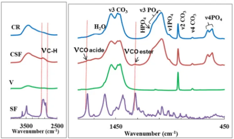
The X-ray diffraction patterns of the cement prepared in the absence (CR7) or presence of sodium fusidate (CSF7) are presented inFig. 1. The XRD pattern of the reference cement (CR7) showed that it was com-posed of carbonate apatite and residual vaterite, revealing the formation of apatite through the reaction of part of the vaterite with all of the DCPD powder. The results also indicated that adding SF to the cement paste did not significantly affect the end product, as the XRD patterns of both cements are similar. FTIR spectrum of drug containing cement (CSF) compared to the spectrum of the reference cement (CR) con-firmed that addition of drug into cement did not change significantly the composition of cementfinal composition despite the presence of the drug (Fig. 2). However FTIR spectroscopy allowed to identify on CSF spectrum bands of very low intensity, which are characteristic of
sodium fusidate: at 1265 cm−1(ester C\\O), 1720 cm−1(acid C_O)
and 2845–3000 cm−1(C\\H).
Fig. 3shows the SEM micrographs of the reactive powders (Fig. 3a and b for brushite and vaterite, respectively) and reference and antibiotic-loaded cements (Fig. 3c and d, respectively). We can observe a microporous microstructure for both hardened cements (Fig. 3c and d). In addition, the typicalfine plate-like morphology of nanocrystalline apatite was clearly visible whereas the brushite platelet crystals were no more visible in both hardened cement micrographs. Furthermore, as vaterite particles showed lentil-like shape with a mean size of about 2–3 μm (Fig. 3b); it seems that these particles served as a support for nanocrystalline apatite nucleation and growth, as we can see nanocrystals of apatite covering the surface of quite spherical particles of about 2–3 μm in diameter (Fig. 3c and d). Finally, we can see that the morphology of the apatite and vaterite crystals and their arrange-ment did not appear to be altered by the introduction of sodium fusidate in the cement formulation.
We can further analyse the microstructure of the cement by examin-ing the porosity measurements performed on the reference and SF-loaded cements. The obtained results are summarised inTable 1. The ce-ments exhibit a total porosity ranging between 69 and 85% depending on the formulation (i.e., the L/P ratio and the presence or absence of an-tibiotic). As expected, we can see that an increase in L/P from 0.7 to 0.8 enhanced the cement total porosity: for the reference cement, an aug-mentation in the total porosity of 11% was observed whereas a lower in-crease in total porosity (5%) was observed when increasing the L/P ratio for the cements loaded with the antibiotic (Table 1). If the increase in the cement total porosity appears also determined by the presence of the drug, the increase of submicron-size porosity, seems however only related to the L/P ratio used. Indeed, whatever the considered cement (CR or CSF) submicron-size porosity increases of 10% when the amount of water used for the preparation of the cement paste increases of 14% (from an L/P of 0.7 to 0.8).
In addition whatever the L/P ratio, the proportion of smaller pores (submicron porosity: 5 nm–2 μm), which represents the major
Fig. 1. X-ray diffraction patterns of the cements prepared with L/P = 0.7 for (a) the refer-ence cement and (b) SF-loaded cement, compared to the XRD patterns of the reactive powders (c) vaterite and (d) brushite.
Fig. 2. FTIR spectra of sodium fusidate (SF), vaterite (V), reference cement (CR), and SF-loaded cement (CSF).
contribution (91 to 98%) in the total porosity of the studied cements was lower for SF-loaded cements. Indeed, the addition of the drug into ce-ment induces a decrease of 7% of the submicron porosity. This decrease could be related to thefilling of the pores and voids with the antibiotic. Furthermore, if we examine the pore-size distributions for the reference cements (L/P = 0.7 and 0.8) presented inFig. 4a, the presence of two types of pore sizes within both cements can clearly be distinguished; thefirst group of pore sizes ranged between 7 and 60 μm and represent-ed a very small pore volume (b4% of the total porosity of the cement), whereas the second group of pore sizes ranged between 5 and 200 nm and represented around 95% of the total porosity of the refer-ence cements for both L/P ratios tested. The increase in L/P induced a slight increase in the submicron-pores size (i.e., the peak between 5 and 150 nm slightly shifted towards higher pore size; maximum shifted from 16 to 21 nm), indicating the determinant role of water in the po-rosity and microstructure of the cement. The pore-size distribution of SF-loaded cements (Fig. 4b) appears to be significantly different from that of the reference cements, and is also determined by the L/P ratio. We observed a broader range of submicron-pore sizes for the SF-loaded cements than for the reference cements; this broadening is more pronounced for higher L/P ratios, and pores with sizes ranging be-tween 1 and 20μm are created.
These observations confirmed that, not only the amount of water (L/P ratio), but also the presence of sodium fusidate were determi-nant parameters for the cement microstructure.
3.2. Cement paste setting time
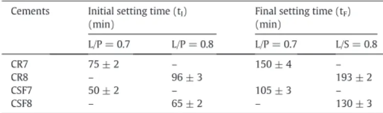
The measured setting times for cements prepared with different L/P ratios in the presence or absence of antibiotic are reported inTable 2. The increase in L/P ratio affected both the initial andfinal setting times of both types of cements studied. Thus, when the L/P ratio in-creased from 0.7 to 0.8, the initial setting time (tI) increased from 75
to 96 min for the reference cement and from 50 to 65 min for antibiotic-loaded cement.
In addition, the incorporation of sodium fusidate into the cement in-duced a decrease in the initial setting time for both L/P ratios tested: tI
decreased from 75 to 50 min for L/P = 0.7 and from 96 to 65 min for L/P = 0.8. A similar evolution occurred for thefinal setting time with re-spect to L/P ratio and drug incorporation.
3.3. Cement compressive strength
The effect of the L/P ratio and the introduction of sodium fusidate on the compressive strength of the cements are presented onFig. 5. As gene-rally observed, the compressive strength decreased significantly when the L/P increased from 0.7 to 0.8. The presence of the antibiotic in the cement slightly decreased the compressive strength of the cement from 11.8 to 9.3 MPa for L/P = 0.7 and from 4 to 3.4 MPa for L/P = 0.8. How-ever, this decrease does not appear significant in the latter case.
3.4. Paste injectability
Fig. 6shows that the injectability of the reference cements (CR7 and CR8) was poor, as the load needed to extrude the volume of paste in-creased during piston displacement by up to 21 kg for L/P = 0.7. The two curves obtained were characteristic of the occurrence of a filter-pressing phenomenon during paste extrusion. Interestingly, the filter-pressing phenomenon was much less pronounced for SF-loaded cement pastes, as the two curves wereflatter and the maximum load needed to extrude the paste was 6.8 kg for L/P = 0.7. These results indicated that antibiotic-loaded cement pastes were injectable, as the load needed to extrude the paste was less than 12 kg in all cases, which is the accepted limit of manual injectability. The injectability of the paste was signi fi-cantly improved by an increase in the L/P ratio in all cases.
3.5. Kinetics of antibiotic release
The release of fusidate from the cements is presented inFigs. 7and 8 as a cumulative percentage of fusidate released and as the amount of fusidate released per day in 1 L of NaCl solution as a function of time, respectively.
Table 1
Total porosity and microporosity of the reference (CR) and SF-loaded (CSF) cements pre-pared with different L/P ratios (0.7 or 0.8).
Reference cement (CR) Sodium fusidate-loaded cement (CSF) L/P = 0.7 L/P = 0.8 L/P = 0.7 L/P = 0.8 Total porosity (%) [5 nm–300 μm] 74 ± 2 85 ± 3 69 ± 1 74 ± 1 Submicron porosity (%) [5 nm–2 μm] 70 ± 2 80 ± 3 63 ± 2 72.7 ± 0.4
Fig. 4. Pore-size distribution of the reference (a) and the SF-loaded (b) cements, both pre-pared with L/P ratios of 0.7 and 0.8.
Table 2
Initial andfinal setting times of reference (CR) and SF-loaded (CSF) cements prepared with different L/P ratios (0.7 or 0.8).
Cements Initial setting time (tI)
(min)
Final setting time (tF)
(min) L/P = 0.7 L/P = 0.8 L/P = 0.7 L/S = 0.8 CR7 75 ± 2 – 150 ± 4 – CR8 – 96 ± 3 193 ± 2 CSF7 50 ± 2 – 105 ± 3 – CSF8 – 65 ± 2 – 130 ± 3
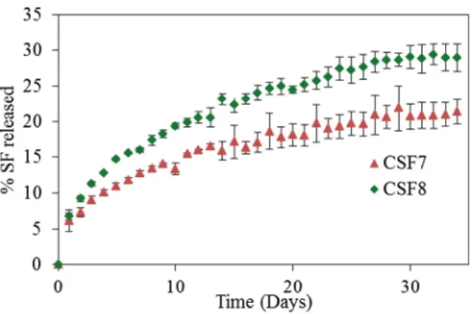
The sustained release of the antibiotic was evidenced over more than 1 month (between 20 and 30% released at day 30), no matter what the L/P ratio (Fig. 7). The release rate was significantly faster for cement prepared with the highest L/P ratio. The release rate can be eva-luated by considering the time required to release 20% (t20%) of the
so-dium fusidate; thus, 27 days was the necessary time to release 20% of the antibiotic from CSF7 cement, whereas it took only 12 days for the CSF8 cement to reach the same rate of release (20% of the drug re-leased). Finally, at the end of the test (day 34), the cumulated SF release was about 30 and 22% from CSF8 and CSF7 cements, respectively.
The dissolution profiles (CSF7 and CSF8) highlight an initial burst re-lease of fusidate (3–10 mg/day) during the first days (Fig. 8), but then the daily SF release significantly decreased until a sustained and quite constant release rate was achieved (1–2 mg/day).
4. Discussion
Generally, the addition of a drug to bone cement formulation mod-ifies the physicochemical properties of the material; besides, drug re-lease is defined by specific parameters, that is, the porosity of the matrix, solubility of the drug and its interaction with the cement. The ef-fects of the L/P ratio and antibiotic loading on the physicochemical pro-perties and microstructure of the cement and on the drug-release ability have been identified through the presented results. Hereafter, the vari-ous parameters controlling the cement properties and the mechanism of antibiotic release in vitro are discussed.
4.1. Effect of L/P and antibiotic on cement microstructure and properties 4.1.1. Setting time
It has been reported that the setting time of cement for orthopaedic applications should be within the 10–40 min range[16]. In the present study, the setting time determined for the reference and SF-loaded ce-ments exceed this range for any L/P ratio tested, but, interestingly, it de-creased when SF was introduced into the cement formulation; furthermore, this decrease can be further enhanced when the well-known CPC setting-time promoter, disodium hydrogen phosphate (Table 3), is also dissolved in the liquid phase (SF aqueous solution) used to prepare the paste.
Several authors have shown, on the contrary, that the incorporation of an antibiotic in phosphocalcic cement formulations generally delayed the setting time[6,17–22]; this effect was attributed to the interaction of the drug with the cement mineral matrix during the cement setting reaction. It shall be considered, however, that physical setting times, de-termined using Gilmore needles, and the chemical setting times, estimated through the advancement of the setting reaction, are two pa-rameters that are not connected by simple relationships. The mode of interaction of the fusidate ions with the cement constituents was not elucidated in this study, although, the formation of insoluble amor-phous calcium fusidate seemed to be possible. Thus, the preliminary re-sults on SF interactions with calcium phosphate materials and cements (data not shown) revealed that, in the range of concentration investi-gated (0–0.21 mM), the antibiotic exhibited a high affinity for the mine-ral surface, and for higher drug contents the possible formation of a complex involving fusidate molecules and matrix mineral ions could not be discarded.
4.1.2. Injectability
Several studies have indicated the effect of the L/P ratio, not only on the rheological properties of the cement paste, but also on its porosity
[23]. The main concern for the implementation of bone mineral cement using minimally invasive surgery techniques (including a syringe sys-tem) is related to thefilter-pressing phenomenon, also called the phase-separation phenomenon (i.e., a paste with a higher L/P ratio is ex-trudedfirst and then the L/P ratio of the extruded paste decreases)[24], which leads to a heterogeneous composition of the extruded paste and
also to an increase in the extrusion load, over the limit of manual extru-sion (considered as 12 kg). The present study demonstrated that the injectability of SF-loaded cement paste is higher than that of the refer-ence cement; in addition, thefilter-pressing phenomenon can be avoided by combining a L/P ratio of 0.8 with a SF load. An analogous be-haviour has been revealed for another CPC loaded with another antibio-tic, that is, cephalexin; the authors suggested a lubricating effect of the antibiotic, favouring the extrusion of the CPC suspension[6]. Regarding the effect of the L/P ratio on the mineral cement injectability, our results are in agreement with the data reported by many authors[2,25–26]. In addition, the improvement in the injectability with the additive can be related to the high water solubility of sodium fusidate, which can in-crease the viscosity of the liquid phase (SF solution) and, thus, the cohe-sion of the paste that, in turn, facilitates its homogeneous extrucohe-sion. The improvement of the paste cohesion has been noticed when preparing the cement paste using SF solution compared to the paste prepared using only water.
4.1.3. Mechanical properties
Several studies have reported the effect of the introduction of an an-tibiotic on the mechanical properties of CPC, especially their compres-sive strength, which is more or less altered depending on the concentration of the drug[4,9,17,22–23,27–28]. This decrease in com-pressive strength can be attributed to several factors, such as an increase in cement porosity or the interaction of the drug with the reactive ce-ment powders, which could inhibit the setting reaction[29].
Fig. 5. Compressive strength of the reference (CR) and the SF-loaded (CSF) cements, both prepared with different L/P ratios of 0.7 and 0.8.
Fig. 6. Influence of the L/P ratio and the presence of sodium fusidate on the injectability of the cement paste.
In the present study, we showed that the introduction of sodium fusidate leads to a decrease in the total porosity of the cement, which did not improve the cement compressive strength. However the com-pressive strength noted for the reference or SF-loaded cements remains close to 9 MPa, corresponding to trabecular bone[30].
4.1.4. Porosity
Porosity is a key factor affecting mechanical properties of cement as well as its biological properties. The pore size and interconnection can control cell activity. CPCs are intrinsically mesoporous and this porosity is mainly related to the L/P ratio. A high porosity can alter the mecha-nical properties; however it could also enable the penetration of biolog-icalfluids, vessels and cells, favouring material resorption and bone neo-formation.
In the case of SF-loaded cement, a decrease in porosity without an improvement in cement compressive strength was observed. We can hypothesise that the consumption of water during the cement setting reaction and water elimination upon cement drying lowered the amount of water available to keep the SF solubilised; when the solubi-lity limit was reached, the SF could precipitate within the pores. Another hypothesis could be related to the precipitation of calcium fusidate within the pores; however, FTIR spectroscopy and XRD analyses could not allow distinguishing calcium fusidate from sodium fusidate (data not presented). Furthermore, it appears that the smaller pores (around 15 nm in diameter) are mainly affected by the decrease in porosity for SF-loaded cement; the proportion of pores with nanometric sizes was lowered, because they werefilled with the antibiotic (as sodium and/ or calcium fusidate).
The results of this study confirmed that, not only the amount of water (L/P ratio), but also the presence of sodium fusidate were deter-minant parameters for the cement microstructure.
4.2. Antibiotic release kinetics and mechanism
To identify the mechanisms that govern the release of the antibiotic from the cement, the release data were analysed using several models
[31].
Korsmeyer and Peppas developed a simple, semi-empirical model, relating drug release with the time elapsed, according to Eq.(1) [32,33]: Q¼ Mt=M∞¼ ktn ð1Þ
where t is the release time (min), Q (or Mt./M∞) is the fraction of drug
released, k is a kinetic constant, and the diffusional exponent, n, gives an indication of the drug-release mechanism with limits depending in the geometry of the release device; values of n below 0.5 correspond to Fickian diffusion, whereas values from 0.5 to 1.0 are attributed to non-Fickian transport (i.e. a mixed diffusion and erosion mechanisms). Higuchi developed several theoretical models to study drug release
[34]. In summary, a simplified Higuchi model can be expressed as Eq.(2):
Q¼ Mt=M∞¼ at0:5 ð2Þ
where t is the release time, Mt.is the amount of drug released at time t,
M∞is the total amount of drug released, and a is the Higuchi diffusion
constant. This model showed good correlation to the release profiles, in which the predominant mechanism is simple Fickian diffusion.
The Kopcha model can be used to quantify the contributions of diffu-sion (A) and erodiffu-sion (B).[35]. This model is described by Eq.(3): Mt¼ A
ffiffi t p
þ Bt ð3Þ
where Mtis the amount of drug released at time t, A is a diffusional term
and B is the erosional term. The diffusion phenomenon is predominant when A/BN 1.
To determine the constants, the experimental SF release data (Fig. 7) werefitted with the three models presented (i.e. Eqs. (1), (2) and (3). The parameters obtained afterfitting the SF release data with each model are reported inTable 4. Examining these parameters suggests that, regardless of the L/P used, the mechanisms involved in the release of sodium fusidate were mainly owing to diffusion process. Indeed values of n lower than 0.5 for the Korsmeyer–Peppas model indicated that the release followed Fick's law. In addition, A/B ratios higher than
Fig. 7. Cumulated fusidate released (%) from cements prepared with an L/P ratio of 0.7 or 0.8.
Fig. 8. Amount of fusidate released per day from cements prepared with an L/P ratio of 0.7 or 0.8.
Table 3
Initial andfinal setting times of SF-loaded cements (CSF) prepared with L/P = 0.7 and a liquid phase including different amounts of dissolved disodium hydrogen phosphate (0– 0.5 M).
Concentration of Na2HPO4
Initial setting time (tI)
(min)
Final setting time (tF)
(min) CSF7 CSF7 0 50 ± 2 105 ± 3 0.25 M 34 ± 1 76 ± 1 0.5 M 25 ± 1 52 ± 2 Table 4
Parameters of the SF release profile fitting, according to the different models.
Higuchi Korsmeyer–Peppas Kopcha
a r2 n r2 A/B r2
CSF7 0.600 0.970 0.36 0.988 ≫1 0.970
1 (obtained by considering the Kopcha model) confirmed that diffusion was the determinant phenomenon for SF drug release from the studied cements. Moreover, the release rate constants for the Higuchi model showed that increasing the L/P ratio used to prepare the cements caused an increase in the speed of release. The release is directly linked to the porosity of cement and especially to the ability of a cement to favour the diffusion phenomenon.
It is important to note that the in vitro release test was performed at physiological pH and temperature, but in the absence of macrophage cells. Under these conditions, the erosion process can only occur by chemical dissolution of the cement in buffer solution, and this phenom-enon is considered to be very low. When implanted, the biological activ-ity of cells shall favour cement resorption and, consequently, enhance the release of the antibiotic.
Our study showed that, irrespective of the L/P ratio, a burst release of fusidate (3–10 mg/day) was followed by a sustained daily release of SF (1–2 mg/day) over the next 30 days. The amount of drug released during thefirst stage of release kinetics can ensure the loading dose, whereas, after few days, the amount released can be considered as a maintenance dose.
It has been shown in the literature that fusidic acid concentrations be-tween 0.03 and 0.12 mg/L inhibit the growth of most of staphylococcus
strains [14,36–38]; however, for Staphylococcus aureus that is resistant to methicillin (SARM), the minimum inhibitory concentrations (MICs) are slightly higher (0.03–0.8 mg/L).
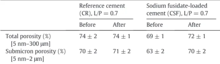
If we compare the evolution of the porosity of the reference and antibiotic-loaded cements before and after the release test (Table 5), we note an increase in the total porosity after 34 days of testing only for SF-loaded cement, suggesting that the dissolution and the release of the antibiotic created a secondary porosity in addition to the existing porous volume of the cement before the in vitro release tests. Examina-tion of the pore-size distribuExamina-tion of the reference and SF-loaded ce-ments before and after release testing (Fig. 9) revealed that the immersion of the reference material into the solution for 34 days induces a slight increase in pore size and the apparition of a secondary porosity domain. This modification of reference cement pore-size distri-bution during immersion could be due to the evolution of its chemical composition (vaterite, which is metastable in aqueous solution, could partly dissolve during the release testing) as well as crystals organisa-tion in cement matrix. Concerning the drug-containing cement, the po-rosities obtained after release testing highlight an increase in the total porosity with modification of the porous network. The dissolution and release of sodium fusidate induce secondary porosity, owing to the dis-appearance of drug crystals.
5. Conclusion
This study shows the determinant role of water (L/P ratio) and the presence of the drug on calcium carbonate–calcium phosphate biometic cement properties (especially cement paste injectability) and mi-crostructure, which, in turn, control the diffusion of the antibiotic and, consequently, its release rate. Based on the in vitro release study, this local antibiotic delivery bone-substitute material could ensure the de-livery of a loading dose (3–10 mg/day) for the first 2 days after surgery and then a maintenance dose (1–2 mg/day) for more than 1 month, making this SF-loaded cement a promising candidate as a local antibiot-ic delivery bone-substitute material, especially for the prevention of bone-implant-associated infections.
Acknowledgements
The authors thank the Campus France Agency (PHC Volubilis Pro-gramme 2011–2013; MA/11/263 project) for supporting this research work, Dr Boubker ESSADKI and Dr. Said ZAYANE, the two orthopaedic surgeons from the University of Marrakech (Morocco) who helped us in the choice of the antibiotic and its doses, and the International Drug Development (IDD) company for providing sodium fusidate samples.
References
[1] M.P. Ginebra, in: J.A. Planell (Ed.), Cements as Bone Repair Materials Bone Repair Biomaterials, Woodhead Publishing Limited, Cambridge, England 2009, pp. 271–308.
[2] M. Bohner, G. Baroud, Injectability of calcium phosphate pastes, Biomaterials 26 (2005) 1553–1563.
[3] A. Sugawara, K. Asaoka, S.J. Ding, Calcium phosphate-based cements: clinical needs and recent progress, J. Mater. Chem. B 1 (2013) 1081–1089.
[4] U. Joosten, A. Joist, T. Frebel, B. Brandt, S. Diederichs, C. Von Eiff, Evaluation of an in situ setting injectable calcium phosphate as a new carrier material for gentamicin in the treatment of chronic osteomyelitis: studies in vitro and in vivo, Biomaterials 25 (2004) 4287–4295.
[5] W.C. Liu, C.T. Wong, M.K. Fong, W.S. Cheung, R.Y.T. Kao, K.D.K. Luk, W.W. Lu, Gentamicin-loaded strontium-containing hydroxyapatite bioactive bone cement— an efficient bioactive antibiotic drug delivery system, J. Biomed. Mater. Res. B Appl. Biomater. 95 (2010) 397–406.
[6] S. Hesaraki, R. Nemati, Cephalexin-loaded injectable macroporous calcium phos-phate bone cement, J. Biomed. Mater. Res. B Appl. Biomater. 89 (2009) 342–352.
[7] T. Sasaki, Y. Ishibashi, H. Katano, A. Nagumo, S. Toh, In vitro elution of vancomycin from calcium phosphate cement, J. Arthroplast. 20 (2005) 1055–1059.
[8] P.J. Jiang, S. Patel, U. Gbureck, R. Caley, L.M. Grover, Comparing the efficacy of three bioceramic matrices for the release of vancomycin hydrochloride, J. Biomed. Mater. Res. B Appl. Biomater 93 (2010) 51–58.
Fig. 9. Pore-size distribution of cements, prepared with an L/P ratio of 0.7, before and after the release test: (a) reference cement (CR7) and (b) SF-loaded cement (CSF7). Table 5
Total porosity and microporosity of reference (CR) and SF-loaded (CSF) cements (L/P ratio = 0.7) before and after 34 days of release testing.
Reference cement (CR), L/P = 0.7
Sodium fusidate-loaded cement (CSF), L/P = 0.7
Before After Before After
Total porosity (%) [5 nm–300 μm] 74 ± 2 74 ± 1 69 ± 1 72 ± 1 Submicron porosity (%) [5 nm–2 μm] 70 ± 2 71 ± 2 63 ± 2 70 ± 2
[9] M.H. Alkhraisat, C. Rueda, J. Cabrejos-Azama, J. Lucas-Aparicio, F.T. Mariño, J. Torres García-Denche, L. Blanco Jerez, U. Gbureck, E. Lopez Cabarcos, Loading and release of doxycycline hyclate from strontium-substituted calcium phosphate cement, Acta Biomater. 6 (2010) 1522–1528.
[10]M.P. Ginebra, C. Canal, M. Espanol, D. Pastorino, E.B. Montufar, Calcium phosphate cements as drug delivery materials, Adv. Drug Deliv. Rev. 64 (2012) 1090–1110.
[11] M. Titécat, E. Senneville, F. Wallet, H. Dezèque, H. Migaud, R.J. Courcol, C. Loïez, Bac-terial epidemiology of osteoarticular infections in a referent center: 10-year study, Orthop. Traumatol. Surg. Res. 99 (2013) 653–658.
[12]R. Maviglia, R. Nestorini, M. Pennisi, Role of old antibiotics in multidrug resistant bacterial infections, Curr. Drug Targets 10 (2009) 895–905.
[13] C.A. Aboltins, M.A. Page, K.L. Buising, A.W. Jenney, J.R. Daffy, P.F. Choong, P.A. Stanley, Treatment of staphylococcal prosthetic joint infections with debridement, prosthe-sis retention and oral rifampicin and fusidic acid, Clin. Microbiol. Infect. 13 (2007) 586–591.
[14]J. Turnidge, Fusidic acid pharmacology, pharmacokinetics and pharmacodynamics, Int. J. Antimicrob. Agents 12 (1999) S23–S34.
[15] C. Combes, R. Bareille, C. Rey, Calcium carbonate–calcium phosphate mixed cement compositions for bone reconstruction, J. Biomed. Mater. Res. A 79 (2006) 318–328.
[16] F.C.M. Driessens, M.G. Boltong, O. Bermudezet, J.A. Planell, Formulation and setting times of some calcium orthophosphate cements: a pilot study, J. Mater. Sci. Mater. Med. 4 (1993) 503–508.
[17]A. Ratier, M. Frèche, J.L. Lacout, F. Rodriguez, Behaviour of an injectable calcium phosphate cement with added tetracycline, Int. J. Pharm. 274 (2004) 261–268.
[18] F. Tamimi, J. Torres, I. Tresguerres, L. Blanco Jerez, E. López Cabarcos, Vertical bone augmentation with granulated brushite cement set in glycolic acid, J. Biomed. Mater. Res. A 81 (2007) 93–102.
[19] M. Takechi, Y. Miyamoto, Y. Momota, T. Yuasa, S. Tatehara, M. Nagayama, K. Ishikawa, K. Suzuki, The in vitro antibiotic release from anti-washout apatite ce-ment using chitosan, J. Mater. Sci. Mater. Med. 13 (2002) 973–978.
[20] C.J. Wang, T.Y. Chen, J. Zhang, C.S. Liu, An in vivo study of tobramycin-impregnated calcium phosphate cement as an artificial bone material repairing bone defect, Fudan Univ. J. Med. Sci. 28 (2001) 473–475.
[21] K. Ishikawa, Y. Miyamoto, M. Takechi, T. Toh, M. Kon, M. Nagayama, K. Asaoka, Non-decay type fast-setting calcium phosphate cement: hydroxyapatite putty containing an increased amount of sodium, J. Biomed. Mater. Res. 36 (1997) 393–399.
[22] A. Ratier, S. Best, M. Freche, J. Lacout, F. Rodriguez, Behaviour of a calcium phosphate bone cement containing tetracycline hydrochloride or tetracycline complexed with calcium ions, Biomaterials 22 (2001) 897–901.
[23]C. Canal, D. Pastorino, G. Mestresa, P. Schulera, M.P. Ginebra, Relevance of micro-structure for the early antibiotic release of fresh and pre-set calcium phosphate ce-ments, Acta Biomater. 9 (2013) 8403–8412.
[24]M. Habib, G. Baroud, F. Gitzhofer, M. Bohner, Mechanisms underlying the limited injectability of hydraulic calcium phosphate paste. Part II: particle separation study, Acta Biomater. 6 (2010) 250–256.
[25] I. Khairoun, M.G. Boltong, F.C.M. Driessens, J.A. Planell, Some factors controlling the injectability of calcium phosphate bone cements, J. Mater. Sci. Mater. Med. 9 (1998) 425–428.
[26] N.J. Dunne, R. O'Hara, F. Buchanan, J. Orr, Effect of liquid/powder ratio on the setting, handling and mechanical properties of collagen–apatitic cements, Key Eng. Mater. 493-494 (2011) 415–421.
[27] O. Kisanuki, H. Yajima, T. Umeda, Y. Takakura, Experimental study of calcium phos-phate cement impregnated with dideoxy-kanamycin B, J. Orthop. Sci. 12 (2007) 281–288.
[28] M.P. Hofmann, A.R. Mohammed, Y. Perrie, U. Gbureck, J.E. Barralet, High-strength re-sorbable brushite bone cement with controlled drug-releasing capabilities, Acta Biomater. 5 (2009) 43–49.
[29] C. Canal, M.P. Ginebra, Fibre-reinforced calcium phosphate cements: a review, J. Mech. Behav. Biomed. Mater. 4 (2011) 1658–1671.
[30]D.R. Carter, W.C. Hayes, The compressive behavior of bone as a two-phase porous structure, J. Bone Joint Surg. 59A (1977) 954–962.
[31]P. Costa, J.M. Sousa Lobo, Modeling and comparison of dissolution profiles, Eur. J. Pharm. Sci. 13 (2001) 123–133.
[32] N.A. Peppas, Analysis of Fickian and non-Fickian drug release from polymers, Pharm. Acta Helv. 60 (1985) 110–111.
[33] R.W. Korsmeyer, R. Gurny, E. Doelker, P. Buri, N.A. Peppas, Mechanisms of solute re-lease from porous hydrophilic polymers, Int. J. Pharm. 15 (1983) 25–35.
[34]T. Higuchi, Mechanism of sustained-action medication. Theoretical analysis of rate of release of solid drugs dispersed in solid matrices, J. Pharm. Sci. 52 (1963) 1145–1149.
[35]M. Kopcha, N. Lordi, K.J. Tojo, Evaluation of release from selected thermosoftening vehicles, J. Pharm. Pharmacol. 43 (1991) 382–387.
[36] P. Collignon, J. Turnidge, Fusidic acid in vitro activity, Int. J. Antimicrob. Agents (Suppl. 2) (1999) S45–S58.
[37] O. Tunger, A. Arlsoy, S. Kurutepe, S. AkcalI, B. Ozbakkaloglu, In vitro susceptibility of Staphylococcus aureus and coagulase-negative Staphylococcus strains to fusidic acid, Int. J. Antimicrob. Agents 18 (2001) 445–447.
[38] L.A. Mandell, G.L. Mandell, J.E. Bennett, R. Dolin, Fusidic acid, Mandell, Douglas and Bennett's Principles and Practice of Infectious Diseases, 5, Churchill Livingstone, Philadelphia, 2000.