Université de Montréal
Neurotoxicity and Neurobehavioral Effects of Manganese Phosphate/Sulfate Mixture in Male $prague-Dawley Rats following $ubchronic Inhalation Exposure
Par
Fariba Salehi
Département de santé environnementale et santé au travail faculté de Médecine
Thèse présentée à la faculté des études supérieures en vue de l’obtention du grade de Philosophiœ Doctor (Ph.D.)
en santé publique
Option : toxicologie de l’environnement
janvier 2005 © Fariba Salehi, 2005
I j L
Université
de Montréal
Direction des bibliothèques
AVIS
L’auteur a autorisé l’Université de Montréal à reproduire et diffuser, en totalité ou en partie, par quelque moyen que ce soit et sur quelque support que ce soit, et exclusivement à des fins non lucratives d’enseignement et de recherche, des copies de ce mémoire ou de cette thèse.
L’auteur et les coauteurs le cas échéant conservent la propriété du droit d’auteur et des droits moraux qui protègent ce document. Ni la thèse ou le mémoire, ni des extraits substantiels de ce document, ne doivent être imprimés ou autrement reproduits sans l’autorisation de l’auteur.
Afin de se conformer à la Loi canadienne sur la protection des
renseignements personnels, quelques formulaires secondaires, coordonnées ou signatures intégrées au texte ont pu être enlevés de ce document. Bien que cela ait pu affecter la pagination, il n’y a aucun contenu manquant.
NOTICE
The author of this thesis or dissertation has granted a nonexciusive license allowing Université de Montréal to reproduce and publish the document, in part or in whole, and in any format, solely for noncommercial educational and research purposes.
The author and co-authors if applicable retain copyright ownership and moral rights in this document. Neither the whole thesis or dissertation, flot substantial extracts from it, may be printed or otherwise reproduced without the author’s permission.
In compliance with the Canadian Privacy Act some supporting forms, contact information or signatures may have been removed from the document. While this may affect the document page count, it does flot represent any Ioss of content from the document.
Université de Montréal Faculté des études supérieures
Cette thèse intitulée:
Neurotoxicity and Neurobehavioral Effects of Manganese
Phosphate/Sulfate Mixture in Male Sprague-Dawley Rats Following
Subchronic Inhalation Exposure
Présentée par:
Fariba Salehi
A été évalué par un jury composé des personnes suivantes:
Président rapporteur: Kannan Krishnan Directeur de recherche: Joseph Zayed Codirecteur de recherche: Daniel Krewski Membre dujury: Gaétan Carrier
Examinateur externe: Claude Ostiguy
Résumé
Le méthylcyclopentadiényle manganèse tricarbonyle (MMT) est un dérivé organique de manganèse (Mn) utilisé comme antidétonant dans l’essence sans plomb au Canada. Plusieurs inquiétudes ont été soulevées quant à la neurotoxicité des principaux produits de combustion du MMT, à savoir: le phosphate de Mn, le sulfate de Mn et un mélange de sulfate/phosphate de Mn. L’objectif de cette recherche vise à évaluer et à comparer les concentrations tissulaires à la suite d’une exposition subchronique par inhalation à des particules d’un mélange de sulfate/phosphate de Mn chez des rats exposés à des concentrations faible, moyenne et élevée. Cette recherche vise aussi à évaluer la neuropathologie et les effets neurocomportementaux associés à l’exposition au mélange.
Ainsi, un groupe contrôle et trois groupes de 30 rats de $prague-Dawley mâles ont été exposés dans des chambres d’inhalation, pendant 6 heures/jour, 5 jours/semaine durant 13 semaines consécutives. Les concentrations d’exposition étaient de l’ordre de 3000, 300, et 30 1g/m3. Pour l’étude de la neuropathologie, le décompte des cellules neuronales a été réalisé au niveau du globus pallidus, du noyau caudé-putamen, et du cortex frontal. Quant au changement neurocomportemental, il a été évalué à l’aide de l’activité locomotrice et du tremblement. Pour ce faire, la moitié de chaque groupe des rats a été implantée avec des électrodes EMG dans le muscle de gastrocnémien de la patte arrière. L’évaluation du tremblement a été réalisée environ une heure après la dernière exposition pourune durée approximative de 5 minutes quand le rat était au repos, quand il marchait et quand il était en position verticale sur un écran grillagé de 39 cm.
À
la toute fin de l’étude, l’activité locomotrice a été évaluée pendant 36 heures enutilisant un autotrack informatisé. Les rats ont ensuite été sacrifiés par exsanguination et les concentrations de Mn dans les tissus (foie, poumon, testicule et rein), dans le sang et dans le cerveau (noyau caudé- putamen, globus pallidus, bulbe olfactif, cortex frontal, et le cervelet) ont été déterminées par activation neutronique.
Des concentrations plus élevées de Mn ont été observées dans le sang, le rein, le poumon, le testicule et dans toutes les sections du cerveau chez le groupe le plus exposé. Le Mn dans le poumon et dans le bulbe olfactif était dose-dépendant. Qui plus est, le bulbe
olfactif constitue la région du cerveau qui accumule davantage le Mn. Les paramètres
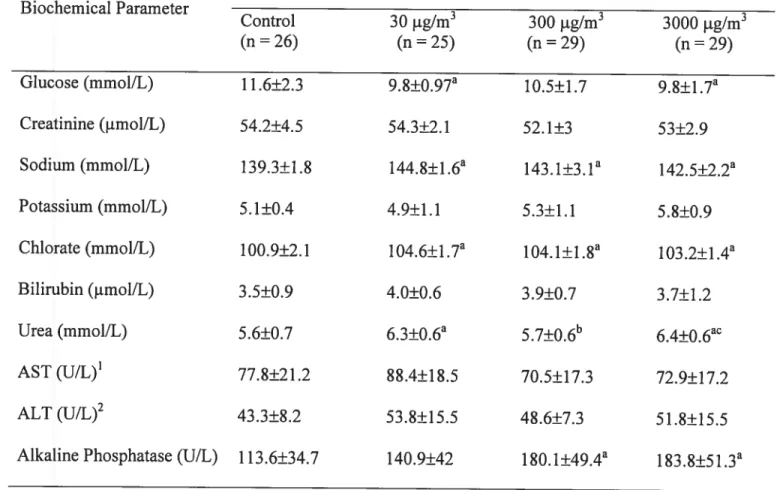
biochimiques sanguins ont révélé aussi quelques différences significatives entre les groupes, en particulier pour la phosphatase alcaline, l’urée, et le chlorate.
L’activité locomotrice était significativement plus élevée chez les groupes exposés à 30 jtg/m3 et à 3000 jig/m3. Chez ce dernier groupe, on observe aussi une perte significative de cellules neuronales dans les trois régions investiguées du cerveau. Par ailleurs, les résultats n’indiquent aucun signe de tremblement. En conclusion, l’exposition à un mélange de phosphate/sulfate de Mn entraîne des changements neuropathologiques au niveau du cerveau et des effets neurocomportementaux chez les rats.
Mots-clés: mélange de phosphate/sulfate de manganèse, rats Sprague-Dawley, exposition
Abstract
Methylcyclopentadienyl manganese tricarbonyl (MMT) is an organic manganese (Mn) compound added to unleaded gasoline in Canada. The primary combustion products of MMT are Mn phosphate, Mn sulfate, and Mn phosphate /sulfate mixture. Concems have been raised that the combustion products ofMMT could be neurotoxic, even at low levels
of exposure. The objective of this study is to assess and compare tissue concentration
following subchronic inhalation exposure to a mixture of Mn phosphate/sulfate particles in rats exposed at Ïow, intermediate and high levels. This research aims also to assess the neuropathology and neurobehavioral effects associated with exposure to the mixture.
A control group and three groups of 30 male Sprague-Dawley rats were exposed in inhalation chambers for a period of 13 weeks, 5 days per week, 6 hours a day. Exposure concentrations were 3000, 300, and 30 pg/m3. The neuropathology aspect of this study was to count neuronal celis in globus pallidus, caudate putamen, and frontal cortex. Also the neurobehavioral change was measured by assessing locomotor activity and tremor among groups. In each group, half of the rats were implanted with chronic EMG electrodes in the gastrocnemius muscle of the hind lamb for the purpose of measuring tremor. Tremor assessment was conducted one hour afier the last exposure by recording muscle EMG activity for approximately 5 minutes with implanted EMG electrode rats whule resting in the cage, walking on the floor, and standing on a vertical 39 cm metal screen grid.
At the end, locomotor activity test was conducted for 36 hours using a computerized autotrack system. Rats were then sacrificed by exsanguinations, and Mn concentration in different tissues (liver, lung, testis, and kidney), blood and brain (caudate putamen, giobus pallidus, oifactory buib, frontal cortex, and cerebellum) were determined by neutron activation analysis. Increased manganese concentrations were observed in blood, kidney, lung, testis, and in ail brain sections in the highest exposure group. Mn in the lung and in the oifactory bulb was dose-dependent. Our data indicate that the olfactory bulb accumulated more Mn than other brain regions following inhalation exposure. Biochemical profiles also reveaied some significant differences in certain parameters, specifically aikaline phosphatase, urea, and chlorate.
Locomotor activity was significantly increased at 30 and 3000 jig/m3. The neuronal cell loss was significantly different in ail three areas of interest for rats exposed to the highest level of exposure (3000 p.g/m3). The result does flot show any sign of tremor. In conclusion, exposure to Mn phosphate/sulfate mixture leads to onset the neuropathological changes in particular area of the brain and causes some neurobehaviorai differences among the rats.
Key words: Manganese phosphate/sulfate mixture, Sprague-DawÏey rats, inhalation exposure, neuropathology, neurobehavioral effect
Table of Contents
Résumé iii
Abstract y
List of Tables xi
List of Figures xii
Abbreviations xiii
Acknowledgements xiv
CHAPTER I: Introduction 1
1.1 MANGANESE 2
1.2 SOURCES AND LEVEL 0F ExPosuRs 3
1.3 T0xIc0KINETIc5 6 1.3.1 Absorption 6 1.3.2 Distribution 7 1.3.3 Metabolism 9 1.3.4 Excretion 10 1.4 HEALTH EFFECTS 11 1.4.1 Beneficial Effects 11 1.4.2 ToxicEffects 12 1.5 MECHANISMS 0F T0xIcITY 19
1.7 PuBLIc HEALTH CONCERN .22
1.8 SPECIFIC CONCERN RELATED TO MMT 24
1.9 OBJECTIVES 28
1.10 SPECIAL C0N5IDERATI0N IN TRIS STUDY 28
CHAPTER II: Bioaccumulation and Locomotor Effects of Manganese Phosphate / Sulfate Mixture in Sprague-Dawiey Rats Foilowing $ubchronic (90 Days) Inhalation
Exposure 31
2.1 ABSTRACT 33
2.2 INTRODUCTION 35
2.3 MATERIALS AND METHODS 37
2.3.1 Chemicals 37
2.3.2 Animais 37
2.3.3 Inhalation Exposure 38
2.3.4 Locomotor Activity Assessment 39
2.3.5 Tissue Concentrations 39
2.3.6 Chemicai Anaiysis 39
2.3.7 Biochemical Profile 40
2.3.8 Statistical Analysis 40
2.4 RESULTS 41
2.4.1 Mn in the Inhalation Chamber 41
2.4.2 Tissue Concentrations 41
2.4.3 Body Weight 42
2.4.5 Biood Biochemicai Analysis .42
2.5 DISCUSSION 43
2.6 ACKNOWLEDGMENTS 49
2.7 REFERENCE$ 50
CHAPTER III: Neurotoxicity and Neurobehaviorai Effects of Manganese Phosphate /
Sulfate Mixture on Sprague-Dawley Rats 61
3.1 ABSTRACT 63 3.2 INTRoDucTIoN 65 3.3 MATERIALS ANDMETHODS 67 3.3.1 Chemicals 67 3.3.2 Animais 67 3.3.3 Inhalation Exposure 68
3.3.4 Electrode Implant Procedures 68
3.3.5 Oxotremorine Administration 69
3.3.6 Tremor Assessment 69
3.3.7 Neuropathologicai Assessment 70
3.3.8 Statistical Anaiysis 70
3.4 RESULTS 71
3.4.1 Mn Concentrations in the Inhalation Chamber 71
3.4.2 Neuropathological Assessment 71
3.4.3 Tremor Assessment 71
3.7 REFERENCES .81
CHAPTER IV: Discussion .97
DISCUSSION 9$
CHAPTER V: Conclusion 106
CoNcLusioN 107
CHAPTER VI: BIBLIOGRAPHY 10$
REFERENCES 109
APPENUIX: Physical and Chemical Characterization of Mn Phosphate/Sulfate Mixture
List of Tables
Table 2-l: Particles size distribution of Mn phosphate!sulfate mixture in the inhalation
chamber 55
Table 2-2: Mean manganese concentrations (.ig/g ± SD) in different tissues and blood following subchronic (90 days) inhalation exposure to a manganese phosphate!
sulfate mixture 56
Table 2-3: Mean manganese concentrations (tg/g ± SD) in brain regions afier subchronic (90 days) inhalation exposure to manganese phosphate/sulfate mixture 57 Table 2-4: Mean distance traveled and resting time over 36 hour period after subchronic (90 days) inhalation exposure to manganese phosphate/sulfate mixture 58 Table 2-5: Biochemical parameters in serum following subchronic (90 days) inhalation exposure to a manganese phosphate!sulfate mixture 59 Table 3-1: Mean percentage of the maximum power of EMG signals in oxotremorine
injected rats 90
Table 3-2: Mean percentage of maximum power of EMG signal for rats in the walking
position at different levels of Mn exposure 91
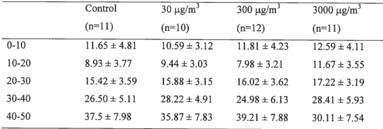
Table 3-3: Mean percentage of maximum power of EMG signal in rats in the resting
position at different levels of Mn exposure 92
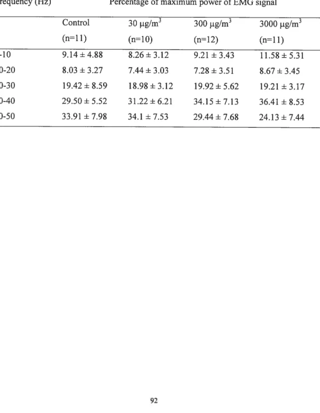
Table 3-4: Mean percentage of maximum power of EMG signal in rats when standing on a vertical grid for different levels of Mn exposure 93
List of Figures
Figure 2-1: Mean body weight of rats following subchronic (90 days) inhalation exposure
to a manganese phosphate/sulfate mixture 60
figure 3-l: Mean neuronal ceil counts in the giobus pailidus, caudate putamen, and frontal cortex of rats afler subchronic inhalation exposure of Mn (+ $D) 94 f igure 3-2: Tremor signais in oxotremorine injected rats. A shows a pattem of frequency (Hz) of EMG signais per time (ms). B reperesents rectified signals. C shows the
power (volt) of frequency of signais 95
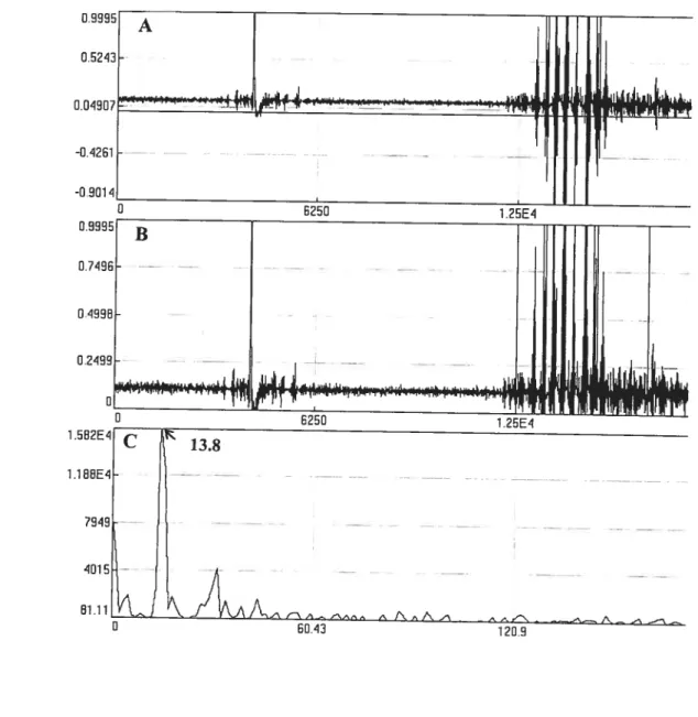
Figure 3-3: Typical EMG signals in rats exposed to Mn. A shows a pattern offrequency (Hz) of EMG signais per time (ms). B reperesents rectified signais. C shows the
Abbreviations
ACGIIET American Conference of Governmental Industrial Hygienists
ATSDR Agency for Toxic Substances and Disease Registry
EMG Electromyography
EPA Environmental Protection Agency
HSDB Hazardous Substances Data Bank
INAA Instrumental Neutron Activation Analysis
LOAEL Lowest Observed Adverse Effect Level
MMI Methylcyclopentadienyl Manganese Tricarbonyl
MRI Magnetic Resonance Imaging
NOAEL No Observed Adverse Effect Level
PCA Portacaval Anastomosis
RfC Reference Concentration
U.S.EPA U.S. Environmental Protection Agency
Acknowledgements
God knows that ail I have done was just his compassion and his faciiitate and I am so grateful for that.
This thesis is a symbol of collaboration efforts of many excellent people and I wouid like to express my genuine appreciation to these peopie particularly to my supervisors Dr. Joseph Zayed and Dr. Daniel Krewski for their numerous supports and supervision ail through this study.
Also, so many thanks to the several fantastic members of the department of Environmental and Occupational Health, especialiy Mrs. Linda-Ann Beaupré, and Lise Gareau for their assistance in this study.
Continuousiy, I am so thankful to Drs. Gaétan Carrier and Pbilip Gardiner who contributed to the achievement of this study.
Immeasurable thanks to my husband Mehdi and my kids Nazanin and Nivad for their love, support, patience and understanding during my study.
Finally, I would like to dedicate my thesis to rny father Mohammad and in memory of my mother, Tahereh who just biessed me plenty ftom far away.
1.1
Manganese
Manganese (Mn) is a heavy metal that is an essential element in the diet of humans and animais. Mn is widely distributed in the environment and can be found in rock, sou, air, water, and food (WHO, 1999). Mn plays an important role for biologicai functions including bone mineralization, metabolic regulation, celiular protection from damaging by ftee radical species, and the formation of glycosaminoglycans. Mn can be found as a component of over 100 minerais and does flot exist naturally in its pure form. It exists in various forms as silicates, carbonates, phosphates. sulfides, borates and oxides (ATSDR, 2000).
Mn compounds are produced from Mn ores and they have a variety of applications. Metallic Mn is used principally in steel production along with cast iron and superalloys to improve hardness, stiffness, and strength in the production of manganese-iron alloy (HSDB, 199$). Mn dioxide is commonly utilized in the production of dry-cell batteries, matches, fireworks, and glass-bonding materials (U. S. EPA, 1984). Mn chloride is employed as a precursor for other Mn compounds, as a catalyst in the chlorination of organic compounds, and in animal feed (HSDB, 199$). Mn sulfate is used primariÏy as a fertilizer, livestock supplement, ceramic, and fungicide. Potassium permanganate has been used as an oxidizing agent, disinfectant, antialgal agent, metal cleaning, water purifier, and a preservative for flowers and fruits (HSDB, 1998). Any ofthese uses may result in the release of Mn to the environment.
Mn compounds can exist as solids in the soi! and as solutes or small particles in water. Most of the Mn saits are soluble in water except the phosphate and the carbonate (WHO, 1999). Mn exists in 11 oxidative states and the most common valences are +2, +4, and + 7 while the most stable and biologically important valence state is +2 (ATSDR, 2000). Due to Iow vapor pressure, inorganic Mn compounds exist in the particu!ate matter (NICNAS, 2003).
1.2
Sources and Level ofExposure
food is the most significant source of Mn exposure in the general population. The total dietary Mn intake among individuals might vary depending upon nutritional intake. Most people have a daily intake with a reported range of 0.9 to 10 mg per day (Finley and Davis, 1999). Grains, tea, nuts, and green leafy vegetabÏes contain the highest amounts of Mn. The daily intakes of 1 to 5 mg Mn from food have been estimated for humans to stay healthy. The food and Nutrition Board has set an adequate intake level of Mn at 2.3 rng/day for men and 1.8 mg/day for women. The current recommendations for infants and children are 0.03 to 0.6 and 1.2 to 1.9 mg/day, respectively (food and Nutrition Board, 2002). The tolerabte upper intake level for Mn has been set at 11 mg/day, which is the highest level of daily intake that is like!y to cause no adverse health effects in the general population (f ood and Nutrition Board, 2002).
Mn represents about 0.1% of the earth’ s crust and it is a naturally occurring component of ail sous in a range of 2 to 7000 mg/kg. Voicanic eruption and soit erosion can release Mn
underground water at low levels ranging ftom 0.4 to 10 mg/L. The primary sources for surface and underground water Mn contamination are industriai facilities discharge, Iandfihl disposai sites, and sou ieaching. Mn levei in water can be as high as 2000 mg/L in areas close to industriai sources or waste disposai iands (ATSDR, 2000). Using potassium permanganate in drinking water treatment, industrial wastewater purification and odor abatement is another source of Mn release into water. The average Mn concentration in drinking water ranges from 0.02 to 0.15 mg/L (EPA, 2002).
In addition to natural existence, Mn can be introduced into the environment by human activity. The most important sources of Mn released into the air are industrial emissions associated with ferroaiioy production, iron and steel foundries, power piant, coke oven, and dust from uncontrolled mining operation (Lioy, 1983). Average Mn in urban and rural areas without significant pollution sources is in the range of 0.01 to 0.007 tg/m3 (WHO, 1999). U.S. EPA has established a mean Mn background concentration of 0.04 jig/rn3 for urban area. According to data summarized by EPA, $0 % of U.S. cities have Mn ieveis well beiow 0.1 tg/m3, and oniy 4.7 % have levels above 0.3 tg/m3. Higher ambient leveis, up to 5 or even 10 lg/m3 occur near industriai point sources, such as steel plants (EPA, 2002).
Another main source of inorganic Mn contamination in the urban environment is the combustion of methylcyclopentadienyi manganese tricarbonyl (MMT) a fuel additive used in unieaded automotive gasoline, particulariy in areas with high traffic density (Joselow et al., 1978). The average atmospheric Mn concentrations measured in a high
traffic area in Montreal were 0.05 and 0.024 tg!m3 for total and respirable (MnR) fractions respectively (Loranger and Zayed, 1997; Zayed et al., 1999c).
According to a recent study in Montreal, the average outdoor MnR concentration in the urban area (0.025 jig/m3) was significantly different compared to the rural area (0.005 tg/m3). Also, the indoor urban MnR concentration (0.017 .ig/m3) was significantly different from indoor concentration in rural areas (0.007 tg/m3). This study has also suggested that a higher outdoor atmospheric Mn concentration Ieads to higher indoor concentration (Boité et al., 2004). Besides, in specific environments, such as houses near highway, the atmospheric Mn concentration could exceed EPA’s reference concentration (0.05 jiglm3) (Zayed et al., 1999a).
Another source of exposure to Mn is at the workplaces. The occupational exposure to Mn is most likely occurring by inhalation of Mn fumes or dust. There is a potential for workers to be exposed to Mn in the industrial facilities. For instance, workers in the ferroalloy production sites, welding operations, and the dry battery manufacturing facilities are exposed to Mn concentrations ranging from 300 to 20000 tg/m3. It has been reported that the workers in mining operation are exposed to much higher Mn concentrations, up to 45000 1g/m3 (Gibbs et al., 1999). Mn levels in certain workplace environments such as garages are due to the use of MMI in gasoline. A study in Montreal showed that blue-collar workers are exposed to mean Mn levels of 0.04 tg/m3, while garage mechanics are exposed to 0.43 jig/m3 (Sierra et al., 1995).
1.3
Toxicokinetics
Mn absorption occurs primarily from the gastrointestinal tract afier ingestion and from the alveolar lining afier inhalation. The dermal absorption is flot a considerable route for inorganic Mn since they caimot penetrate through the skin. The dermal pathway is most likely important for absorption of organic Mn compounds such as maneb, mancozeb, and MMT. Age, chemical species, dose, dietary condition, and route of exposure ail have an effect on the extent and rate of Mn absorption and retention (Greger, 1999). Inhaled Mn may be absorbed more rapidly and to a greater extent than ingested Mn (Ijalve et al., 1996). The solubility of Mn species is another determinant of Mn absorption and distribution (Roels et al., 1997).
1.3.1 Absorption
The animal studies indicated that important determinants of Mn absorption are the route ofexposure and the chemical form of Mn (Smith et al., 1995; Tjilve et al., 1996; Roels et al., 1997; Anderson et al., 1999). Under normal conditions, only about 3-5 % of ingested Mn is absorbed by the gastrointestinal tract in the human body (f inley et al., 1994). The absorption of dietary Mn appears to be controlled by biliary excretion, intestinal absorption, and intestinal elimination. Homeostatic mechanisms control the absorption of Mn from the gut. There are many other factors that have been found to affect Mn absorption, including dietary Mn level, dietary levels of various minerais, age and developmental state of the individual and iron status (Greger, 1999). Low-protein and iron-deficiency increase the absorption, whereas increased calcium and phosphorus levels
The absorption of inhaled Mn is a function of particle size and its solubility. Particles that are deposited in the lower airway are mainly absorbed, while particles deposited in the upper airways may be moved by mucociliary transport to the throat, where they are swallowed and enter the stomach. Thus Mn may be absorbed both from the lungs and in the gastrointestinal tract following inhalation exposure (ATSDR, 2000).
Roels et al. (1997) measured Mn levels in blood following intranasal administration of 1.22 mg/kg as either MnC12 or Mn02 in rats. MnCl2, a soluble form, is quickly absorbed into the blood compared to the insoluble Mn02. Blood Mn concentrations were increased by 68% and 41 % following MnC12 and Mn02 exposure, respectively. Moreover, the soluble particles were more readily delivered to the brain than insoluble Mn compounds (Roels et al., 1997). Dorman et al. (2001) also reported that inhaled MnSO4 was cteared from lung more rapidly than the less soluble phosphate or tetroxide compounds.
1.3.2 Distribution
The highest concentrations of Mn in the body with no excessive Mn exposure are found in the liver, pancreas, adrenal gland, and kidney. The lowest concentrations are obsewed in fat and bone (ATSDR, 2000). It has been suggested that tissues rich in mitochondria may accumulate higher levels of Mn (Kato, 1963). Tjlve et al. (1996) investigated the uptake of Mn in brain regions of rats following intranasal administration of 4 tg/kg. The whole body autoradiography of rats at different time points indicated that the olfactory bulb contained the highest concentration of Mn at 1, 3, and 7 days post-dosing (90, 69,
uptake of Mn by other brain regions was flot observed until the third day, when the basal forebrain, cerebral cortex, hypothalamus, and striatum had 21, 2, 3, and 1 % of the labeled Mn, respectively. By contrast, liver and kidneys, each contained approximately 1 % ofmeasured Mn at 1, 3, and 7 days with values decreasing consistently after 12 weeks.
The absorbed Mn exists in divalent and trivalent states in the plasma ($cheumamer and Cherian 1983). Both forms can enter the central nervous system via the blood-brain barrier and the blood-cerebrospinal fluid barrier. Divalent Mn can be transported to brain capillary endothelial ceils and choroidal epithelial celis via undefined transporters (Takeda, 2003). Mn may also circumvent the bÏood-brain barrier and pass directly into the central nervous system via olfactory pathways (Tjalve et aï., 1996; frumkin and Solomon, 1997).
A large portion of divalent Mn may exist in non-protein-bound form as the free ion (May et al., 1997). This free ion can be transported into the brain via calcium channels (Narita et al., 1990) or tbrough Na and Ca exchange pathways (Frame et al., 1991). Trivalent Mn is able to connect to transferrin and pass through the brain barrier via receptor-mediated endocytosis. Mn entering the brain can combine with metalloproteins such as glutamine, which are synthesized in astrocytes (Takeda et al., 2003).
Pharmacokinetic factors that may contribute to the increased efficiency of brain Mn delivery following inhalation include greater Mn absorption from the lungs and slower clearance of absorbed Mn from the circulation (Andersen et al., 1999). Moreover, inhalation exposure to soluble forms of Mn resuits in higher brain Mn concentration
compared with insoluble form of Mn (Dorman et al., 2001). Another study has shown that afier intratracheal instillation, a surrogate for inhalation exposure, Mn concentrations were higher in brain for soluble MnCI2 than for the insoluble Mn02. Striatal Mn concentrations increased by 205% and 48% following MnC12 and Mn02 administration, respectively (Roels et al., 1997).
Absorbed Mn is transported to other organs via the iron-binding protein transfenin, Œ2-macroglobulin, and albumin. Manganese readily crosses the blood-brain barrier, and is largely distributed to the central nervous system (Aschner et al., 1999). Following chronic exposure to high level of Mn in humans and other primates, the metal preferentially accumulates in the thalamic nuclei, substantia nigra, pallidum, and other brain regions that also accumulate iron (Barbeau et al., 1984). Under normal plasma concentrations, Mn enters the brain mainly across the cerebral capillaries. As the plasma concentration increases, entry of Mn across the choroids plexus becomes more important (Murphy et al., 1991).
1.3.3 Metabolism
Mn as
a
metallic element does flot go undergo metabolic transformation. Mn hasa
potential to exist in
a
number of oxidation states in biological systems, and may transfer from Mn (II) to Mn (III) in the body (ATSDR, 2000). The Œ-globulin protein, ceruloplasmin, appears catalyze the alteration of Mn (II) to Mn (III) (Andersen et al., 1999). The high affinity of the iron-transporting protein transfenmn for Mn (III) probablywhich readily forms complexes with some organic and inorganic ligands, albumins, and proteins such as transfenin and a-macroglobulin (EPA, 2002). Mn also plays a role in enzymatic reactions with hydrolases, kinases, transferases, and decarboxylases (Keen et al., 1999; Lee, 2000).
1.3.4 Excretion
Several animal studies demonstrated that biliary/fecal excretion is the main route for Mn elimination (Thompson et al., 1982; Davis et al., 1993; Malecki et al., 1996). In addition, Mn might be excreted in the urine, pancreatic fluid, hair, sweat, and breast milk (WHO, 1981). Excretion of inorganic Mn from the body is rapid and does flot require active processes. Most of inorganic Mn rapidly appears in the bile and feces, with a portion undergoing enterohepatic recirculation.
Blood levels of Mn are increased in patients with impaired Mn clearance due to liver dysfunction (Hauser et al., 1996). The biological haif-time in blood ranges from about 12 days in healthy miners to approximately 40 days in healthy volunteers (Mahoney and $mall, 196$). Experiments in animais indicated that the elimination of Mn from the brain is much siower compared with the whoie body (Newiand et al., 1989). Cotzias et ai. (1968) demonstrated that the biologicai haif-time for intravenously injected Mn is 37.5 days in healthy subjects, 15 days in healthy miners, and 28 days in subjects with chronic Mn exposure.
Occupational exposure to high concentrations of Mn as well as hepatic failure appears to alter Mn distribution in the central nervous system (Layrargues et al., 1995). People with impaired clearance of Mn resulting from liver dysftmnction or portal systemic shunting show increase of Mn concentration in the brain. The average 70 kg man carnes a total body burden of about 12 to 20 mg of Mn. Iron status (assessed as serum concentration of sodium ferritin) may possibÏy affect Mn excretion. The biological haif-time of Mn for subjects with low ferritin was twice as of the rate determined for high ferritin status subjects (Finley, 1999).
1.4
Health Effects
Mn is an important constituent of normal diet for humans and animals. Thus, at low levels it has sorne beneficial effects while at higher levels of exposure via inhalation or ingestion, it can cause adverse health effects to pulmonary, reproductive and nervous systems (Komura and Sakamoto, 1991; Chang, 1996).
1.4.1 Beneficial Effects
Mn is a key element for normal physiological functioning in humans and all animal species (U.S. EPA, 1996). It plays an important role in bone mineralisation, protein and energy metabolism, metabolic regulation, celi protection from oxidative stress, and nervous system function (ATSDR, 2000). Mn is a regulatory and catalytic factor for enzymes including hydrolases, kinases, decarboxylases and transferases (Keen et al., 1999).
Animal studies have demonstrated that Mn deficiency associated with some physiological effects such as skeletal abnormalities, impaired growth, testicular degeneration, and impaired reproductive function (Strause, 1986). Rats with low Mn in their diet experienced more oxidation of mitochondrial membranes of the heart (Malecki and Greger, 1996), and ultrastructural changes in the retina (Gong and Amemiya, 1996). Mn deficiency has not been seen in the general population since human diet has relatively high content of Mn. According to a human study, Mn deficiency may decrease level of clotting proteins, serum cholesterol, retarded growth of hair and nails, and reddening of black hair (Friedman et al., 1987).
1.4.2 Toxic Effects
Mn dust in different forms can irritate lungs of humans and animais and result in an inflammatory response (Roels et al., 1987). There are significant adverse health effects induced by Mn particulate inhalation in workers such as impotence and loss of libido (AT$DR, 2000). The neurotoxic effects are primarily observed following chronic inhalation exposure to high Mn concentration (Mergler et al., 1994). Ingestion of contaminated well water (2 mg MnJL) has also been reported to induce neurological effects (Kondakis et al., 1989). Since the inhaled Mn bypasses the typical excretory mechanisms in the gastrointestinal tract it probably accumulates more in the brain and is more available to the nervous system and other target organs. Therefore, inhaled Mn may be more toxic than ingested Mn (Mirowitz and Westrich, 1992; ATSDR, 2000).
1.4.2.1 Single Exposure
Studies in animais and humans indicate that the inorganic Mn compounds have very low acute toxicity by any route of exposure. Acute inhalation exposure to high concentrations of Mn dust (> 0.97 mg/m3) can cause an inflammatory response in the lung, which can lead to impaired lung function in humans (Roels et al., 1987). This response however, is characteristic ofnearly ail inhalable particulate matter (EPA, 2002) and not specific to the Mn content of particles. Animal study has also revealed that acute inhalation exposure to Mn oxide at high concentrations (>40 mg/m3) can induce respiratory effects and increase susceptibiiity to pneumonia (Shiotsuka, 1984). The dermal pathway must also be considered during spillage of MMT in gasoline on the skin.
Following single oral exposure, LD50 ranged from 275 to 804 mg/kg body weight for Mn chloride in different rat strains. Also, LD50 from single exposure to Mn sulfate and Mn acetate in rats were 782 and 1082 mg/kg body weight, respectively (Singh and Junnarkar, 1991). Age possibly plays a role in susceptibility to acute Mn toxicity. In an animal study, it was found that Mn chioride induced greatest oral toxicity in the youngest than in the oidest (Kostial et al., 1978). Data on the irritant and contact sensitivity properties of Mn compounds are not available. The organic Mn compounds (MMT, mancozeb, and maneb) have been reported to be sensitizers in animais (Thomas et al., 1990).
1.4.2.2 Short-term Exposure
$hort-term inhalation exposure in animal studies shows that the lungs and nervous system are the major target organs (Rodier, 1955). Inhalation exposure to 69 mg/m3 Mn dioxide increased susceptibility to pneumonia (Maigetter et al., 1976). Subchronic inhalation studies in animais have yielded NOAELs or LOAELs values for systemic (0.7 to 3.9 mg/m3), neurological (1.1 to 72 mg/m3). and reproductive effects (61 mg/m3). The range of exposure levels associated with these effects is too wide to define a threshold. It has been demonstrated that subchronic oral exposure of rats to MnC12 and MnSO4 (> 22 mg/kg) might induce the neurological, reproductive, and systemic effects (Desole et al., 1994; Grant et al., 1997; Dorman et al., 2000). Systemic effects have been reported following subchronic oral exposure to Mn sulfate (40 mg/kg) and Mn chloride (11 mg/kg) including changes in blood celi counts, reduced liver weight, and decreased body weight in animais (Komura and Sakamoto, 1991).
1.4.2.3 Long-term Exposure
Available data from animal and epidemiological studies conceming the oral and the inhalation exposure have suggested that the nervous system is the most sensitive target for Mn (Mena et al., 1967; Wennberg et al., 1991). Chronic oral exposure to Mn (0.59 mg/L) in human has illustrated mild neuroÏogical signs (Kondakis et al., 1989). In contrast, animal studies have determined that the oral exposure to Mn chloride and sulfate may be associated with neurological (40 to 275 mg/kg) and reproductive effects (275 mg/kg) (Nachtman et al., 1986; Komura and Sakamoto, 1992). Chronic inhalation
exposure to high concentrations of Mn-containing dust can induce adverse neurological (0.027 to 5 mg/m3), respiratory (0.18 to 3.6 mg/m3), and reproductive (0.97 to 82.3 mg/m3) effects in humans (Iregren, 1990; Roles, et al., 1992; Wu et al., 1996).
1.4.2.3.1 Neurotoxicity
Occupational studies of miners, industrial and agricultural workers have established that the lungs and brain are the primary target organs following chronic exposure to Mn. The nervous system is the critical target organ of Mn toxicity. Long-term exposure to Mn in occupational setting can resuit in a progressive neurological dysfunction, which can produce a disabling syndrome referred to as manganism. Progression of manganism is related to the dose and duration of exposure, as well as individual susceptibilities. The striatum and associated brain structures are important target sites of Mn neurotoxicity in humans (Came et al., 1994).
The clinical symptoms of manganism occur in three stages. The initial stage is subjective and nonspecffic such as anorexia, apathy, asthenia, headaches, bad temper, and weakness in the lower extremities. Gradually, an intermediate phase of developing neurological signs associated with speech disturbance, facial expression, alterations of gait, loss of balance, and fine tremor sets in. The final phase is characterized by persistent, ofien irreversible neurological deficits characterized by muscle rigidity, a staggering gait, dysphasia and a course intention tremor. Because some of these symptoms resemble those of Parkinson’s disease, many investigators have used terms such as “Parkinsonism
Although, symptoms of manganism look like those of Parkinson’s disease significant differences have been noted. The tremor and hypokinesia present in patients suffering from manganism are different from those seen in Parkinson’s disease (Barbeau, 1984). Also psychiatric disturbances early in the disease such as “cock walk” and a propensity to fali backward, less frequent resting tremor, more dystonia, and failure to respond to dopaminomimetics are characteristics of manganism (Came et al., 1994; Olanow et al., 2004).
There are some alterations in pathological findings between manganism and Parkinson’s disease. Lesions in manganism are more distributed in the pallidum, the caudate nucleus, the putamen, and even the cortex. In contrast, in Parkinson’s disease lesions are found in the substantia nigra and other pigmented areas (Came et al., 1994). Using brain magnetic resonance imaging (MRI) techniques, researchers have demonstrated that manganism patients have elevated Mn concentrations in their basal ganglia, which has not been seen in patients with Parkinson’s disease (Nagatomo et al., 1999). Mn can induce high signal intensity on T1-weighted MRI that is related to its chemical property. The abnormality of MRI signais has been reported in workers exposed to high concentrations of Mn (41.4%). However, measurement obtained with this technique is qualitative rather than quantitative (Kim, 2004).
More specific clinical signs of basal ganglia dysfunction characterize a slow or halting speech without tone or inflection, a dull and emotionless facial expression, altered gait, and fine tremor (Hochberg et al., 1996; Mergler and Baldwin, 1997). Severely affected
manganism patients also develop progressive, irreversible loss of dopaminergic neurons in globus pallidus and nigrostriatal pathway (Came et al., 1994).
Animal studies also revealed that chronic Mn exposure by the oral route could develop muscular weakness and rigidity of the lower limbs. Oral exposure to Mn chloride in rodents increases spontaneous locomotor activity, rigid and unsteady gait (Chandra et al., 1983; Nachtman et ai.. 1986; Kristensson et al., 1986; Ah et al., 1995). The neurological effects have been reported at lower airborne Mn concentrations in humans than in animais (Newiand and Weiss, 1992). Although, there are dissimilarities in the variety of neurological responses between animais and humans, it can be suggested that animal modeis, mainly rodents, are likely useful for determining quantitative dose-response relationships for neurological effects.
Occupational studies indicated that neurological symptoms were more significantly associated with exposure to airbome Mn between 0.2 to 5 mg 1m3 for more than 20 years (Crump and Rousseau, 1999; Gorehi et al., 1999). The alteration ofmotor effects has been evidentiy attributed to an abnormal basal ganglia function (Huang et al., 1998). The neuropathological feature of these symptoms includes neuronal loss in basal ganglia (Jog et al., 1995).
1.4.2.3.2 Puimonary toxicity
include bronchitis, cough, pneumonitis, and reduction in lung function (Roels et al., 1987; Abdel-Hamid, 1990; Akbar-Khanzadeh, 1993). Increases in respiratory symptoms, pneumonia and bronchitis have been reported among workers with occupational exposure and in people living near manganese alloy production facilities (WHO, 1999). One study has suggested an increase in prevalence of respiratory ilinesses in school chiidren residing near a point source of atmospheric Mn pollution (Nogawa et al., 1973). A higher incidence of pneumonia was observed arnong people living close to ferromanganese factory as well as factory worker (Tanaka, 1994). The increased susceptibility to respiratory infection might be related to the lung irritation and inflammation caused by inhaled particulate Mn.
1.4.2.3.3 Reproductive and Developmental Toxicity
Limited data are available on developmental toxicity of Mn following oral exposures. Several studies have reported developmental effects in animal models with orally administered Mn. These studies show a decrease in serum testosterone, reduced fetal body weight, and malformations in pups (Laskey et al., 1985; $zkmary et al., 1995; Dorman et al., 2000). Male mice exposed to elevated dietary Mn (< 20 mg/kg) also show decreased size of the testicles, seminal vesicles, and preputial glands, even without frank neurological signs such as tremor and ataxia (Gray and Laskey, 1980).
Some inhalation data from occupational studies revealed that male reproductive dysfunction is a major adverse health effect of Mn toxicity. The symptoms include loss of
of chiidren have been associated with male workers exposed to Mn dust for many years (Laskey et al., 1985). A human study in an isolated island population with high Mn intake, found an apparent excess of fetal malformations (Kilbum, 1987). Impaired fertility has been observed in male workers exposed for 1-19 years to Mn dust at levels (0.97 mg/rn3) that did flot produce any neurological effects (Lauwerys et al., 1985). No information was found regarding reproductive effect in women.
1.5
Mechanisms ofToxicity
The mechanism of Mn-induced adverse effects has been studied for decades, but it is not clearly established. Several hypotheses have been advanced to explain the relation between Mn exposure and its neurotoxicity. It is clear that the main portais of Mn absorption are the gastrointestinal tract and the lung (Aschner et al., 1991).
Absorbed Mn exists as divalent and trivalent Mn in plasma (Scheumamer and Cherian 1985). Both forms may distribute to the central nervous system via the blood-brain and the blood-cerebrospinal ftuid barriers (Takeda, 2003). Mn may also circumvent ifie blood brain barrier, and pass directly into the central nervous system via olfactory pathways (Tjilve et al. 1996; Frumkin and Solomon, 1997). The olfactory bulb in rats can play a significant role in the uptake of inhaled Mn and transport to brain (Tjiive et ai., 1999). However, this route of Mn delivery to the brain is not clear in humans.
non-protein-bound forms as free ion (May et al., 1997). This free ion can be transported into the brain via calcium channels (Narita et al., 1990) or through Na and Ca exchange pathways (Frame et al., 1991). Trivalent Mn is able to bind to transferrin and be transported across the brain barrier via the receptor-mediated endocytosis (Takeda et al., 2003).
When Mn enters the brain, it can bind to Mn metalloproteins such as glutamine, which are synthesized in astrocytes. Several studies have documented that increased Mn concentrations in the brain may alter the level and metabolism of neurotransmitters such as dopamine, y-aminobutyric acid, and glutamate (Erikson et al., 2003). Glutamate is the most common excitatory neurotransmitter, while -y-aminobutyric acid is the most abundant inhibitory neurotransmitter. These neurotransmitters are responsible for the control of motor behaviors (Carlsson, 1990). Astrocytes may play a key rote in Mn induced neurological effects in the brain (Hazeli, 2002). Hazeil (2002) suggested that the involvement of energy metabolism of mitochondria in astrocytes could cause the Mn induced neuropathology.
Other studies have suggested that glutamate-mediated excitotoxicity may be responsible for the neurological effects of Mn in the brain (Brouillet et al., 1993). HazelI and Norenberg (1997) have studied the oxidant capacity of Mn in reduction of glutamate uptake. In a further study, they found that increased Mn concentrations in the basal ganglia could lead to oxidative damage in astrocytes, with a concomitant reduction of glutamate uptake (Hazeil and Norenberg, 199$). Mn accumulation in the brain may therefore cause major changes in astrocytes, including: reduction of glutamate uptake,
reduction in mitochondria energy metabolism in astrocytes, increased excitotoxicity, and proliferation of astrocytes. These effects may play a key role in impaired astrocytic neuronal interaction, which resuits in neuronal celi death (Hazeli, 2002).
OveralÏ, brain Mn deposition is region-selective, being predominant in the basal ganglia, which is involved in the control of movement arid of some cognitive functions. Acute exposure to Mn is associated with an increase in dopamine neurotransmission, which is also manifested as hyperactivity. Nevertheless, long-term exposure results in a loss of dopamine in the brain, and the neuronal celi damage could be expressed as an increase in motor activity or variation in neurobehavioral effects (Bonilla, 1984; Nachtman et al.,
1986).
1.6
Sensitive Population
Sensitive populations are defined as those that demonstrate an enhanced reaction to a chemical compared with other exposed population. Sensitivity may be attributed to many factors, for instance genetic, age, developmental stage, life style, and health status. Infants and children with developing organs as well as the elderly with declining organ function are expected to be more sensitive to Mn toxicity than healthy aduits. Mn is excreted more rapidly in adults than in children (Fechter, 1999). Also there is evidence of increased sensitivity for neurotoxic effects following Mn exposure in neonatal rats compared to adult rats (Dorman et al., 2000).
U.S. EPA (1996) has identified other sensitive subpopulations for Mn. These are groups who may have greater potential for increased body burden due to increased absorption or different excretion mechanisrn. However, pregnant women, elderly, iron or calcium deficient individuals, and people with impaired liver function are considered as sensitive population (Davis and Elias, 1996).
Patients with chronic liver disease could be more vulnerable to the neurotoxic effects of Mn while they have a decrease in biliary excretion of Mn. In a study, portacaval anastomosis (PCA) rats flot exposed to Mn, Mn content was increased in globus pailidus by 57% and in caudate putamen by 67% compared to a control group (Therrien et al., 1995). The relative influence of portal-systemic shunting and cholestasis on Mn accumulation and neuronal cell loss in the brain was determined in rats with chronic liver failure (Rose et al., 1999; Salehi et al., 2001). These studies indicated that compromised Mn clearance might influence Mn bioaccumulation and Mn neurotoxicity. It has been hypothesised that a relatively high exposure to Mn may be tolerated by normal animais and humans but may lead to an overload and neurological problems in those with hepatic dysfunction.
1.7
Public Health Concern
Mn toxicity is a well-recognized occupational hazard in human following exposure to high concentrations of respirable Mn dust. Respiratory symptoms such as cough, impaired pulmonary function, bronchitis, and pneumonitis (Roels et al., 199$) are related to respirable Mn. Although the respiratory effects are important, the greatest public health
concem is the neurotoxic effects such as manganism. Exposure to concentrations between 0.027 to 1 mg MnIm3 among miners and other industrial workers has been shown to provoke adverse respiratory, neurological, and reproductive effects (Iregren, 1990; Mergier et al., 1994; Lucchini et al., 1995).
The U.S. EPA has determined the potential health risks following exposure to Mn by setting an inhalation reference concentration. The RfC for chronic inhalation exposure was derived by using data ftom two epidemiological occupational studies (Roels et al., 1987; Roels et al., 1992). The significant effect was destruction of neurobehavioral function, as assessed by medical questionnaire, audio-verbal short-term memory, visual reaction time, eye-hand coordination, and hand steadiness. According to Roels et al. (1992), the LOAEL was identified to be 0.15 mg/m3 and the RfC was calculated by dividing the LOAEL with a total uncertainty factor of i&, allowing 10 for each of the following factors: 1) potentially greater susceptibility of the elderly and chiidren, 2) extrapolation of LOAEL to NOAEL, 3) for chronic duration. Then the resuit was divided by a factor of 3 for converting workday exposure (8 hr) to continuous exposure (24 hr), which yielded 0.05 tg/m3 (U.S. EPA, 1996). This RfC is an estirnate ofa daily exposure to human population that is likely to be without a signfficant risk during lifetime.
Presently, the public health concem of airbome Mn is increased due to the replacement of lead with MMT. The use of MMI as a gasoline additive has led to an increase of inorganic Mn, mainly Mn phosphate/sulfate mixture, Mn phosphate, and Mn sulfate in
the atmosphere. Ihus, increased level of Mn in the air could lead to potential health risks associated with chronic exposure.
Consequently, susceptible populations, such as children, individuals with chronic liver disease, people with sub-optimal Mn or iron intake, and those with other medical states (e.g., pre-parkinsonian state, aging) may have altered Mn metabolism and could be at greater risk for Mn toxicity (Pujol et al., 1993; Spahr et al., 1996; Malecki et al., 1999).
Although, human data are most useful for the assessment of potential health hazards such data conceming the long-term and low-level inhalation exposure to Mn species related to MMI combustion products are not available. Consequently, to provide adequate toxicological information on Mn species for risk assessment purposes, it is necessary to develop dose-response data using animal models.
1.8
Specific Concern Related to MMI
MMI (C9H7MnO3) was developed by the Ethyl Corporation in the 1950s. MMI is an organometallic compound used as an antiknock agent in intemal combustion engine fuels to increase the octane level of gasoline and therefore to improve the antiknock properties ofthe fuel (Davis, 199$). MMI has been used in the United States since 1976. Although in 1977, the adoption of the Clean Air Act limited the use of MMT to gasoline and U.S. EPA denied Ethyl’s waiver for the use of MMT until October 1995, when a court decision allowed the company to offer it for sale to refiners for use in unleaded gasoline
and since that time MMT has been used freely (Wallace and Slonecker 1997, Landrigan, 2001).
The use of MMI in gasoline has been permitted in Canada since 1978 (Health and Welfare Canada, 1978). Its use increased steadily until it completely replaced tetraethyl lead in gasoline in 1990 (Hurley et al., 1992). Since 1997, trade of MMT was limited in Canada under the Mn-based Fuel Additive Act that effectively banned the importation of MMI into Canada. Eventually in 1998, the Govemment of Canada had removed restriction and continues to use it (Davis, 1998).
MMI is allowed to use in France (Ministre de L’Aménagement du Territoire et de L’Environnement, 1999), UK (British Standards Institute, 1999), China (China State Bureau of Quality and Technology Supervision, 2000), Russia (Ministiy of Fuel and Energy of Russian Federation, 1997), and Argentina (Norma Argentina, 1999). In New Zealand, according to the Petroleum Products Specification Regulations 2002, automotive fuel must contain no more than 2.0 mg MnIL.
Usually, 72 mg MMT/L is used in gasoline that corresponds to 1$ mg MnIL (Garrison et al., 1995). The maximum concentration of MMT allowed by the U.S. EPA is 8.3 mg MnJL of gasoline. According to Health Canada, MMI is currently used in 80% of Canadian gasoline at various concentrations and the mean concentration is approximately 10 mg MnIL (Wood and Egyed, 1995). However, Zayed and colleagues have found that
the MMT-added gasoline in Canada has a mean concentration of 6.5 mg MnJL (Zayed et al., 1999c).
MMT is marketed as AK-33X (antiknock agent-33X) or Hilec 3062 (Jaques, 1987). It is a yellow, volatile liquid that has a very low vapor pressure at room temperature (0.05 mmHg
@
20°C) and is completely miscible in gasoline and very slightly soluble in water. MMT is extremely unstable in light and degrades rapidly in air; thus, exposure via gasoline exhausts is expected to be more to the oxidative products such as Mn oxides, phosphate, and sulfate rater than to the parent compound (Garrison et al., 1995; Ressier, 2000; Molders et al., 2001; NICNAS, 2003).Generally, the combustion products of MMT depend on fuel composition, engine and catalytic converter thermodynamics. The actual portion of Mn emitted is also very variable and depends on various factors including, overail distance driven and driving condition (urban vs. highway) (Ardeleanu et al., 1999).
More recently, samples of particles emitted from several cars that has accumulated significant mileage on MMT-additive gasoline determined that there were tbree maj or Mn compounds including a mixture of Mn phosphate, Mn5(P04)2[P03-(OH)]2.4H20, MnSO4.H20, and Mn304 (Ressler et al., 2000; Molder et al., 2001). The ratio for each compound is dependent on the driving cycle and vehicle, however the percentage of combusted product stays relatively constant with the 80% of Mn phosphate and sulfate and less then 20 % of Mn oxide (Pfeifer et al., 2004).
It is also recognized that most ofthe emitted Mn in the form ofphosphate/sulfate mixture in the tailpipe has particle sizes ranging between 0.2 and 10 im (Zayed et al., 1994; Zayed et al., 1 999b). More than 99 ¾ of Mn particles are in the respirable fraction (< 5 im) and 86 % are smaller than 1 im (Ardeleanu et al., 1999; Zayed et al., 1999b). In conclusion, Mn particles emitted from MMT are more likely to reach the alveolar region. Many studies have established a positive relation between traffic density and Mn concentrations in the biotic and abiotic components of ecosystem (Loranger et al., 1996; Zayed et aÏ., 2003). Ardeleanu suggested that motor vehicles would add 350 tons of Mn into the Canadian environment each year (Ardeleanu et al., 1999). The U.S. EPA (1994) estimated that if MMT used in gasoline increases by 100%, the level of Mn in ambient air will increase to 0.1 tg/m3, way more than the RfC.
Nevertheless, exposure of the general population to Mn originating ftom MMT seems to be negligible compared to the contribution of the Mn from other industrial and natural sources (Loranger and Zayed, 1997). However, experience with lead in gasoline demonstrated that significant widespread exposure of the general population might occur by adding relatively small amounts of a substance to gasoline (Davis et al., 1996). To determine the risk posed to general population by inhaled Mn species, it is required to have more information about the potential toxicological hazards and the accepted levels of exposure.
1.9
Objectives
The main hypothesis in this study is that the exposure to Mn phosphate/sulfate mixture may resuit in different bioaccumulation, neuropathological, and neurobehavioral changes that could be dose-dependent. The objectives of this study are to evaluate the effects of Mn phosphate/sulfate mixture on $prague-Dawley rats at low (30 jiglm3), intermediate (300 j.tg/m3) and high levels (3000 jig/m3) on:
the deposition and the bioaccumulation of Mn in different tissues and organs, > the blood biochemical parameters,
the neurobehavioral status (locomotor activity and muscular tremor), and > the neuropathology effect in the brain.
The resuits of this study may correspond to another step forward to better understand the relationship between exposure and nervous system affects for Mn compounds resulting from the use of MMI in gasoline.
1.10 Special Consideration in this Study
To date, studies to evaluate the neurotoxic effects following inhalation exposure to Mn phosphate/sulfate mixture have not been undertaken. This mixture is the primary combustion product of MMT, receiving increased attention in relation to environmental contamination and public health. This study is unique in that it is the first study to investigate the adverse effects of the Mn phosphate/sulfate mixture following inhalation,
which is the main route of exposure associated with neurotoxicity of Mn in humans and animais.
In this study, equal weights of Mn phosphate and Mn sulfate were introduced into the inhalation chamber. However, the subsequent analysis of air samples by electron microscopy showed that 39 % of the particles were Mn sulfate and 61 % were Mn phosphate. This ratio is acceptable since it is near the ideal ratio of 1:1. Prior to the experiment, the molecular weight (MW) of each substance was taken into account to maximize the probability of obtaining a ratio of 50:50 for particles in the inhalation chamber. Since the MW was 728.65 and 169.03 for Mn phosphate and Mn sulfate respectively, representing a ratio of approximately 4:1, the inverse ratio was used for w/w (4 Mn sulfate / 1 Mn phosphate). Nevertheless, the proportion of particles for each Mn species was far away from the target of 1:1. In fact, more than $0 % of the particles were related to Mn sulfate compound. The details can be found in the report of Beaupré et al. (2004) in the Appendix ofthis thesis.
The exposure level of 3000 ig/m3 was derived from the inhalation study of Coulston and Griffin (1977) that used 100 jtg/m3 of Mn compounds for 24 br. Thus, 3000 j.ig/m3 was derived by multiplying 100 jtg/m3 with 3 (24/8) and a correction factor of 10 in consideration of exposure duration. b help adequately evaluate the dose-response relationship in subchronic study, this level of exposure was divided by a factor of 10 sequentially, which yielded 300 and 30 tg/m3.
Human exposure to high levels of Mn in mining and occupational exposure has been reported to induce neurobehavioral and neuropathological effects. Experimental studies in rodent and other anina1 species have been carried out to produce these effects. Rats seem to be a usefu! mode! to study neurotoxicity effects of Ivin and its compounds (Russeli,
2
CHAPTER II: Bioaccumulation and Locomotor Effects of
Manganese Pliosphate/Sulfate Mixture in Sprague-Dawley Rats
Following Subchronic (90 Days) Inhalation Exposure
Bioaccumulatïon and Locomotor Effects of Manganese Phosphate/Sulfate Mixture in Sprague-Dawley Rats Following Subchronic (90 Days) Inhalation Exposure
Fariba $alehi”2, Daniel Krewski2, Donna Mergler3, Louise Normandin’, Greg Kennedy1, Suzanne Philippe1, Joseph Zayed’
‘TOXHUM (Human Toxicology Research Group) and Departrnent of Environmental and Occupational Health, facuhy ofMedicine, University ofMontreal
2McLaughuin Center for Population Health Risk Assessment, University ofOffawa
pour l’étude des interactions biologiques entre la santé et l’environnrnent (CINBIOSE)
Running titÏe: Manganese bioaccumulationand locomotor activity
To whom requests forreprints should be sent: Joseph Zayed, Ph.D.
Department of Environmental and Occupational Health faculty of Medicine Université de Montréal C.P. 612$ Succursale Centre-Ville, Montréal, Québec Canada, H3C 3J7 Telephone: (514) 343-5912 Fax: (514) 343-2200 E-mail: joseph.zayed@umontreal.ca
2.1
Abstract
Methylcyclopentadienyl manganese tricarbonyl (MMT) is an organic manganese (Mn) compound added to unleaded gasoline in Canada. Ihe primary combustion products cf MMI are Mn phosphate, Mn sulfate, and a Mn phosphate /sulfate mixture. Concems have been raised that the combustion products of MMT containing Mn could be neurotoxic, even at low levels cf exposure. The objective cf this study is to investigate exposure response relationships for bioaccumulation and locomotor effects following subchronic inhalation exposure to a mixture cf manganese phosphates/sulfate mixture. A control group and three groups cf 30 male $prague-Dawley rats were exposed in inhalation chambers for a period cf 13 weeks, 5 days per week, 6 hours a day. Exposure concentrations were 3000, 300, and 30 1g/m3. At the end cf the exposure period, locomotor activity and resting time tests were conducted for 36 h using a computerized autotrack system. Rats were then sacrificed by exsanguination and Mn concentrations in different tissues (liver, lung, testis, and kidney), blood and brain (caudate putamen, globus pallidus, olfactory bulb, frontal cortex, and cerebellum) were determined by neutron activation analysis. Increased manganese concentrations were observed in blood, kidney, lung, testis, and in all brain sections in the highest exposure group. Mn in the lung and in the olfactory bulb was dose-dependent. Our data indicate that the olfactory bulb accumulated more Mn than other brain regions following inhalation exposure. Locomotor activity was increased at 30 and 3000 j.ig/m3, but no difference was observed in resting time among the exposed groups.
At the end of the experiment, rats exposed to 300 and 3000 jig/m3 exhibited significantly decreased body weight in comparison with the control group. Biochemical profiles also revealed some significant differences in certain parameters, specifically aikaline phosphatase, urea, and chlorate. This study is one of several ongoing studies in our laboratory that address the toxicity of different Mn species.
Keywords: Manganese phosphate/sulfate mixture, inhalation exposure, Sprague-Dawley rats, bioaccumulation, locomotor activity
2.2
Introduction
Methylcyclopentadienyl manganese tricarbonyl (MMT) is one of the main sources of inorganic manganese contamination in urban air, mainly in areas with high traffic density (Joselow et al. 1978). The main combustion products of MMT are essentially Mn phosphate, Mn-sulfate, and Mn-phosphate/sulfate mixture (Zayed et al., 1999). Exposure to high concentrations of atmospheric Mn can lead to adverse health outcomes, notably respiratory and neurological effects. Exposure to concentrations >1 mg Mnm3 among miners and other industrial workers has been shown to persuade adverse respiratory, neurological, and reproductive effects (Iregren, 1999).
The clinical syndrome of manganese neurotoxicity (manganism) can be divided into an early phase characterized by obvious mood and behavior changes, and a later stage somewhat similar to Parkinson’s disease that is characterized by dystonia and severe gait disorder (Pal et al., 1999). However, little is known about the potential health effects that may result from long-term low-level exposure of populations through ambient air. Certain subpopulations such as children and patients with chronic liver disease could be more susceptible to different levels of Mn contamination.
It is clear that the route of exposure can influence the distribution, metabolism, and potential for neurotoxicity of Mn-containing compounds (Roels et al., 1997). Inhalation exposure is more efficient than ingestion at transporting Mn to the brain. Pharmacokinetic factors that may contribute to the increased efficiency of brain Mn
clearance of absorbed Mn from the circulation (Andersen et al., 1999). Moreover, inhalation exposure to soluble forms of Mn results in higher brain Mn concentration compared with insoluble form of Mn (Dorman et al., 2001). One study has shown that after intratracheal instillation, a surrogate for inhalation exposure, Mn concentrations were higher in brain following the administration of the soluble salt MnCl2, than following the administration of the insoluble oxide Mn02. $triatal Mn concentrations increased by 205% and 48% following MnC12 and Mn02 administration, respectively (Roels et al., 1997).
The main brain target for Mn toxicity is the basal ganglia (caudate nucleus, globus pallidus, and putamen), which is involved in motricity. Disturbances of the basal ganglia can lead to unintentional contraction of the skeletal muscles, such as tremor and muscular rigidity, as in Parkinson’s disease. f ew studies have been conducted to describe the distribution of brain Mn following inhalation of different Mn species, the main route by which Mn intoxication occurs in workers. It seems likely that the neurotoxicity of inhaled manganese possibly be related to an uptake of this metal into the brain via olfactory neurons. The olfactory bulb in rats can make a significant role to uptake of inhaled manganese and then delivery to the brain (Tjilve et al., 1999). However, this route of delivery of manganese to the brain is not well clear in human. The primary objective of this study is to determine the effects of subchronic exposure to Mn phosphate/sulfate mixture on Mn tissues concentrations, and locomotor activity.
2.3
Materials and Methods
2.3.1 Chemicats
The manganese phosphate/sulfate mixture, a fine crystalline powder which includes Mn5(P04)2(P03(OH))24H20 hureaulite minerai form, and manganese (II) sulfate monohydrate (MnSO4. I-120), were obtained from Alfa Aesar (Johnson Matthey Company), and combined in a 50:50 ratio. Whereas manganese sulfate is relatively water soluble, manganese phosphate is insoluble.
2.3.2
AnimaIs
One hundred and twenty, 6 week-old male Sprague-Dawley rats weighting 125-150 g were purchased from Charles River Laboratories (St.Constant, Quebec, Canada) and marked on the tail for identification purposes. Rats were acclimated for approximately 2 weeks in HEPA filtered air. Animais were given NIH-07 chow ad libitum when they were not exposed. All experiments were undertaken with the consent ofthe animal ethics committee of the University of Montreal, and were conducted in accordance with the guidelines set out by the Canadian Council on Animal Care. Rats were individually housed in a polycarbonate cage with stainless steel wire lids under constant conditions of temperature and humidity, with 12:12 h day/night cycles. The subjects were randomly divided into 4 groups with 30 animals in each group for two different periods. Body weight and food and water consumption were measured weekly.
2.3.3 Inhalation Exposure
A control group and three groups of 30 male Sprague-Dawley rats each were respectively exposed to 3000, 300 and 30 jig/m3 manganese phosphate/sulfate mixture. Exposure was conducted in two inhalation chambers with a total volume of 1 m3 each (Hazelton Systems Company, Inc., Kalamazoo, MI) over 13 consecutive weeks, 5 days per week, 6 hr/day. The chamber received (HEPA) filtered air and was maintained at a constant temperature (22-25 °C) and relative humidity (25-40%) throughout the study. Mn aerosol was generated by a fluidized Bed Aerosol Generator (Model 3400, TSI Inc., $t. Paul, MN) with a flow rate of 200-250 L/min. Stainless steel A$ME pressure tanks was used in order to obtain fine particles.
Concentrations were verified continuously using a Dust Track (Model $520) aerosol monitor. In both chambers, air samples were collected on a daily basis to monitor Mn concentrations, using a sampling system consisting of Gilian pumps (Gilian Corp., West Caldwell, NJ) with standard 3-piece cassettes and 37-mm diameter filters. The filters were Teflon (manufactured for SKC Inc., Gelman Sciences, Ann Arbor, MI) 0.45 tm pores. Pumps were used at a constant flow rate of 1.5 L/min. The flow rate was calibrated each day with a Gilibrator (Gilian Corp., West Caldwell, NJ). The particle size distribution of manganese phosphate! sulfate mixture was measured by using a six-stage Marple Personal Cascade impactor (Series 290). Rats were individually housed during the non-exposure period and were weighed weekly. Food and water were available ad libitum when the rats were flot exposed.
2.3.4 Locomotor Activity Assessment
four hours after the last Mn inhalation exposure, the rats were tested for motor activity. A computerized Auto-Track System (Columbus instruments, Ohio, U.S.A.) was used to measure locomotor activity for a period of 36 consecutive hours. This system is consisted of a 15 by 15 inftared beam array with an interbeam distance of 2.4 cm along the X and Y-axes. Data was collected every 0.1 second and the motor activity categorized as distance traveled by rats and resting time. The system was installed in a quiet isolated room with 12 h light / 12 h dark cycles.
2.3.5 Tissue Concentrations
Following locomotor activity assessment, rats were anesthetized in a glass-walled container containing a pledget wet with metofane, an inhalation anesthetic. As soon as the animals were anesthetized, they were sacrificed by exsanguinations and the following tissues and organs weighed and analyzed: liver, lung, kidneys, testis, and one hemisphere of the brain. (olfactory bulb, globus pallidus, caudate/putamen, frontal cortex, and cerebellum). Blood samples were taken from the abdominal aorta for chemical and biochemical analyses and determination of Mn concentrations.
2.3.6 Chemical Anatysis
Manganese concentrations in blood, food, water, and tissues were measured by fNAA (Kennedy, 1990) using a flue nuclear reactor and an EG&G ORTEC (mode! DSPec) digital gamma-ray spectrometer incorporating a high-resolution large-volume germanium
2.3.7 Biochemicat Profite
Blood samples were colÏected from the abdominal aorta of anesthetized rats. Blood serum was obtained by centrifugation at 4°C for 15 minutes at 3000 g. The biochemical tests were performed using standard techniques for glucose, urea nitrogen, serum creatinine, bilirubin, transaminases (AST, ALT), and alkaline phosphatases (ALP).
2.3.8 Statisticat Anatysis
The Dunnet T3 test was used to compare among ail pair treatment groups for mean concentrations of Mn in blood, kidney, liver, lung and testis. A simiiar analysis was conducted for Mn concentrations in different regions of the brain (cerebellum, frontal cortex, globus pallidus, caudate putamen, and oifactory bulb), motricity (distance travelled and resting time), the ten serum biochemicai parameters, and body weight in each of the four treatment groups. Homogeneity of variances for the treatment groups was assessed using Levene’s test. All analyses were performed using SPSS statistical software (version 11.0). The level of statisticai significance was set at P< 0.05 for the set of 6 pairwise comparisons performed among the treatment groups using Dunett’s test. Data are presented as group mean+ standard deviation ($D).