Université de Montréal
Modulation of the 5-HT3 Receptor as a Novel
Anti-Dyskinetic Target in Parkinson’s Disease
par Cynthia Kwan
Département de pharmacologie et physiologie Faculté de Médicine
Mémoire présenté
en vue de l’obtention du grade de maîtrise en pharmacologie
option neuropharmacologie
Décembre, 2017
i
Résumé
La L-3,4-dihydroxyphénylalanine (L-DOPA) est le traitement le plus efficace de la maladie de Parkinson. Cependant, avec une administration chronique de L-DOPA, les patients développent des complications motrices telles que les dyskinésies. Des études antérieures ont montré que le blocage des récepteurs type 3 de la sérotonine (5-HT3) réduit les niveaux de dopamine dans les ganglions de la base, suggérant qu'il pourrait atténuer la libération de dopamine qui caractérise l'état dyskinétique. Ici, nous avons étudié les effets de l’ondansétron, un antagoniste hautement sélectif du récepteur 5-HT3 à diminuer et à prévenir le développement des dyskinésies induites par L-DOPA chez le rat lésé a la 6-hydroxydopamine. Dans la première expérience, les rats sensibilisés avec L-DOPA pour induire des mouvements involontaires anormaux (AIMs), ont reçu L-DOPA en combinaison avec l'ondansétron ou un véhicule. Dans la seconde expérience, les doses efficaces d'ondansétron ont été administrées simultanément avec L-DOPA pendant 22 jours, et la sévérité des dyskinésies a été évaluée. Après 3 jours d’élimination, L-DOPA a été administré en aigu et la sévérité des dyskinésies évaluée. Nous avons trouvé que l'ondansétron 0,0001 mg/kg en combinaison avec L-DOPA, a significativement diminué la sévérité des dyskinésies par rapport à L-DOPA seul. Ondansétron 0,0001 mg/kg, administré en même temps que L-DOPA, a retardé le développement des dyskinésies. L'action anti-dyskinétique de l'ondansétron n'a pas compromis le bénéfice thérapeutique conféré par la L-DOPA. Ces résultats suggèrent que l'antagonisme des récepteurs 5-HT3 est une stratégie thérapeutique potentiellement nouvelle et efficace pour soulager la sévérité et prévenir le développement des dyskinésies.
Mots-clés : maladie de Parkinson, dyskinésie, sérotonine, récepteur 5-HT3, L-DOPA, 6-OHDA, rat
ii
Abstract
L-3,4-dihydroxyphenylalanine (L-DOPA) is the most effective treatment for Parkinson’s disease However, with chronic administration of L-DOPA, patients develop motor complications such as dyskinesia. Previous studies have shown that 5-HT3 receptor blockade reduces dopamine levels within the basal ganglia, suggesting that it could mitigate the aberrant dopamine release that characterises the dyskinetic state. Here, we investigated the effects of the highly-selective 5-HT3 antagonist ondansetron at diminishing the expression of established, and preventing the development of L-DOPA-induced dyskinesia in the 6-hydroxydopamine-lesioned rat. In the first set of experiments, rats were primed with L-DOPA to induce abnormal involuntary movements (AIMs), after which L-DOPA was administered, in combination with ondansetron or vehicle. The effect of ondansetron on L-DOPA anti-parkinsonian action was subsequently determined by the cylinder test. In the second set of experiments, rats were administered effective doses of ondansetron, started concurrently with L-DOPA for 22 days, during which dyskinesia severity was monitored. After a 3-day washout period, an acute challenge of L-DOPA was administered and AIMs severity was assessed. We found that acute challenges of ondansetron 0.0001 mg/kg in combination with L-DOPA, significantly diminished the severity of AIMs compared to L-DOPA alone. Ondansetron 0.0001 mg/kg, when started concurrently with L-DOPA, attenuated the priming process leading to the development of dyskinesia. The anti-dyskinetic action of ondansetron did not compromise the therapeutic benefit conferred by L-DOPA. These results suggest that 5-HT3 receptor antagonism is a potentially new and effective therapeutic strategy to alleviate the severity, and prevent the development of dyskinesia.
Keywords: Parkinson’s disease, dyskinesia, serotonin, 5-HT3 receptor, L-DOPA, 6-OHDA, rat
iii
Table of content
Résumé ... i
Abstract ... ii
Table of content ... iii
List of tables ... vii
List of figures ... viii
List of abbreviations ... ix
Acknowledgements ... xiii
I. Introduction ... 1
1. General Introduction ... 2
2. Parkinson’s Disease ... 3
2.1. Epidemiology of Parkinson’s Disease ... 3
2.2. Aetiology of Parkinson’s Disease ... 4
2.3. Risk factors of Parkinson’s disease ... 5
2.4. Pathophysiology of Parkinson’s disease ... 14
2.5. Dopaminergic system in Parkinson’s disease ... 17
2.6. Clinical features of Parkinson’s disease ... 20
2.7. Current pharmacotherapy for Parkinson’s disease ... 22
2.8. Surgical interventions for Parkinson’s disease ... 25
3. L-DOPA induced dyskinesia ... 25
3.1. Clinical characteristics of dyskinesia ... 25
3.2. Timing of dyskinesia ... 26
3.3. Risk factors for the induction of dyskinesia ... 28
3.4. Risk factors for developing dyskinesia ... 30
3.5. Dyskinesia rating scales in Parkinson’s disease ... 33
3.6. Pharmacological management of dyskinesia ... 34
3.7. Surgical options for dyskinesia ... 34
iv
3.9. Dopaminergic system in dyskinesia ... 45
3.10. Serotonergic system in dyskinesia ... 47
4. 5-HT3 receptor ... 52
4.1. Localization of 5-HT3 receptors ... 53
4.2. 5-HT3 receptor subtypes and properties ... 54
4.3. Physiology and pharmacology of 5-HT3 receptors ... 55
4.4. 5-HT3 receptors in Parkinson’s Disease and L-DOPA-induced dyskinesia ... 57
5. Animal models of Parkinson’s disease ... 59
5.1. The 6-OHDA-lesioned rat ... 60
5.1.1. Injection of 6-OHDA into the MFB ... 61
5.1.2. Compensation ... 61
6. Behavioural testing ... 62
6.1. Cylinder test ... 62
6.2. ALO AIMs ... 63
7. Objectives and hypotheses ... 65
II. Material and methods ... 67
Animals ... 68
Dose-finding pharmacokinetic study ... 68
Unilateral 6-OHDA lesion ... 68
Cylinder test ... 69
Drug treatments ... 69
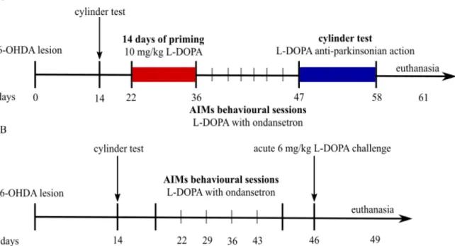
Experimental design ... 70
Acute challenges of ondansetron study ... 70
De novo ondansetron study ... 70
Ratings of AIMs ... 71
Assessment of L-DOPA anti-parkinsonian action ... 72
Perfusions ... 72
LC-MS/MS analysis for dopamine and its metabolites ... 73
Statistical Analysis ... 73
v
Cylinder test ... 74
Acute challenges of ondansetron study ... 74
De novo ondansetron study ... 74
III. Results ... 75
Pharmacokinetic profile of ondansetron ... 76
Extent of dopaminergic denervation assessed in the cylinder test ... 77
Acute challenges of ondansetron at 0.0001 mg/kg significantly alleviated the severity of established AIMs ... 78
Duration of axial AIMs ... 78
Duration of limbs AIMs ... 78
Duration of orolingual AIMs ... 79
Duration of AL AIMs ... 79
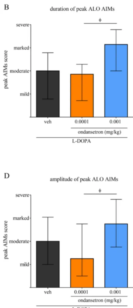
Duration of ALO AIMs ... 79
Amplitude of axial AIMs ... 82
Amplitude of limbs AIMs ... 82
Amplitude of orolingual AIMs ... 82
Amplitude of AL AIMs ... 83
Amplitude of ALO AIMs ... 83
De novo study ... 86
De novo treatment with ondansetron attenuates the development of L-DOPA-induced AIMs ... 86
De novo treatment with ondansetron attenuates the development of ALO AIMs ... 92
Administration of ondansetron does not impair the therapeutic efficacy of L-DOPA in the cylinder test ... 94
IV. Discussion ... 97
Limitations and future directions ... 98
Pharmacokinetic study of clinically relevant doses of ondansetron ... 99
5-HT3 blockade with ondansetron alleviates previously established AIMs without impairing the anti-parkinsonian efficacy of L-DOPA ... 100
vi
Effect of ondansetron and ALO AIMs on L-DOPA anti-parkinsonian action ... 105
Effect of ondansetron on the development of ALO AIMs ... 105
V. Conclusion ... 108
VI. Bibliography ... 110
vii
List of tables
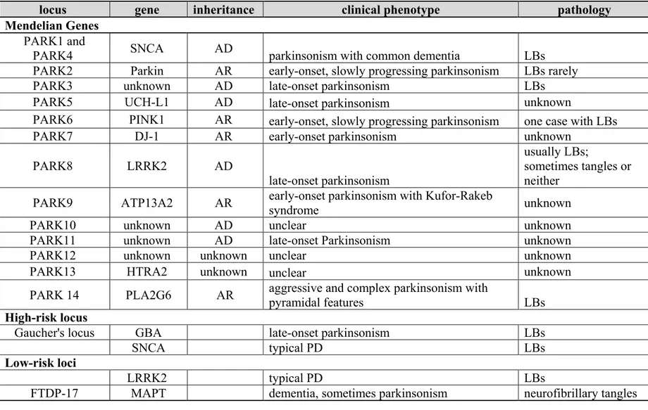
Table I: Genes and loci associated with Parkinson’s Disease ... 9 Table II: Ondansetron pharmacokinetic parameters in the 6-OHDA-lesioned rat ... 76 Table III: Duration rating scale of ALO AIMs in the 6-OHDA-lesioned rat ... I Table IV: Amplitude rating scale of ALO AIMs in the 6-OHDA-lesioned rat ... II Table V: Glass delta’ of right forepaw use across ondansetron treatments ... III Table VI: Glass delta’ of both forepaw use across ondansetron treatments ... IV
viii
List of figures
Figure 1: Dopaminergic synapse ... 19
Figure 2: Schematic diagram of the classical BG circuitry describing different states ... 39
Figure 3: Schematic representation of the experimental design ... 71
Figure 4: Plasma levels of ondansetron in a preliminary pharmacokinetic study ... 76
Figure 5: Performance in the cylinder test in drug-naïve lesioned animals ... 77
Figure 6: Effect of acute challenges of ondansetron on the duration of established L-DOPA induced AIMs ... 81
Figure 7: Effect of acute ondansetron treatment on the amplitude of established L-DOPA induced AIMs ... 85
Figure 8: Time course of the development of AIMs during the 22-day priming phase of the de novo ondansetron study ... 90
Figure 9: Effect of ondansetron on the duration of cumulative and peak AIMs severity during an acute 6 mg/kg L-DOPA challenge ... 93
Figure 10: Use of forepaws across treatment conditions ... 96 Figure 11: Equation to calculate Glass’ delta ... V Figure 12: Time course of the development of AIMs during the 22-day priming phase of the
ix
List of abbreviations
α-synuclein alpha-synuclein 2-DG 2-deoxyglucose 5,7-DHT 5,7-dihydroxytryptamine 5-HT serotonin 5-HT1A serotonin 1A receptor 5-HT2A serotonin 2A receptor 5-HT3 serotonin 3 receptor5-HIAA 5-hydroxyindoleacetic acid 5-HTP 5-hydroxytryptophan 6-OHDA 6-hydroxydopamine
8-OHDPAT 8-hydroxy-2-(di-n-propilamino) tetralin AADC aromatic acid decarboxylase
AIMs abnormal involuntary movements AIMS Abnormal Involuntary Movement Scale
AL axial limbs
ALO axial limbs orolingual ATP adenosine triphosphate
ATP13A2 adenosine triphosphatase 13A2 BBB blood brain barrier
BG basal ganglia
cAMP cyclic adenosine monophosphate Cmax peak plasma concentration CNS central nervous system
Complex I NADH:ubiquinone oxidoreductase COMT catechol-O-methyltransferase
DA dopamine
DAT dopamine transporter DNA deoxyribonucleic acid
DOPAC 3,4-dihydroxyphenylacetic acid
x
ERK extracellular signal-regulated kinase
EP entopeduncular nucleus
FBXO7 F-box only protein 7 GABA γ-amino butyric acid
GBA glucocerebrosidase
GI gastrointestinal
GPCR G protein-coupled receptor GWAS genome-wide association studies GPe globus pallidus pars externa GPi globus pallidus pars interna
HD Huntington’s disease
HVA homovanillic acid
L-DOPA L-3,4-dihydroxyphenylalanine
LB Lewy body
LC-MS/MS liquid chromatography-tandem mass spectrometry
LN Lewy neurite
LID L-DOPA-induced dyskinesia
LRRK2 leucine-rich repeat kinase 2 MAO-B monoamine oxidase-B
MAPT microtubule-associated protein tau mCPBG 1-(m-chlorophenyl)-biguanide MFB medial forebrain bundle
MPTP 1-methyl-4-phenyl-1,2,3,6-tetrahydropyridine
MSN medium spiny neuron
NMDA N-methyl-D-aspartate
PD Parkinson's disease
PINK1 PTEN-induced putative kinase 1
PK pharmacokinetic
PLA2G6 phospholipase A2, group VI PLTS persistent low-threshold spiking RBD REM sleep behaviour disorder
xi
RN raphe nucleus
RNA ribonucleic acid
ROS reactive oxygen species SEM standard error of the mean SERT serotonin transporter
SNARE soluble N-ethylmaleimide-sensitive factor attachment protein receptor SNc substantia nigra pars compacta
SNCA alpha-synuclein gene
SNr substantia nigra pars reticulata
STN subthalamic nucleus
t1/2 plasma half-life
tmax time at maximal plasma levels
TH tyrosine hydroxylase
UDysRS Unified Dyskinesia Rating Scale VMAT2 vesicular monoamine transporter type 2 VPS35 vacuolar protein sorting 35
xii
“You can’t ever reach perfection, but you can believe in an asymptote toward which you are ceaselessly striving.” -Paul Kalanithi
xiii
Acknowledgements
First and foremost, I thank my supervisor, Dr Philippe Huot, for giving me the opportunity to pursue my graduate studies in his research group and for going above and beyond in his duties as a supervisor. His extensive knowledge on movement disorders, attention to detail, open-ended questions and critique on my project have constantly furthered my scientific critical thinking skills and my development as a researcher. His welcoming attitude to questions and encouraging support and advice on scholarship submissions were also greatly appreciated. I am also grateful for Dr Huot’s willingness to meet and have discussions, often with little notice, in spite of his busy schedule. I sincerely thank him for his feedback on the Thesis and his insightful comments that forced me to further my thoughts. Thank you for imparting some of your valuable knowledge and for your patience, work ethic and understanding.
I would also like to thank all the past and current members of the Huot lab, whom I have had the pleasure of working with, for their camaraderie and support really made my MSc journey a positive experience. I thank our lab manager, Dr Adjia Hamadjida, who was really a co-supervisor in terms of responsibilities, for his guidance throughout my project. Dr Hamadjida’s expertise in various techniques, particularly in stereotaxic surgeries and behavioural testing, was instrumental in my training as a graduate student, especially when I first started. I thank Dominique Bédard for her help, as without it, the behavioural studies and pharmacokinetic study wouldn’t have been possible. I thank Imane Frouni for being such a wonderful colleague and a compassionate friend and for assisting with some behavioural studies. We have shared countless discussions on methods and troubleshooting experiments, and life outside research, and even travelled to our first international conference together. I look forward to our future adventures! I thank Sébastien Belliveau for his help with the necropsy and I thank Élodie Bourgeois-Cayer for the French translation of my abstract. I really appreciated their great energy and infectious enthusiasm that created such a lively atmosphere in the lab. I thank Lamia Sid-Otmane for her insightful comments and thought-provoking discussions. I thank Vaidehi Nafade for all the lengthy scientific and non-scientific discussions and for making the long commute enjoyable and worthwhile. Moreover, I would like to thank all my friends who were so supportive and always willing to lend an ear whenever I needed it.
xiv
To my mentor Dr Michel Panisset, although our interactions were few, I am grateful for your invaluable advice and recommendations that allowed me to reflect and develop my ideas. I would also like to express my thanks to all of my collaborators, Dr Francis Beaudry and Ms Fleur Gaudette and Dr Lehka Sleno.
Last but not least, I am extremely grateful to my immediate family for their endless support and encouragement throughout this challenging but rewarding adventure! I thank my grandparents for their continued support in my pursuit of higher learning and for being so understanding of my occasional early/late hours. I thank my parents for their unconditional love, support and understanding, I know it wasn’t easy being my on call chauffeur to the train station every day. I thank my brother Henry for his lively disposition and humour that has always left me in good spirits. I thank my sister Karina for sharing her knowledge on global and humanitarian issues, and for our deep philosophical discussions that are often hilarious and outrageous. I look forward to your journey of higher learning.
2
1. General Introduction
Parkinson’s Disease (PD) was initially described in An Essay on Shaking Palsy by the British physician James Parkinson in 1817 (1). However, it was only until 1861 that the French neurologist Jean-Martin Charcot, known as “the founder of modern neurology”, coined the term “Parkinson’s disease” and distinguished bradykinesia as a separate clinical feature of the illness (2). PD is one of the most common neurodegenerative disorders that affects nearly 1% of the population over 65 years of age (3, 4). PD can be defined by four cardinal motor features: tremor at rest, akinesia (or bradykinesia), rigidity and postural instability (5). In addition to these symptoms, many patients are also affected by non-motor symptoms including dementia, autonomic dysfunction, and sleep disorders (6). The onset of PD is gradual and clinical manifestations do not appear until there is a loss of approximately 40-60% of the dopamine (DA) neurons in the substantia nigra (SN) pars compacta (SNc) and about 80% of striatal nerve terminals (7-10). In more severe stages PD, neuronal loss spreads to outside the SNc to regions including the locus coeruleus, raphe nuclei (RN), olfactory bulb and cerebral cortex, and this widespread neurodegeneration may be responsible for the progression of non-motor symptoms of PD (11) (12). The pathological hallmark of the disease is the presence of intracellular proteinaceous inclusions, known as Lewy bodies (LBs), however, the role of LBs in the pathogenesis of PD is still unknown (13). Alpha-synuclein (α-synuclein) is a major component of LBs and recent studies demonstrate that specific α-synuclein conformations are directly toxic to neurons (14, 15) and can propagate via a “prion-like” mechanism of pathogenesis (16).
Currently, the most effective symptomatic drug for PD is the biochemical precursor to DA, L-3,4-dihydroxyphenylalanine (L-DOPA), which helps to relieve motor symptoms by restoring striatal DA levels. However, as L-DOPA is also converted into DA in the peripheral nervous system, chronic L-DOPA therapy results in adverse effects, notably debilitating involuntary movements, termed L-DOPA induced dyskinesia (LID) (17). Moreover, the longer the duration of treatment, the greater the number of PD patients that develop LID and approximately 80 to 90% of patients suffer from LID after 10 years of treatment (18, 19). Patients with advanced PD suffer from these erratic movements that cannot be adequately controlled with existing therapies (20), which underscores the urgent need to develop therapies that attenuate dyskinesia. In recent years, the understanding of neuronal mechanisms that
3
underlie the pathophysiology of LID has grown and has been associated with events including the pulsatile stimulation of dopaminergic receptors, downstream changes in proteins and genes and abnormalities in non-dopaminergic transmitter systems, which modify the activity of the basal ganglia (BG) circuitry (21).
2. Parkinson’s Disease
2.1. Epidemiology of Parkinson’s Disease
Epidemiological studies show that PD is an age-related disease with men at higher risk of developing the disease than women (3). In Canada, it is estimated that about 99,000 individuals are living with PD and by 2031, the number of is expected to increase by 65% to 163,700 (22). In addition, Canadians with PD tend to be older individuals with an average age of diagnosis of 66.2 years of age (22).
The global prevalence PD is estimated at between 18 to 300 per 100,000 individuals while the incidence of PD is between 10 and 50 cases per 1000,000 individuals per year, respectively (23, 24). Incidence of PD is heavily age-dependent, and onset is rare before 50 years of age until a sharp increase of incidence is observed after 60 years of age (25). The disease prevalence is estimated at 1% in subjects over 65 years of age and increases to 4.3% in those over the age of 85 (26). Most of the increase is attributed to the general trend of an increasingly ageing population (27). Despite the worldwide distribution of PD, incidence rates may vary among populations. The conflicting results between individuals studies may reflect differences in research methodologies, particularly in case definitions, diagnostic criteria, and the age distribution of the study population (28). A collaborative study in four European countries using similar case-finding methods and diagnostic criteria did not reveal any differences (29). In contrast, a meta-analysis of six studies found a lower prevalence in Africa than in Europe or North America (30), but no significant difference was reported between African Americans and Caucasians living in Mississippi (31). In addition, an autopsy study found that African Americans showed the same prevalence of incidental LB disease when compared with Caucasian populations (32). Similarly, lower prevalence rates have also been reported in some Asian countries (23, 33-35) but some studies have found similar estimates to Western countries (36, 37). Accordingly, differences in environmental exposure or interethnic distribution of
4
susceptibility genes may also contribute to the ethnic differences in estimates of PD prevalence and incidence (38, 39). Moreover, the variation in PD prevalence reported amongst studies may be related to differences in response rates, survival and case certainty rather than ethnic differences in PD prevalence (31, 38, 40, 41). Thus, the relative contribution of genetic or environmental variations to population differences in PD incidence is still unclear (26).
Some studies report a higher prevalence of PD in men than in women (42-45) with a male-to-female ratio of about 1.5 (46-48) but other studies found no significant differences in PD prevalence between men and women (29, 49, 50). Consistent with prevalence studies, prospective studies have reported a higher incidence of PD in men than in women (29, 42, 51-53). The neuroprotective effects of oestrogens in women and X-linked genetic factors may account for the higher risk of PD in men but their role is still controversial (54).
2.2. Aetiology of Parkinson’s Disease
The aetiology of PD is poorly understood but considerable advances in sequencing technology, genetics (55) and clinical studies (20) have contributed to a greater comprehension on the pathogenic processes occurring in PD. The common view today is that PD is a multifactorial disease that arises from the combined effects of exposure to environmental risk factors, genetic susceptibility, and complex genetic-environmental interactions (26, 39). Ageing is the strongest risk factor of developing PD (56) and can be explained by the increasing failure of normal physiological and biochemical processes that lead to the increased vulnerability of DA neurons to toxic insult (57). Growing evidence suggests that impairments in the regulation of protein homeostasis including processes such as protein aggregation, intracellular protein and membrane trafficking and disruptions to the ubiquitin-proteasome and lysosome-autophagy are implicated in the pathogenesis of PD (58). In addition, it has been suggested that the genetics of PD are involved in aberrations in synaptic structure and function (58), which confirmed the importance of mitochondrial dysfunction in toxin models of PD (59).
5
2.3. Risk factors of Parkinson’s disease
2.3.1. Non-genetic risk factors
2.3.1.1. Environmental hypothesis
Evidence linking exposure to agrochemicals, including pesticides and herbicides to an increased risk of PD has been postulated for many years (60, 61). In particular, it has been demonstrated that rotenone (62) and paraquat (63-65) cause nigral dopaminergic cell death in rodents. Furthermore, individuals exposed to pesticides had a 70% higher incidence of developing PD those not exposed (66). Additional environmental factors identified include industrialization, rural environment (67), use of well water (68), intake of various metals, etc. (69-71) but studies show conflicting outcomes (72). Although several studies report a positive association between environmental risk factors and PD, no factor has been consistently implicated as the sole causative agent (73). Similarly, the degree of pesticide exposure that may lead to PD is unknown.
2.3.1.2. Discovery of MPTP-induced Parkinson’s disease
The discovery of 1-methyl-4-phenyl-1,2,3,6-tetrahydropyridine (MPTP)-induced parkinsonism (74) stimulated an interest in exposure to environmental risk factors for PD. MPTP causes the degeneration of nigrostriatal dopaminergic neurons with the loss of striatal dopamine in various species, including primates, cats and mice (75-80). In 1982, several young people developed an acute and severe form parkinsonism, due to MPTP produced during their illegal synthesis process of heroin (74, 81). MPTP is highly lipophilic, and can be metabolized into 1-methyl-4-phenylpyridinium (MPP+), the active toxic molecule, by brain monoamine oxidase B (MAO-B) (75-80, 82). MPP+ is accumulated by high affinity DA transporters (DAT) into the mitochondria of dopaminergic neurons (83). Once inside the mitochondria, MPP+ binds to and inhibits NADH-ubiquinone oxidoreductase I (complex I) of the mitochondrial electron transport chain (84). This results in an impairment of ATP production, elevated intracellular calcium concentration and free radical generation (81). Accumulation of MPP+ in dopaminergic neurons causes neurodegeneration via reactive oxygen species-mediated oxidative stress and results in DA neuron death (85-87). MPTP-induced parkinsonism exhibits similar phenomena to PD, particularly the preferential loss of DA nerve terminals of the putamen and DA neuron
6
loss of the SNc. Primates exposed to MPTP are responsive to L-DOPA treatment, and develop motor complications after chronic administration (88). Despite the contributions of the MPTP-lesioned animal models to the knowledge of pathways implicated in PD pathogenesis, they do not fully capture all the features of the disease. For example, MPTP-induced parkinsonism is not progressive, an acute rather than chronic increase in α-synuclein occurs and LB formation is absent (57, 64, 89). In addition, the investigation of agents based on MPTP models in clinical trials has not been successful thus far. Thus, the underlying pathways in the MPTP models of PD may not all be shared with those in PD patients (64).
A recent systematic review of meta-analyses identified several environmental factors that are associated with a risk of developing PD (90). Two factors, physical activity and constipation, presented with convincing evidence for a strong association with PD. Several cohort studies support the protective effect of physical activity for PD (91, 92). Also, constipation may be an early premorbid manifestation of PD (93) and laboratory studies and laboratory studies reported an abnormal deposition of α-synuclein within the submucosal and myenteric plexuses of the enteric nervous system (94, 95). Highly significant association for increased PD risk included head injury (96), anxiety or depression (97), while decreased risk is associated with smoking (97) and high uric acid levels (98). Additional non-genetic risk factors significantly associated with development of PD include a decrease in risk with alcohol (99) and coffee consumption (100), whereas pesticides (101), well water (97), and male sex (102, 103) are linked to an increase in risk. Although substantial evidence supports the association of environmental risk factors and PD, the heterogeneity amongst the examined meta-analyses suggests that some associations may reflect reverse causation, residual confounding, information bias and sponsor conflicts. In addition, the variation in cohort studies and case-control studies, differences in exposure assessment (frequency and exposure types) may account for the biased estimates of association. Furthermore, the authors emphasize that mechanisms of several putative risk factors are poorly understood, and additional studies are required to clarify the association between these factors and the risk of developing PD.
2.3.2. Genetic risk factors
Clinical observation of increased prevalence of PD amongst relatives of patients (104, 105) and the discovery of families with genetic forms of parkinsonism (106-109) heightened
7
interest in the heritability of PD. However, familial aggregation does not necessarily imply genetic causation (110, 111) and a large PD twin study found no increased concordance for PD amongst monozygotic twins (112). However, for subjects with an onset before 50 years of age, a significant concordance rate in identical twins was identified, which suggests young onset PD has a greater genetic component. In contrast, a later twin study using clinical assessment and fluoro-dopa positron emission tomography (PET) to image dopaminergic function reported increased concordance amongst monozygotic twins (113), which supports a role of genetics in PD aetiology.
Significant advances in understanding the pathogenic processes of PD in the past few decades have been made due to the identification of genetic mutations and chromosomal loci associated with parkinsonism (Table I, page 9) (114-116). The majority of PD cases are sporadic (117) but Mendelian loci and the high-risk glucocerebrosidase (GBA) locus collectively account for approximately 10 – 40% of disease risk depending on the population under study (114). Genetic factors that have been identified include mutations in the genes for α-synuclein (SNCA), and leucine-rich repeat kinase 2 (LRRK2), which are responsible for autosomal-dominant PD forms, whereas mutations in the genes PARK2 (Parkin), PARK7 (PTEN-induced putative kinase 1, PINK1), PARK7 (DJ-1), and PARK9 (ATPase 13A2, ATP13A2) account for early-onset autosomal recessive PD forms (117). Recently, two other autosomal dominant PD genes, vacuolar protein sorting 35 (VPS35) and eukaryotic translation initiation factor 4-gamma (EIF4G1), have been identified (118-120). Early candidate gene studies and subsequent meta-analyses provided conclusive evidence demonstrating that polymorphisms in SNCA (121), LRRK2 (122), microtubule-associated protein tau (MAPT) (123) and glucocerebrosidase (GBA) (124) impact PD susceptibility.
High-density arrays of single nucleotide polymorphisms identified genetic susceptibility factors in genome-wide association studies (GWAS), where the frequencies of putative risk alleles are compared in patients and controls (125). Genetic variations may be susceptibility factors or disease modifiers, affecting penetrance, age at onset, severity, and progression (126). The most commonly studied candidate genes include genes involved in DA metabolism, mitochondrial metabolism, detoxification, other neurodegenerative diseases and familial PD (3, 127) and genes associated with putative risk factors for PD including oestrogen receptor gene polymorphisms (128), the tau HI haplotype (129) and the apolipoprotein E epsilon 2 allele (130).
8
Although the significance of many loci identified with an increase in PD risk is still unknown, they account for a population attributable risk of > 60% (131).
2.3.2.1. Risk Loci
2.3.2.1.1. GBA
The GBA gene encodes a lysosomal enzyme β-glucocerebroside, which is involved in glycolipid metabolism (116). Homozygous GBA mutations cause Gaucher’s disease, an autosomal recessive lysosomal storage disease caused by accumulation of glucocerebrosides (132). In contrast, heterozygous mutations in GBA are associated with a higher risk of PD where approximately 5-10% of PD patients have GBA mutations as opposed to an estimated frequency of 1% in healthy controls but this may be underestimated in certain populations (133-137). Carriers of only one mutated allele have a 5-fold increased risk to develop PD compared with non-carriers, which makes GBA one of the strongest genetic risk factors reported to date (134). The high prevalence of PD amongst GBA mutation carriers, which is also age-dependent and rises up to 30% at 80 years of age (138), has led to the suggestion that GBA mutations can act as dominant factors with reduced penetrance rather than simple risk variants (139). The mechanism underlying the association of mutations in GBA with the development of PD and other LB disorders (140) is not known, but may be caused by alterations in lipid metabolism or autophagy and lysosomal function (115). In fact, the mechanism of pathogenicity may be linked to α-synuclein as intracellular glucocerebrosides facilitate the aggregation of α-synuclein into toxic oligomers and fibrils (141), which are the main constituent of LBs (142). Moreover, α-synuclein is primarily degraded through autophagy and GBA mutations interfere with autophagic clearance of α-synuclein fibrils (143, 144). Consequently, fibrils are likely to accumulate in the cell (145, 146), following which they may propagate through cell to cell transmission (147, 148).
9
Table I: Genes and loci associated with Parkinson’s Disease
Adapted from (114-116).
locus gene inheritance clinical phenotype pathology
Mendelian Genes
PARK1 and
PARK4 SNCA AD parkinsonism with common dementia LBs
PARK2 Parkin AR early-onset, slowly progressing parkinsonism LBs rarely
PARK3 unknown AD late-onset parkinsonism LBs
PARK5 UCH-L1 AD late-onset parkinsonism unknown
PARK6 PINK1 AR early-onset, slowly progressing parkinsonism one case with LBs
PARK7 DJ-1 AR early-onset parkinsonism unknown
PARK8 LRRK2 AD
late-onset parkinsonism
usually LBs;
sometimes tangles or neither
PARK9 ATP13A2 AR early-onset parkinsonism with Kufor-Rakeb syndrome unknown
PARK10 unknown AD unclear unknown
PARK11 unknown AD late-onset Parkinsonism unknown
PARK12 unknown unknown unclear unknown
PARK13 HTRA2 unknown unclear unknown
PARK 14 PLA2G6 AR aggressive and complex parkinsonism with pyramidal features LBs
High-risk locus
Gaucher's locus GBA late-onset parkinsonism LBs
SNCA typical PD LBs
Low-risk loci
LRRK2 typical PD LBs
10
2.3.2.1.2. MAPT
Most of the gene loci discovered through GWAS are present in more than 5% of the population (allele frequencies of > 10%) and carriers of the risk allele have a less than two-fold increase of disease risk over the general population average (114). The majority of these low-risk loci appear to mediate their effect by altering gene expression rather than through translational changes. The MAPT is a protein that can form aggregates similar to α-synuclein and beta-amyloid. Mutations in the MAPT gene cause a range of neurodegenerative phenotypes but some can lead to a typical PD presentation (149). The H1 haplotype at the MAPT locus has been consistently suggested as a risk factor for PD (150, 151) and gene duplications at MAPT cause frontotemporal dementia (152), which suggests that the pathogenic cascades in the tauopathies can provoke severe neurodegeneration leading to parkinsonism (153, 154). Moreover, MAPT promotes the formation of α-synuclein oligomers and fibrils (115) and in transgenic mice that exhibit the LB variant of Alzheimer’s disease, cognitive decline is accelerated and associated with amyloid beta, tau and α-synuclein pathologies compared to age-matched control animals (155). Thus, synergistic interactions between α-synuclein and tau may promote their fibrillization and the formation of pathological inclusions characteristic of neurodegenerative diseases.
2.3.2.2. Autosomal dominant forms of Parkinson’s disease 2.3.2.2.1. LRRK2
The most common cause of autosomal-dominant Mendelian form of PD is mutations in the LRRK2 at the PARK8 locus, which account for nearly 10% of all familial dominant inherited forms (156). The G2019S kinase domain mutation is the most frequent LRRK2 mutation (157), and responsible for 5-40% of sporadic or dominantly inherited PD, depending on the population studied (158-160). Higher G20119S prevalence rates have been reported in more isolated populations, such as the Ashkenazi Jewish (161) and North African Berber Arab (162) populations, which can be explained by a genetic founder effect (163). Patients with LRRK2 mutations tend to display late-onset PD with symptoms indistinguishable from those of sporadic PD, even though LB pathology is sometimes absent or lacking (164, 165). Thus, the disconnect between clinical manifestations of PD and the presence of LBs (166, 167) supports the theory
11
that inclusions may not be necessary for neurodegeneration and may instead be a consequence of PD (115). The mechanism underlying the neurodegeneration caused by LRRK2 mutations and its natural substrate are still unknown. However, cell culture studies suggest that neurotoxicity in vitro requires intact kinase activity (168, 169), prompting increased interest towards LRRK2 kinase inhibitors (170) as a potential neuroprotective strategy.
2.3.2.2.2. α-synuclein
SNCA mutations are the second most common cause of dominant PD (171) and various studies have reported a link between familial PD and duplications or triplications in the SNCA gene (172). The SNCA genes encodes α-synuclein, which accumulates in LBs predominantly within the brainstem. As LB pathology is also the dominant pathology observed in most cases of LRRK2-related PD, this suggests that SNCA and LRRK2 affect a common pathway that leads to α-synuclein aggregation (139). Moreover, gene triplication leads to earlier onset and faster progression of disease than duplication, which suggests a gene-dose effect between α-synuclein levels and disease severity (173). The link between α-α-synuclein expression levels and the appearance of PD is well-established across studies, and leads to the hypothesis that a gain-of-function by α-synuclein underlies pathogenesis of PD (115). In addition, recent in vivo evidence shows that it binds to and promotes assembly of soluble N-ethylmaleimide-sensitive factor attachment protein receptor (SNARE) complexes, which are required for the fusion of vesicles to the pre-synaptic membrane (174, 175). Triple knock-out mice lacking α-synuclein also exhibit deficits in SNARE complex assembly and develop accelerated age-associated motor impairments and early-onset mortality, but do not show neurodegeneration (168, 174).
2.3.2.2.3. VPS35
VPS35 gene encodes a major component of the retromer complex involved in endosomal trafficking to the trans Golgi (119, 176). Recent studies have identified a single missense (D620N) mutation in VPS35 as a new cause of autosomal-dominant PD in two independent exome sequencing studies on Swiss (118) and Austrian families (119). Frequency of mutation carriers is low and has been estimated to represent about 0.1% of the PD population (177). Patients with a VPS35 mutation exhibit classical late-onset, L-DOPA responsive parkinsonism similar to that of sporadic PD, with a slightly earlier age at onset (139). Specific deletion
12
of VPS35 in DA neurons of mice results in PD-like deficits, including loss of DA neurons and accumulation of α-synuclein and early degeneration at 2-3 months of age (178). Consistent with this data, overexpression of human D620N VPS35 variants induce the marked degeneration of SNc DA neurons and axonal pathology (179). In addition, mutations in VPS35 caused extensive mitochondrial fragmentation and cell death as well as functional deficits in vitro, in mouse SNc neurons in vivo, and in human fibroblasts from PD patient bearing the D620N mutation (180). Defects in macroautophagy, aminomethylphosphonic acid (AMPA) receptor trafficking to dendritic spines or alterations in mitochondrial dynamics and turnover have been proposed as the mechanism underlying VPS35-induced neurodegeneration (181). Although the mode of action by which it causes PD is unclear, modulation of the development of DA neurons via the wingless-related integration site pathway (182, 183) and aberrant brain iron accumulation (184, 185) have been suggested as possible mechanisms. Furthermore, recent studies demonstrate that VPS35 may interact with other PD-linked gene products including LRRK2, SNCA and Parkin (186-190) in a common pathway that leads to the neurodegeneration observed in PD.
2.3.2.3. Autosomal recessive forms of Parkinson’s disease 2.3.2.3.1. Parkin, PINK1, DJ-1
Mutations in Parkin are the most common cause of autosomal recessive forms of PD, whereas mutations in PINK1 and DJ-1 are relatively less prevalent (139). Parkin gene mutations account for almost 50% of early-onset recessive familial PD and up to 15% of early onset sporadic cases (191, 192). Pathology underlying Parkin-related PD does not tend to show LBs, unlike the autosomal dominant and idiopathic forms. Clinical manifestation of Parkin mutations is often indistinguishable from that of the sporadic disease except for the earlier age at onset (generally before 45 years of age) (139). Wild-type Parkin, PINK1 and DJ-1 are involved in processes of mitochondrial quality control and regulation such as mitogenesis, mitophagy and mitochondrial homeostasis and transport (139, 163). Studies suggest that the function of these proteins in a mitochondrial quality control pathway is impaired in PD, leading to the accumulation of bioenergetically compromised mitochondria, however, it is unclear how this might give rise to substantial nigral degeneration and PD (193).
13
2.3.2.3.2. ATP13A2, FBXO7 and PLA2G6
More rarely, recessively inherited forms of atypical parkinsonism are caused by mutations in the ATP13A2, F-box only protein 7 (FBXO7) and phospholipase A2, group VI (PLA2G6) genes (131). Mutations in ATP13A2 were first identified from families with Kufor-Rakeb syndrome, a rare hereditary disease with typical signs of PD that also includes symptoms of more extensive neurodegeneration (194). Loss of function mutations of ATP13A2 underlie an autosomal recessive form of early-onset parkinsonism with pyramidal degeneration and dementia (194) while heterozygous mutations may be a risk factor for PD (195). ATP13A2 mutations likely play a role in lysosome degradation (131) and recent studies demonstrate that ATP13A2 can rescue against α-synuclein toxicity in a yeast, Caenorhabditis elegans (C. elegans) and neuronal culture model of PD (196). FBXO7 mutations cause early-onset autosomal recessive parkinsonism with pyramidal signs and after an initial favourable response to L-DOPA, patients often develop dyskinesia (197). Most of the reported FBXO7 mutations are loss of function (198) but no neuropathology has been described thus far. Mutations in PLA2G6 cause an early-onset recessive degenerative disorder characterized by spasticity, ataxia and dystonia but adult onset forms can manifest as dystonia-predominant parkinsonism (199) that is responsive to L-DOPA (200). PD associated with PLA2G6 is caused by the homozygous or compound heterozygous inheritance of various missense mutations (201-203).
The clinical and sometimes pathological resemblance of genetic PD to sporadic disease make it a suitable human model to identify at-risk individuals in earlier and possibly prodromal phases of the disease (116). However, monogenic causes of PD represent less than 10% of PD cases in most populations (204) whereas the majority of cases seem to arise from complex interactions among genes and between genes and environmental factors (39). Thus, environmental factors appear to be more important determinants than ethnic and genetic factors in the aetiology of PD. Further efforts are warranted to understand how genetic causes and risk factors of PD play a role in the underlying pathophysiology in hopes of developing targeted therapies that alter disease course (139).
14
2.4.
Pathophysiology of Parkinson’s disease
Based on autopsy findings in PD patients, Braak and colleagues postulated that α-synuclein aggregates form in the periphery in early stages of PD before α-α-synuclein aggregation in the brain (205) and also propose a six-stage system for PD based on the stereotypic pattern of α-synuclein spreading (206). The Braak model is based on the presence of LBs and Lewy neurites (LNs) where the pathogenic process begins in the lower brainstem in the dorsal motor nucleus of the vagus nerve and anterior olfactory structures (206). The disease then spreads rostrally from the dorsal motor nucleus of the vagus nerve through the medulla, pontine tegmentum, midbrain and basal forebrain before ultimately reaching the cerebral cortex. This process follows a specific pattern where susceptible regions are affected in a predictable topographic sequence (207) where severity of the lesions and the clinical manifestations of the disease increase as the pathology ascends from the brainstem (208). Accordingly, in vitro (209, 210), in vivo (211, 212) and clinical evidence (213, 214) suggest that cell types in the central nervous system (CNS) exhibit a propensity for developing Lewy pathology that shares common features. In spite of the support for Braak’s hypothesis, there is criticism around whether it accurately reflects the development of PD in all patients as studies report that Braak staging fails to describe the disease progression in upwards of 50% of α-synuclein immunoreactive cases (215-217). Moreover, the absence of information on the loss of neurons and synaptic connections in the original Braak papers has been the subject of scrutiny (218, 219) as the scientific premises underlying the model remain unclear (220). Thus, the inconsistencies between the Braak model and conflicting reports of the spread of pathology require further study to determine the relationship and likely also require a deeper understanding of the mechanisms underlying the role of α-synuclein in disease progression (207).
Several lines of evidence have implicated dysfunctions in the ubiquitin-proteasome system in PD pathogensis (221-223), which have been further supported by the identification of disease-causing mutations in genes encoding proteins involved in protein degradation in PD (224). Impairment of ubiquitination pathways and proteasomal function could result in defects in the clearance of toxic aggregates and result in their accumulation and degeneration of DA neurons (223, 225). Although systemic administration of proteasomal inhibitors modelled a behavioural and pathological phenotype reminiscent of PD (226), this model has been met with
15
great scrutiny due to the extensive variability in the consequences of in vivo proteasomal inhibition (227). Moreover, questions on the molecular connections between these systems and pathogenesis of PD remain, including the divergent fate of misfolded proteins for degradation or inclusion formation, and further studies, that likely exploit advances in genetics and technologies (228), are warranted to clarify this relationship.
2.4.1. Lewy bodies in Parkinson’s Disease
Idiopathic or sporadic PD is characterized by the selective loss of neurons and appearance of abnormal cytoplasmic proteinacious aggregates called LBs in the soma or LNs in the processes in DA neurons (173). Immunohistochemistry shows that LBs consist primarily of the protein α-synuclein (229), as well as other proteins such as ubiquitin (230) and parkin co-regulated gene (231). Studies have suggested that misfolded α-synuclein and the deposition of LBs within midbrain neurons could contribute to neuronal damage and cell death (232). In addition to SNc DA neurons, neuronal loss and Lewy pathology also occurs extensively in locus coerulus noradrenergic neurons, RN serotonergic neurons, enteric DA neurons, post-ganglionic sympathetic noradrenergic neurons and olfactory neurons (11, 12).
2.4.2. Alpha-synuclein in Parkinson’s disease
α-synuclein is of the main constituents of LBs and LNs, and accumulates widely in central and peripheral neurons of PD patients (233). Given it predominates in pre-synaptic terminals and the nuclear envelope, it plays a role in SNARE-mediated exocytosis and synaptic vesicle transport (234). Moreover, α-synuclein is present in mitochondria in PD brain and may affect mitochondrial function both in vitro and in vivo, possibly leading to the vulnerability of nigrostriatal DA neurons in PD (235-238). Accordingly, it has been demonstrated that α-synuclein aggregation may be associated with oxidative or nitrosative stress (239-241), which may be important in the pathogenesis in neurodegenerative disorders with LBs, like PD (242). Converging evidence also supports the hypothesis that α-synuclein oligomers (243, 244) and fibrils (245, 246), the pathologic form of α-synuclein, may participate in the propagation of neurodegeneration observed in PD (247). Thus, the misfolded α-synuclein fibrils present in LBs (142) and their self-propagation and spread, reminiscent of a “prion-like” process, suggest that their mode of cell-to-cell transmission is not in disagreement with the Braak staging and a
16
possible peripheral origin of Lewy pathology (210, 211, 248-250). Furthermore, the prion hypothesis of α-synuclein transmission is supported by evidence from transgenic mouse models of synucleinopathy and viral vector-mediated α-synuclein overexpression in rats (251, 252) and nonhuman primates (253, 254). However, no data from transgenic mice models has reported the spontaneous pathological α-synuclein (255) and conflicting findings surround the specificity of peripheral α-synuclein for PD in humans (255), for instance, some studies have found similar levels of α-synuclein accumulation in the colon of patients with PD compared with healthy controls (256-258).
2.4.3. Oxidative stress in Parkinson’s disease
Oxidative stress defines a disequilibrium between the levels of reactive oxygen species (ROS) produced and the ability of a biological system to detoxify the reactive intermediates, ultimately creating a perilous state contributing to cellular damage (259). An increasing body of evidence suggests that in PD, oxidative stress and mitochondrial damage contribute to a sequence of events that lead to the degeneration of DA neurons in the SNc (57, 260-262). In addition to mitochondrial dysfunction, DA metabolism (263), neuroinflammation (259), iron (264), calcium (265) and ageing (266) also contribute to ROS production in the PD brain. Indeed, post-mortem brain analyses consistently show increased oxidative damage to lipids (267, 268), proteins (269), deoxyribonucleic acid (DNA) and ribonucleic acid (RNA) (270, 271). Further support for the link between oxidative stress and DA neuronal degeneration has been demonstrated by modelling motor symptoms of PD in toxin-induced animal models that cause oxidative stress such as 6-hydroxydopamine (6-OHDA), MPTP, rotenone and paraquat (272). It has also been suggested that mechanisms that contribute to neurodegeneration act in a feed forward manner where primary insults lead to oxidative stress, which damages key cellular proteins and disrupts lipid membranes that in turn cause more ROS production (259).
2.4.4. Mitochondrial dysfunction in Parkinson’s Disease
In addition to the dual role of mitochondria as both a source and target of ROS (273-275), compelling evidence suggests that mitochondrial dysregulation is critical in the pathogenesis of PD (259). Mitochondria are dynamic organelles with important functions in cellular respiration, energy metabolism, calcium homeostasis, stress response and apoptosis
17
pathways (260). Several groups have reported decreased complex I activity in the SN of PD patients (276-278), and the finding of the downregulation of genes encoding mitochondrial proteins further supports the role of mitochondrial dysfunction in PD (279). In addition, PD-related proteins, including DJ-1, PINK1, Parkin, α-synuclein and LRRK2, are also involved in mitochondria quality control leading to exacerbations of ROS generation and susceptibility to oxidative damage (259).
2.5. Dopaminergic system in Parkinson’s disease
DA neurons form four major systems within the mammalian brain: the nigrostriatal, mesolimbic, mesocortical and tuberoinfundibular systems (280) that originate from the A9, A10, and A8 groups of dopamine-containing cells, respectively (281, 282). In the nigrostriatal pathway, projections from dopaminergic neurons with cell bodies in the SN terminate in the striatum (283). The mesolimbic pathway consists of dopaminergic neurons that originate in the ventral tegmental area (VTA) and project to the nucleus accumbens and related limbic regions, whereas VTA neurons that project to the prefrontal cortex establish the mesocortical pathway (284). Last, the tuberohypophyseal pathway consists of dopaminergic projections from the hypothalamus to the pituitary gland, and its secretions regulate prolactin (285). Due to their wide connectivity within distinct pathways, DA neurons exert a variety of functions including locomotion, addiction, reward, learning and memory, cognition, stress and movement (286).
DA is a monoamine neurotransmitter synthesized in a series of enzymatic reactions (287), beginning with the conversion of the amino acid tyrosine into L-DOPA via the rate-limiting enzyme tyrosine hydroxylase (TH) (Figure 1, page 19). L-DOPA is subsequently decarboxylated into DA by the enzyme aromatic acid decarboxylase (AADC). DA is then packaged into pre-synaptic vesicles by the vesicular monoamine transporter type 2 (VMAT2) and released at nerve terminals into the synapse upon stimulation. Released DA bind to DA receptors to elicit a response in the post-synaptic cell and this interaction is important in the modulation of motor function through the BG circuitry. Extracellular DA is either metabolized by MAO-B and catechol-O-methyl transferase (COMT) in the cytosol or transported back into the pre-synaptic terminal via the DAT. Following re-entry of DA into the pre-synaptic neuron, DA can be repackaged into vesicles and recycled or degraded into the metabolite homovanillic acid (HVA).
18
A deficit in the number of nigrostriatal dopaminergic neurons, characteristic of PD, disrupts the dopaminergic transmission and produces abnormal motor features in affected subjects.
2.5.1. Nigrostriatal dopaminergic pathway
Dopaminergic terminals in the striatum consist of dense innervation of fibres from two specific groups of neurons in the brainstem (282, 288) . The first group are neurons with cell bodies in the VTA that project to the nucleus accumbens and olfactory tubercle. The second group have cell bodies in the SNc and project primarily to the putamen and the caudate nucleus. The tegmental and nigral afferents form the nigrostriatal dopaminergic pathways.
2.5.2. Classification of dopamine receptors and their distribution in the basal ganglia
DA receptors are a family of G protein-coupled receptors (GPCRs) with five subtypes, D1-D5, that are divided into two groups (289). D1-like receptors are comprised of D1 and D5 receptors and mainly couple G proteins (290), which stimulate adenylyl cyclase and cyclic adenosine monophosphate (cAMP) production (291, 292). In contrast, D2-like receptors comprise D2, D3 and D4 receptors; they couple with Gαi /Gαo and inhibit adenylyl cyclase and negatively regulate cAMP production (293, 294). D1-like receptors have an excitatory effect by stimulating cAMP production, whereas activation of D2-like receptors is inhibitory. D2 receptors are the presynaptic receptors of the dopaminergic system and are responsible for the negative feedback on levels of synaptic DA (295).
Both D1Rs and D2Rs are highly expressed by striatal medium spiny neurons (MSNs) (296, 297) and are present at lower levels in the cortex as compared to the striatum (298). D1 receptors are expressed in striatonigral neurons containing substance P and dynorphin that project to the SN pars reticulata (SNr) and to the globus pallidus (GP) pars interna (GPi), which constitute the direct striatal output pathway (299). In contrast, D2 receptors are predominantly localized in striatofugal neurons expressing encephalin, which project to the GP pars externa (GPe), constituting the indirect pathway (300, 301). D1 receptors are post-synaptic whereas D2 receptors are also localized on pre-synaptic nigrostriatal dopaminergic terminals, on SNc neurons, and on pre-synaptic cortico-striatal terminals where they can inhibit striatal glutamate
19
release (296, 302, 303). In humans and nonhuman primates, D3 receptors are mostly found in the nucleus accumbens and caudate nucleus-putamen complex but are also localized in the GPi, anterior thalamus, amygdala, hippocampus and cortex (304-306). In the human striatum, there is approximately D3:D2 receptors is approximately 1:2, and D3 receptors can co-localize with both D1 and D2 receptors (305, 307).
Figure 1: Dopaminergic synapse. After DA is synthesized in the pre-synaptic neuron and
released into the nerve terminal, extracellular levels of DA are regulated through several mechanisms. DAT is responsible for the reuptake of DA back into the pre-synaptic neuron, VMAT2 packages DA back into synaptic vesicles, pre-synaptic D2 receptors control DA synthesis and release, and MAO-B and COMT are involved in the extracellular metabolism of DA. Following the release of DA at synaptic terminals, DA can bind to two types of DA receptors on post-synaptic neurons. The D1 receptor is coupled to Golf and activates cAMP-dependent signalling pathways while the D2 receptor is coupled to Gi and inhibits the same pathways. AADC: aromatic L-amino acid decarboxylase; AC: adenylate cyclase; COMT: catechol-o-methyl-transferase; DAT: dopamine transporter; MAO-B: monoamine oxidase B; TH: tyrosine hydroxylase; VMAT2: vesicular monoamine transporter 2. Modified from (308).
20
2.6. Clinical features of Parkinson’s disease
Traditionally described as a motor disorder, numerous brain structures are affected at different time points along the course of the disease manifestation, and both motor and nonmotor symptoms are observed in PD.
2.6.1. Motor symptoms of Parkinson’s disease
The four cardinal motor features of PD are the following: bradykinesia, muscular rigidity, resting tremor and impairment of postural balance leading to disturbances in gait and falls. Movement can be normal in early disease (309) due to the redundancy in BG activity and the capacity of the striatum to compensate functionally for lower degrees of DA deficiency (7). However, after the loss of approximately 80% striatal DA and loss 60% SNc DA neurons, motor symptoms begin to appear (7-10). Initially, the symptoms are mild and are usually confined to one side of the body but over the disease course, symptoms are increasingly impairing and involve the contralateral side as well (310). Motor features are heterogeneous and in spite of the lack of consensus on the classification of subtypes, empirical clinical observations suggest the existence of two major subtypes: tremor-dominant PD with a relative absence of other motor symptoms and non-tremor-dominant PD, a phenotype described as akinetic-rigid syndrome and postural instability gait disorder (311). Bradykinesia is defined as difficulty in planning, initiating and executing movements, as well as with performing sequential and simultaneous tasks (312). This often manifests as deficits in fine motor control (312), slower reaction times(313, 314) and slowness in performing daily activities (315-318). Moreover, bradykinesia leads to impairment of the power of voluntary movement (319, 320). Muscle rigidity is described as increased resistance to passive joint movement, and can often lead to a flexed posture (321). The combination of these motor symptoms along with disturbances in gait, leads to greater disability in PD patients. In addition to motor symptoms, PD patients also experience debilitating nonmotor symptoms.
2.6.2. Non-motor symptoms of Parkinson’s disease
The spectrum of non-motor features encompasses olfactory dysfunction, sleep disturbances, autonomic dysfunction, gastrointestinal (GI) distress, memory loss and dementia, as well as neuropsychiatric conditions (6, 322-324) (325, 326). Virtually all patients with PD
21
exhibit at least one nonmotor symptom with an average of 7.8–11.9 nonmotor symptoms per patient (322, 327-330). Moreover, nonmotor symptoms have been reported to affect the quality of life of PD patients to a greater extent than motor features (330-332).
Olfactory dysfunction is of one of the most common nonmotor symptoms in PD that affects over 80% of patients with PD (333), a prevalence with greater sensitivity for PD compared to many other clinical markers. Although robust evidence indicates that olfactory loss precedes PD (334-340), the lead time for olfactory loss is variable, and some patients may develop detectable loss before or after developing parkinsonism (309). In addition, strong evidence supports rapid eye movement (REM) sleep behaviour disorder (RBD) as a predictor of synucleinopathies, most commonly PD with or without dementia LBs (309). RBD is defined as apparent enactment of dreams during REM sleep, associated with a loss of normal REM sleep atonia (341). Five prospective studies have reported that synucleinopathies, i.e. PD, dementia with LBs or multiple system atrophy develop in up to 80-90% of patients with RBD (342-347). Moreover, one study showed that α-synuclein deposition was present in brains of 98% of subjects with neurodegenerative disease who also had RBD confirmed by polysomnography (348).
Additional symptoms relating to autonomic dysfunction include constipation, orthostatic hypotension, urinary and sexual dysfunction and somnolence. α-synuclein is abundant in the GI system (256), which led some to speculate that prion-like spreading might occur from the GI tract to the brain (207), and a delay in colon transit time that results in constipation could theoretically facilitate this spreading (309). Although this remains controversial, pathological evidence supports the capacity of α-synuclein to spread (349) and a preliminary report showed that vagotomy may reduce the risk of PD (350). Depression and anxiety are commonly comorbid in PD, however, their potential as a marker is limited by the low relative risks (351-355) and predictive value (309), and highly variable lead times for psychiatric manifestations (353-356). Cognitive impairment is traditionally associated to late stages of PD but mild cognitive changes are also observed in de novo PD (357). In general, nonmotor features precede the appearance of motor symptoms in PD, including olfactory dysfunction, RBD, constipation, urinary and sexual dysfunction, which show promise as potential markers of prodromal PD (358).
22
2.7. Current pharmacotherapy for Parkinson’s disease
Currently available therapies are not disease-modifying or neuroprotective and they provide only symptomatic relief for motor features of the disease (359). There are non-pharmacological, pharmacological and surgical treatments that attempt to restore dopaminergic activity using L-DOPA and DA receptor agonists.
2.7.1. L-DOPA
Administration of L-DOPA with a peripheral AADC inhibitor such as carbidopa or benserazide is the most effective treatment for relief of motor symptoms of parkinsonism, particularly for controlling bradykinesia (360). The addition of carbidopa or benserazide enhances the therapeutic benefits of L-DOPA, reduces the dose of L-DOPA required, and minimize peripheral adverse effects (361).
2.7.1.1. Pharmacology of L-DOPA
DA was first synthesized in 1910 (362) and its biochemical precursor was synthesized the following year (363). In a seminal study, Ehringer and Hornykiewicz discovered that DA levels were reduced in the striataum of PD patients (364). Moreover, Hornykiewicz observed a correlation between most of the PD motor symptoms and striatal DA depletion (365). The introduction of DA replacement therapy with L-DOPA in the early 1960s revolutionized symptomatic treatment of PD (366). Unlike DA, L-DOPA crosses the blood brain barrier (BBB), and is effective at alleviating motor features of PD during early stages of treatment (367). Barbeau and colleagues reported an improvement of parkinsonism, mostly with respect to rigidity, after oral administration of L-DOPA to patients with PD (368). Furthermore, Cotzais and colleagues reported that high doses of L-DOPA had marked beneficial effect on the motor symptoms of parkinsonism, mostly bradykinesia and rigidity (369-371).
As AADC is also present outside of the brain, peripheral metabolism of L-DOPA can cause adverse side effects including hypotension, nausea and vomiting (372, 373). Thus, a peripheral acting AADC inhibitor, such as carbidopa or benserazide, that does not cross the BBB is often co-administered, to limit the peripheral decarboxylation of L-DOPA so that more L-DOPA is available to enter the brain, while minimizing the aforementioned peripheral adverse
23
effects. The addition of peripherally acting AADC inhibitors allowed a reduction in the required dose of L-DOPA up to 60-80% (374, 375), potentiated its efficacy, led to a faster onset of anti-parkinsonian benefit and a reduction of cardiovascular and GI side effects (371, 373, 376).
L-DOPA is absorbed in in the duodenum and proximal jejunum by active transport via the large neutral amino acid system (377, 378). L-DOPA enters the body and brain by active transport (379, 380) and competes with dietary proteins and amino acids (381). Thus, high protein intake can reduce L-DOPA anti-parkinsonian action and indeed, intraduodenal delivery of L-DOPA leads to a decline of motor performance following oral protein intake (382). In the clinic, DOPA has a short half-life of 1.5 to 2 hours (383-388). After oral administration, L-DOPA plasma levels reach a maximum about one hour after intake, although this may vary due to the unpredictable absorption (387-389). L-DOPA plasma levels are 10–15-fold higher than L-DOPA levels in the ventricular cerebrospinal fluid (390). COMT inhibitors are often used to extend the duration of L-DOPA anti-parkinsonian action and indeed, tolcapone, entacapone and opicapone increase the area under the curve when administered with L-DOPA (391, 392).
L-DOPA is converted into DA by AADC in DA neurons from the SN and projections (393), serotonin (5-HT) neurons from the raphe complex and their striatal projections (394, 395) and noradrenergic neurons from the locus coeruleus and their projections (396). Due to the characteristic degeneration of the nigrostriatal system in PD, L-DOPA is converted in DA mostly by raphe-striatal 5-HT neurons, and to a lesser extent, by striatal intrinsic AADC-containing interneurons (397-399).
Although DOPA is the most effective treatment for PD, chronic administration of L-DOPA is associated with the development of motor complications including motor fluctuations and LIDs (400). LID represent a major limitation of current pharmacotherapy for PD as the majority of patients experience dyskinesia after a few years of treatment (400, 401) and underscores the need to develop effective therapeutic strategies for patients suffering from these involuntary movements.
2.7.2. Dopamine agonists
DA receptor agonists such as ropinirole, pramipexole and rotigotine are commonly employed and their main advantages over L-DOPA are: they do not require enzymatic activation
24
and have a longer duration of action (402). However, due to their action on DA receptors, their adverse effect profile includes: hallucinations, confusion, nausea, and increased incidence of impulse control disorders including pathological gambling and hypersexuality (403). Apomorphine, a DA agonist, is primarily used as rescue therapy for temporary relief of off-periods of akinesia in patients with fluctuating response to dopaminergic therapy (404), but is not available in Canada.
2.7.3. MAO-B inhibitors
Selective MAO-B inhibitors like selegiline and rasagiline delay the breakdown of DA in the striatum (287). As their efficacy is modest, they can be used as a monotherapy in early PD and in advanced stages of the disease, they can be administered as an adjunct to reduce off time in patients with declining response to L-DOPA (405).
2.7.4. COMT inhibitors
COMT inhibitors block the peripheral degradation of LDOPA, which leads to its increased half-life and enhanced central bioavailability (406). Two COMT inhibitors are available in Canada, tolcapone and entacapone, while opicapone is also available in Europe, although entacapone is preferred because it is not associated with hepatotoxicity (407-409). They are used as adjunctive treatment in patients who develop motor fluctuations to prolong the effect of L-DOPA (410).
2.7.5. Anti-cholinergics and amantadine
Anti-cholinergic agents like trihexyphenidyl and benztropine were historically used for the treatment of PD before the introduction of L-DOPA. Their main therapeutic effect is on tremor and they are only indicated in early PD or as an adjunct to DA replacement therapy. Amantadine modulates dopaminergic and cholinergic transmissions and is also a non-competitive N-methyl-D-aspartate (NMDA) receptor antagonist with a modest efficacy and improves parkinsonian symptoms in mildly affected patients with early disease and reduces LIDs in patients with advanced disease.