This is an author-deposited version published in:
http://oatao.univ-toulouse.fr/
Eprints ID: 5644
To link to this article:
DOI:10.1016/J.DESAL.2008.11.037
URL:
http://dx.doi.org/10.1016/j.desal.2008.11.037
To cite this version:
Pierre, Gwenaelle and Causserand, Christel and
Roques, Christine and Aimar, Pierre (2010) Adsorption of MS2
bacteriophage on ultrafiltration membrane laboratory equipments.
Desalination pp. 762-766. ISSN 0011-9164
O
pen
A
rchive
T
oulouse
A
rchive
O
uverte (
OATAO
)
OATAO is an open access repository that collects the work of Toulouse researchers and
makes it freely available over the web where possible.
Any correspondence concerning this service should be sent to the repository
administrator:
staff-oatao@listes.diff.inp-toulouse.fr
Adsorption of MS2 bacteriophage on ultrafiltration membrane
laboratory equipments
☆
Gwenaelle Pierre
a, Christel Causserand
a,⁎
, Christine Roques
b, Pierre Aimar
aa
Laboratoire de Génie Chimique (UMR 5503), Université de Toulouse, Université Paul Sabatier, 31062 Toulouse cedex 09, France
bEquipe Adhésion Bactérienne et Formation de Biofilms (EA 3036 – IFR 31), Université Paul Sabatier, Toulouse, France
a b s t r a c t
Virus adsorption has been quantified (i) on different materials including various containers made of glass, plastic or stainless steel and hollow fiber membranes made of polyethersulfone, polysulfone, PVDF or cellulose acetate (with two configurations: in/out and out/in and various molecular weight cut-offs); and (ii) on the filtration equipment. The selected virus is MS2 bacteriophage used as a model to estimate viral survival in water or to quantify virus elimination by membrane filtration. A series of experiments have been conducted with suspensions of MS2 at different concentrations prepared in a sterilized saline solution (8 g/L of KCl) or in sterilized distilled water. This study has shown that the most appropriate material to be used as a filtration test tank is Pyrex glass. We show that an addition of a virus solution 15 min after the beginning of the experiment allows keeping the virus concentration at a high level in the system (including tank, tubing, and pump). No adsorption was observed on membrane materials tested during soaking.
1. Introduction
The integrity of membrane processes used in drinking water production and waste water treatment has to be assessed in relation to their capacity to retain viruses. Therefore the development of a protocol is a prerequisite. This protocol has to take into account phenomenon that could affect (enhance or decrease) apparent virus removal such as virus aggregation or virus adsorption. The assessment of adsorption on any solid surface of the testing equipment and on the membrane is of major importance considering the very low concentration of viruses involved. The kinetics of adsorption is important so as to evaluate the time to reach saturation without which an accurate evaluation of viruses' retention by the membrane would not be possible.
Fewer studies report on virus adsorption on the walls of reactors than on membrane materials. Madaeni et al.[1]show by electron microscopy that poliovirus 1 is retained by adsorption onto the membrane (MF, UF). As a consequence the apparent virus retention changes over time: it is initially high (ca. 100%) due to adsorption and drops over time (to 91%). Gassilloud et al. [2,3]indicate that when poliovirus 1 is put in a hydrophobic container (polypropylene) and maintained in a dark environment at 20 °C, a significant viral
transfer (1.5 log10) from the aqueous phase onto the hydrophobic
walls occurred during the first 20 days of incubation. However no general conclusion can yet be drawn from such studies and an experimental validation remains necessary at this stage.
We first present and discuss a series of experiments that have been carried out to quantify the adsorption of viruses on various containers and hollow fiber membranes used in drinking water production. Adsorption on the filtration equipment with a feed tank made of stainless steel or of Pyrex and with PVC and polyamide tubing is then evaluated.
MS2 bacteriophage was selected in this project as it is a standard used by many membrane manufacturers to challenge membranes. MS2 bacteriophage is a non enveloped RNA virus with an icosahedral capsid measuring 23–30 nm in diameter and with an isoelectric point of 3.9[4,5]. This RNA phage specially infects bacterial strains, such as Escherichia coli, exhibiting F-pili structures in their cell wall. The advantages of MS2 are many: it is one of the smallest viruses (then should reveal smaller defects or pores), close in size and shape (icosohedron) and nucleic acid (single-stranded RNA) to the enteric virus hepatitis A and poliovirus and especially non-pathogenic[6]. For these reasons, MS2 is used as a model to estimate viral survival in water or to quantify virus elimination by membrane processes.
This study provides guidelines to propose a preconditioning protocol allowing the determination of the “true” virus retention by the membranes. This should enable us to acquire a better under-standing of the mechanisms of transmission of micro-organisms in membrane processes.
☆ Presented at the Conference on Membranes in Drinking and Industrial Water Production, 20–24 October 2008, Toulouse, France.
⁎ Corresponding author. Tel.: +33 (0) 561 55 86 90. E-mail address:caussera@chimie.ups-tlse.fr(C. Causserand).
2. Materials and methods
2.1. MS2 phage, cultivation and characterization 2.1.1. MS2 phage stock preparation
All tests were performed with MS2 phage virus (ATCC 15597-B1) and E. Coli (W 1485) obtained from Pasteur Institute (Paris). The bacteriophage was replicated according to the Pasteur Institute procedure (P-PH6003 MS2 Phage; 03/05/2006, CNCM – lot 8848). Briefly, 1 mL of MS2 stock was mixed to 1 mL of E. coli suspension in 10 mL Tryptone yeast extract broth and 0.8 mL of this preparation was placed into inclined base agar and incubated for 22 h at 37 °C. After replication, viral suspension was centrifuged (4000 rpm/ 20 min/10 °C) and the supernatant was filtered through a 0.22 μm filter (Millipore) to remove any remaining residuals. The phage was then stored at 4 °C until addition to the system. Typically, a stock concentration of 1011plaque forming units (PFU)/mL was obtained.
2.1.2. MS2 phage concentration determination
The plaque assay procedure was used to determine the concen-tration of the prepared MS2 phage stock suspension as well as one of the samples collected during adsorption experiments. Samples were diluted, when necessary, using phosphate-buffered saline (PBS) in order to decrease the concentration of MS2 phage under 200 PFU/mL. This concentration of MS2 phage could easily be counted on each plate with the naked eye. Approximately 3 mL of soft agar was dispensed into each glass vial with 0.9 mL of E. coli broth prepared in PBS (ATCC, 15597) to the concentration approximately 108 bacteria/mL. This
concentration was obtained by measuring the optical density at 640 nm with UV spectrophotometer (Secomam Prim). Then, 0.1 mL of each MS2 phage sample was added to individual vial. The contents of the vials were then emptied onto 15 × 100 mm petri dishes, contain-ing base agar, and allowed to harden. Then, the plates were incubated overnight at 37 °C for a minimum of 18 h. Next, the plates were removed from the incubator and the plaques were counted. The number of plaques observed on each plate was assumed to correspond to the concentration of MS2 phage in PFU/mL.
2.2. MS2 phage characterization
The cultures obtained were characterized in terms of shape and size by transmission electron microscopy and dynamic light scattering. The MS2 phage characterization makes it possible to show that isolated and non aggregated viruses are obtained after amplifi-cation and to check the reproducibility of the suspensions thus prepared. It is necessary that the viruses are isolated to determine the conditions of adsorption in the “worst cases”.
2.2.1. Transmission electron microscopy (TEM)
Transmission electron microscopy (TEM) is the most commonly used because it allows visualization of particle morphology and size. Two microliters of samples were applied to glow-discharged, 200-mesh carbon coated grids. The remaining liquid on the grids was blotted with filter paper after 2 min. Grids were washed with water, stained with 2% (w/v) uranyl acetate for 30 s, dried and visualized by TEM (Hitachi HU-12A).
2.2.2. Dynamic light scattering
Samples were analyzed at room temperature with a Malvern Zetasizer Nano ZS equipped with a He–Ne laser (633 nm). The sen-sitivity of the apparatus requires a minimum viral suspension con-centration of 109PFU/mL[7].
2.3. Experimental procedure for adsorption assessment in test vessels, membrane materials and filtration equipment
2.3.1. Test vessels
In the bench scale study, three different materials that are supposed to be used in the filtration equipment (feed tank, tubing) were compared in terms of quantity of MS2 bacteriophage adsorbed. The selected materials are glass (Pyrex beaker of 50 mL), plastic (polypropylene beaker of 50 mL), and stainless steel (a stainless beaker of 1 L). MS2 suspensions at a concentrations of 108, 104and
103PFU/mL were prepared in a sterilized saline solution (8 g/L of KCl,
salt conditions under which the phage is stored) or in sterilized distilled water to test the impact of salt on adsorption. The suspension was placed under stirring (300 rpm) in beakers made of each material (by keeping equal to 1 the ratio contact area/solution volume) during 6 h. A sample of the suspension was collected every hour approx-imately and an enumeration was carried out in order to determine the new concentration of the suspension.
2.3.2. Membrane materials
Experiments on the adsorption of the virus on various UF hollow fibers were conducted. Each membrane was soaked under stirring (300 rpm) during 6 h in virus suspensions at a concentration 108PFU/
mL in sterilized saline solution or in sterilized distilled water. The vessel used for these experiments is made of glass. A sample of the suspension was collected every hour approximately and an enumer-ation was carried out in order to determine the concentrenumer-ation of the suspension. The selected membranes are made of polyethersulfone, polysulfone, PVDF or cellulose acetate, with two configurations (in/ out and out/in types) and with various molecular weight cut-offs ranging from 60 to 200 kDa (different porosities which play a role in the contact area), the same mass quantity of fibers is added in the vessel.
Fig. 1. Image by TEM of amplified MS2 phages.
Fig. 2. Measurement of size distribution of amplified MS2 phages by dynamic light scattering (nanosizer).
In parallel with these experiments, a glass vessel was filled with the same virus suspension but without membrane sample to be used as blank.
2.3.3. Filtration equipment
The first series of experiments was performed using an empty filtration module shell (without membrane) to determine how much of the initial virus could be adsorbed by the filtration rig with a feed tank made of stainless steel or of Pyrex and with PVC and polyamide tubing
[8]. A feed suspension of MS2 at a concentration of 108PFU/mL was used
in sterilized distilled water. The suspension was circulated in the system (60 L/h Re= 1800) with recycling of retentate and permeate in the feed
tank. Samples were collected every hour in the permeate stream and counting carried out in order to determine the virus concentrations over time. Then, “doping” experiments have been conducted by adding 2 mL of MS2 at a concentration of 1011PFU/mL, 15 min, 30 min or 60 min
after the beginning of the run. The equipment was cleaned after each experimental run by circulating sodium hypochlorite solution at 20 ppm. The system was then rinsed with sterile water until no hypochlorite was detected in permeate and concentrate streams.
All experiments were performed in replicate at ambient temper-ature (20+/−2 °C) with a neutral pH. The conductivity of the suspension was measured and ranged between 22 μS/cm (feed suspension) and 44 μS/cm (after doping).
Fig. 4. Adsorption to the (A) Pyrex, (B) polypropylene and (C) stainless beaker of MS2 at a feed concentrations of 108
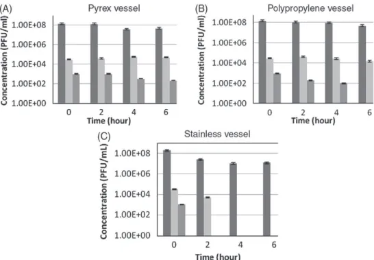
PFU/mL in a saline solution of KCl and in sterilized distilled water . Fig. 3. Adsorption to the (A) Pyrex, (B) polypropylene and (C) stainless beaker of MS2 at feed concentrations 108
, 104
and 103
3. Results and discussion
3.1. Characterization of MS2 suspension with TEM and DLS
The size of viral particles of MS2 phage stock was determined with a suspension at a concentration of 1011PFU/mL. Dynamic light
scattering revealed a single size distribution peak with a z-averaged hydrodynamic diameter of 26.0 nm when negatively-stained prepa-rations (TEM) showed a diameter of approximately 30 nm (Figs. 1 and 2). These results are in agreement with previous studies[9], in which the diameter of the spherical MS2 is found to be 30 nm. Moreover, these measurements show the absence of any par-ticle aggregation, despite the fairly high viral concentration used (1011PFU/mL).
3.2. Adsorption to the test vessel walls
Figs. 3–5 show the virus adsorption to the vessel walls from suspensions at various concentrations prepared in a saline solution containing 8 g/L of KCl. In the aim of limiting MS2 adsorption, Pyrex glass and polypropylene seem to be more appropriate than stainless steel, as no significant change in suspension concentration was observed for initial concentrations larger than 104PFU/mL.
We also observed that adsorption was reversible (experiment not shown); since adsorbed viruses tend to desorb when the virus suspension was replaced by distilled water. This desorption should involve a change in concentration of the suspension in the filtration cell over time. Nevertheless if the adsorption step has been conducted with a virus suspension at high concentration, the quantity desorbed is negligible. A suspension concentration of 108PFU/mL was then
chosen for further experiments.
In order to evaluate the effect of salt on the adsorption in conditions similar to those in drinking water production (i.e. at rather low salt concentration), the same adsorption tests with a suspension concen-tration of 108PFU/mL were performed by using distilled water as
solvent. The same behavior was observed with and without salts in the suspension.
As a conclusion, at high virus concentration (108PFU/mL) no
adsorption is observed on Pyrex and on polypropylene when a light adsorption is obtained on the stainless beaker. In the tested conditions, the quantity adsorbed at neutral pH is independent of the salt concentration of the suspension.
3.3. Adsorption to the membrane materials
This series of experiments was conducted with a virus suspension at a concentration of 108PFU/mL in saline solution. No adsorption on
the membranes was observed (Fig. 5) whatever the material and the molecular weight cut-off ranging from 60 to 200 kDa. Similar results have been obtained in sterilized distilled water (data not shown). 3.4. Adsorption to the filtration equipment
In the first step, tests were performed by circulating (60 L/h; Re = 1800) the virus suspension in the filtration equipment which contains an empty filtration module (shell without membrane).
Results reported inFig. 6show the evolution over time of virus concentration in the permeate streams when two different feed tank materials are used: glass and stainless steel. We observe that the adsorption over time is more important with a stainless steel feed tank than with a Pyrex glass one, that is in accordance with the results presented insection 3.2. As a consequence, the tank made of Pyrex was chosen for further experiments.
Fig. 5. Adsorption of MS2 to membranes made of polyethersulfone , polysulfone , PVDF or cellulose acetate , with various configurations (in/out and out/in) and molecular weight cut-offs. Glass vessel filled with the same virus suspension but without membrane sample was used as blank .
Fig. 6. Initial test using an empty filtration module (shell without membrane) and a feed tank made of stainless steel or of glass .
Fig. 7. Test using an empty filtration module (shell without membrane) and a feed tank made of glass — doping at 15 min.
In a second series of experiments, we aimed at keeping the virus concentration in the rig almost constant and as close as possible to 108
PFU/mL over a sufficiently large period of time in order to be able to evaluate “true” membrane sieving of viruses in future experiments. Experiments have been conducted by adding (doping) 2 mL of solution at 1011PFU/mL in MS2 to the feed suspension, 15, 30 or
60 min after the beginning of the run (Figs. 7–9). According to the results obtained, the most stable conditions in terms of concentration after doping are obtained in the case of virus addition after 15 min (concentration was kept in the range 108
–107PFU/mL over 30 min). 4. Conclusion
The results obtained show that feed reservoirs made of Pyrex glass are the most appropriate of those tested as no virus adsorption was observed on this material in the tested conditions (virus concentra-tion ranging from 103to 108PFU/mL). Doping the feed solution in the
tank 15 min after the beginning of an experiment allows keeping, in the equipment (in the absence of membrane), the virus concentration almost constant and close to 108PFU/mL over 30 min approximately,
which is the range of time necessary to characterize the membrane sieving properties.
According to the present work, the experimental conditions for future tests with membrane are: MS2 suspension prepared in sterilized distilled water at a concentration of 108PFU/mL (neutral
pH) in the feed tank. After 15 min of circulation of this feed suspension in the filtration equipment, 2 mL of the suspension stock of MS2 at the concentration of 1011PFU/mL will be added. The suspension will be
sampled in the permeate and retentate and a counting carried out in order to determine the virus concentration in each stream by the plaque forming unit (PFU) method. In this last step, virus adsorption on membrane structure during filtration has to be evaluated but we
can expect a negligible contribution as no adsorption on membranes was observed after soaking whatever the membrane material and the molecular weight cut-off ranging from 60 to 200 kDa.
Acknowledgment
The authors wish to acknowledge the French National Agency of Research (ANR project) for its support in the framework of the PRECODD research program.
References
[1] S.S. Madaeni, A.G. Fane, G.S. Grohmann, Virus removal from water and wastewater using membranes, J. Memb. Sci. 102 (1995) 65–75.
[2] B. Gassilloud, C. Gantzer, Adhesion–aggregation and inactivation of poliovirus 1 in groundwater stored in a hydrophobic container, Appl. Environ. Microbiol. 71 (2004) 912–920.
[3] B. Gassilloud, L. Huguet, A. Maul, C. Gantzer, Development of a viral concentration method for bottled water stored in hydrophobic support, J. Virol. Methods 142 (2007) 98–104.
[4] G. Herath, K. Yamamoto, T. Urase, Removal of viruses by microfiltration membranes at different solution environments, Wat. Sci. Tech. 40 (1999) 331–338. [5] J. Langlet, F. Gaboriaud, J.F.L. Duval, C. Gantzer, Aggregation and surface properties
of F-specific RNA phages: implication for membrane filtration processes, Water Res. 42 (2008) 2769–2777.
[6] F. Lucena, X. Mendez, A. Moron, E. Calderon, C. Campos, A. Guerrero, M. Cardenas, C. Gantzer, L. Shwartzbrood, S. Skraber, J. Jofre, Occurrence and densities of bacteriophages proposed as indicators and bacterial indicators in river waters from Europe and South America, J. Appl. Microbiol. 94 (2003) 808–815. [7] J. Langlet, F. Gaboriaud, C. Gantzer, Effects of pH on plaque forming unit counts and
aggregation of MS2 bacteriophage, J. Appl. Microbiol. (2007) 1364–5072. [8] J.Y. Hu, S.L. Ong, L.F. Song, Y.Y. Feng, W.T. Liu, T.W. Tan, L.Y. Lee, W.J. Ng, Removal
of MS2 bacteriophage using membrane technologies, Wat. Sci. Tech. 47 (2003) 163–168.
[9] E. Arkhangelsky, V. Gitis, Effect of transmembrane pressure on rejection of viruses by ultrafiltration membranes, Separation and Purification Technology 62 (2008) 621–630.
Fig. 8. Test using an empty filtration module (shell without membrane) and a feed tank made of glass — doping at 30 min.
Fig. 9. Test using an empty filtration module (shell without membrane) and a feed tank made of glass — doping at 60 min.