Université de Montréal
Rôle de la tomodensitométrie à double énergie/double source pour la
personnalisation des traitements de radiothérapie
Par Houda Bahig, MD
Directeurs
Dr Jacques de Guise
Dr David Roberge
Sciences Biomédicales, Faculté de Médecine
Thèse présentée en vue de l’obtention du grade de doctorat en
Sciences Biomédicales, option Médecine Expérimentale
Septembre 2018
TABLES DES MATIÈRES
1 RÉSUMÉ IV 2 ABSTRACT VII 3 LISTE DES TABLEAUX/LIST OF TABLES IX 4 LISTE DES FIGURES/LIST OF FIGURES X 5 REMERCIEMENTS XI 6 LISTE DES SIGLES ET ABBRÉVIATIONS XIII 7 INTRODUCTION GÉNÉRALE 1 7.1 LA RADIOTHÉRAPIE PERSONNALISÉE 1 7.2 PRINCIPES GÉNÉRAUX DU DECT 2 7.3 PRINCIPES GÉNÉRAUX DU DSCT 3 7.4 HYPOTHÈSE ET RATIONNELLE DU PROJET 5 8 PREMIER ARTICLE SCIENTIFIQUE 9 8.1 ABSTRACT 10 8.2 INTRODUCTION 12 8.3 MATERIAL & METHODS 14 8.4 RESULTS 18 8.5 DISCUSSION 21 8.6 CONCLUSION 25 8.7 TABLES ET FIGURES 26 9 DEUXIÈME ARTICLE SCIENTIFIQUE 34 9.1 ABSTRACT 35 9.2 INTRODUCTION 37 9.3 MATERIAL AND METHODS 39 9.4 RESULTS 42 9.5 DISCUSSION 44 9.6 CONCLUSION 49 9.7 TABLES ET FIGURES 50 10 TROISIÈME ARTICLE SCIENTIFIQUE 58 10.1 ABSTRACT 59 10.2 INTRODUCTION 61 10.3 MATERIAL AND METHODS 62
10.4 RESULTS 64 10.5 DISCUSSION 65 10.6 CONCLUSION 68 10.7 TABLES ET FIGURES 69 11 QUATRIÈME ARTICLE SCIENTIFIQUE 72 11.1 ABSTRACT 73 11.2 INTRODUCTION 75 11.3 MATERIAL AND METHODS 76 11.4 RESULTS 79 11.5 DISCUSSION 80 11.6 CONCLUSION 84 11.7 TABLES ET FIGURES 86 12 DISCUSSION GENERALE 91 12.1 CARACTÉRISATION DES TISSUS SAINS 91 12.2 CARACTÉRISATION TUMORALE 95 12.3 IMAGERIE CARDIAQUE 96 12.4 LIMITATIONS DU DECT/DSCT 97 13 CONCLUSION GÉNÉRALE 100 14 BIBLIOGRAPHIE 101
1 RÉSUMÉ
Le futur de la radiothérapie réside dans le développement de stratégies visant à adapter les traitements à chaque individu. La tomodensitométrie à double énergie/double source (DECT/DSCT) est une technologie d’imagerie permettant de caractériser avec précision les tissus(sains et tumoraux) et d’imager le cœur en mouvement. En raison de ses fonctionnalités, la technologie du DECT/DSCT a le potentiel de jouer un rôle important dans la personnalisation des traitements de radiothérapie. Nous avons exploré le rôle du DECT/DSCT dans 3 études cliniques prospectives, relatives à la planification des traitements de radiothérapie.
Dans une première approche, nous avons évalué le rôle du DECT pour l’évaluation de la fonction du parenchyme pulmonaire en radiothérapie thoracique (conventionnelle et stéréotaxique). Nous avons émis l’hypothèse qu’une quantification précise de la concentration d’iode du parenchyme pulmonaire, dérivée du DECT, permettrait de déterminer les régions pulmonaires les plus fonctionnelles à éviter lors de la planification de la radiothérapie. Nous avons démontré la faisabilité et la validité d’une méthode de quantification de la fonction pulmonaire en utilisant la cartographie d’iode du DECT. De plus, nous avons montré que l’incorporation de cette information en planification de radiothérapie peut réduire significativement la dose aux régions pulmonaires fonctionnelles dans le but de réduire les toxicités.
Dans une deuxième approche, nous avons évalué le rôle du l’imagerie à double-source (DSCT) pour une évaluation individualisée du mouvement cardiaque lors de la planification des traitements de radiothérapie. Nous avons montré que le DSCT permettait de visualiser et quantifier le mouvement des sous-structures cardiaques notamment les veines pulmonaires et les artères coronaires, et ainsi déterminer un volume cible personnalisé pour chaque patient. De plus, nous avons montré le bénéfice dosimétrique d’une irradiation du sein gauche avec synchronisation cardiaque (limitée à la phase systolique) pour épargner les sous-structures cardiaques, notamment de l’artère antérieure descendante gauche, une structure critique dans le développement des toxicités cardiaques post-radiques.
Finalement, utilisant à nouveau la capacité de quantification précise de la concentration d’iode, nous avons évalué le rôle du DECT pour dériver la perfusion des tumeurs du larynx et de l’hypopharynx traitées par radiothérapie. Dans un contexte exploratoire de 25 patients, nous avons démontré que les statistiques quantitatives dérivées des cartes d’iodes tumorales étaient prédictives du contrôle locorégional chez ces patients, suggérant un rôle de ces cartes d’iode comme bio-marqueurs prédisant l’agressivité tumorale.
Les résultats de nos travaux centrés sur ces 3 stratégies démontrent que le DECT/DSCT a le potentiel de jouer un rôle important à divers niveaux dans la personnalisation de la planification des traitements radiothérapie, notamment: 1) pour l’évaluation de la fonction des tissus sains; 2) pour la détermination personnalisée du mouvement cardiaque, et 3) comme outil prédictif du contrôle tumoral.
Mots clés: Tomodensitométrie à double énergie (DECT); tomodensitométrie à double source (DSCT); radiothérapie; cancer; toxicités; médicine personnalisée; imagerie fonctionnelle; imagerie cardiaque.
2 ABSTRACT
The future of radiotherapy lies in the development of strategies to adapt treatments to each individual. Dual energy / dual source computed tomography (DECT/DSCT) is an imaging technology that allows for accurate tissue characterisation (organs at risk or tumors) and that can capture precisely the anatomy of the heart in motion. DECT/DSCT technology has the potential to be important player in personalized radiotherapy. We explored the role of DECT/DSCT in radiotherapy planning in the context of 3 prospective clinical studies.
First, we evaluated the role of DECT imaging for the assessment of lung function in lung cancer radiotherapy planning (both conventionally fractionated and stereotaxic radiotherapy). We hypothesized that accurate quantification of DECT iodine concentration could be used in treatment planning to assesses and preserve functional parenchyma. We demonstrated the feasibility and validity of a novel lung function quantification method using DECT iodine mapping. In addition, we showed that incorporating this information into radiotherapy planning could help improve lung dosimetry, and thus potentially reduce toxicities.
In a second approach, we evaluated the role of DSCT for individualized assessment of cardiac motion in radiotherapy planning. We showed that the DSCT allows visualization and quantification of motion of cardiac sub-structures, including the pulmonary veins and coronary arteries, and therefore can be used to determine personalized target volume for each patient. In addition, we quantified the dosimetric impact of cardiac-gated radiotherapy in left breast cancer radiotherapy and demonstrated significant sparing of cardiac sub-structures with this
method, in particular sparing of left anterior descending artery, a critical structure involved in radiation-induced cardiovascular toxicities.
Finally, we assessed the role of DECT in determining tumor perfusion in larynx/hypopharynx cancers using the iodine concentration quantification method. In an exploratory prospective cohort of 25 patients with cancer of the larynx or hypopharynx, we demonstrated that histogram statistics derived from tumor iodine maps could predict locoregional control in these patients. This finding supports the role of iodine concentration maps as functional biomarkers to predict tumor aggressiveness.
The results of our work focused on these various strategies demonstrate that DECT/DSCT has the potential to play an important role in the following 3 avenues of personalized radiotherapy: 1) for the evaluation of functional healthy tissues; 2) for individualized determination of optimal margins or cardiac-gating window in radiotherapy involving the heart and, 3) for prediction of cancer control outcomes.
Key words: Dual-energy computed tomography (DECT); dual source computed tomography (DSCT); radiotherapy; cancer; toxicities; personalized medicine; functional imaging; cardiac imaging.
3 LISTE DES TABLEAUX/LIST OF TABLES
TABLE 1- CHARACTERISTICS OF PATIENTS WITH LUNG CANCER ENROLLED IN THE STUDY ... 26
TABLE 2- DECT VS. SPECT/CT- DERIVED LOBAR FUNCTION AND SUMMARY OF WL VS. FL DOSIMETRY ... 28
TABLE 3- VARIATION OF CARDIAC STRUCTURES VOLUME ALONG THE CARDIAC CYCLE AND HAUSDORFF BETWEEN CARDIAC STRUCTURES AND TANGENTIAL FIELDS ... 50
TABLE 4- DOSE VARIATION FOR WH, LV, AND LMCA-LAD WITH ALONG THE CARDIAC CYCLE .. 51
TABLE 5- MEAN DOSE TO WH, LV, MIDDLE AND DISTAL LAD IN EACH PHASE OF THE CARDIAC CYCLE ... 52
TABLE 6- BASIC DSCT RELATED PARAMETERS ... 69
TABLE 7- MEAN DISPLACEMENTS OF RSPVA, RIPVA, LSPVA AND LIPVA ... 69
TABLE 8- PATIENTS AND TUMORS’ CHARACTERISTICS ... 86
TABLE 9- UNIVARIATE COX REGRESSION ANALYSIS OF ALL VARIABLES FOR RISK OF LRR FOR ENTIRE COHORT ... 87
TABLE 10-COX REGRESSION ANALYSIS FOR RISK OF LRR FOR SUBGROUP OF PATIENTS TREATED WITH UPFRONT RADIATION +/- CHEMOTHERAPY (N=17) ... 88
4 LISTE DES FIGURES/LIST OF FIGURES
FIGURE 1- MANUAL SEGMENTATION OF LUNG LOBES ON DECT AND SPECT/CT ... 29 FIGURE 2- CORONAL SLICE SHOWING 6 FUNCTIONAL LUNG SUB-VOLUMES WITH INCREASING
IODINE CONTENT. ... 30 FIGURE 3- DECT-IODINE AND SPECT/CT MAPS ... 31 FIGURE 4- LINEAR CORRELATION BETWEEN DECT-IODINE MAP AND SPECT/CT LOBAR
FUNCTION ... 32 FIGURE 5- RELATIVE VARIATIONS IN MLD, V5 AND V20 BETWEEN WL VS. FL DOSIMETRIES. ... 33 FIGURE 6- CONTOURS OF THE WH, LV AND DISTAL LAD ... 53 FIGURE 7 - EXAMPLE OF 4D DOSE VOLUME HISTOGRAM SHOWING MAXIMUM SYSTOLIC AND
DIASTOLIC DOSES TO LAD SEGMENTS ... 54 FIGURE 8- CORRELATION BETWEEN MEAN HAUSDORFF DISTANCE TO TANGENTIAL FIELDS AND
DOSE VARIATION WITH CARDIAC CYCLE FOR WH, LV, MIDDLE AND DISTAL LAD. ... 55 FIGURE 9- PLOTS OF MAXIMUM CARDIAC DISPLACEMENT OF RIGHT SUPERIOR VS. RIGHT INFERIOR
PULMONARY ANTRUMS (A) AS WELL AS LEFT SUPERIOR VS. LEFT INFERIOR PULMONARY
VEIN ANTRUMS (B) ... 70 FIGURE 10- ROI REPRESENTING THE RSPVA DEFINED AS A 5 MM AXIAL SURFACE AT THE
ANTERO-SUPERIOR INTERSECTION OF THE SUPERIOR PULMONARY VEIN WITH THE LEFT
ATRIUM ... 71 FIGURE 11- WEIGHTED AVERAGE AXIAL DECT FROM PATIENT WITH T2 SUPRAGLOTTIC SCC. ... 89 FIGURE 12- KAPLAN MEIER CURVES OF LOCO-REGIONAL CONTROL (LRC) AS A FUNCTION OF
TIME ... 90 FIGURE 13- KAPLAN MEIER CURVES OF RELAPSE-FREE SURVIVAL AS A FUNCTION OF TIME ... 90
5 REMERCIEMENTS
Premièrement, j’aimerai remercier mes 2 formidables directeurs de recherche : Les Drs Jacques de Guise et David Roberge. Je n’aurai pu espérer un meilleur duo pour supporter mon début de carrière en recherche et je les remercie pour la confiance et la liberté intellectuelle qu’ils m’ont accordée. Le Dr Jacques de Guise m’a accueilli les bras ouverts dans son monde de l’imagerie, m’a donné une grande autonomie tout en étant toujours disponible, mais surtout, il a été un rôle modèle inestimable. Le Dr Roberge, ce génie de la radio-oncologie qui « pense plus vite que son ombre », a été un mentor exceptionnel sans qui je n’aurai pu naviguer à travers les défis rencontrés. Je lui suis reconnaissante pour son support continuel et pour toutes les opportunités qu’il a mis sur mon chemin.
Je tiens également à remercier l’équipe de physique médicale, incluant les Drs Stéphane Bedwani, Andréanne Lapointe et Hugo Bouchard, ainsi que Danis Blais. Leur rigueur et leur créativité à solutionner les problèmes ont été cruciales à l’aboutissement de ce doctorat. De la même manière, j’aimerais remercier les cliniciens, notamment les Drs Edith Filion, Marie-Pierre Campeau, Toni Vu, Félix-Phuc Nguyen-Tan et Louise Lambert qui ont facilité l’implémentation des études cliniques, le recrutement de patients et qui ont offert leur support au quotidien. De plus, j’aimerais remercier les collaborateurs en radiologie et médecine nucléaire, notamment les Drs Carl Chartrand Lefebvre, Laurent Létourneau-Guillon et Martin Lord, qui ont généreusement offert leur expertise et ont été indispensables à assurer la validité des méthodes proposées.
incomptables sacrifices, notamment leur courageux et altruiste parcours migratoire avec pour seul objectif d’offrir à leur enfants un avenir meilleur. Merci à mon fiancé pour sa patience inébranlable, ses encouragements au quotidien et son sens de l’humour imbattable.
Enfin, je voudrai souligner les sources de financement reçus pour la réalisation de ce projet, qui incluent : une bourse d’étude des Fonds de Recherche Santé Québec, une bourse de projet pilote du Réseau de Bio-Imagerie du Québec, les bourses de Support Professoral du département de Radiologie, Radio-oncologie et Médecine nucléaire de l’Université de Montréal, ainsi qu’une bourse de recherche de l’industrie de Varian Medical Systems®.
6 LISTE DES SIGLES ET ABBRÉVIATIONS
4D-CT: Four-dimensional computed tomography 99mTc-MAA: 99m-Technetium macro-aggregated albumin
CI: Confidence Interval
COPD: Chronic obstructive pulmonary disease
CK: CyberKnife®
CT: Computed Tomography
DECT: Tomodensitométrie à double énergie DSCT: Tomodensitométrie à double source DIBH: Deep-inspiration breath-hold
DLCO: Diffusing capacity of the lungs for carbon monoxide DVH: Dose-volume histograms
ECG: Electrocardiogramme FAZA: Fluoroazomycin arabinoside
FEV1: Forced expiratory volume in 1 second
FDG: Fluorodeoxyglucose
FMISO: Fluoromisonidazole
FVC: Forced vital capacity
FL: Functional lung
HR: Hazard ratio
IC: Iodine concentration
ITV: Internal target volume
IMRT: Intensity-modulated radiotherapy LAD: Left anterior descending artery LLL: Left lower lobe
LMCA: Left main coronary artery LRR: Loco-regional recurrence
LUL: Left upper lobe
LV: Left ventricle
M: Mean
MLD: Mean dose
MRI: Magnetic resonance imaging
N: Number
NaCl: Sodium chloride
NSCLC: Non small cell lung cancer PET: Positron emission tomography PTV: Planning target volume
QUANTEC: Quantitative Analysis of Normal Tissue Effects in the Clinic RUL: Right upper lobe
RML: Right middle lobe RLL: Right lower lobe
SABR: Stereotactic ablative radiotherapy SCLC: Small cell lung cancer
TPS: Treatment planning system
V5: Percent lung volume receiving 5 Gy V20: Percent lung volume receiving 20 Gy
WL: Whole lung
7 INTRODUCTION GÉNÉRALE
7.1 LA RADIOTHÉRAPIE PERSONNALISÉE
L’objectif global de la radiothérapie est d’administrer la dose prescrite à la tumeur tout en réduisant la dose aux organes sains environnants. Dans les 2 dernières décennies, l’introduction de techniques comme la radiothérapie par modulation d’intensité (IMRT), la radiothérapie stéréotaxique et la radiothérapie guidée par l'imagerie ont permis d’augmenter significativement la précision des traitements (1, 2). Dans le contexte de l’utilisation grandissante d’imagerie multi-modalités pour la planification des traitements, il existe un intérêt croissant pour le concept d’une « radiothérapie personnalisée » visant à élargir l’indice thérapeutique (3). L’une des potentielles approches de « personnalisation » des traitements de radiothérapie consiste à intégrer non seulement l’information anatomique, mais également l’information fonctionnelle lors de la planification des traitements (4). Parmi ces applications, on compte par exemple le rôle de bio- marqueurs d’imagerie (résonnance magnétique, tomodensitométrie de perfusion ou tomodensitométrie par émission de positron) pour déterminer le volume cible à traiter ou prédire le comportement des tumeurs et le pronostic des patients (5-7). On compte également l’utilisation de bio-marqueurs pour évaluer la fonction des tissus sains environnants et estimer plus judicieusement la réserve de chaque patient pour minimiser les dommages radio-induits (8-10). D’autres applications comptent l’évaluation individualisée du mouvement tumoral ou des tissus sains, induits par exemple par la respiration, le péristaltisme, le mouvement aléatoire des organes ou les contractions cardiaques, dans le but d’utiliser des marges individualisés permettant de traiter la cible en
mouvement, tout en protégeant maximalement les organes environnants (11-13). L’utilisation de plusieurs de ces applications en clinique demeure expérimentale. Ces diverses stratégies ont pour objectif global l’amélioration du contrôle tumoral, la réduction des complications post-radiques et la préservation de la qualité de vie des patients traités.
7.2 PRINCIPES GÉNÉRAUX DU DECT
L’imagerie par tomodensitométrie à double énergie (DECT) est basée sur le principe que l’utilisation des rayons X à 2 énergies différentes donne lieu à une atténuation spécifique à un matériau donné (14, 15). Cette caractéristique permet de distinguer et de mesurer la quantité des différentes substances comme l’iode ou le calcium du reste des tissus mous (16).
Aux énergies utilisées en imagerie, l’effet photoélectrique et l’effet Compton sont les 2 composantes les plus importantes d’atténuation des rayons X passant par un matériau donné (17). La composante d’atténuation de l’effet photoélectrique est fortement dépendante du numéro atomique Z du matériau traversé et inversement proportionnelle à l’énergie du faisceau. L’effet photoélectrique survient lorsqu’un photon incident a suffisamment d’énergie pour causer l’éjection d’un électron de la couche K (la couche la plus profonde) d’un atome. Les chances que l’effet photoélectrique se produise augmentent avec le rapprochement de l’énergie du photon incident de celle de l’énergie de liaison de la couche K (17, 18). Le coefficient d’extinction K d’une substance fait référence au pic d’atténuation observé à un niveau d’énergie juste au dessus de celui de l’énergie de liaison de la couche K - en raison de l’augmentation drastique de l’absorption photoélectrique à ce niveau d’énergie. Le coefficient d’extinction varie pour chaque matériau et est plus élevé avec l'augmentation des numéros
atomiques (17).
Alors qu’avec un CT à simple énergie, la différentiation des différents matériaux n’est basée que sur leur atténuation à cette énergie spécifique (quantifiée en Unité Hounsfield) (19), le DECT permet la différentiation de matériau par l’interdépendance entre l’effet photoélectrique, l’énergie et la variabilité des coefficients d’extinction K pour chaque élément. L’acquisition de 2 séries d’images à différentes énergies permet donc de mesurer la différence d’atténuation entres les acquisitions de faible et haute énergie et ainsi de décomposer la matière en caractérisant les éléments qui la composent. Il devient ainsi possible de séparer par exemple l’iode, le calcium ou le Xénon du reste des tissus mous, en dérivant le contenu de chaque voxel (équivalent 3D du pixel, le voxel correspondant à la plus petite entité distinguable sur une imagerie tridimensionnelle) (16).
Il existe plusieurs types de DECT qui diffèrent dans la technique d’acquisition des séries de haute et faible énergie. Les images peuvent être acquises par l’utilisation de 2 sources de rayons X à 2 énergies différentes (dit, double-source) ou par l’utilisation d’une seule source (soit par alternance rapide de kV ou par utilisation d’un détecteur à 2 couches) (18). Dans le cadre de notre projet, nous utilisons une tomodensitométrie à double-source (DSCT ; SOMATOM® Definition Flash, Siemens Healthineers).
7.3 PRINCIPES GÉNÉRAUX DU DSCT
Bien que la tomodensitométrie à double source (DSCT) permette l’acquisition d’images à des énergies différentes et de bénéficier de l’information relative à l’atténuation différentielle, le
DSCT a été initialement développé pour l’imagerie cardiaque. En effet, les 2 tubes à rayon X disposés à 90o
degrés l’un par rapport à l’autre, associés à leurs détecteurs correspondants, permettent une augmentation de la vitesse d’acquisition d’image et ainsi une résolution temporelle supérieure (20, 21) avec, dans le cas du DSCT utilisé dans le cadre de ce projet (SOMATOM® Definition Flash, Siemens Healthineers) un temps de rotation de 0.28 seconde et une résolution temporelle de 75 milisecondes. Le principal défi de l'imagerie cardiaque non invasive est celui de la visualisation précise des structures cardiaques, notamment des petites structures comme les artères coronaires et la paroi vasculaire, qui sont sujets à des artéfacts de mouvements liés à la contraction cardiaque. Les études initiales en imagerie diagnostique ont montré que l’acquisition de DSCT synchronisé à l’électrocardiogramme (ECG) permettait d’imager le cœur en mouvement indépendamment de la fréquence ou du rythme cardiaque, de visualiser les artères coronaires ainsi que d’évaluer l’étendue de leur mouvement, sans artéfacts de mouvement (20, 22-28). Le signal de l’ECG du patient est surveillé pendant l’examen et l’acquisition est déclenchée à une phase prédéfinie du cycle cardiaque. Bien que ceci n’ait pas été évalué en radiothérapie préalablement, l’utilisation du DSCT pour évaluer le déplacement du cœur et des sous-structures du cœur de la diastole à la systole aurait plusieurs implications pertinentes pour la personnalisation des traitements. Ces applications incluent notamment de déterminer des marges de traitements personnalisées dans les cas d’une irradiation thoracique où le coeur est un organe à risque (par exemple en radiothérapie du sein, poumon, ou autre structures thoracique), ou de faciliter le traitement de pathologies cardiaques en permettant une visualisation appropriée des structures cardiaques et de leur marge de mouvement. Enfin, à l’image de ce qui est fait en imagerie 4D respiratoire (29, 30), le DSCT pourrait permettre de déterminer de façon individualisée la fenêtre d’irradiation optimale
pendant le cycle cardiaque, et ainsi élargir l’indice thérapeutique et minimiser les toxicités cardiaques liées à la radiothérapie.
7.4 HYPOTHÈSE ET RATIONNELLE DU PROJET
Actuellement, le flux de travail standard des traitements de radiothérapie débute par l’acquisition d’une tomodensitométrie dite de planification. Cette tomodensitométrie est utilisée pour préparer les traitements de radiothérapie, pour définir les volumes cibles, pour définir les organes sains à éviter et pour faire le calcul dosimétrique. Dans le contexte de la radiothérapie, le DECT/DSCT a l’avantage de pouvoir être facilement intégré dans le flux de travail des traitements tout en ajoutant l’information de la caractérisation tissulaire (en mode DECT) ou de l’évaluation du mouvement cardiaque (en mode DSCT). Contrairement aux autres multi-modalités d’imagerie souvent intégrées à la planification des traitements de radiothérapies comme l’IRM ou le PET scan par exemple, le DECT/DSCT à l’avantage particulier qu’il ne constitue pas un examen supplémentaire, mais qu’il peut simplement servir de tomodensitométrie de planification. Ainsi, l’information fournie par le DECT/DSCT a l’avantage particulier d’être exactement en position de traitement et donc de ne pas nécessiter l’étape supplémentaire de fusion. En plus de faciliter le flux de travail, ce dernier point a aussi l’avantage d’une meilleure précision anatomique, comparativement a une imprécision de plusieurs millimètres associée à l’étape de fusion du scan de planification avec un autre modalité comme l’IRM par exemple (31).
L’hypothèse centrale de notre projet est que l’utilisation du DECT/DSCT comme tomodensitométrie de planification dans le flux de travail de la radiothérapie, peut jouer un rôle important dans l’individualisation des traitements de radiothérapie pour la réduction de la dose aux organes à risques, de même que pour la prédiction de la réponse tumorale. Dans ce cadre, nous avons procédé à l’évaluation de cette technologie dans 3 contextes cliniques pertinents à la planification de la radiothérapie :
a) Première approche: Le DECT pour l’évaluation de la fonction pulmonaire (publication 1)
La première approche évaluée est celle du rôle de l’imagerie à double-énergie pour l’évaluation de la fonction des tissus sains en radiothérapie pulmonaire (conventionnelle et stéréotaxique). Lors d’une irradiation thoracique, les contraintes de dose utilisées pour les poumons dans le but de minimiser le risque de toxicités ont pouvoir de prédiction variable, avec des taux de pneumonite estimés variant entre 5 and 40%, dépendamment des techniques, des doses et du volume pulmonaire irradié (32). Les contraintes de dose utilisées en clinique sont exprimées sous forme de dose moyenne aux poumons et/ou d’histogramme dose-volume. Un problème important de ces contraintes est qu’elles considèrent que chaque voxel pulmonaire contribue également à la fonction des poumons; elles ne prennent donc pas en compte la fonction pulmonaire souvent asymétrique d’une majorité de patients atteints de maladie pulmonaire obstructive chronique. Dans le cadre de cette première étude, nous avons émis l’hypothèse que la caractérisation tissulaire et la quantification précise de l’iode dans le parenchyme pulmonaire au DECT permettraient d’établir une corrélation anatomo-fonctionnelle précise, qui pourrait être utilisée lors la planification des traitements afin de
préserver le parenchyme fonctionnel. L’objectif de cette étude prospective est de quantifier la fonction pulmonaire basée sur la cartographie d’iode dérivée du DECT chez les patients traités pour un cancer pulmonaire ainsi que d’évaluer l’impact dosimétrique de l’intégration de ces cartes d’iode en planification de radiothérapie. Étant donné que la littérature actuelle sur les toxicités pulmonaires démontre une relation linéaire entre la réduction de dose aux poumons et les risques de pneumonites post-radiques, le potentiel clinique d’une telle méthode est celui de réduire les risques de toxicités.
b) Deuxième approche: Le DSCT pour évaluer le mouvement cardiaque (publications 2 et 3)
Dans un deuxième temps, explorant le fait que le DSCT offre la possibilité d’évaluer le mouvement cardiaque et de visualiser les coronaires et le myocarde, nous avons évalué le rôle du DSCT pour une évaluation personnalisée du mouvement cardiaque lors de la planification des traitements de radiothérapie. Le coeur étant fréquemment inclus dans les champs tangentiels d’une radiothérapie du sein gauche, il a été démontré que le risque de maladies ischémiques est augmenté suite à une irradiation du sein gauche (33). Dans une cohorte prospective de 10 patientes traitées par radiothérapie limitée au sein gauche, nous avons évalué 2 questions cliniques. Dans une première étude, nous avons quantifié le potentiel bénéfice dosimétrique d’une irradiation du sein gauche avec synchronisation cardiaque, permettant une épargne des sous-structures cardiaques. Plus précisément, nous avons évalué la variation de dose aux différents segments de l’artère antérieure descendante gauche, à l’artère coronarienne gauche, au ventricule gauche et au cœur pendant le cycle cardiaque (de la systole à la diastole). En utilisant la même cohorte de patientes dans une seconde étude, nous avons
évalué l’amplitude de déplacement de l’antrum veines pulmonaires lors du battement cardiaque dans le but de déterminer un volume cible personnalisé dans la nouvelle avenue du traitement par radiothérapie stéréotaxique d’arythmies cardiaques réfractaires.
c) Troisième approche: Le DECT pour évaluer la perfusion tumorale (publication 4)
Comme deuxième approche, nous avons évalué le rôle du DECT pour évaluer la perfusion tumorale dans les cancers de la tête et du cou. Nous avons émis l’hypothèse que suite à l’acquisition d’un DECT injecté en phase artérielle, la concentration tumorale d’iode dérivée du DECT serait représentative de la perfusion tumorale. Une concentration d’iode plus élevée serait indicative d’une augmentation du volume sanguin et d’une hyperperméabilité capillaire représentant une néoangiogenèse augmentée. Dans une cohorte prospective de 25 patients avec des cancers du larynx supraglottique ou de l’hypopharynx, nous avons investigué le rôle des données statistiques quantitatives dérivées des cartes d’iodes tumorales du DECT pour prédire les récidives locoregionales.
8 PREMIER ARTICLE SCIENTIFIQUE
Phase I-II Study of Dual-Energy Computed Tomography (DECT) for Assessment of Pulmonary Function in Radiotherapy Planning
Houda Bahig, MD1 , Marie-Pierre Campeau, MD1 , Andréanne Lapointe MSc1 , Stephane Bedwani PhD1 , David Roberge MD1 , Jacques de Guise PhD2 , Danis Blais MSc, 1 Toni Vu MD1 , Louise Lambert MD1 , Carl Chartrand-Lefebvre MD MSc3 , Martin Lord MD4 , Edith Filion MD1 1
Radiation Oncology department, Centre Hospitalier de l’Université de Montréal, Montreal, Quebec, Canada
2
Imaging and Orthopaedics Research Laboratory (LIO), École de Technologie Supérieure, Centre de Recherche du Centre Hospitalier de l'Université de Montréal (CRCHUM), Montreal, Quebec, Canada
3
Radiology departement, Centre Hospitalier de l’Université de Montréal, Montreal, Quebec, Canada
4
Nuclear medicine department, Centre Hospitalier de l’Université de Montréal, Montreal, Quebec, Canada
Keywords: Dual energy CT, Lung, Thoracic radiotherapy, SBRT, IMRT, functional imaging, iodine contrast, tissue characterization, dosimetry, radiation pneumonitis.
Publication:
Int J Radiat Oncol Biol Phys. 2017 Oct 1;99(2):334-343. doi: 10.1016/j.ijrobp.2017.05.051. Epub 2017 Jun 7.
CONTRIBUTIONS: Rédaction du protocole de recherche, coordination et gestion de l’implémentation du protocole de recherche en clinique, récole et analyse des données, interprétation des données, rédaction du manuscrit.
8.1 ABSTRACT
Purpose: To quantify lung function based on a dual energy computed tomography
(DECT)-derived iodine map in patients treated with radiotherapy for lung cancer, and to assess the dosimetric impact of its integration in radiotherapy planning.
Methods: Patients treated with stereotactic ablative radiotherapy (SABR) for early stage or
intensity-modulated radiotherapy (IMRT) for locally advanced lung cancer were prospectively enrolled in this study. A DECT in treatment position was obtained at time of treatment planning. The relative contribution of each voxel to the total lung function was based on iodine distribution. The composition of each voxel was determined based on a two-material decomposition. DECT-derived lobar function was compared to Single Photon Emission Computed Tomography / Computed Tomography (SPECT/CT). A functional map was integrated in treatment planning system using 6 sub-volumes of increasing iodine distribution levels. Percent lung volume receiving 5Gy (V5), V20 and mean dose (MLD) to whole lungs (anatomical) vs. functional lungs were compared.
Results: 25 patients, including 18 patients with lung cancer treated with SABR and 7 patients
with IMRT (locally advanced), were included. Eighty-four percent had chronic obstructive pulmonary disease. Median forced expiratory volume in 1 second was 62% of predicted (29-113%) and median diffusing capacity of the lung for carbon monoxide was 56% (39-91%). There was a strong linear correlation between DECT and SPECT/CT-derived lobar function (Pearson’s coefficient correlation r= 0.89, p<0.00001). Mean differences in V5, V20 and MLD between anatomical and functional lung volumes were 16% (0-48%, p=0.03), 5% (1-15%, p=0.12) and 15% (1-43%, p=0.047), respectively.
Conclusion: Lobar function derived from DECT iodine map correlates well with SPECT/CT
and its integration in lung treatment planning is associated with significant differences in V5 and MLD to functional lungs. Future work will involve integration of weighted functional volume in the treatment planning system, along with integration of iodine map for functional lung-sparing IMRT.
8.2 INTRODUCTION
The goal of lung cancer radiotherapy is to administer the prescribed dose to the tumor while minimizing the dose to the surrounding healthy lungs and other organs at risk. Radiation-induced pulmonary toxicities include a wide spectrum of events ranging from subclinical radiological changes to fatal complications of respiratory failure (34). Rates of clinical radiation pneumonitis (RP) after radiotherapy for lung cancer vary between 5 and 50% in the literature (35). In lung stereotactic ablative radiotherapy (SABR) as well as in conventional radiotherapy, several dose parameters are used to predict the risk of RP, the most commonly used being: mean lung dose (MLD) (32) as well as volume receiving 5 Gy (V5), 20 Gy (V20), 30 Gy (V30) and 60 Gy (V60) (36, 37). However, these parameters were shown to have a highly variable predictive power for RP risk estimation. This was hypothesised to be due to heterogeneities in patient-related (comorbidities/smoking habits) and treatment-related factors, which significantly decrease the sensitivity of these parameters (35, 37). Nonetheless, data from the Quantitative Analysis of Normal Tissue Effects in the Clinic (QUANTEC) (32) suggest that there is no threshold below which RP risk is null and highlights the importance of lung-sparing strategies.
More than half of lung cancer patients have concomitant pulmonary disease (38), and therefore have reduced and inhomogeneous lung function. Marks et al. (39) showed that exclusion of patients with severe chronic obstructive pulmonary disease (COPD) from the QUANTEC analysis was associated with markedly increased sensitivity of lung dose-volume histograms (DVH). This suggested that functional lung volume information may be more useful than anatomical lung volume for these patients. In the context of both 3D conformal
radiotherapy and intensity modulated radiotherapy (IMRT), functional lung avoidance has been proposed in several feasibility studies (40). While Single Photon Emission Computed Tomography (SPECT) perfusion scintigraphy is the most studied method for evaluation of relative pulmonary function (41), in recent years, several alternative methods have emerged, including the use of hyperpolarized gas magnetic resonance imaging (MRI) (42), four-dimensional (4D) CT ventilation maps (43) (44) (45) and 4D positron emission tomography(PET)/CT (46). These methods have diverse potential advantages in comparison to SPECT/CT, including avoidance of ionizing radiation and improvement of anatomical precision.
Dual-energy computed tomography (DECT) is an advanced imaging technique, recently introduced in radiation oncology. Based on the principle that the use of two different X-ray energies results in an attenuation specific to a given material, this technique has the potential to provide better tissue characterization (15). This characteristic of the DECT allows for precise voxel-to-voxel determination of iodine partial electronic density, which can be used to derive a regional blood volume map (16). Our hypothesis is that a DECT-iodine map of the lungs derived from a contrast-injected CT at time of planning would provide precise lung anatomical and functional information. This information can be used at time of treatment planning for a better estimation of delivered dose to functional lung. Compared to previously developed methods, the proposed technique has the advantage of deriving lung function directly from the treatment planning CT, therefore simplifying workflow, at no additional ionising radiation cost. The purpose of the current study was to quantify relative lung function using DECT-iodine map and correlate these results with SPECT/CT (current standard). The
secondary objective was to compare whole (anatomical) lung to functional lung dosimetry in patients treated for lung cancer.
8.3 MATERIAL & METHODS Patient population
Patients were prospectively enrolled in this study from September 2014 to June 2016. Eligibility criteria included: (1) a clinical diagnosis of lung cancer; (2) Eastern Cooperative Oncology Group performance status 0-2; (3) planned curative radiotherapy. Exclusion criteria were: previous radiotherapy, iodine allergy or other contra-indications to iodine injection. Patients with locally advanced lung cancer treated with concurrent or sequential chemotherapy could be included in this study. Baseline work-up included: thoracic computed tomography (CT) scan, bronchoscopy, 18-fluoro-2-deoxy-d-glucose positron emission tomographic (FDG-PET) scan, pulmonary function tests and, when available, histological confirmation. In selected early stage cases where histological confirmation could not be obtained, tumor progression on CT and increased FDG-PET uptake were used to establish the clinical diagnosis of malignancy. All cases were discussed in the context of a multidisciplinary tumor board. The protocol and patient consent form were reviewed and approved by our institutional ethics committee and all patients signed a consent form.
Planning CT, DECT and SPECT/CT
The dual-source DECT available in our department (SOMATOM Definition Flash; Siemens Healthineers) was used for planning purposes. Planning CT was acquired in supine position, with the arms along the sides. Immobilisation at the time of treatment planning was
individualized based on treatment technique and involved a custom vacuum cushion or a double vacuum immobilization device (BodyFix, Elekta, Stockholm, Sweden). All patients had a 2 mm slice thickness single-energy non-contrast four-dimensional (4D) supine planning CT-scan in free breathing. A 3D contrast enhanced DECT in suspended breathing (end-expiration) was subsequently obtained in treatment position, using an angiography protocol for optimal visualisation of lung perfusion (47). Iopamidol contrast bolus was injected (Isovue 370 mg/mL) in the antecubital vein at a rate of 4 mL / seconds over 16 seconds (total of 64 mL). Immediately after, a 24 mL saline solution with 50% contrast concentration and a 40 mL saline solution were sequentially injected at a rate of 4 mL / sec. DECT was acquired in distal arterial phase, 7 seconds after contrast bolus tracking in the pulmonary trunk (47), with acquisition from inferior to superior direction to limit artefacts caused by contrast accumulation in the superior vena cava and supraclavicular veins. Two sets of spiral CT data were simultaneously acquired with the following parameters: slice thickness of 2 mm, pitch of 0.55, rotation time of 0.28 seconds, collimation of 64 x 0.6 mm and X-ray tube potentials of 100 kVp and 140Sn kVp (Sn= additional tin filtration), a matrix of 512 × 512 pixels, a field of view of 50 cm and a voxel size of 1 x 1 x 1 mm3
. A Q30 filter kernel was used for image reconstruction. Care Dose (Siemens Healthineers) technology was used for modulation of tube current in order to maintain constant image quality.All patients also underwent pre-treatment 99m-Technetium macro-aggregated albumin (99mTc-MAA) SPECT/CT. All studies were performed on a SPECT/CT camera (Discovery NM/CT 670, GE Healthcare) after intravenous administration of 99mTc-MAA (mean dose of 292 Megabecquerel). The SPECT was acquired in supine position with the arms elevated over the head in order to limit artefact at the thorax (48) and in free breathing, with the following parameters: 20% energy window centered
at 140 keV, 60 projections/360o
, matrix 128 x 128 and voxel size 3.45 x 3.45 x 3.45 mm3
. SPECT and CT were subsequently fused using rigid co-registration. A low-dose non-enhanced CT was acquired immediately before the perfusion SPECT acquisition, in same position as SPECT and in free breathing. Low dose CT parameters were as follows: slice thickness of 1.25 mm, pitch of 1.375, rotation time of 0.5 s, collimation of 16 x 0.625 mm and X ray tube potential of 120 kVp.
Radiotherapy
Patients with early stage lung cancer were treated with either: (1) CyberKnife (CK) near-real time tumor tracking (Synchrony or Xsight Lung, VSI 2013-2014, Accuray, Sunnyvale, CA), or (2) linac-based volumetric arc modulated radiotherapy (RapidArc®, Varian Medical Systems, Palo Alto, CA) using an internal target volume (ITV). Multiplan Treatment planning system (TPS) (Accuray, Sunnyvale, CA) with Monte Carlo calculation algorithm was used for CK plans whereas Eclipse TPS (Varian Medical Systems, Palo Alto, CA) with Analytical Anisotropic Algorithm was used for volumetric arc modulated radiotherapy. Patients with locally advanced disease were treated on a Varian iX linear accelerator and were planned on Eclipse TPS system using Analytical Anisotropic Algorithm. Patients underwent standard treatment planning with optimization on whole lung volume. All patients were treated in free breathing; motion management involved either tumor tracking for patients treated with robotic SBRT or use of an internal target volume based on 4DCT motion analysis for all other patients. For lower lobe tumors with > 1 cm motion treated with an internal target volume, abdominal compression was used as tolerated by the patient. For dosimetric purposes, whole lung volume was defined as total lung minus planning treatment volume. Dose limits to lungs
and other organs at risk were as per RTOG 0236 and RTOG 0813 constraints in SABR, and as per RTOG 0617 in IMRT.
Iodine map vs. SPECT/CT
A radiation oncologist (HB) proceeded to manual segmentation of right upper lobe (RUL), right middle lobe (RML), right lower lobe (RLL), left upper lobe (LUL) and left lower lobe (LLL) on both DECT and SPECT/CT, for each patient (Figure 1). Using an in-house script (Matlab — MathWorks, Natick, MA), iodine concentration was extracted by determining the iodine partial electron density from each voxel of the lungs, using a two-material decomposition method (contrast agent and lung tissue) (49). Lung perfusion was hypothesized to be linearly correlated to iodine content in the lung parenchyma. Larger blood vessels were automatically removed using an upper threshold of -250 Hounsfield units, in order to isolate lung parenchyma. Total lung function was defined as the sum of iodine content (in mg/mL) of all the voxels within the lung parenchyma (50). Relative function of each lung lobe was defined as the sum of iodine content of each voxel in the given lobe divided by the iodine content of the whole lung parenchyma. Similarly, for SPECT maps, relative function of each lobe was defined as the sum of radioactive count per voxel within the given lobe relative to total lung parenchyma count.
Whole lung vs. functional lung dosimetry
Treatment planning data from all patients were digitally transferred to the TPS (Varian Medical Systems, Palo Alto, CA). Because our clinical treatment planning systems do not support weighted volumes, functional lung volume was imported under 6 functional
sub-
volumes; for each patient, the range from minimum to maximum iodine concentration was subdivided into 6 equal intervals of iodine concentration within the map (Figure 2). For the entire cohort, mean iodine concentration was 0.2 mg/mL (0.2-0.3), 0.9 mg/mL (0.7-1.1), 1.5 mg/mL (1-1.9), 2.1 mg/mL (1.3-2.5), 2.7 mg/mL (1.7-3.3) and 4.2 mg/mL (3.3-7.6), for sub-volumes 1 to 6. Each of these sub-sub-volumes was considered a structure (organ at risk) with a weighted contribution to whole lung function. Delivered radiation plans (optimised on anatomical lungs) were retrospectively applied to functional sub-volumes. Dose-volume histograms of each sub-volume were added according to their weight, to obtain total functional V5, V20 and MLD. Standard whole lung V5, V20 and MLD derived from anatomical lungs were also assessed.
Statistics
Pearson coefficient was used to correlate relative function of individual lung lobes derived from SPECT/CT vs. DECT iodine maps. To detect a strong correlation between the 2 methods (r ≥ 0.6), at a threshold of 5% (for a bilateral test) and with a power of 80%, 24 patients were necessary. Whole lung dosimetry was compared to functional dosimetry using nonparametric Wilcoxon signed rank test for paired sample, with p values p<0.05 considered statistically significant, from two-sided tests. SPSS 24 (IBM, Armonk, NY) was used for statistical analysis.
8.4 RESULTS
Patients and treatment characteristics are summarized in Table 1. In total, 25 patients were prospectively enrolled. Among these, 72% presented early stage lung cancer (TNM stage IA-IB), whereas the remaining 28% had stage IIIA disease. In addition, 68% had non-small cell lung cancer histology, and 1 patient was treated for limited stage small cell lung cancer. Histological confirmation could not be obtained for 7 patients with early stage disease because of poor lung function or a technically difficult biopsy. Eighty-four percent of patients had a prior diagnosis of chronic obstructive lung disease. For the whole group, median percent forced expiratory volume in 1 second (FEV1), absolute FEV1, percent forced vital capacity (FVC), absolute FVC, and diffusing capacity of the lungs for carbon monoxide (DLCO) were 60% (19-131%), 1.2 L (0.4-3.9 L), 76% (45-130), 2.3 L (1.4-4.4) and 56% (39-91%) of predicted, respectively. Mean volume CT dose index for DECT acquisition was 20.2 mGy (4.4-28.8 mGy). All patients with locally advanced disease treated with IMRT received concurrent chemotherapy. SABR and IMRT median dose and fractions were 60 Gy in 3 fractions and 66 Gy in 33 fractions, respectively. Patient and treatment characteristics are summarized in Table 1.
DECT-iodine map vs. SPECT/CT for lung function assessment
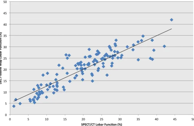
Mean iodine concentration in whole lungs volume was 1.7 mg/mL (1.1-2.3 mg/mL). Median relative functions for each lung lobe, derived from DECT-iodine map and SPECT/CT, are detailed in Table 2. Figure 3 shows an example of DECT-iodine map and the corresponding SPECT/CT in a patient with COPD and severe functional asymmetry. As shown in Figure 4, a strong correlation was found between lobar function derived from DECT-iodine map vs. SPECT/CT (r= 0.89, p<0.00001). There were only weak correlations between mean iodine
value of the overall lung parenchyma and percent FEV1 (r= 0.38), absolute FEV1 (L) (r=0.49), percent FVC (r= 0.28), absolute FVC (L) (r= 0.064) and DLCO (r= 0.47).
Whole lung vs. functional lung dosimetry
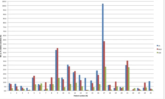
Table 2 summarizes whole lung vs. functional lung dosimetry (mean MLD, V5 and V20) for the entire cohort, as well as for SABR and IMRT patients specifically. Looking at differences between whole vs. functional lung V5, we find a mean relative variation of +/-16% (0-102%) and a mean absolute variation of +/- 3% (0-10%), p=0.047. Similarly, for mean relative MLD variation was +/-14% (1-58%) and mean absolute MLD variation was 1 Gy (0.0-5.4), p=0.03. Lastly, mean relative V20 variation was 6% (1-28%) and mean absolute V20 variation was 1% (0-9%), p=0.087. Relative variations in MLD, V5 and V20 between whole lung vs. functional lung dosimetries for each patient are shown in Figure 5. Figure 3 shows an example of a patient treated with SABR where whole lung dosimetry significantly underestimated dose to functional lung (whole lung V5 of 10% vs. functional lung V5 of 20%).
More specifically per treatment technique, mean absolute variation of MLD was 1.5 Gy (0.1-5.4 Gy) in the IMRT group vs. 0.5 Gy (0.0-1.7 Gy) in the SABR group (p= 0.046). Mean absolute variation in V20 was 3% (0-9%) in the IMRT group vs. 1% (0-3%) in the SABR group (p=0.048). Mean absolute V5 variation was the same in patients treated with IMRT and SABR (3% (0-10%)).
8.5 DISCUSSION
Although current clinical evidence for the use of DECT in radiotherapy is limited, potential applications in radiotherapy planning are numerous and include more precise dose calculation (51-53), metal artefact reduction (54) as well as improved tumor delineation and normal tissue characterization (15, 55-57). This study is the first report on the use of functional DECT imaging in radiotherapy planning. Single-energy CT differentiates materials based on their attenuation at a specific energy (quantified in Hounsfield Unit). The acquisition of 2 series of images at different energies (usually 80-100 kV and 120-140 kV) exploits the principle of variable energy-dependant photoelectric effect and allows to determine material-specific difference in attenuation between low and high energy acquisitions (15). With precise material decomposition, it becomes possible to distinguish and quantify different substances such as iodine from the remaining soft tissues on DECT imaging (16). In the current study, a thoracic DECT in treatment position was used to extract the iodine fraction of each lung voxel, serving as a surrogate for regional blood volume, in order to derive split lung function.
Quantitative radionuclide pulmonary perfusion scintigraphy has been widely studied in the context of pre-operative assessment of parenchymal lung disease (58, 59) and remains the standard for evaluation of relative lung function (60-62). Because the combination of SPECT with low dose CT provides better spatial resolution and 3D anatomical information, SPECT/CT was chosen as the standard for the evaluation of lung function in the context of this study. We showed that DECT-iodine maps were strongly correlated with SPECT/CT for lung function assessment. This is in concordance with several studies showing that iodine
distribution from DECT could be used to detect perfusion defects in the context of pulmonary embolisms (63-67) and parenchymal lung disease (68, 69). Pansini et al. (68) used DECT-iodine maps in 57 patients, including 37 patients with emphysema; their study showed that areas of decreased iodine distribution significantly matched areas of emphysema and that the severity of perfusion alteration was correlated with severity of parenchymal destruction. To date, the role of DECT-iodine maps for assessment of pre-operative lung function has been investigated in only one study (70). In that study, Chae et al. compared pre- and post-operative DECT and perfusion scintigraphy for prediction of post-operative pulmonary function tests (70). Their group reported a 15% vs. 18% error in post-operative estimation of FEV1 with DECT vs. scintigraphy, respectively, suggesting that DECT may have a higher accuracy. Integration of functional information in lung radiotherapy planning has the potential to better predict lung toxicity as well as to spare most functional lung areas. Use of SPECT/CT-derived function has previously been extensively reported in lung radiotherapy (40, 71, 72). In recent years, several alternative lung function assessment methods including hyperpolarized gas MRI (73-76), 4DCT-based ventilation (43, 44, 77), thin section quantitative CT (78) or 4D PET/CT (46) have also been reported in the context of small feasibility studies. Although direct comparison between these studies is limited by the use of widely different methodologies, these studies have reported variable improvements of lung dosimetry parameters with the use of functional-avoidance (46, 74, 79-84). In our study, while no functional-avoidance optimisation was performed, dosimetric assessment of standard radiation plan showed that integration of iodine map led to significant differences in V5 and mean dose to anatomic vs. functional lung in both SABR and IMRT. The inaccuracy (over or under-estimation) of actual dose to functional parenchyma could explain, at least partly, the variable predictive power of
current whole lung dosimetric parameters (32). Importantly, the use of functional information has so far never been evaluated in lung SABR. In our study, although absolute dose variations between functional and anatomical dosimetry were larger in the IMRT group, we report that significant variations in V5 and MLD also occur in SABR, suggesting that functional information integration is also important in these patients. In fact, the large number of radiation field entries and the increased conformity of SABR treatments, along with a pre-selection of non-operable or borderline operable patients with poorer lung function, makes function-sparing SABR particularly appealing.
The clinical benefit of the integration of functional information in IMRT (for locally advanced disease) as well as in SABR (for early stage disease) remains to be demonstrated. In a recent literature review that included 6 clinical studies integrating perfusion SPECT in lung radiotherapy planning, use of functional dosimetry was most beneficial in patients with emphysema or large areas of perfusion deficit and functional dose-volume histograms appeared to improve RP risk estimation (85). An important prospective study by Farr et al. involving 58 lung cancer patients treated with IMRT showed that SPECT-based functional MLD, V5, V10, V20 and V30 provided superior RP risk estimation compared to the standard whole lung dosimetric counterparts (86). Similarly, Kimura et al. looked specifically at functional MLD and V20 and reported a strong correlation with development of grade 2 ≥ RP (87). Currently, at least 4 clinical trials assessing the clinical impact of functional information in lung radiotherapy are on-going: 1) a double blind randomized trial evaluating hyperpolarized gas MRI (NCT02002052); 2) a single blind randomized trial using 4DCT (NCT03077854); 3) a feasibility trial on the use of 4DCT ventilation in treatment planning
(NCT02528942) and; 4) a phase II trial investigating SPECT/CT-based functional avoidance for toxicity prediction (NCT02773238).
The use of DECT-iodine map is particularly attractive in radiotherapy as it offers several advantages. First, DECT-iodine map can be derived from a contrast-injected scan in treatment position, and therefore provides perfect fusion with precise anatomical and functional correlation. As contrast-injected scans are routinely obtained at time of radiotherapy planning, notably for central or node-positive tumors, integration of DECT-iodine mapping would be straightforward in the current workflow as it obviates the need for an additional exam in nuclear medicine, which would also increase unnecessary radiation dose. It is estimated that up to 70% of patients with lung cancer have concurrent chronic obstructive lung disease (88) and therefore would have some excepted degree of parenchymal asymmetry. DECT scanners being slowly adopted in radiation oncology (most notably in proton centers), the DECT-iodine map has the potential to allow global acquisition of functional information in any patient undergoing thoracic radiotherapy; systematic, individualized plan optimization could then be performed by sparing of the most contributing lung areas
.
However, an evident downside of this method is the necessary use of iodine contrast, which limits its use in any patients with contra-indications to contrast agent.This preliminary study on the role of DECT for assessment of lung function in radiotherapy planning has several limitations but should be seen as a first step towards the clinical implementation of this innovative, dose neutral and easily implementable method in the global
process of radiotherapy planning. First, as the primary goal was to assess the validity of DECT-based functional information and feasibility of iodine-map integration in planning, this study did not involve planning re-optimization based on lung function. Further clinical data is needed to define if there is a threshold of iodine below which parenchyma needs not to be spared at all. Our use of discrete functional sub-volumes in the treatment planning system decreases the voxel-to-voxel precision of the initial iodine-map. The development of methods allowing the use of weighted volumes in clinically approved TPS is needed for the best use of function-sparing optimization. As current TPS are not conceived for optimization of low doses, early integration could involve selecting beam angles or arcs to pass through less functional lung. Another limitation is the current lack of 4D functional information accounting for respiratory motion. In fact, dose calculation is typically achieved on the average 4DCT whereas DECT was obtained in end-expiratory position. While this likely decreased the precision of the registration, end-expiratory position represents the worst-case scenario for lung dosimetry and is routinely used in our institution for whole lung volume delineation. In addition, the added use of 4DCT could potentially allow for ventilation information. Despite these limitations, this study represents an important step towards the promising clinical use of a simple, low-cost and dose-neutral method for lung function assessment in radiotherapy planning.
8.6 CONCLUSION
We have reported innovative and promising integration of functional DECT imaging in radiotherapy. DECT-iodine maps strongly correlate with standard SPECT/CT for evaluation
of differential lung function. The use of functional imaging reveals significant variation in functional dosimetric parameters as compared to standard anatomical dosimetry in SABR and IMRT plans. These discrepancies may partially explain the suboptimal predictive power of standard anatomical DVH parameters and future work will involve integration of weighted functional volumes in the planning process. Such integration should lead to better sparing of lung function in pulmonary radiotherapy.
8.7 TABLES ET FIGURES
Table 1- Characteristics of patients with lung cancer enrolled in the study Patients characteristics
Median age (years) 69 (53-81)
Gender (n ; %) Female 14 (56%) Male 11 (44%) Histology (n ; %) NSCLC 17 (68%) SCLC 1 N/A 7 (28%) TNM stage (n ; %) IA 16 (64%) IB 2 (8%) IIIA 7 (28%) Localisation (n ; %) Inferior lobe 7 (28%) Middle lobe 4 (16%)
Superior lobe 14 (56%) Pulmonary function Median FEV1 (%) 60 (19-131) Median FEV1 (L) 1.2 (0.4-3.9) Median FVC (%) 76% (45-130) Median FVC (L) 2.3 (1.4-4.4) Median DLCO (%) 56 (39-91) COPD (n ; %) 21 (84%) SPECT/CT function Left lung (%) 45 (21-63) Right lung (%) 55 (37-79)
Median time between SPECT/CT
and planning CT (days) 11 (1-60)
Treatment characteristics
SABR (n ; %) 18 (72%)
ITV technique 15 (84%)
Tumor tracking 3 (16%)
Median dose (Gy, range) 60 (50-60) Median fractions (n, range) 3 (3-5)
Median PTV (cm3) 30 (5-53)
IMRT (n ; %) 7 (28%)
Median dose (Gy, range) 66 (40-66) Median fractions (n, range) 33 (15-33) Median PTV (cm3, range) 250 (6-1012)
Concurrent chemotherapy (n ; %) 7 (28%)
NSCLC= Non small cell lung cancer; SCLC= Small cell lung cancer; N/A= not available; FEV1= Forced expiratory volume in 1 second; FVC= Forced vital capacity; DLCO= Diffusing capacity for carbon monoxide; COPD= Chronic obstructive lung disease; SPECT/CT= Single photon emission computed tomography; SABR=
Stereotactic ablative radiotherapy; IMRT= Intensity modulated radiotherapy; Gy= Gray; ITV= Internal target volume; PTV= Planning target volume; n= number.
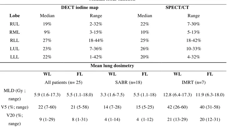
Table 2- DECT vs. SPECT/CT- derived lobar function and summary of WL vs. FL dosimetry
Median lobar function
DECT iodine map SPECT/CT
Lobe Median Range Median Range
RUL 19% 2-32% 22% 7-30%
RML 9% 3-15% 10% 5-13%
RLL 27% 18-44% 25% 18-42%
LUL 23% 7-36% 26% 10-33%
LLL 22% 1-42% 20% 4-32%
Mean lung dosimetry
WL FL WL FL WL FL
All patients (n= 25) SABR (n=18) IMRT (n=7)
MLD (Gy ;
range) 5.9 (1.6-17.3) 5.5 (1.1-18.0) 3.3 (1.6-7.5) 5.5 (1.1-18) 12.8 (6.4-17.3) 11.9 (6.3-18.0) V5 (%; range) 22 (7-60) 21 (5-58) 14 (7-28) 15 (5-25) 42 (26-60) 40 (31-58)
V20 (%;
range) 9 (1-29) 8 (1-31) 4 (1-14) 4 (1-12) 21 (13-29) 20 (12-31) RUL= Right upper lobe; RML= Right middle lobe; RLL= Right lower lobe; LUL= Left upper lobe; LLL= left
lower lobe; WL = Whole lung; FL = Functional lung; n= number of patients; MLD=mean lung dose; Gy= Gray; V5= percent lung volume receiving 5 Gy or more; V20=percent lung volume receiving 20 Gy or more; SABR= stereotactic body radiotherapy; IMRT = intensity modulated radiotherapy.
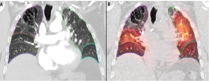
Figure 1- Manual segmentation of lung lobes on DECT and SPECT/CT
Legend:
Coronal slice showing manual segmentation of lung lobes on (A) DECT and (B) SPECT/CT. Magenta= right superior lobe; yellow= right middle lobe; blue= right inferior lobe; green= left superior lobe; cyan= left inferior lobe.
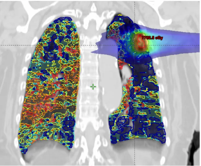
Figure 2- Coronal slice showing 6 functional lung sub-volumes with increasing iodine content.
Legend:
Dark blue = least functioning lung; red = most functioning lung; light blue, yellow, orange and pink represent intermediate regions with increasing lung function. PTV (dark green), GTV (red) as well as 5 Gy dose distribution also shown.
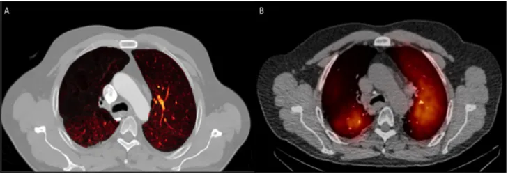
Figure 3- DECT-iodine and SPECT/CT maps
Legend:
Example of a DECT-iodine map (A), and the corresponding SPECT/CT map (B) in a patient with known COPD showing a large perfusion deficit in the superior lobe of the right lung.
DEUXIÈME ARTICLE SCIENTIFIQUE 9
In a Heartbeat: An Assessment of Dynamic Dose Variation to Cardiac Structures using Dual Source Computed Tomography
Houda Bahig1,2 , Jacques de Guise2 , Toni Vu1,2 , Danis Blais1 , Carl Chartrand-Lefebvre2,3 , Nhu Tram Nguyen4, Sophie Lavertu1,2, Jean-Pierre Guay1,2, David Roberge1,2
1Radiation Oncology Department, Centre Hospitalier de l’Université de Montréal, Montreal, QC. Canada
2Centre de recherche du Centre Hospitalier de l’Université de Montréal, Montreal, QC. Canada 3Radiology Department, Centre Hospitalier de l’Université de Montréal, Montreal, QC. Canada 4Radiation Oncology Department, Juravinski Hospital and Cancer Centre, Hamilton, ON. Canada
Key Words: Left breast, Tangential irradiation, Coronary arteries, Dual-source computed tomography, Left anterior descending artery, Cardiac dose, Cardiac-gated imaging. Publication:
Int J Radiat Oncol Biol Phys. 2018 Jan 31. pii: S0360-3016(18)30115-9. doi: 10.1016/ j.ijrobp. 2018.01.049. PMID: 29559290
CONTRIBUTIONS: Rédaction du protocole de recherche, coordination et gestion de l’implémentation du protocole de recherche en clinique, récole et analyse des données, interprétation des données, rédaction du manuscrit.
9.1 ABSTRACT
Purpose: To assess radiation dose variation to the left anterior descending artery (LAD), left main coronary artery (LMCA), left ventricle (LV) and whole heart (WH) during the cardiac cycle using dual source computed tomography (DSCT).
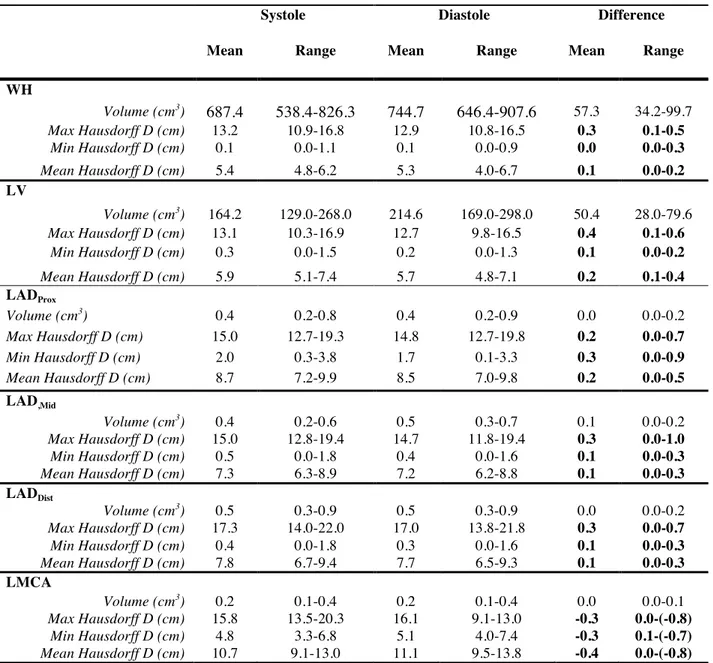
Methods: This prospective study included patients with left breast cancer planned for tangential radiotherapy. An electrocardiogram-synchronized contrast-injected DSCT was obtained in treatment position, in deep-inspiration breath-hold (DIBH), using retrospective sequential acquisition. WH, LV, LMCA as well as proximal, middle and distal LAD segments were contoured on each phase of the cardiac cycle. Maximum, minimum and mean Hausdorff distance between each structure and the tangential fields was assessed in ventricular systole vs. diastole. Four-dimensional dose-volume histograms were used to compare systolic vs. diastolic dosimetries.
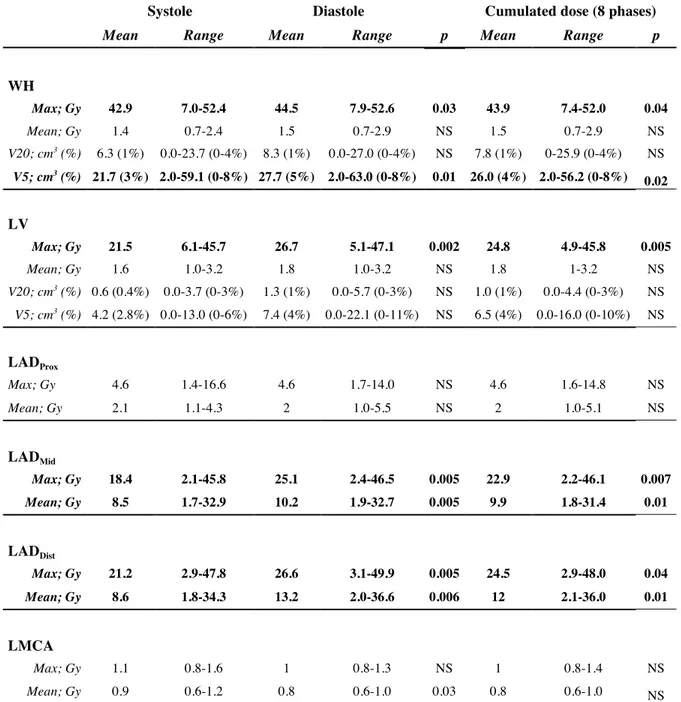
Results: Ten patients were enrolled. Average maximum, minimum and mean Hausdorff distance variation from systole to diastole was ≤4 mm for LV and LMCA, and ≤3 mm for the heart and LAD segments. WH maximum dose and volume receiving 5Gy were decreased in systole vs. diastole (42.9 Gy vs. 44.5 Gy, p=0.03 and 21.7 cm3
vs. 27.7 cm3
, p=0.01), but mean dose remained similar throughout the cycle. Maximum and mean dose to distal LAD was 21.2 Gy vs. 26.6 Gy (p=0.005) and 8.6 Gy vs. 13.2 Gy (p=0.006), in systole vs. diastole, respectively. Maximum and mean dose to middle LAD was 18.4 Gy vs. 25.1 Gy (p=0.005) and 8.5 Gy vs. 10.2 Gy, in systole vs. diastole (p=0.005). Maximum dose to LV was lower in systole vs. diastole (21.5 Gy vs. 26.7 Gy, p=0.005).
Conclusion: Beyond DIBH, systolic irradiation would be associated with decrease in dose to LAD, LV and WH. In addition to potential use in planning for cardiac gating, DSCT imaging can be used to help define a planning organ at risk volume for clinically important cardiac sub-structures.