HAL Id: hal-03004328
https://hal-cnrs.archives-ouvertes.fr/hal-03004328
Submitted on 20 Nov 2020
HAL is a multi-disciplinary open access
archive for the deposit and dissemination of
sci-entific research documents, whether they are
pub-lished or not. The documents may come from
teaching and research institutions in France or
abroad, or from public or private research centers.
L’archive ouverte pluridisciplinaire HAL, est
destinée au dépôt et à la diffusion de documents
scientifiques de niveau recherche, publiés ou non,
émanant des établissements d’enseignement et de
recherche français ou étrangers, des laboratoires
publics ou privés.
The molecular puzzle of two-component signaling
cascades
Marie Foussard, Stéphanie Cabantous, Jean-Denis Pedelacq, Valérie Guillet,
Samuel Tranier, Lionel Mourey, Catherine Birck, Jean-Pierre Samama
To cite this version:
Marie Foussard, Stéphanie Cabantous, Jean-Denis Pedelacq, Valérie Guillet, Samuel Tranier, et al..
The molecular puzzle of two-component signaling cascades. Microbes and Infection, Elsevier, 2001, 3
(5), pp.417-424. �10.1016/s1286-4579(01)01390-9�. �hal-03004328�
Marie Foussard, Stéphanie Cabantous, Jean-Denis Pédelacq, Valérie Guillet, Samuel Tranier,
Lionel Mourey, Catherine Birck, Jean-Pierre Samama*
Groupe de Cristallographie Biologique, IPBS-CNRS, 205, route de Narbonne, 31077 Toulouse, France
ABSTRACT – Two-component systems constitute prevalent signaling pathways in bacteria and
mediate a large variety of adaptative cellular responses. Signaling proceeds through His-Asp
phosphorelay cascades that involve two central partners, the histidine protein kinase and the
response regulator protein. Structural studies have provided insights into some design principles
and activation mechanisms of these multi-domain proteins implicated in the control of virulence
gene expression in several pathogens. © 2001 Éditions scientifiques et médicales Elsevier SAS
two-component systems / signal transduction / functional domains1. Introduction
Efficient signaling cascades are necessary for microor-ganisms in order to face limited resources and survive in any local environment. Microorganisms must be masters at accommodating potential sources of carbon, nitrogen or energy, at resisting poisons of their metabolic and regulatory processes and at establishing intra- and inter-species communications. This adaptation requires that the organism senses the multitude of extracellular signals and responds, in most instances, by controlling the expression of an adequate repertoire of genes.
The study of sensory–response systems has firmly estab-lished the ’two-component paradigm’ as prevalent signal-ing cascades in bacteria [1]. These pathways involve phosphorylation of two key effector proteins. The modular sensor histidine-kinase is the primary signal transduction protein, and detection of the signal via the input domain(s) controls either the inhibition or the activation of the kinase. Active kinases hydrolyse ATP and autophosphorylate on a histidine residue (figure 1). Each sensor kinase has a cog-nate response regulator (figure 2). Phosphotransfer from the phospho-histidine to an aspartate residue in the receiver domain of the response regulator activates the protein and the cellular responses.
The modular organization of the sensors and of the response regulators is a key feature of two-component systems. The variety of input and output functional domains and their arrangements in different
configura-tions built many types of phosphorelay circuits. In the most sophisticated cases, activation occurs through multistep His-Asp phosphorelay cascades [2–4]. In several instances, additional protein partners are involved that contribute to the control of the phosphorylation state of the response regulator, the ’on-off’ switch of the biological response. This diversity reflects the specific localizations, functions and regulatory mechanisms of two-component systems in the cell.
The understanding of the signaling cascades raises chal-lenging questions. How do essential pathways have com-mon mechanisms of chemical activation and avoid cross-talk? Could the diversity of phosphorelay circuits be rationalized in terms of a puzzle of functional modules? How are individual functions modulated by protein–pro-tein interactions? What does activation by phosphoryla-tion mean? Recent structural approaches provide some insights into these questions.
2. Organization and functional modules
in histidine protein kinases
Kinases operate as homodimers, and two classes of enzymes have been defined, based on the organization of their functional domains (figure 1). Class I histidine kinases are the most commonly encountered. Canonical con-structs typically contain a variable N-terminal sensing region which can be more than 500 residues long and a C-terminal core region, also called the transmitter domain. This 250-residue long region exhibits sequence finger-prints that are generally conserved in the histidine protein kinase superfamily. These homology boxes have been
*Correspondence and reprints.
E-mail address: samama@ipbs.fr (J.P. Samama).
termed the H, N, G1, F and G2 boxes. Hybrid kinases derive from this canonical type and display a phospho-accepting domain and a histidine phosphotransfer (HPt) domain fused at the C-terminal end. In some cascades, this HPt domain is distinct from the kinase and constitutes an isolated module.
Class II (CheA) histidine kinases are specialized in chemotaxis responses (figure 1). They contain well-characterized domains linked by flexible polypeptides of variable lengths. In these proteins, the phospho-accepting
histidine residue (H box) is located at the N terminus of the protein, in the so-called P1 domain. The next module in these kinases, the P2 domain, is dedicated to the recogni-tion of the cognate response regulators. It is followed by the transmitter domain that only carries the N, G1, F and G2 boxes. The C-terminal region of these kinases is involved in the regulation of the autokinase activity through the coupling to the transmembrane receptor-transducer proteins and to CheW [5, 6].
2.1. The H box
As illustrated in figure 1, the phospho-accepting histi-dine residues are found in two distinct locations: the N-terminal region of the transmitter core-domain and dedicated protein modules. However, the study of several two-component proteins showed that the H box always belongs to a similar structural motif, a finding that may be considered as a design principle in His-Asp phosphorelay cascades.
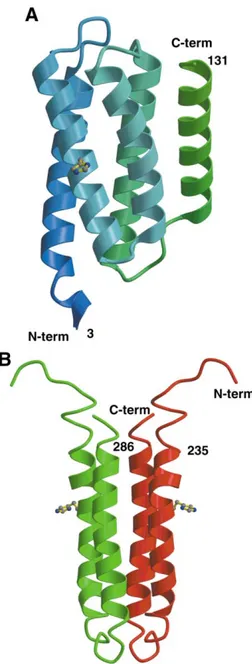
Nuclear magnetic resonance (NMR) investigation [7] and X-ray structure determination in our laboratory of the P1 domain from the chemotactic class II CheA kinase revealed that the phosphorylatable histidine residue belongs to a four-helix bundle motif (figure 3A). A similar architecture was found for the HPt domain fused at the C terminus of the hybrid class I kinase ArcB [8] and for YPD1 [9, 10], the isolated HPt domain in the two-component system that controls the HOG1-dependent MAP kinase in
Saccharomyces cerevisiae [11]. In these three cases, the
four-helix bundles are built from continuous polypeptide stretches that share less than 10% sequence homology.
From a topological point of view, a common way to design a four-helix bundle is by protein dimerization. It is precisely how HPt domains are designed in class I histi-dine kinases where each transmitter core-domain contrib-utes two N-terminal helices. This finding was first revealed by NMR studies on EnvZ (figure 3B), the osmosensor
Figure 1. Modular organization of the histidine
kinases. In class I enzymes (a, b, c), input domains are of variable length and sense a variety of signals. In most cases, they display putative transmembrane regions. The transmitter core-domain is conserved in the super-family of histidine kinases. Hybrid kinases (b, c) con-tain a phosphorylatable receiver module and in some cases, a phosphotransfer unit. Class II (CheA) histidine kinases (d) are involved in chemotaxis and coupled to membrane receptors.
Figure 2. Modular organization of the response regulators. These
proteins share a common phosphorylatable receiver domain. Response regulators usually carry additional functional modules whose activity may be regulated by the receiver domain.
Review Foussard et al.
kinase from Escherichia coli [12]. The H box is evidently present on each subunit, and the four-helix bundle there-fore carries two phosphorylatable histidine residues. This setting satisfies trans-phosphorylation, since histidine kinases phosphorylate the histidine residue on the partner subunit within the dimer.
As already mentioned, CheA kinases do not display the H box in the N-terminal region of the transmitter core-domain. Nevertheless, the X-ray structure of the homodimer of CheA revealed that this region also folds as two helices that mediate association of the two subunits through a four-helix bundle [13]. The finding that dimer-ization of class I and CheA histidine kinases is mediated by
be isolated modules. In multistep His-Asp phosphorelay cascades, HPt domains may allow further control of the activation cascade and/or may collect the signals from independent upstream activation pathways.
Interestingly, these principles hold for other two-component kinases and for partners of the signaling cas-cades. Sequence analysis of genome databases indicates that in some proteins the H box has been replaced by other residues including serine or tyrosine. In the two-component kinase DivL of Caulobacter crecentus, it was shown that the tyrosine residue is phosphorylated [14]. Spo0B is a phosphotransferase, and relays the phosphoryl group between two response regulators (Spo0F and Spo0A) in the cascade that controls sporulation in bacilli [2]. As shown by the X-ray structure [15], dimerization of the protein is achieved through the formation of a four-helix bundle that carries two phosphorylatable histidine resi-dues.
2.2. The N, G1, F and G2 boxes of the catalytic domain
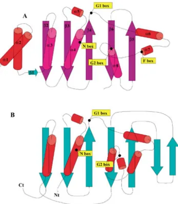
In all histidine kinases, the catalytic ATP-binding domain follows in sequence the dimerization four-helix bundle structural unit. The three-dimensional structure of this domain has been revealed by X-ray crystallography for CheA [13] and by NMR spectroscopy for EnvZ [16]. Its topology (figure 4A) is completely unrelated to that of Ser/Thr or Tyr kinases but strikingly similar to that of heat-shock protein 90 (figure 4B) [17], to the DNA mis-match repair protein MutL [18] and to gyrase B [19] showing that the catalytic domain of histidine kinases belongs to a superfamily of ATPases. From the structure-based sequence alignment (figure 5), it is apparent that the N, G1, F and G2 boxes are among the very few invariant residues in all sequences. These boxes delineate a cavity where ATP binds [20, 21]. The fingerprints of these homol-ogy boxes were thoroughly analyzed in 348 histidine protein kinases [22] and led to the categorization of these proteins into 11 subfamilies. This study pointed out that in some bacterial strains most of the histidine protein kinases belong to a single subfamily, suggesting a single lateral gene transfer event. In other cases, such as Bacillus
subti-lis, the 31 histidine protein kinases are nearly evenly
distributed over eight subgroups.
The sequence alignment (figure 5) also shows that the number of residues between the G1 and G2 boxes, located at the end of β4 and before α8, respectively (figure 4), varies among procaryotic histidine kinases, and between kinases and Hsp90. This variability is also a major differ-ence between procaryotic histidine kinases and the Sln1 histidine kinase from the yeast S. cerevisiae [23], in which 120 amino acids are inserted in this region. In contrast,
Figure 3. Four-helix bundle topology of HPt domains. (A) The
P1 domain in the S. typhimurium CheA kinase. The phosphory-latable histidine residue is carried by the second helix of the bundle. (B) The four-helix bundle generated by dimerization in EnvZ, a class I histidine kinase.
deletion of 50 residues in the region that includes the F and G2 boxes, occurs in the C-terminal domain of the phos-photransferase Spo0B. This domain displays the fold of the catalytic domain of the kinases but is devoid of ATPase activity, and one may speculate that these proteins have evolved from a common ancester. This latter hypothesis may also hold for SpoIIAB, an anti-sigma F factor from
B. subtilis, which exists independently in nature [24, 25].
This phosphokinase contains all homology boxes of the catalytic domain but acts as a serine protein kinase on the anti-anti-sigma factor spoIIAA [26].
The conservation of the fold and of the invariant resi-dues in the catalytic domains of histidine kinases and gyrases now raises the need for better insight into what determines the kinase versus the ATPase activities in these proteins.
In all two-component histidine kinases, the four-helix bundle is the substrate of the ATP-binding catalytic domain. The modular and respective locations of these functional units (figures 1 and 4) imply movements of these two domains with respect to each other, and a highly specific molecular recognition to prevent cross-talk. These two aspects remain to be documented, but some evidence suggests that the hinge regions between domains may play an important role in the modulation of signal transduction and in the properties of the kinases [27].
3. Modules and functions
in response regulator proteins
Response regulators may contain one, two or three domains (figure 2). They all contain a conserved amino-terminal receiver module of about 125 residues that car-ries the phosphorylatable and invariant aspartate amino acid. A significant number of response regulators only consist of this receiver domain, but the majority of response regulators carry carboxy-terminal output domains. In most instances, one of these domains binds promoter sequences in DNA to negatively or positively control transcription. The phosphorylation state of the receiver domain is, in most cases, the ’on-off’ switch of the cellular response, although transcriptional regulation of an alternate set of genes by the unphosphorylated response regulator has been reported [28].
The receiver domains in response regulators display a doubly wound five-strandedα/β fold (figure 6). The active site is a conserved acidic pocket and contains the phos-phorylatable aspartate residue, which argues for a com-mon Mg2+-dependent mechanism of phosphorylation for
all members of this superfamily. Phosphorylation may, in most cases, be achieved in vitro, by small phosphodonors such as acetyl phosphate or phosphoramidate, suggesting that the active site of the response regulators catalyses phosphoryl transfer. In vivo, the receiver modules are the substrates of the cognate phosphorylated HPt domains. The half-life of the phosphorylated response regulators ranges from a few seconds to over 1 h, a property related to the type of cellular response that must be achieved. This autophosphatase activity of the receiver domain may be stimulated by specific phosphatases [29, 30] or by the kinase itself. Complex reversal of phosphotransfer was demonstrated in the ArcA/ArcB system [31], and in the regulation of chemotaxis in Sinorhizobium meliloti. In this system, a second response regulator, CheY1, assumes the role of a phosphate sink whenever phosphorylated CheY2, the master switch for chemotaxis, and unphosphorylated kinase are present. The kinase in this system may also act as a phosphorelay protein between the two response regulators [32].
Receiver domains mediate various protein–protein interactions, both in their unphosphorylated and phos-phorylated states. The functional significance of these interactions has been revealed in a few cases. Unphospho-rylated receiver domains may act negatively [33–35] and inhibit the function of the output domains. X-ray structure determinations of NarL and CheB revealed that in each response regulator the interface between the two protein modules sterically prevents DNA binding and methyl-esterase activity, respectively [36, 37]. From biochemical and genetic studies it was shown that phosphorylation disrupts previous interactions and fosters new ones that are required for optimal biological function [38]. This modulation of the function by the phosphorylation state of the regulatory domain has been a central question in the field. It was postulated that phosphorylation induces con-formational changes in the receiver domain, and recent advances have provided new insights into the events that drive signal transduction.
Figure 4. The similar topology of the ATP-binding domain in
(A) the transmitter core-domain of the CheA histidine kinase (B) Hsp90 ATPase. The location of the conserved boxes in histidine kinases is indicated. The two N-terminal helices in CheA mediate dimerization of the protein through formation of a four-helix bundle but do not carry the H box. It could be noticed that the first strand and helix (β2 and α3) of the ATP-binding domain in CheA is provided by the C-terminal part of the protein in Hsp90.
Review Foussard et al.
4. Phosphorylation-induced activation
of the receiver domain
As phosphoaspartates are inherently unstable, it seemed at first an impossible task to determine crystal structures of phosphorylated receiver domains. In the past few months, the X-ray structures of the phosphorylated receiver domains of Spo0A and FixJ were reported [39, 40], and could be compared to the structures of the unphosphory-lated domains solved independently [41, 42]. In the phos-phorylated structures, the phosphoryl group bound to the active site aspartate is stabilized by polar interactions that involve main chain atoms and invariant side chains. This geometry is therefore representative of the environment of the acylphosphate group in any phosphorylated receiver domain. Phosphorylation of FixJ induces a large confor-mational change and dimerization of the protein. The major changes involve a switch of the conformation of the β4-α4 loop adjacent to the active site, the relocation of two highly conserved side chains and the modification of theα4-β5 surface of the protein (figure 7). This remodeling provides the interface that mediates dimerization of the phosphorylated FixJ response regulator. This change in
oligomeric state for FixJ is an essential feature for binding to the target DNA and thus for regulation of the transcrip-tion [43].
NMR studies of activated receiver domains (phospho-NtrC, BeF3-CheY and phosphono-CheY) also showed
structural transitions in line with those described for FixJ, as for instance the relocation of the conserved residues that connects the active site to the α4-β5-α5 signaling surface of the protein [44–46]. The structural transitions from inactivated to activated species are significant enough to disrupt previous interactions, such as those established between receiver domains and effector C-terminal domains [36, 37], or between CheY and its recognition domain in CheA [47].
Activated proteins intriguingly display significantly vari-able extents of conformational changes. This finding raised the question of why receiver domains use the aspartyl-phosphate, a high-energy bond just like the anhydride linkage in ATP, and not phosphotransfer from ATP to serine or threonine that would generate enough energy to account for the observed conformational changes of the proteins. One hypothesis is that the change in structure that decreases the free energy of hydrolysis of the
acylphos-Figure 5. Structure-based sequence alignment
of the transmitter core-domain of histidine kinases (CheA_thema and EnvZ) and of topo-logically related proteins (Spo0B and Hsp90). The homology boxes (H, N, G1, F and G2) are indicated. The secondary structure elements are illustrated by helices and arrows. Regions that display significant differences in the pro-tein structures are underlined.
phate only occurs during complex formation between the response regulators and their targets [48]. This proposal provides a possible explanation for the small structural changes observed in phosphorylated Spo0A or activated CheY, which contrast with the drastically altered phospho-rylated FixJ structure. Indeed, the target for FixJ is FixJ itself,
and dimerization constitutes FixJ activation. Isolated phos-phorylated Spo0A which targets DNA, and activated CheY which targets FliM, would only be high-energy species expected to promote and/or undergo significant confor-mational changes upon binding to their respective targets. The structures of all conserved pieces of the puzzle in two-component systems have revealed some key func-tional features and design principles. However, the com-plexity and the fine tuning of the activation of the cellular response remain to be documented. The next challenges concern the communication between domains and the characterization of complexes with downstream targets of the response regulators. The variety of signal transduction input domains and the regulation of the histidine kinase activity that determines transmission of the information also represent a formidable task. The growing amount of data from genomic sequencing, proteomic and transcrip-tome analysis, structural and biocomputing approaches will likely reveal new and more complete pictures of the regulatory aspects of two-component systems.
These signaling cascades have been, in several instances, implicated in virulence and pathogenicity. For example, in Streptococcus pneumoniae, loss of function of the VncRS system produced tolerance of the bacteria to vancomycin and other classes of antibiotics [49], and a systematic program of mutagenesis identified two-component systems that are essential for viability and pathogenicity [50]. Interestingly, it was also found that systems which apparently play a role in the maintenance of the respiratory tract infection are conserved in other Gram-positive bacteria. The PhoPQ two-component sys-tem in Salmonella typhimurium modulates expression of a suite of genes in response to external Mg2+concentration.
A complex regulatory cascade involving the PhoPQ and the PmrAB systems allows S. typhimurium to integrate multiple signals for macrophage survival and resistance to antimicrobial peptides [51, 52]. Clostridium perfringens is
Figure 6. The common fold of receiver domains. The highy
conserved residues are shown. An arrow indicates the phosphory-latable aspartate in the active site.
Figure 7. Illustration of theα4-β5-α5 surface of the
receiver domain in FixJ according to the structures of the unphosphorylated and phosphorylated species. The arrows indicate the phosphorylation-induced displace-ments of the highly conserved Thr82 and Phe104 side chains and of theβ4-α4 loop. These changes affect the signaling surface of receiver domains and provide the protein-protein interface for dimerization in the case of phosphorylated FixJ.
Review Foussard et al.
Acknowledgments
This work was supported by Hoechst Marion Roussel (HMR), an HMR grant to M. F., by the Programme de Recherches Fondamentales en Microbiologie (MENRT) and by CNRS.
References
[1] Parkinson J.S., Kofoid E.C., Communication modules in bacterial signaling proteins, Annu. Rev. Genet. 26 (1992) 71–112.
[2] Burbulys D., Trach K.A., Hoch J.A., Initiation of sporula-tion in B. subtilis is controlled by a multicomponent phos-phorelay, Cell 64 (1991) 545–552.
[3] Appleby J.L., Parkinson J.S., Bourret R.B., Signal trans-duction via the multistep phosphorelay: not necessarily a road less traveled, Cell 86 (1996) 845–848.
[4] Mizuno T., His-Asp phosphotransfer signal transduction, J. Biochem. 123 (1998) 555–563.
[5] Bourret R.B., Davagnino J., Simon M.I., The carboxy-terminal portion of the CheA kinase mediates regulation of autophosphorylation by transducer and CheW, J. Bacteriol. 175 (1993) 2097–2101.
[6] Morrison T.B., Parkinson J.S., A fragment liberated from the Escherichia coli CheA kinase that blocks stimulatory, but not inhibitory, chemoreceptor signaling, J. Bacteriol. 179 (1997) 5543–5550.
[7] Zhou H., Dahlquist F.W., Phosphotransfer site of the chemotaxis-specific protein kinase CheA as revealed by NMR, Biochemistry 36 (1997) 699–710.
[8] Kato M., Mizuno T., Shimizu T., Hakoshima T., Insights into multistep phosphorelay from the crystal structure of the C-terminal HPt domain of ArcB, Cell 88 (1997) 717–723.
[9] Xu Q., West A.H., Conservation of structure and function among histidine-containing phosphotransfer (HPt) domains as revealed by the crystal structure of YPD1, J. Mol. Biol. 292 (1999) 1039–1050.
[10] Song H.K., Lee J.Y., Lee M.G., Moon J., Min K., Yang J.K., Suh S.W., Insights into eukaryotic multistep phosphorelay signal transduction revealed by the crystal structure of Ypd1p from Saccharomyces cerevisiae, J. Mol. Biol. 293 (1999) 753–761.
[11] Maeda T., Wurgler-Murphy S.M., Saito H., A two-component system that regulates an osmosensing MAP kinase cascade in yeast, Nature 369 (1994) 242–245.
tion, Proc. Natl. Acad. Sci. USA 96 (1999) 13068–13073. [15] Varughese K.I., Madhusudan M., Zhou X.Z., White-ley J.M., Hoch J.A., Formation of a novel four-helix bundle and molecular recognition sites by dimerization of a response regulator phosphotransferase, Mol. Cell 2 (1998) 485–493.
[16] Tanaka T., Saha S.K., Tomomori C., Ishima R., Liu D., Tong K.I., Park H., Dutta R., Qin L., Swindells M.B., Yamazaki T., Ono A.M., Kainosho M., Inouye M., Ikura M., NMR structure of the histidine kinase domain of the E. coli osmosensor EnvZ, Nature 396 (88) (1998) 92. [17] Prodromou C., Roe S.M., O’Brien R., Ladbury J.E.,
Piper P.W., Pearl L.H., Identification and structural char-acterization of the ATP/ADP-binding site in the Hsp90 molecular chaperone, Cell 90 (1997) 65–75.
[18] Ban C., Yang W., Crystal structure and ATPase activity of MutL: implications for DNA repair and mutagenesis, Cell 95 (1998) 541–552.
[19] Wigley D.B., Davies G.J., Dodson E.J., Maxwell A., Dod-son G., Crystal structure of an N-terminal fragment of the DNA gyrase B protein, Nature 351 (1991) 624–629. [20] Yang Y., Inouye M., Requirement of both kinase and
phos-phatase activities of an Escherichia coli receptor (Taz1) for ligand-dependent signal transduction, J. Mol. Biol. 231 (1993) 335–342.
[21] Stewart R.C., VanBruggen R., Ellefson D.D., Wolfe A.J., TNP-ATP and TNP-ADP as probes of the nucleotide binding site of CheA, the histidine protein kinase in the chemotaxis signal transduction pathway of Escherichia coli, Biochemistry 37 (1998) 12269–12279.
[22] Grebe T.W., Stock J.B., The histidine protein kinase super-family, Adv. Microb. Physiol. 41 (1999) 139–227. [23] Ota I.M., Varshavsky A., A yeast protein similar to
bacte-rial two-component regulators, Science 262 (1993) 566–569.
[24] Min K.T., Hilditch C.M., Diederich B., Errington J., Yud-kin M.D., Sigma F, the first compartment-specific tran-scription factor of B. subtilis, is regulated by an anti-sigma factor that is also a protein kinase, Cell 74 (1993) 735–742. [25] Kang C.M., Brody M.S., Akbar S., Yang X., Price C.W., Homologous pairs of regulatory proteins control activity of
Bacillus subtilis transcription factor sigma(b) in response to
environmental stress, J. Bacteriol. 178 (1996) 3846–3853. [26] Najafi S.M., Willis A.C., Yudkin M.D., Site of phosphory-lation of SpoIIAA, the anti-anti-sigma factor for sporulation-specific sigma F of Bacillus subtilis, J. Bacteriol. 177 (1995) 2912–2913.
[27] Hsing W., Russo F.D., Bernd K.K., Silhavy T.J., Mutations that alter the kinase and phosphatase activities of the two-component sensor EnvZ, J. Bacteriol. 180 (1998) 4538–4546.
[28] Msadek T., Kunst F., Rapoport G., in: Hoch J.A., Silhavy T. (Eds.), Two-Component Signal Transduction, ASM Press, Washington D.C., 1995, pp. 447–471.
[29] Perego M., Hoch J.A., Protein aspartate phosphatases con-trol the output of two-component signal transduction sys-tems, Trends Genet. 12 (1996) 97–101.
[30] Hess J.F., Oosawa K., Kaplan N., Simon M.I., Phosphory-lation of three proteins in the signaling pathway of bacterial chemotaxis, Cell 53 (1988) 79–87.
[31] Georgellis D., Kwan O., De Wulf P., Lin E.C., Signal decay through a reverse phosphorelay in the Arc two-component signal transduction system, J. Biol. Chem. 273 (1998) 32864–32869.
[32] Sourjik V., Schmitt R., Phosphotransfer between CheA, CheY1, and CheY2 in the chemotaxis signal transduction chain of Rhizobium meliloti, Biochemistry 37 (1998) 2327–2335.
[33] Gu B., Lee J.H., Hoover T.R., Scholl D., Nixon B.T.,
Rhizobium meliloti DctD, a sigma 54-dependent
transcrip-tional activator, may be negatively controlled by a subdo-main in the C-terminal end of its two-component receiver module, Mol. Microbiol. 13 (1994) 51–66.
[34] Ireton K., Rudner D.Z., Siranosian K.J., Grossman A.D., Integration of multiple developmental signals in Bacillus
subtilis through the Spo0A transcription factor, Genes Dev.
7 (1993) 283–294.
[35] Kahn D., Ditta G., Modular structure of FixJ: homology of the transcriptional activator domain with the -35 binding domain of sigma factors, Mol. Microbiol. 5 (1991) 987–997. [36] Baikalov I., Schroder I., Kaczor-Grzeskowiak M., Cas-cio D., Gunsalus R.P., Dickerson R.E., NarL dimerization? Suggestive evidence from a new crystal form, Biochemistry 37 (1998) 3665–3676.
[37] Djordjevic S., Goudreau P.N., Xu Q., Stock A.M., West A.H., Structural basis for methylesterase CheB regu-lation by a phosphoryregu-lation-activated domain, Proc. Natl. Acad. Sci. USA 95 (1998) 1381–1386.
[38] Cervin M.A., Spiegelman G.B., The Spo0A sof mutations reveal regions of the regulatory domain that interact with a sensor kinase and RNA polymerase, Mol. Microbiol. 31 (1999) 597–607.
[39] Lewis R.J., Brannigan J.A., Muchová K., Barák I., Wilkin-son A.J., Phosphorylated aspartate in the structure of a response regulator protein, J. Mol. Biol. 294 (1999) 9–15. [40] Birck C., Mourey L., Gouet P., Fabry B., Schumacher J., Rousseau P., Kahn D., Samama J.P., Conformational changes induced by phosphorylation of the FixJ receiver domain, Structure Fold. Des. 7 (1999) 1505–1515.
[41] Lewis R.J., Muchová K., Brannigan J.A., Barák I., Leonard G., Wilkinson A.J., Domain swapping in the sporulation response regulator Spo0A, J. Mol. Biol. 297 (2000) 757–770.
[42] Gouet P., Fabry B., Guillet V., Birck C., Mourey L., Kahn D., Samama J.P., Structural transitions in the FixJ receiver domain, Structure Fold. Des. 7 (1999) 1517–1526. [43] DaRe S., Schumacher J., Rousseau P., Fourment J., Ebel C., Kahn D., Phosphorylation-induced dimerization of the FixJ receiver domain, Mol. Microbiol. (1999) 504–511. [44] Yan D., Cho H.S., Hastings C.A., Igo M.M., Lee S.Y.,
Pelton J.G., Stewart V., Wemmer D.E., Kustu S., Beryllof-luoride mimics phosphorylation of NtrC and other bacte-rial response regulators, Proc. Natl. Acad. Sci. USA 96 (1999) 14789–14794.
[45] Cho H.S., Lee S.Y., Yan D., Pan X., Parkinson J.S., Kustu S., Wemmer D.E., Pelton J.G., NMR structure of activated CheY, J. Mol. Biol. 297 (2000) 543–551. [46] Halkides C.J., McEvoy M.M., Casper E., Matsumura P.,
Volz K., Dahlquist F.W., The 1.9 Å resolution crystal structure of phosphono-CheY, an analogue of the active form of the response regulator, CheY, Biochemistry 39 (2000) 5280–5286.
[47] Welch M., Chinardet N., Mourey L., Birck C., Samama J.P., Structure of the CheY-binding domain of histidine kinase CheA in complex with CheY, Nat. Struct. Biol. 5 (25) (1998) 29.
[48] Stock J., DaRe S., Response regulators on and off, Curr. Biol. 10 (2000) 420–424.
[49] Novak R., Henriques B., Charpentier E., Normark S., Tuomanen E., Emergence of vancomycin tolerance in
Strep-tococcus pneumoniae, Nature 399 (1999) 590–593.
[50] Throup J.P., Koretke K.K., Bryant A.P., Ingraham K.A., Chalker A.F., Ge Y., Marra A., Wallis N.G., Brown J.R., Holmes D.J., Rosenberg M., Burnham M.K., A genomic analysis of two-component signal transduction in
Streptococ-cus pneumoniae, Mol. Microbiol. 35 (2000) 566–576.
[51] Garcia Vescovi E., Soncini F.C., Groisman E.A., Mg2+as an
extracellular signal: environmental regulation of Salmo-nella virulence, Cell 84 (1996) 165–174.
[52] Soncini F.C., Groisman E.A., Two-component regulatory systems can interact to process multiple environmental signals, J. Bacteriol. 178 (1996) 6796–6801.
[53] Shimizu T., Ba-Thein W., Tamaki M., Hayashi H., The virR gene, a member of a class of two-component response regulators, regulates the production of perfringolysin O, collagenase, and hemagglutinin in Clostridium perfringens, J. Bacteriol. 176 (1994) 1616–1623.
Review Foussard et al.