Université de Montréal
Establishing a Robust In Vitro Embryonic Stem Cell
Differentiation Assay to Monitor the Hematopoietic
Potential of DELES Clones
par Swati Shetty
Biologie moléculaire, Faculté de Médecine
Mémoire présenté à la Faculté de Médecine
en vue de l'obtention du grade de Maître ès sciences (M.Sc.) en Biologie moléculaire, option générale avec mémoire
Mémoire présenté à la Faculté
January, 2016
Résumé
Afin d’effectuer des études fonctionnelles sur le génome de la souris, notre laboratoire a généré une bibliothèque de clones de cellules souches embryonnaires (ESC) présentant des suppressions chromosomiques chevauchantes aléatoires – la bibliothèque DELES. Cette bibliothèque contient des délétions couvrant environ 25% du génome murin.
Dans le laboratoire, nous comptons identifier de nouveaux déterminants du destin des cellules hématopoïétiques en utilisant cet outil. Un crible primaire utilisant la benzidine pour démontrer la présence d'hémoglobine dans des corps embryoïdes (EBS) a permis d’identifier plusieurs clones délétés présentant un phénotype hématopoïétique anormal. Comme cet essai ne vérifie que la présence d'hémoglobine, le but de mon projet est d'établir un essai in vitro de différenciation des ESC permettant de mesurer le potentiel hématopoïétique de clones DELES. Mon hypothèse est que l’essai de différenciation hématopoïétique publié par le Dr Keller peut être importé dans notre laboratoire et utilisé pour étudier l'engagement hématopoïétique des clones DELES. À l’aide d’essais de RT-QPCR et de FACS, j’ai pu contrôler la cinétique de différenciation hématopoïétique en suivant l’expression des gènes hématopoïétiques et des marqueurs de surface comme CD41, c-kit, RUNX1, GATA2, CD45, β-globine 1 et TER-119. Cet essai sera utilisé pour valider le potentiel hématopoïétique des clones DELES candidats identifiés dans le crible principal.
Mon projet secondaire vise à utiliser la même stratégie rétro-virale a base de Cre-loxP utilisée pour générer la bibliothèque DELES pour générer une bibliothèque de cellules KBM-7 contenant des suppressions chromosomiques chevauchantes. Mon but ici est de tester si la lignée cellulaire leuémique humaine presque haploïde KBM-7 peut être exploitée en utilisant l'approche DELES pour créer cette bibliothèque. La bibliothèque de clones KBM-7 servira à définir les activités moléculaires de drogues anti-leucémiques potentielless que nous avons identifiées dans le laboratoire parce qu’elles inhibent la croissance cellulaire dans plusieurs échantillons de leucémie myéloïde aiguë dérivés de patients. Elle me permettra également d'identifier les voies de signalisation moléculaires qui, lorsque génétiquement perturbées, peuvent conférer une résistance à ces drogues.
Mots-clés: cellules souches embryonnaires, différenciation hématopoïétique, cellules KBM-7, suppressions chromosomiques.
Abstract
To carry out functional studies on the mouse genome, our laboratory has generated a library of Embryonic Stem Cell (ESC) clones harboring random nested chromosomal deletions – DELES library. This library contains deletions covering ~ 25% of the mouse genome.
In the lab, we are interested in identifying novel hematopoietic cell fate determinants using this resource. A primary screen using benzidine to demonstrate the presence of hemoglobin in embryoid bodies (EBs) was able to identify several DELES clones exhibiting abnormal hematopoietic phenotype. Since this assay only tested for the presence of hemoglobin, the goal of my project is to establish a robust in vitro ESC differentiation assay to monitor the hematopoietic potential of DELES clones. My hypothesis is that the hematopoietic differentiation assay published by Dr. Keller can be used to observe hematopoietic commitment of the DELES clones. Using QRT-PCR and FACS assays I was able to monitor the kinetics of hematopoietic differentiation by observing the expression of hematopoietic genes and surface markers including CD41, C-KIT, RUNX1, GATA2, CD45, β-GLOBIN 1 and TER-119. This assay will be used to validate the hematopoietic potential of the candidate DELES clones identified in the primary screen.
My secondary project aims to use the same retro-viral Cre-loxP strategy used for the DELES library, in order to generate a library of KBM-7 leukemic cells harboring nested chromosomal deletions. My goal here is to test if the human near haploid KBM-7 cell line can be exploited using the DELES approach to create this library. The library of KBM-7 clones will be used to delineate the molecular activities of potential anti-leukemic drugs that we have identified in the lab to inhibit cell growth in several patient-derived acute-myeloid leukemia specimens. It will also allow me to identify molecular signaling pathways that, when genetically disrupted, can confer resistance to these drugs.
Keywords: Embryonic stem cells, hematopoietic differentiation, KBM-7 cells, chromosomal deletions.
Table des matières
Résumé ... i
Abstract ... ii
Table des matières... iii
Liste des tableaux ... v
Liste des figures ... vi
Liste des sigles ... viii
Liste des abréviations ... xi
Acknowledgements ... xii
1. Introduction ... 1
1.1 Genomic approaches used to conduct functional studies on ESCs. ... 4
1.1.1 Gene targeting ... 4
1.1.2 siRNA and shRNA screens ... 5
1.1.3 ZFNs, TALENs and CRISPR ... 6
1.1.4 Cre-loxP ... 7
1.1.5 DELES approach ... 9
1.2 Hematopoiesis ... 15
1.2.1 Ontogeny of hematopoiesis... 17
1.2.2 In- Vitro derivation of hematopoietic cells ... 18
Goal, Hypothesis and Objectives: ... 25
2. Materials and Methods ... 27
Embryonic Stem Cell maintenance and differentiation ... 27
Embryoid body differentiation ... 27
Benzidine Staining Assay ... 28
RNA Extraction ... 28
cDNA synthesis ... 28
QRT-PCR ... 29
Methylcellulose preparation used in viral stock titration ... 32
Transfection ... 33
3. Results ... 35
3.1.1 Establishment of a hematopoietic differentiation protocol. ... 35
3.1.2 Kinetics of the hematopoietic differentiation. ... 36
3.1.3 Titration of CD45 and Ter-119 antibodies... 38
3.2 Validation of the DELES clones using the hematopoietic differentiation assay. ... 44
3.2.1 Preliminary screen of DELES library identified clones having reduced hematopoietic potential. ... 44
3.2.2 Validation of the 5241 and 5035 DELES families. ... 45
3.2.5 Validation of 5035 family using hematopoietic differentiation assay. ... 50
3.3 Towards the generation of a KBM-7 cells library harboring nested chromosomal deletions using DELES methodology. ... 53
3.3.1 Growth kinetics of KBM-7 cells. ... 54
3.3.2 Clonogenicity of KBM-7 cells. ... 55
3.3.3 Antibiotic selection tests. ... 56
3.3.4 Retrovirus infection of KBM-7cells. ... 58
4. Discussion ... 62
5. Conclusion ... 69
Liste des tableaux
Table 1: cDNA synthesis program ... 29
Table 2: Composition of Master Mix for qRT-PCR. ... 29
Table 3:Sequence of primers used for qRT-PCR ... 30
Table 4: qRT-PCR program̊ ... 31
Table 5: Methylcellulose Mix ... 32
Table 6: Methylcellulose Mix with cytokines ... 32
Table 7: Serial Dilution of Viral supernatant. ... 58
Liste des figures
Figure 1: Illustration of the origin and differentiation of the three germ layers ... 1
Figure 2: Illustration of the origin of stem cells.. ... 3
Figure 3: Retro-viral design of the primary A1 and secondary S1 Saturation virus... 10
Figure 4: Preliminary hematopoietic screen using benzidine staining assay. ... 14
Figure 5: Hierarchy of hematopoietic stem cells ... 16
Figure 6: Ontogeny of hematopoiesis. ... 18
Figure 7: Methods for deriving hematopoietic cells in-vitro. ... 19
Figure 8: SMAD signaling at different stages during hematopoietic commitment ... 23
Figure 9: Lineage-specific gene markers used to assess hematopoietic differentiation from ES cells. ... 24
Figure 10: Schematic of the hematopoietic differentiation assay.. ... 36
Figure 11: Kinetics of hematopoietic differentiation. ... 37
Figure 12 : Illustration of the distribution of different blood cell types on a SSC vs FSC FACS plot. ... 39
Figure 13: Ter-119 Antibody titration. ... 40
Figure 14: CD45.2 Antibody titration. ... 42
Figure 15: FACS analysis of the differentiation assay . ... 43
Figure 16: Benzidine Staining Assay ... 44
Figure 17: Representation of the nested chromosomal deletions in family 5241 ... 46
Figure 18: Embryoid body formation and Benzidine staining assay of 5241 family ... 47
Figure 19: Representation of the nested chromosomal deletions in family 5035 ... 49
Figure 20 : Embryoid body formation and Benzidine staining assay of 5035 family ... 50
Figure 21: Kinetics of hematopoietic differentiation for 5035 family. ... 51
Figure 22: Rationale . ... 54
Figure 23: Growth curve of KBM-7 cells ... 55
Figure 24: Limiting diluting assay ... 56
Figure 25: Dose response selection tests for puromycin, hygromycin and G418. ... 57
Liste des sigles
AGM - Aorta Gonad Mesonephros APC/Cy7 - Allophycocyanin/Cyanine7 BAC - Bacterial Artificial Chromosomes BM - Bone Marrow
BMP4 - Bone Morphogenetic Protein 4 bp - basepair
BSA - Bovine Serum Albumin CCC- Compound Correlation Clusters cDNA - complementary DNA
CLP - Common Lymphoid Progenitors CML – Chronic Myeloid Leukemia CMP - Common Myeloid Progenitors
CRISPR - Clustered Regularly Interspaced Short Palindromic Repeats Dctn6 - dynactin 6
DELES – Deletion in Embryonic Stem Cells DMEM - Dulbecco's Modified Eagle Medium DNA – deoxyribonucleic acid
DSB – double stranded break
dsRNA – double stranded Ribonucleic Acid EB – Embryoid Body
EPO - Erythropoietin ESC – Embryonic Stem Cell
EUCOMM - European Conditional Mouse Mutagenesis Program FACS - Fluorescence-activated cell sorting
FBS - Fetal bovine serum
FDA - Food and Drug Administration FSC – Forward Scatter
GATA 1- GATA Binding protein 1 GATA2 - GATA Binding protein 2 GFP - Green fluorescent protein
GM-CSF - Granulocyte-macrophage colony-stimulating factor Gsr - glutathione-disulfide reductase
Gtf2e2 - General transcription factor IIE subunit 2 HAT - Hypoxanthine-Aminopterin-Thymidine HDR - Homology Directed Repair
HEK293 - Human Embryonic Kidney 293 cells
HPRT - Hypoxanthine-guanine phosphoribosyltransferase HPRT - Hypoxanthine-guanine phosphoribosyltransferase HSC – Hematopoietic Stem Cell
ICM – Inner Cell Mass
IKMC - International Knockout Mouse Consortium IL-3 - Interleukin 3
IL-6 - Interleukin 6
IMDM - Iscove's Modified Dulbecco's Medium INDELs - Insertions and/or Deletions
I-PCR - Inverse - Polymerase Chain Reaction iPS - Induced Pluripotent Stem Cell
JAK/STAT - Janus kinase/signal transducers and activators of transcription KDR - Kinase Insert Domain Receptor
KOMP - Knockout Mouse Project Repository
Leprotl1 - Leptin Receptor Overlapping Transcript-Like 1 LIF - Leukemia inhibitory factor
Mboat4 - Membrane Bound O-Acyltransferase Domain Containing 4 MEF – Mouse Embryonic Fibroblast
mESC – Mouse Embryonic Stem Cell
MMLV-RT - Moloney Murine Leukemia Virus Reverse Transcriptase MPP - Multi-Potent Progeny
MSC – Mesenchymal Stem Cell MSCV - Murine Stem Cell Virus NHEJ - Non Homologous End Joining
NorCOMM - North American Conditional Mouse Mutagenesis Project PBS - Phosphate-buffered saline
PFHM II - Protein Free Hybridoma Medium II PGC – Primordial Germ Cell
PIGF- Placental Growth Factor
qRT-PCR - Quantitative Real Time Polymerase Chain Reaction Rbpms - RNA Binding Protein with Multiple Splicing
RISC - RNA-induced silencing complex RNA - Ribonucleic Acid
ROS - Reactive oxygen species
RUNX1- Runt-related transcription factor 1 S.D – Standard Deviation
SCF - Stem Cell Factor shRNA – Short hairpin RNA
siRNA – Small Interfering Ribonucleic Acid SSC - Side Scatter
T - Brachyury
TALEN- Transcription activator-like effector nucleases TALEs - Transcription activator- like effectors
TGF-β - Transforming Growth Factor- β Tmem 66 - Transmembrane Protein 66 TPO - Thrombopoietin
VEGF – Vascular Endothelial Growth Factor WT – Wild-type
Liste des abréviations
µg - microgram ̊C - Celsius et al - et alia mL – millilitre mm - millimetre ng - nanogram nm - nanometreRpm - Revolutions per minute T.U - Transducing Unit
Acknowledgements
It is indeed a moment of pleasure for me to express my sincere thanks and sense of profound gratitude to all the people who have had a helping hand in making my Master Memoir at Institute of Research in Immunology and Cancer, University of Montreal a great learning experience for me.
I am grateful to Professor Guy Sauvageau who rendered me his valuable assistance, able guidance and an opportunity which made this training actually possible.
I am also indebted to Jalila Chagraoui, Berhnard Lehnertz and Simon Fortier who helped me in my training and made me always learn by observation.
I would also like to thank all the members of the Sauvageau Lab who have always been a constant source of inspiration and guidance for me.
Last, but not the least, I’d like to thank my Family and Friends, who have supported me throughout the entire process, both by keeping me harmonious and by helping me put pieces together. I will be grateful forever for their love and support.
1. Introduction
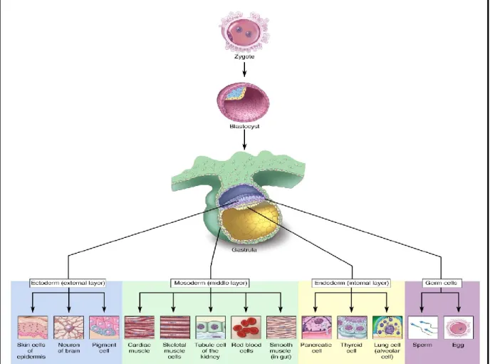
Embryonic stem cells (ESCs) have two main characteristics – self renewal and pluripotency. Self-renewal is a mechanism by which a cell can generate a daughter cell identical to itself. ESCs undergo symmetric cell division to generate two identical daughter cells. Pluripotency refers to the ability of a cell to differentiate into the three primary germ layers – ectoderm, mesoderm and endoderm. [1]
Figure 1: Illustration of the origin and differentiation of the three germ layers. (Permission obtained from Terese Winslow
http://stemcells.nih.gov/StaticResources/info/scireport/images/figure11.jpg)
Mouse Embryonic Stem Cells (mESCs) are pluripotent cells isolated from the inner cell mass (ICM) of day 3.5 preimplantation blastocyst. The ICM isolated from the blastocyst has the potential to give rise to all the three germ layers. It is however unable to contribute to the trophectoderm and primitive endoderm lineages. [2] When maintained under appropriate conditions these ICM cells give rise to ESC cell lines that have normal karyotype and high telomerase activity. Each cell line is defined by colonies derived from a single blastocyst; therefore all ESC cell lines are genetically unique. Most ES cell lines are 40XY, this is because in XX ES cell lines there is a global reduction of DNA methylation due to the presence of two active X chromosomes (one X chromosome eventually becomes inactive). This hypomethylation affects the long term maintenance of these cell lines in culture. [3, 4] One of the most debated questions in the embryonic stem cell field is whether ES cells are artifacts of tissue culture or/and whether they relate to a specific cell type in-vivo? ESCs exhibit some characteristics that are not shown in vivo by cells within an embryo; an example of one such property is that, no cell in the embryo demonstrates long-term self-renewal and pluripotency. When embryonic cells are brought to culture and exposed to specific signals (to which they wouldn’t have been exposed to in vivo), they adapt and procure novel functions that enable them to maintain their pluripotency and self-renewal abilities indefinitely. Studies have suggested that within the embryo there is a transient population of cells that are pluripotent and possess self-renewal capabilities, however, this population is present in vivo for only a limited period of time. These cells become more restricted in their potency as development progresses. Therefore, one can say that ES cells are a tissue culture artifact. [5]
ES cell lines can be derived from the ICM, primitive ectoderm and possibly from early germ cells. (Figure 3) mESCs are not equivalent to ICM cells because the ICM cells are capable of differentiating into the trophoblast whereas mESCs cannot. [5] mESCs are not entirely equivalent to germ cells as early germ cells do not self-renew for an extended period of time. It is shown that primordial germ cells (PGCs) do not contribute to chimeras when injected into blastocysts. [5] Studies have shown a founder population of cells that emerges from the primitive ectoderm before gastrulation that passes through the primitive streak to give rise to many structures of the extraembryonic mesoderm and the germ cells. The inductive signaling
involved in this process might be reproducible in vitro. ESCs might be related to this founder population. [5]
Figure 2: Illustration of the origin of stem cells. (Fiona M. Watt, Ryan R. Driskell. Phil. Trans. R. Soc B (2009).365 155-163, Permission obtained from the journal).
Embryonic stem cells are an interesting model in research for several reasons. Due to their self-renewal ability, they can be cultured in vitro for an extended period of time - serving as a constant supply of starting material. [6] mESCs can be used to generate mouse chimeras when re-introduced into a blastocyst. The pluripotency of ESCs enable them to differentiate into the three germ layers, making them an excellent model to study most developmental processes. Finally their genome is well characterized and easily modifiable using different mutagenesis strategies. [6, 7]
1.1 Genomic approaches used to conduct functional studies on
ESCs.
Over the years, several genome editing strategies have been developed to conduct functional studies on the mouse genome. In this section, I will briefly explain a few strategies that have gained popularity in the past decade.
1.1.1 Gene targeting
Gene targeting is an approach that relies on homologous recombination to modify an endogenous target gene. [8] This strategy requires the creation of a vector that contains a part of the gene to be modified, a reporter gene and a selection marker gene flanked by two homology arms. [8] To prevent unwanted effects caused by the presence of selection marker genes, Cre-loxP or Flp-frt technologies can be used to excise these genes.
The type and exact location of the mutation is known to us when we use this strategy. This offers us an advantage when we wish to recover these mutations. One of the drawbacks associated with this approach is that it requires germ-line competent ES cells. When recombinant ES cells are injected into the blastocoel cavity of 3.5 day old embryos to generate chimeric mice, it is important they colonize the germ cells of the animal to propagate the targeted mutation to subsequent generations. When using any genetic mutation strategy we must keep in mind that genetic redundancy can result in compensatory effects thereby possibly impeding the functional analysis of the mutation. [9] Sometimes the targeted gene of interest is involved in embryonic viability in the early stages of development and has several different functions at the later stages of development. [9] Abrogation of such genes results in embryonic lethality preventing the study of the genes’ function in later developmental processes. The design of conditional recombinase systems, allowing the abrogation of gene expression in a spatio-temporal controlled manner has helped overcome this limitation. [10]
The International Knockout Mouse Consortium (IKMC), Knockout Mouse Project Repository (KOMP), European Conditional Mouse Mutagenesis Program (EUCOMM) and North American Conditional Mouse Mutagenesis Project (NorCOMM) are some of the international
1.1.2 siRNA and shRNA screens
Though RNA interference technology is not a genome editing strategy, it allows us to study loss of function effects of specific genes in the mammalian genome by degrading the specific gene mRNAs. [11] Two approaches that have gained interest for achieving gene silencing are ds-small interfering RNA (siRNA) and vector based short hairpin RNA (shRNAs). While both approaches result in gene silencing, they differ in their mechanisms of action. In siRNA screens, dsRNA is synthesized complementary to the gene of interest and is introduced into the cytosol of the cells through transfection. Upon introduction to the cell, a ds-RNA specific enzyme Dicer forms a complex with long dsRNAs and cleaves them into shorter fragments of 20-25bp. The cleaved fragments are then incorporated into the RISC complex which is comprised of Arogonaute 2 (Ago-2), Dicer and TAR-RNA binding protein (TRBP). The RNA duplex is dissociated, the strand with the lowest duplex stability remains in complex with the RISC while the other strand is removed.[12]
shRNAs can be delivered to the cells by infection with virally produced vectors or through transfection with plasmid vectors encoding them. shRNAs consists of two complementary 19-22 bp RNA sequences linked by a loop of 4-11 nucleotides. shRNAs are transcribed by RNA polymerase II or III based on the promoter driving their expression. Drosha and its dsRNA-binding partner DGCR8 processes these initial precursors to pre-shRNAs which is then exported to the cytoplasm by Exportin-5. Dicer and TRBP/PACT cleaves these pre-shRNAs, removing the hairpin and creating a 20-25 nucleotide ds-siRNA which is then loaded onto the RISC complex.[12]
Once the fragments are loaded onto the RISC, the process by which siRNA and shRNA recognizes and degrades the target mRNA is the same. As part of the RISC, siRNA binds to the target mRNA mediated by complementary base pairing. This leads to the cleavage of the target RNA phosphate backbone around the center of the duplex by the action of Ago-2. RNAi-mediated cleavage products are degraded by the same enzymes responsible for degrading the bulk cellular mRNA.
Factors such as cell type and requirement for a transient or stable integration determine the method of choice to be employed. Off-target effects need to be considered when working with siRNAs and shRNAs. These off-target effects can be mitigated by using more than 3
siRNAs/shRNAs for the gene of interest. [12] Additional controls include non- targeted/scrambled siRNAs and shRNAs.
Several siRNA and shRNA screens have been carried out in ESCs using different readouts such as reporter assays, cell morphology and proliferation assays to study ESC pluripotency and self-renewal. [13, 14]
1.1.3 ZFNs, TALENs and CRISPR
Previously, Zinc Finger Nucleases (ZFNs) and Transcription activator-like effector nucleases (TALENs) were the most widely used targeted genome editing strategies. Both these technologies use engineered nucleases, i.e. artificial proteins that are composed of a customizable sequence-specific DNA binding domain that is fused to a nuclease that cleaves DNA non-specifically. [15]
ZFN is a heterodimer, where each subunit consists of a zinc finger DNA binding domain and a Fok1 endonuclease domain. To achieve a double strand break (DSB) the Fok1 domains must dimerize. Each Zinc Finger (ZF) motif folds into a ββα structure that consists of ≃ 30 amino
acids. The binding of ZF motifs to DNA takes place by the insertion of the α- helix into the major groove of the DNA double helix. Each zinc finger binds to a triplet within the substrate DNA. The amino acid residues at -1,+1,+2,+3,+4,+5 and +6 positions to the start of the α-helix contribute to the sequence-specific interactions between the ZF motifs and the DNA site. To generate ZF motifs with different sequence specificities, amino acids at these positions can be changed while maintaining the remaining amino acids as a consensus backbone. Several ZF motifs can be linked in tandem to form Zinc Finger Proteins (ZFPs) to enable binding to longer DNA sequences.[16]
Specificity and affinity of ZF sets can be a challenge. To obtain adequate affinity, atleast 3 fingers in each ZFNs are required. Addition of fingers can improve specificity as well as affinity , however, there is a possibility that they might mediate binding to off-target sites.
Zinc finger technology is relatively expensive and can be tricky to synthesize. [15, 17]
consists of highly conserved repeats derived from transcription activator- like effectors (TALEs). TALEs can be designed to bind to any target DNA sequence. TALEs are proteins secreted by the Xanthomonas bacteria to alter transcription genes in host plant cells. The DNA binding domain consists of 33-34 amino acid sequence which is highly conserved. Within this sequence, the 12th and the 13th amino acid location is highly variable and is involved in specific nucleotide recognition. TALENs, like ZFNs function as dimers, thereby it requires two unique DNA binding domain sites in the genome with the correct orientation and spacing to induce a DSB. ZFNs and TALENs induced DSB can be repaired by cellular Non Homologous End Joining (NHEJ) or Homology Directed Repair (HDR) pathways. NHEJ-mediated repair of a DSB in a gene results in insertions and/or deletions (INDELs) causing frameshift mutations that disrupts gene function. HDR repair pathway can also be exploited to introduce specific nucleotide modifications by supplying the double stranded DNA of interest as template. [15, 17]
Currently, clustered regularly interspaced short palindromic repeats (CRISPR) is the most popular targeted genome editing tool, allowing to edit genomes with unprecedented efficiency, precision and flexibility. CRISPR/Cas9 is part of the bacterial adaptive immune system. Cas9 is an endonuclease that makes a double stranded break in the DNA. The site for DNA break is determined by a guide RNA of ~20 nucleotides. Cas9 induced DSB are repaired via the NHEJ pathway. The NHEJ pathway is more error prone compared to the HDR pathway and thereby introduces INDELs resulting in impaired gene function. Alternatively, precise replacement mutations can also be made if a donor template having a mutation and homology to the targeted locus is supplied. [18]
Although genome wide specificities of the CRISPR-Cas9 system has yet to be entirely defined, several modifications of this system such as inducible Cas9 strategies are being employed to provide a platform to study loss of function phenotypes. [19]
1.1.4 Cre-loxP
The Cre-loxP recombination strategy of Coliphage P1 provides a unique tool to manipulate the mouse genome. The site specific DNA recombinase Cre (cyclization recombination) is a 38 kDa protein recognizing 34bp loxP sites and catalyzes inter and intramolecular recombination
between them. The loxP site consists of a nonpalindromic 8bp core region flanked by two palindromic 13bp sequences. [20]
This strategy brings about chromosomal rearrangements between two loxP sites, referred to as endpoints. The recombination between two loxP sites results in chromosomal deletions, inversions and duplications if the loxP sites are located on the same chromosome whereas recombination between loxP sites located on different chromosomes results in translocations. To create a deletion, two regions on the same chromosome are successively targeted by homologous recombination using different vectors. Each vector contains a sequence for a loxP site. Cre recombinase expression results in the excision of DNA sequences between the loxP sites that have been integrated. [21] To isolate ESC clones in which successful recombination and deletion of genes have occurred, two non-functional halves of a selection marker gene are inserted in each of the vector containing the loxP site. [22, 23] For example the Conlon group previously used a vector that included a non-functional “split” Hprt1 cassette along with the first loxP site. The first loxP site was introduced into a particular locus by homologous recombination whereas the second loxP site and the second half of the Hprt1 cassette was introduced using retro-viral gene transfer. Cre recombinase expression resulted in deletion of chromosomal regions between the loxP sites and reconstitution of the functional Hprt1 gene. These recombinants were selected in Hypoxanthine-aminopterin-thymidine (HAT) medium. [23] HAT is a selection medium for mammalian cell culture. Aminopterin blocks DNA de novo synthesis which is essential for cell division. Hypoxanthine and thymidine provide the cells with essential components required for them to evade the blockage and proceed with the salvage pathway, provided the cells have functional copies of the gene encoding the HPRT enzyme.
The Cre-loxP system can be used to conditionally modulate tissue-specific gene expression. [24] The loxP sites can be introduced around a functionally essential genomic region followed by a tissue-specific Cre recombinase mediated excision of the loxP flanked sequence. A similar strategy can be used for tissue specific overexpression of a transgene, where a strong expressing promoter is separated from the coding region of a gene by “stop” sequences flanked by loxP sites. Although in both cases Cre recombinase transgene allows spatial
control, once Cre expression is induced and recombination has occurred, the resultant gene expression is mostly irreversible.
Animal models for spatial and temporal gene regulation have been generated by combining the Cre-loxP technology with inducible systems. Transgenic mice containing the gene of interest surrounded by loxP sites are crossed with transgenic mice that have tissue-specific expression of the Cre gene. The resulting mice will have the target gene deleted in tissues expressing Cre whereas the target gene will be present and function normally in tissues that do not express the Cre gene. These animals could be used to study specific tumor signature profiles and signaling pathways relevant to tumor supressors and oncogenes during tumor progression. Additionally, studies combining the Cre-loxP inducible system with RNAi have been used to efficiently knock down endogenous genes. [25]
1.1.5 DELES approach
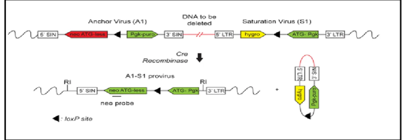
In our laboratory, we used a Cre-loxP strategy to carry out functional studies on the mouse genome. [26, 27] Simon Fortier and Melanie Bilodeau employed a retro-viral based Cre-loxP approach to create a library of ESC clones harboring hemizygous nested chromosomal deletions – the DELES (Deletion in ES cells) library. This strategy was different from the Cre-loxP strategies used by other groups as it did not use homologous recombination to deliver loxP sites to specific sites in the genome. The sequential addition of two retroviruses – anchor virus and saturation virus were used to deliver the loxP sites. The anchor virus consisted of a loxP site, a functional puromycin gene and a truncated ATG-less neomycin gene. The saturation virus consisted of a loxP site, a functional hygromycin gene and a PGK-ATG promoter cassette that on Cre recombination would drive the expression of a functional neomycin gene. (Figure 3) The retrovirus design used to generate the library is as illustrated below:
Figure 3: Retro-viral design of the primary A1 and secondary S1 Saturation virus.
(Fortier, S., et al., Genome-wide interrogation of Mammalian stem cell fate determinants by nested chromosome deletions. PLoS Genet, 2010. 6(12): p. e1001241. , Permission obtained from Simon Fortier.)
R1 ESCs were first infected with the A1 anchor virus to generate primary clones. The ESCs were infected with an infection efficiency of < 1% to achieve single provirus integration per cell. The puromycin resistant clones were next infected with low titer saturation virus to generate hygromycin resistant secondary clones. Cre-recombinase induced gene deletions were carried out by introducing a Cre-plasmid in ESCs through electroporation. Puromycin selection was again performed on G418R tertiary clones to identify clones that harbor gene deletions rather than other re-arrangements such as inversions and duplications. Inverse-PCR was used to map the deletions obtained. This library contains chromosomal deletions covering ~ 25% of the mouse genome with a 3Mb average deletion size. The average genome coverage per autosome is 23% with no deletions on 19, X and Y chromosomes.
To evaluate genome coverage in DELES, the cohorts of mapped deletions were categorized according to their molecular function based on gene ontology analysis. The 25.4% genome coverage of DELES includes 7083 genes, 4265 CpG islands, 128 miRNAs, 470 lincRNAs, 241 ultraconserved elements, 648571 LINE/SINE elements and 108 cancer related genes. Clusters of protein-coding and non-coding elements were also deleted in this library of ES clones, enabling us to analyze synthetic interactions and functional redundancies between
family members (clones that have the anchor virus integration at the same point in the genome but different saturation virus integrations) of DELES. [26]
Advantages of DELES strategy:
I. Integration of the loxP sites was achieved by retro-viral gene transfer and not gene targeting, thereby the library was generated quickly and was less labor-intensive. II. Nested chromosomal deletions were generated enabling us to interrogate protein
coding and non-coding regions associated with a phenotype. III. Alleles of the genes are permanently deleted and not just silenced.
The DELES clones can be used to generate homozygous mutant mice to perform in vivo phenotypic studies of dominant and recessive mutations. For example, in our lab, Melanie Bilodeau wanted to assess the in vivo biological roles of two cystatin proteins Stfa2l1 and Csta, the dysregulation of which has been observed in cancers such gastric cancer, head and neck carcinomas. To this end, she generated chimeric mice by injecting the DELES clone 7-30 harboring a 95-kb chromosomal deletion whose deletion spanned four genes (Fam162a, Ccdc58, Csta and Stfa2l1) into C57BL/6J morulas and blastocysts. To achieve germline transmission of the mutant allele, the two chimeric mice were crossed with C57BL/6J females. Homozygous mutant mice were generated by intercrossing heterozygous mutant mice. PCR studies were conducted on the genomic DNA isolated from mouse tail clips to confirm the loss of the genomic regions covered by the chromosomal deletion in the homozygous mice. She reported that Csta and Stfa2l1 were dispensable for viability, fertility and hematopoietic activity. [27]
Additionally, Simon Fortier exploited the DELES library to identify ES cell fate determinants. The goal of the study was to identify minimal genetic regions which when deleted blocked normal embryoid body development. LIF and BMP signaling maintains ESCs in their undifferentiated state in vitro. Upon removal of these signals, ESCs differentiate into aggregated structures called embryoid bodies (EB). These 3-D aggregate structures contain cells derived from the three germ layers and their formation seems to recapitulate several
differentiation processes observed in vivo in a spatio-temporal manner. Melanie Bilodeau provided evidence that within our DELES library, there is a co-relation between EB formation competency in vitro and contribution to chimeras in vivo. Clones which showed normal in vitro EB differentiation contributed to the generation of chimeras which was documented by coat color whereas clones with abnormal EB differentiation showed undetectable ES-derived contribution in the pups. She also observed that most of the clones with EB differentiation anomalies show formation of EBs at low frequency.[28]
The criteria that a DELES family of clones should meet to determine defects in EB formation are:
I. A clone should have at least 5% of seeding density: Clones were plated in two 96-well plates in parallel, one containing semi-solid differentiation media and the other coated with gelatin. EBs were counted after 8 days of differentiation while the colonies propagated on the gelatinized surface was stained with methylene blue 24 hours after seeding. Metamorph software that analysed methylene blue stained area was used to analyze cell input that produced the corresponding EB number. Clones with methylene blue staining < 5% was excluded. This low value could be the result of a defect in proliferation, cell adhesion or maybe just a technical issue.
II. An abnormal tertiary clone should produce less than 1/5th the number of EBs compared to the primary clone.
III. An abnormal family should include clones with a phenotype that co-relates with deletion sizes.
Simon Fortier reported that three ribosomal protein genes Rps5, Rps14 and Rps28 are haplo-insufficient for EB formation in a p53-independent manner. [29]
Disadvantages of the DELES strategy:
I. Large deletions could involve removal of haplo-insufficient regions that are detrimental to ES cells.
III. Physical constraints such as chromatin organization might affect the efficiency of Cre-loxP recombination.
We propose to use cDNA and BAC complementation approaches to re-introduce deleted regions and identify the deletions associated with an abnormal phenotype. Annotated cDNA libraries that can be manipulated easily are commercially available. These libraries can be used to identify determinants involved in relevant pathways; however, potential roles of non-coding elements cannot be determined. Additionally, cDNA expression is mostly driven from a ubiquitous promoter, which may not reflect the endogenous expression level, temporally or specific to a cell type. To overcome these limitations, different cDNA isoforms might have to be tested using an inducible promoter. BAC complementation is advantageous when large regions (100 ~ 250kb) need to be re-introduced. They contain regulatory elements such as promoters and they also help in identifying non-coding elements that may be associated with an abnormal phenotype. However, due to their large sizes, their transfection in cells is more challenging. A protocol for BAC complementation has been optimized in the laboratory.[28]
Since we are a lab whose focus is hematopoiesis, we would now like to use the DELES library to identify novel determinants of hematopoietic cell fate commitment. A preliminary hematopoietic screen conducted in the lab identified several DELES clones with an abnormal hematopoietic phenotype. To identify chromosomal regions essential for hematopoietic commitment, DELES clones were seeded into 96-well plates and subjected to EB differentiation. Embryoid bodies are able to spontaneously differentiate into cells of the three germ layers, including the hematopoietic cells. To test the presence of primitive hematopoietic cells, day 8 EBs were directly stained with benzidine – a highly selective and efficient procedure to demonstrate the presence of hemoglobin in EBs.
Principle of Benzidine staining:
The “haem” moiety of hemoglobin possesses peroxidase activity which can bring about the decomposition of hydrogen peroxide and liberate nascent oxygen. This nascent oxygen reacts with a chromogen such as benzidine to produce a blue colour product. The equation is as follows:
H2O2 + Chromogen haem oxidized chromogen + H2O Peroxidase activity
The intensity of the blue colour is proportional to the amount of hemoglobin present. Benzidine can also be used to test the presence of erythrocytes. Erythrocytes contain an enzyme called catalase which also possesses peroxidase activity and catalyses the oxidation of hydrogen donors. [30]
Though benzidine staining is a specific, inexpensive and a quick assay to test for the presence of haemoglobin, benzidine is a carcinogen. Skin absorption or inhalation of the powder has shown to lead to bladder cancer, therefore utmost care must be taken when handling benzidine.
Using this assay, DELES clones were screened for the presence of benzidine positive EBs. Out of 88 clones screened, several clones exhibited abnormal phenotype i.e. % of benzidine-positive EBs < 30 % compared to the WT R1 ESCs. Figure 4 shows the DELES families with the highest reduction in benzidine staining.
Figure 4: Preliminary hematopoietic screen using benzidine staining assay - The graph represents the percentage of benzidine positive EBs. The DELES clones were plated in duplicates in a 96-well plate to form embryoid bodies in the absence of LIF. Direct benzidine staining was performed
mentioned on the x-axis. The grey region represents the standard deviation for the average of percentage of benzidine positive EBs for control R1 ESCs.
To identify the stage at which hematopoietic specification is arrested in these DELES clones, we need to screen these clones using a hematopoietic assay that would differentiate ESCs to form hematopoietic cells in a stepwise manner.
1.2 Hematopoiesis
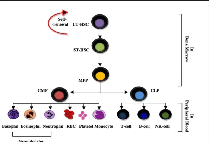
Blood cells are responsible for the maintenance of the immune system of the body. This relentless work requires that these blood cells have the ability to replenish themselves. The production and maturation of blood cells is termed hematopoiesis. [31] The Hematopoietic Stem Cells (HSCs) are ultimately responsible for the constant renewal of blood. [32] HSCs produce differentiated effector cells through a series of increasingly more committed intermediate progenitors. The Long-Term HSCs give rise to Short-Term HSCs which in turn produce multi-potent progeny (MPP) that have limited self-renewal capacity. The MPPs give rise to oligopotent progenitors which contain the Common Lymphoid Progenitors (CLP) and Common Myeloid Progenitors (CMP). CLP and CMP then give rise to lineage-restricted effector cells. [33] (Figure 5)
Figure 5: Hierarchy of hematopoietic stem cells - Illustration of the hierarchy of HSPCs based on self-renewal and differentiation capacity (Permission obtained, .Maha M. Bakhuraysah Christopher Siatskas and Steven Petratos. Hematopoietic stem cell transplantation for multiple sclerosis: is it a clinical reality? Stem Cell Research & Therapy20167:12, (http://creativecommons.org/licenses/by/4.0/)
HSC transplantation is one of the common cell-based therapies applied in clinical settings today. HSCs can be isolated from umbilical cord blood, peripheral blood and bone marrow but their use is limited due to the need for compatibility between donor and recipient and low HSC content. [34] Although, somatic HSCs can be expanded, after ex vivo culture their in vivo potential might be compromised. Pluripotent stem cells are potential sources of HSCs and can serve as an in vitro model to study mechanisms regulating embryonic hematopoietic development. Additionally, human ES cells are more amenable to genetic manipulation compared to the somatic HSCs that are currently used for bone marrow transplantations.
using somatic cell nuclear transfer (SCNT). The genetic defects in these cell lines could be treated by genetic manipulation using siRNA, lentiviral and homologous recombination techniques and can be used to generate transplantable HSCs via in vitro differentiation.
Mature hematopoietic lineages derived from human ES cells can be used for replacement therapies. For example, human dendritic cells (DCs) manipulated in vitro are being explored for the treatment of tumors and autoimmune diseases, however using current techniques, the number of DCs that can be isolated from patients is a limiting factor. Generating DCs from human ES cells can help circumvent this problem. [35] Likewise, it may be possible to generate mature red blood cells from ES cells that could help in erythrocyte transfusions especially in cases where there is limited donor availability for rare blood cell types. [36]
To enable the transit of ES-derived hematopoietic cells from bench to bedside, we will have to understand the 3-D microenvironment composed of stromal contacts and paracrine factors present in vivo during hematopoietic development and be able to recapitulate them in vitro to efficiently produce cells of the hematopoietic lineage.
1.2.1 Ontogeny of hematopoiesis
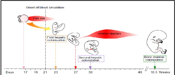
During vertebrate development, two waves of hematopoiesis can be found at distinct anatomical sites and they contribute to both embryonic and adult hematopoiesis. The first wave, known as primitive hematopoiesis, originates in the yolk sac at about E7.5-E11 [37] of mouse development and week 3-6 of human development. [38] At this stage, primitive erythrocytes are formed but the lymphoid lineages are absent. These primitive erythrocytes pool into blood islands soon after the start of gastrulation and these blood islands become enveloped by endothelial cells to form the vascular plexus of the yolk sac. [39] Primitive erythrocytes fulfill the functions necessary for the survival and growth of the post-implantation embryo, however as the embryo grows, it requires more specialized cells to meet the demands of growth and development in later stages. To cater to this demand, the onset of definitive hematopoiesis occurs in the Aorta Gonad Mesonephros (AGM) and then transitions around E10.5 to mouse fetal liver, or between 6-22 weeks to human fetal liver. Definitive erythrocytes are formed during this wave. The definitive erythroid progenitors transition from
the fetal liver towards the newly developed bone marrow at the end of gestation. [37] The generation of all cells of the immune system occurs here, with the exception of T cells, which although originate from precursors derived from the bone marrow, develop in the Thymus. [39]
Figure 6: Ontogeny of hematopoiesis – Illustration of the developmental ontogeny in humans. Reproduced with permission from The International Journal of Developmental Biology (Int. J. Dev. Biol.) (2005) Vol: 49 page numbers 243-250.
1.2.2 In- Vitro derivation of hematopoietic cells
Hematopoietic differentiation from ES cells can be induced by two types of approaches: a) Genetic approaches
Figure 7: Methods for deriving hematopoietic cells in-vitro. (Dan S Kaufman, toward clinical therapies using hematopoietic cells derived from human pluripotent stem cells2009, Blood. Permission obtained from the journal.)
Genetic approaches: Studies carried out by the Humphries and Daley group have shown that ectopic expression of certain genes into ES cells can be used to coax these cells to differentiate into HSCs/HPSCs in an in vitro system. [40, 41]
Ectopic expression of HoxB4 in ES-derived hematopoietic cells in the presence of stromal cells has shown to promote expansion of hematopoietic populations with definitive HSC potential. [42] Studies have shown that these ESC –derived hematopoietic progenitors were rendered competent for engraftment and long-term multilineage reconstitution.
In mouse ES cells ectopic expression of Cdx (family of caudal-related homeobox-containing transcription factors) induced mesodermal specification and enhanced HPC production. Expression of HoxB4 and Cdx on EB-derived cells cultured on OP9 stromal cells had a
synergistic effect on HPC production, enabling engraftment of all hematopoietic lineages in irradiated adult mice. [43]
Recapitulating cellular interactions and paracrine effects occurring in vivo during hematopoietic development will help in the optimal development of ES-derived HPSC/HSCs. Towards this end, 3-D EB formation and stromal feeder co-culture differentiation systems are used in an effort to mimic the in vivo niches that support hematopoietic lineage specification.
Non-genetic approaches: Different protocols are used for generating hematopoietic progenitors from embryonic stem cells. The most commonly used are by:
Formation of Embryoid bodies
Culture on supportive stromal layers (eg. OP9 stromal cells )
Differentiation from embryoid bodies recapitulates many aspects of embryogenesis, it takes advantage of the principle that three-dimensional EBs can spontaneously differentiate into any cell type including ones of the hematopoietic lineage. [44] Gene expression analysis and studies on kinetics of hematopoietic lineage development within EBs have shown that it accurately reflects the early stages of yolk sac hematopoiesis.
Several techniques are used to generate EBs from cultured ESCs: 1) liquid cultures in non-adherent dishes; 2) methylcellulose or other semi-solid media; 3) hanging drop cultures and 4) porous scaffolds. [44]
The 3-dimensional EB method of hematopoietic differentiation is more efficient than the adherent method of ESC hematopoietic differentiation on a gelatinized surface whereas the frequency of hematopoietic progenitors obtained using any method of EB culture is similar. [45] Liu group has shown that dynamic conditions and biomaterial-based scaffolds enhance the efficiency of ESC differentiation. To evaluate the spontaneous differentiation of ESCs, EBs can be cultured in ESC differentiation media without cytokines. In the absence of cytokines, cells within the EB are able to secrete proteins such as CSF, IL-3 and Epo to support the development of early erythroid and macrophage precursors. [46] To induce the
to differentiation media or methylcellulose culture containing hematopoietic cytokines such as SCF, GM-CSF, Epo and interleukins such as IL-1, IL-3,IL-4 and IL-6. [46] The emergence of hematopoietic cells in this system can be tracked through gene expression analysis of lineage specific markers and immunohistochemistry. Additionally, single-cell suspensions obtained by dissociating EBs with trypsin or collagenase can be analyzed by flow cytometry. [47, 48] Within the EBs a transient primitive erythroid population emerges first. The lymphoid progenitors and the HSCs are not generated in the early stages of EB hematopoiesis, further reflecting yolk sac hematopoiesis. Though lymphoid progenitors are not generated in the early stages of EB differentiation, B and T cell lineages have been generated when ES cells are cultured for extended periods of time in the presence of stromal cells. [45]
The bone marrow stroma acts as a three dimensional framework for hematopoiesis. It contains a heterogeneous population of cells including mesenchymal stem cells (MSCs) that play an important part in the bone marrow niche. It helps in the interactions of hematopoietic stem and progenitor cells (HSPCs) with the extra-cellular molecules and soluble factors that are essential for survival and proliferation. [49] With this in mind, primary stromal cell lines were used as feeders to expand HSPCs ex-vivo. The different stromal lines commonly used are OP9 [50], S17 [51] and AM20-1B4 [52]. OP9 is a cell line established from mouse calvaria and lack functional macrophage colony-stimulating factor (M-CSF). The absence of M-CSF enhances hematopoietic differentiation by minimizing preferential differentiation towards the monocyte-macrophage lineage. [53, 54] ESC – derived erythroid, myeloid and B- lineage cells are obtained using OP9 stroma without the addition of exogenous growth factors; however cytokine supplementation influences lineage output. The onset of definitive hematopoiesis and expansion of HSCs takes place in the AGM region; cell lines derived from the AGM region like UG26-1B6 are also used with the aim of providing signals unique to this micro-environment to help with the hematopoietic differentiation. [55]
Culturing on feeder layers have resulted in successful hematopoietic differentiation, however, use of cells in human therapy requires their culture conditions to be defined and “animal-free”. Serum contains several undefined components and we cannot exclude the contamination of the desired hematopoietic cells with the feeder cells or antigens transmitted by the feeder cells even with processes such as cell sorting. Also, since the serum contains undefined factors, the
efficiency of the differentiation assay may differ from batch to batch of the serum used and call into question the robustness of the differentiation protocol. [56]
To overcome these issues, I have adopted a “serum-free, feeder-free” hematopoietic differentiation protocol published by the Keller group. [57] The hematopoietic cells derived here are free of undefined components and interactions.
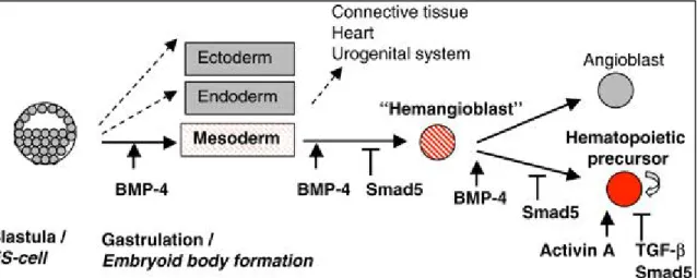
Growth factors and cytokines constitute an important part of the regulatory niche; therefore it is important to know the effect of different cytokines on the developmental pathways in order to nudge the differentiation of mESCs to the desired lineage. In the protocol I set up in the lab, Wnt3a, Activin A, BMP4 and VEGF were used during appropriate time windows to bring about lineage specification step by step.
Transforming Growth Factor – β (TGF-β), Bone Morphogenetic Protein 4 (BMP4) and Activin are members of the TGF-β family of ligands that signal though SMAD pathways to regulate hematopoietic stem and progenitor cells (HSPCs) during hematopoiesis. Upon binding of TGF- β ligands to their receptors, the receptor regulated R-SMADs get activated and bind to SMAD4. This complex translocates to the nucleus and activates several target genes responsible for regulating many signaling pathways. BMP4 is a morphogen that induces ventral mesoderm and promotes hematopoietic commitment. [58] Apart from its role in specification of HSC during development, it also regulates proliferation of adult HSCs [59] and addition of BMP4 in culture has shown to increase the percentage of CD45+ population. [60] BMPs are mainly produced by the osteoclasts in the HSC niche. [61]
Figure 8: SMAD signaling at different stages during hematopoietic commitment - SMAD signaling regulates HSC specification and its subsequent expansion critically at different stages (Jonas Larson et al, 2005, Oncogene. Permission obtained from the journal.)
While BMP4 appears to have a crucial instructive role for the induction and formation of blood cell precursors, other factors are also involved in their regulation and subsequent expansion. Vascular Endothelial Growth Factor (VEGF), a KDR ligand is known to induce the proliferation and differentiation of the BMP4-induced hematopoietic progenitors. [62, 63] VEGF and its homologue PIGF produced by HSCs and erythroblasts influence the bone marrow microenvironment in many ways. They induce bone marrow endothelial cells to express E- selectin and thereby play an important role in HSC migration. Additionally they also act as survival and proliferation factors as VEGF-R1 and VEGF-R2 are expressed on immature HSCs that possess repopulating capacity. [64] Studies have shown that elimination of VEGF genes leads to decreased HSC survival, colony forming ability and hematopoietic repopulation following transplantation. [65] VEGFA is known to promote HSC survival, whose expression levels are dramatically increased in hypoxic conditions. [66]
The Wnt pathway has a varied influence on hematopoiesis. This pathway has 19 different ligands and about 10 different receptors. The different receptor-ligand combination can result in either canonical or non-canonical downstream signalling. [33] Evidence that Wnt signaling plays a role in hematopoiesis came from studies demonstrating the presence of Wnt ligands and receptors in different hematopoietic anatomical sites such as murine yolk sac, aorta-gonad-mesonephros, fetal liver and adult bone marrow. [33] Studies have shown that the
addition of Wnt3a in mESC cultures increases the generation of hematopoietic cells and promotes proliferation of hematopoietic progenitors. Wnt signaling not only plays a role in HSC survival and proliferation but is also involved in the maturation and maintenance of lymphoid progenitor cells such as pro-B cells, pre-B cells, immature thymocytes and mature T-cells. [67]
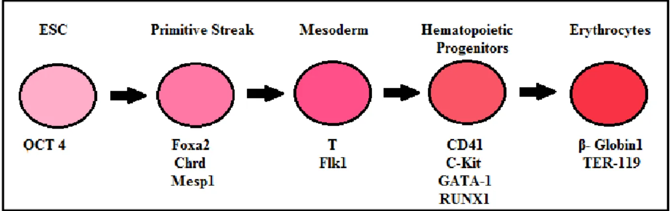
As ES cells differentiate towards the hematopoietic lineage, they express several transcription factors in an orderly manner. Some of the transcription factors and surface markers that can be used to assess the differentiation at specific stages are illustrated in Figure 9.
Figure 9: Lineage-specific gene markers used to assess hematopoietic differentiation from ES cells.
1) Brachyury (T) – It is a transcription factor that binds to a specific DNA palindromic site. Binding occurs through a region called the T-box present in the N-terminus region. On binding, Brachyury regulates the transcription of several genes required for mesoderm differentiation. Brachyury expression is critical for posterial mesoderm development in the mouse. During development, Brachyury expression is observed in early gastrulae next to primitive streak cells destined to form the mesoderm. T-/- embryos die at E.10 due to the absence of a notochord and allantois. [68] 2) CD41 – expression of CD41 in murine development marks the onset of definitive hematopoiesis. CD41 expression precedes that of CD45. In adult hematopoiesis CD41 expression is associated with megakaryocytes and also plays a role in platelet function. Hematopoietic cells emerge from a CD41+ population; these
expression of CD41 separates the hemogenic endothelium from the hemangioblast - which is the common precursor for hematopoietic and endothelial cells. [69] 3) Runt-related transcription factor 1 (RUN1X) plays a crucial role in definitive hematopoiesis. It is involved in the transition of hemogenic endothelial cells to hematopoietic cells. [70] 4) GATA-1 and GATA-2 are members of the GATA family of transcription factors. GATA-1 is involved in erythroid development and is highly expressed in erythroid cells whereas GATA-2 is expressed on early hematopoietic progenitors and plays a role in HSC production in the AGM region and HSC proliferation throughout the ontogeny. [71, 72] 5) Beta-Globin is part of the hemoglobin protein and is involved in erythropoiesis. Mutations in the β-globin gene results in blood disorders such as β-thalassemia and anemias such as sickle cell anemia. [73] 6) C-kit – also known as cytokine tyrosine kinase receptor. C-kit, the receptor for SCF, is present on HSCs and the stromal cells in the bone marrow secrete SCF. Upon binding to the receptor, the receptor homo-dimerizes and auto-phosphorylates at tyrosine residues activating pathways responsible for hematopoiesis and HSC maintenance such as JAK/STAT and RAS/ERK pathways. C-Kit is not essential for the expansion of HSCs in fetal development, although it plays an important role in maintaining the long-term steady state HSCs in adult BM. It also promotes HSC proliferation when in synergy with other hematopoietic cytokines. [74] 7) CD45 is a pan-hematopoietic marker expressed on most hematopoietic cells except erythrocytes and platelets. [75, 76] Due to variable exon splicing, different CD45 isoforms are expressed by different subsets of hematopoietic cells. For example CD45RA is expressed by naïve/resting T cells and medullary thymocytes and CD45RO is expressed on memory/activated T cells and cortical thymocytes. [77]
Goal, Hypothesis and Objectives:
Goal: As mentioned previously, one of our interests in the lab is to take advantage of the DELES library to identify genes or DNA fragments that are required for hematopoietic cell fate. Using clones from the DELES library, we expect to reveal several haploinsufficient and synthetic loci that contribute to the differentiation of ES cells to the hematopoietic lineage. Before this is possible we need to establish a robust hematopoietic differentiation assay which could be exploited to rescreen the DelES collection.
My hypothesis is that the in vitro differentiation assay developed in Dr. Keller’s laboratory could be imported in our laboratory and be used to better characterize the hematopoietic phenotype of DelES clones.
The objectives of my project are to :
Establish in the laboratory a hematopoietic differentiation assay from mouse ES cells which will serve as a more stringent screen to observe hematopoietic
commitment of the candidate DELES clones. This will involve optimizing Q-RT-PCR and FACS parameters for this assay.
Validate candidate DELES clones identified in the preliminary hematopoietic screen using this assay to monitor where the differentiation arrest is occurring. In a secondary project I will test if a human haploid cell line, KBM-7, can be exploited using the DELES approach to create a library of clones comprising random chromosomal deletions. KBM-7 is a human near haploid Chronic Myeloid Leukemia (CML) cell line. Haploid cell lines such as the KBM-7 cell line serve as an excellent model system to carry out functional screens as the phenotype of the mutated allele will not be masked by the allele on the other homologous chromosome.[78]
This library of KBM-7 clones containing nested chromosomal deletions can serve as a platform on which various studies to understand the mechanisms contributing to leukemia development and treatment of leukemia can be conducted. For example, in our lab, this library will be used to delineate the molecular activities of potential anti-leukemic drugs as well as allow me to identify molecular signaling pathways that, when genetically disrupted, can confer resistance to novel and existing drugs.[79]
2. Materials and Methods
Embryonic Stem Cell maintenance and differentiation
R1 ESCs were maintained on irradiated mouse embryonic fibroblasts (MEFs) in the presence of LIF and serum. Hematopoietic differentiation of mESCs was induced using the protocol published by Keller et al. (2012). To set up the differentiation assay, the ES cells previously grown on MEFs were split on dishes coated with 0.1% gelatin. ESCs were treated with trypsin and seeded at a density of 2.5x10^5/mL in a serum-free differentiation medium (75% IMDM, 25% Ham’s F12, supplemented with 1% N2, 0.5% B27, 0.05% BSA, 50 µg/mL ascorbic acid, 4.5 x 10⁻⁴ M 1-thioglycerol, 2 mM glutamine and penstrep) for 48 hours in 60mm bacterial dishes. The embryoid bodies formed were dissociated using 0.25% Accutase for 5 minutes at 37 ̊ C and then re-aggregated at the same density in the serum free media containing 3ng/mL Wnt3a and 1ng/mL Activin A for an additional 48 hours to induce mesoderm differentiation. The aggregates formed were once again dissociated and re-suspended in the serum free medium, this time in the presence of 1ng/mL BMP4 and 5ng/mL VEGF to induce hematopoietic commitment. Recombinant Mouse Activin A (R&D systems, Ref # 338-AC-010), recombinant mouse Wnt3a (R&D systems, Ref # 1324-WN-002), recombinant human BMP-4 (R&D systems. Ref # 314-BP-010) and recombinant human VEGF165 (R&D systems, Ref # 293-VE-010) were used.
Embryoid body differentiation
To improve the differentiation efficiency by getting rid of the MEFs, ES cells were split on 0.1% gelatin prior to the differentiation. A 5X Master mix of the differentiation media was prepared containing IMDM+glutamine, 15% heat-inactivated Fetal Calf Serum, 50µg/mL Ascorbic acid, 5% PFHM II , 0.3Mm 1-thioglycerol and Penstrep. To prepare the semi-solid media, half the media volume was replaced with methylcellulose. Tubes containing methylcellulose and differentiation media were vortexed to attain a homogeneous formulation. The ES cells were trypsinized for 5 minutes at 37 ̊ C and neutralized using the differentiation media. ESCs were seeded at a density of 1250cells/mL in 60mm bacterial- grade tissue culture
dishes. After adding the cells into the semi-solid media, the cells were mixed manually. 5mL syringes and blunt needles were used to slowly transfer the mixture to the dishes. Each dish was placed in a larger dish humidified with sterile water.
Benzidine Staining Assay
A benzidine (Sigma, Cat # B1883-100mg) stock solution of 3% was made by diluting 4, 4’-Diaminobiphenyl in 90% glacial acetic acid and 10% water solution. A fresh working solution of 1 part benzidine stock solution, 1 part hydrogen peroxide (Sigma, Cat # H3410) and 5 parts of sterile water was prepared prior every staining. Day 8 embryoid bodies were stained for the presence of hemoglobin by adding 0.2mL of the working solution per 2mL of methylcellulose culture directly onto the dish.
RNA Extraction
The cell suspension from one 60 mm dish was collected and spun down at 800rpm for 5 minutes. Media was aspirated and the cells were washed with PBS and spun down again. The PBS was aspirated and the cell pellet was re-suspended in 1mL TRIzol reagent (Life Technologies, Cat # 15596-026). The samples were stored at -80 ̊C for subsequent RNA isolation.
RNA Isolation:
RNA was extracted from TRIzol stored samples according the manufacturer’s instructions. Measurement of RNA concentration :
The concentration (260nm) and purity of RNA was assessed using an Implen nanophotometer.
cDNA synthesis
1.5 µg of RNA was used to synthesize DNA using MMLV-RT (Invitrogen, Cat # 28025-013) according to manufacturer’s instructions. The following program was used:
Table 1: cDNA synthesis program Temperature Time 25 ̊C 10 minutes 37 ̊C 50 minutes 70 ̊C 15 minutes
QRT-PCR
Here the DNA was further quantified and simultaneously amplified. The qRT-PCR was carried out using TaqMan® Fast Advanced Master Mix (Applied Biosystems, Cat # 4444558)
Master Mix preparation :
A master mix was prepared and used at a final volume of 8.5µL for each well of a 384- well plate.
Table 2: Composition of Master Mix for qRT-PCR.
Component Volume (µL)
Taqman Mix 5
Primers (F+R) 0.1
Exiqon probe 1
Water 2
o cDNA (1/5 dilution) = 2µL was used for each reaction.
Table 3 – Sequence of primers used for qRT-PCR